early modern experimentation on live animals*hpscdept/people/papers/dbmpaper_earlymodern... · *...
TRANSCRIPT

Early Modern Experimentation on Live Animals*
DOMENICO BERTOLONI MELIIndiana University Bloomington
Bloomington, INUSAE-mail: [email protected]
Abstract. Starting from the works by Aselli (De lactibus sive lacteis venis, 1627) on the
milky veins and Harvey (1628, translated in 1993) on the motion of the heart and thecirculation of the blood, the practice of vivisection witnessed a resurgence in the earlymodern period. I discuss some of the most notable cases in the century spanning fromAselli’s work to the investigations of fluid pressure in plants and animals by Stephen
Hales (Vegetable Staticks, 1727). Key figures in my study include Johannes Walaeus,Jean Pecquet, Marcello Malpighi, Reinier de Graaf, Richard Lower, Anton Nuck, andAnton de Heide. Although vivisection dates from antiquity, early modern experimenters
expanded the range of practices and epistemic motivations associated with it, displayingconsiderable technical skills and methodological awareness about the problemsassociated with the animals being alive and the issue of generalizing results to humans.
Many practitioners expressed great discomfort at the suffering of the animals; however,many remained convinced that their investigations were not only indispensable from anepistemic standpoint but also had potential medical applications. Early modern
vivisection experiments were both extensive and sophisticated and cannot be ignored inthe literature of early modern experimentation or of experimentation on livingorganisms across time.
Keywords: Vivisection, Anatomy, Experiment, Life, Harvey
Introduction
Techniques of investigation are a crucial aspect of anatomical research,as shown for example by studies on microscopy and injections, both
* Previous versions of this essay were delivered at venues on both sides of theAtlantic. I thank Hal Cook, Karin Ekholm, Evan Ragland, Bob Richards, AllenShotwell, Nancy Siraisi, Fernando Vidal, the anonymous referees, and all those who
offered comments on previous versions of my work.
Journal of the History of Biology (2013) 46:199–226 � Springer 2012
DOI 10.1007/s10739-012-9327-7

recent and less recent. Complex techniques require great skills, attentionto detail, careful controls, patient and creative labor: far from beingneutral tools, techniques helped define the horizon of research at dif-ferent periods and interacted problematically with the results attained.Vivisection in particular required great dexterity and involved priorknowledge of the anatomy of the animal to be dissected, including theexact location of its vital parts. The early modern period witnessed aflourishing of anatomical researches based on novel methods of inves-tigation such as microscopy, and the revival and refinement of ancientones such as vivisections and injections, which were used only occa-sionally in the past; these and other techniques were often used incombination. The striking usage of vivisection in the early modernperiod and its interaction with other techniques call for a criticalreflection on how and why anatomists and physicians used it and on thesignificance and implications of the animal being alive.1
Dissection and vivisection were the subjects of debate and contro-versy from antiquity right through the period covered in this study andbeyond: although dissection and vivisection were practiced and theknowledge they provided was deemed essential, some argued thatdissection was useless because the body changed with death, othersbelieved that cutting open a living body altered it to such an extent thatno reliable knowledge could be drawn from studying it, while others stillwere opposed to cutting the animal body altogether, whether dead oralive. Often, however, anatomists assessed the perceived merits andproblems of vivisection implicitly and explicitly not in the abstract butwith regard to specific issues and organs, such as the speed of decay ofgiven body parts and their visibility in a dead or alive body, forexample. At times vivisection involved prolonged observation of pro-cesses in animals and even plants requiring careful analyses from severalperspectives and showing surprising connections between intervention-ist methods of inquiry and observation techniques typical of naturalhistory.2
Anatomists advocated vivisection on the ground of its utility tounderstanding the structure and purpose of many body parts and ofpotential medical benefits. While some anatomists found the suffering ofanimals in artificial and cruel settings unbearable, many defended
1 Cole, 1917–1921; Cook, 2007, pp. 281–285; Wolfe, forthcoming; Wilson, 1995;Ruestow, 1996; Fournier, 1996.
2 Von Staden, 1989, pp. 234, 236; Carlino, 1999, pp. 131, 138, 158–159, 166–167;Rupke, 1987, especially the essay by Maehle and Trohler. [Morgagni], 1705, pp. 166–
169. See the essay by Allen Shotwell in this issue Guerrini (2003).
DOMENICO BERTOLONI MELI200

vivisection on the ground that it is permissible to treat animals the waywe wish; paradoxically, Danish anatomist Nicolaus Steno did both. Myfocus in this paper is primarily on techniques and epistemological issuesrather than on ethical ones, important as they are both historically andphilosophically. In this respect Steno’s observations are especiallyinteresting: in his treatise on the anatomy of the brain, Discours surl’anatomie du cerveau, delivered in 1665, he instantiated the claim thatwe can treat animals the way we want, by arguing that surgeons practiceprocedures like trepanning on them. However, he also warned that thebrains of animals differ considerably from species to species, especiallywhen humans are involved. His passage highlights the links betweensurgical and vivisection practices, as well as of the problem of variabilityin nature (Steno, 1910, vol. 2, pp. 25–26; Guerrini, 1989, p. 406;2006; Oster, 1989; Bartholin, 1663–1667, III, p. 228; Kooijmans, 2010,pp. 32–35; Bertoloni Meli, 2011c).
Our knowledge of vivisection from antiquity includes treatises fromthe Hippocratic corpus, Aristotle, the Alexandria anatomists, and espe-cially Galen. Iacopo Berengario da Carpi, Andreas Vesalius, and RealdoColombo were key figures in the revival of vivisection in the Renaissance.Vivisection was traditionally performed to study motions, such as thoseof the heart, of the thorax in respiration, and of the peristalsis of digestiveorgans, as well as the role of nerves in controlling body parts; in addition,vivisection was used to ascertain the heat of several organs, the precisecondition of body parts that decay rapidly with death, the contents ofvessels, and matters associated with generation, just to mention some ofthe main areas examined by Allen Shotwell in this issue (see also vonStaden, 1989, pp. 147, 247; Cunningham, 1997, Chapter 5 and p. 159;French, 1999, Chapter 6, pp. 193, 207).
The range of the investigations carried out in the early modernperiod was such that this brief essay cannot offer a comprehensiveanalysis; instead I seek here to provide a bird’s eye view of experi-mentation on animals—and occasionally plants—with special focus onits purposes, and to highlight key features in the cases I investigate.Some of the experiments I discuss are well known today, others less so,but seen together, they provide a vivid picture of the issues and prob-lems addressed.
Anatomists were fully aware of the profound changes their field wasgoing through and of each other’s works, which appeared in the widelycirculating scientific journals of the time, in monographs that werefrequently reprinted, typically in the Low Countries, and also in theimposing Bibliotheca anatomica, two huge folio volumes issued in 1685
EARLY MODERN EXPERIMENTATION ON LIVE ANIMALS 201

and in expanded form in 1699 containing a critical compilation of therecent literature. Its contents date overwhelmingly from the second halfof the century, the works by Gasparo Aselli of 1627 andWilliam Harveyof 1628 being the two outstanding exceptions from the first half andthose whence my analysis sets off.
There are additional reasons for starting my analysis from Aselli andHarvey, besides following contemporary perceptions: in their investi-gations they used vivisection in diametrically opposed ways, thus settingthe stage for appreciating the wide range of motivations for whichvivisection was practiced and its surprising outcomes. Aselli examinedstructures whose existence had already been predicted and conceptual-ized but which had not been fully uncovered; Harvey focused onmotions without providing any new structural finding. JohannesWalaeus pursued their researches in creative ways, while Jean Pecquetbrought Aselli’s findings to dramatic new consequences and opened thedoor to the discovery of a new system—the lymphatics. The examples Iselected from Marcello Malpighi’s and Richard Lower’s works reflectthe differences between Aselli’s and Harvey’s, in that Malpighi usedvivisection to investigate structures—not because of their ephemeralnature but to enlarge them as through a microscope—whereas Lowerinvestigated the role and purpose of respiration without any newstructural findings. In the Netherlands, Reinier de Graaf, Anton Nuckand Anton de Heide performed striking vivisections aimed at investi-gating a number of problems, including pathological issues. JohannJakob Wepfer investigated the effects of hemlock and other poisons,while his son-in-law Johann Conrad Brunner excised the pancreas tounderstand its purpose. Lastly, Stephen Hales’s celebrated experimentson blood and sap pressure in animals and plants first published in 1727close a remarkable century, providing rich material for reflection onchanging conceptualizations of life and experimentation more broadly.
This essay highlights the existence of a wide range of purposes,epistemic functions, and technical aspects in vivisection, calling for areevaluation of the role of anatomy in early modern experimentation.Together with other examples in this special issue, the cases I discuss callfor a reflection on changing conceptualizations and practices of animalexperimentation from Antiquity to the Enlightenment.
Gasparo Aselli: Finding a Missing Structure
The posthumous work by the Pavia professor of medicine Aselli, Delactibus sive lacteis venis, announced the discovery of the milky veins,
DOMENICO BERTOLONI MELI202

the vessels allegedly carrying chyle from the intestine to the liver. Whileperforming a vivisection of a dog in front of several witnesses in order tostudy the recurrent nerves, which control the actions of the larynx andthe voice, Aselli decided to investigate the motion of the diaphragm aswell. The recurrent nerves had a dissection and vivisection historystretching from Galen to Vesalius and Colombo (see the essay by AllenShotwell in this issue). Aselli’s curiosity about the motion of the dia-phragm too was quite traditional in a vivisection, Colombo havingdrawn attention to it as well in connection with respiration (Eriksson,1959, pp. 291–293; French, 1999, pp. 207–208). In the course of hisvivisection, however, Aselli noticed many white vessels, whose naturewas at first unclear, but when he saw white milk exuding from an incisionhe had practiced with a sharp scalpel in one of them he exclaimed‘‘eureka!’’, marking his joy at the belief that he had discovered the pas-sage of chyle envisaged by Galenic anatomy from the intestine to theliver. His joy was short-lived, however, since the dog soon died and themilky veins suddenly disappeared in front of his eyes while he wasobserving them. Repeating the experiment on the following day on adifferent dog, Aselli failed to find the vessels altogether. He reasoned atthis point that the dog in his first vivisection had recently eaten; thereforehe performed a third vivisection approximately 6 h after the dog’s lastmeal, finding the milky veins again (Aselli, 1627, pp. 19–21).
Aselli’s finding in the course of what appeared to have been an entirelytypical vivisection in the Galenic tradition was unexpected, more bychance than by design, or as he put it, ‘‘casumagis (ut verum fatear) quamconsilio’’ (Aselli, 1627, p. 18). To his surprise, Aselli realized that vivi-section made a difference not only to the study of actions—as was in hisown and his friends’ intention in revisiting the role of the recurrent nervesand the motion of the diaphragm—but also of structures so ephemeralthat they could suddenly disappear after death. In dissecting a recentlydeceased man in order to investigate the moisture of the pericardium,Vesalius found that the heart was still beating; this borderline casebetween dissection and vivisection highlights the crucial role of fresh-ness—if not life—in dissection (Park, 1994, p. 19. See also the essay byAllen Shotwell in this issue). Aselli’s surprising finding in a live doginvolved a new genre of vessels—the fourth after veins, arteries, andnerves—as well as the reliance on an unusual variable: thus Aselli madenot only an important anatomical find but also highlighted the role andsignificance of the timing of the animal’s last meal, accidentally reviving avivisection technique that had been employed by Galen in order toinvestigate digestion (Galen, On the Natural Faculties, III, 4).
EARLY MODERN EXPERIMENTATION ON LIVE ANIMALS 203

Aselli’s perspective was that of completing the established Galenicsystem rather than subverting it; this, however, was a major contrastwith Harvey.
William Harvey: Understanding Motions and Directionality
Unlike Aselli’s treatise of the previous year, Harvey’s De motu cordis etsanguinis presents no new anatomical structures. Rather, he reinter-preted recent findings, such as the ostiola or little doors in the veinsfound by his teacher Gerolamo Fabrizi, on the basis of anatomicalobservations, quantitative reasoning, and vivisection experiments. Inparticular, the ostiola in the veins and valves in the heart took on amajor role in the study of unidirectionality. While Harvey’s results werestrikingly new, his vivisection techniques were not especially innovative,except possibly for their systematic nature and the wide number ofanimals involved. The originality of Harvey’s findings is clearly not atstake here; in a sense, the largely traditional nature of the methods heemployed would make—if possible—his achievement even moreimpressive.3
De motu cordis et sanguinis treats the motion of the heart in the firstpart, Chapters 1–7, and the circulation of the blood in the second,Chapters 8–17; since Harvey routinely relied on vivisection, my accountdoes not pretend to be exhaustive. In the first part he dissected livecold-blooded animals ‘‘such as toad, serpents, frogs, snails, lobsters,shell-fish, prawns, and all small fishes’’, where the heartbeat is slower.Similarly, he closely observed the motion of the heart in dying warm-blooded animals in order to determine the active phase, or systole, andthe passive one, or diastole, a procedure going back to Galen andemployed also by Colombo. Harvey also experimented on eels, showingthat their hearts keep moving not only after they have been extractedfrom the body but also after they have been chopped to pieces; we aregoing to encounter other instances of experimentation on body partsand cadavers below. It was certainly unusual to dissect such a widerange of animals, but it was not unique. Harvey’s Padua teacher Fabrizihad promoted a broadly neo-Aristotelian comparative anatomy projectand half a century before Harvey the Dutch anatomist and physicianVolcher Coiter had vivisected the hearts of serpents, frogs, fishes, andcats. Harvey’s work, however, stands out for its scope and systematic
3 The study of generation too is relevant to vivisection and poses considerable
problems. On these matters I refer to Ekholm, 2010.
DOMENICO BERTOLONI MELI204

nature (Harvey, 1993, pp. 19 and 26–27; Cunningham, 1985; Bylebyl,1985, pp. 237–242).
Harvey also carefully observed the relation between the motion ofthe heart and arteries, finding that ‘‘arterial diastole is synchronous withcardiac systole’’ and vice versa. Initially he deemed the experiment withthe reed in the artery, performed by Erasistratus and Galen in order toascertain whether arteries fill because of a faculty transmitted by theheart or because of the impulsion of blood, impossible—though later inhis 1649 second response to Riolan the Younger he succeeded in per-forming it, showing that arteries fill like leather bags rather than bel-lows, against Galen and in agreement with Erasistratus. Harveyconfirmed the same result also through an ‘‘experiment’’ performed bynature through disease: he found in a gentleman that the descendingaorta with its two femoral branches had turned into a pipe-like bone.Despite this, Harvey could still feel the pulse in the arteries of the leg, as heknew very well because the gentleman was his patient; indeed, he pre-served a span of the bony artery removed from the corpse. In thisremarkable case Harvey used a diseased state as a form of control of avivisection experiment, confirming his findings through different means.Harvey showed his methodological awareness about the problems ofvivisection in other instances too: he argued that some small shrimpsfound in the Thames have a transparent body, enabling the anatomist tostudy their heart motion ‘‘with the least possible impediment’’, withoutcutting and interfering with the body. In conclusion, in the first part ofDemotu cordis et sanguinisHarvey relied extensively on vivisection, so muchso that he stressed its role in several chapter headings. In one crucialrespectHarvey used vivisection for the same reason for whichGalileo hadused the inclined plane, in order to slow down what he wanted to observeand investigate, in this case the heartbeat rather than free fall.4
In the second part of De motu cordis et sanguinis Harvey establishedthe circulation of the blood through a series of cogent reasons andexperiments, including vivisections. Most brutally, he killed animals bydrawing their blood, something he argued could not happen in less thana quarter hour if blood did not circulate (Harvey, 1993, pp. 9, 50, fromDe motu cordis, 1628). More subtly, he employed ligatures in order toobserve the direction of blood flow inside the body through vivisectionand outside without any need for the knife; the only figure in his bookrelies on this notion, showing the motion of blood in the external veins
4 Bylebyl, 1985, pp. 229, 237, is a valuable study of heart vivisections before Harvey.Harvey, 1993, p. 29, from De motu cordis, 1628, pp. 112–114, from Exercitatio ana-
tomica de circulatione sanguinis, 1649.
EARLY MODERN EXPERIMENTATION ON LIVE ANIMALS 205

of the arm. While traditionally anatomy books focused on structuresand were replete with figures, Harvey’s figure shows a process in fourparts but no new structures. Significantly, the most effective visualrepresentation of his findings is in the form of a film shot in 1928 for thetercentenary of the editio princeps, highlighting motion and processes. InChapter 10, Harvey described a vivisection experiment on a snake,where the vena cava goes into the lower part of the heart and the arteryleaves from the upper part. By seizing with a forceps or ligating the venacava, the heart becomes pale and is emptied of blood by its pulsation;conversely, by releasing the vein and ligating the artery, the heart willturn purple and then livid in color in becoming engorged with blood.Ligatures were well known from antiquity for surgical operations suchas amputations and even simple bloodletting. Contrary to standardbeliefs, Harvey argued that ligatures do not draw or attract—attractiobeing the Latin term—anything, they simply block—one could addmechanically—blood flow: tight ligatures block both arterial andvenous blood, medium tight ones only the latter.5
Although most previous anatomists assumed they already knew thedirection of flow of fluids in the body, using ligatures to investigatedirectionality was not a new procedure: Galen had used them in a vivi-section experiment described inOn the Natural Faculties in order to arguethat urine reaches the bladder via the ureters. Harvey revived Galen’sprocedure to investigate directionality: as we are going to see, his way ofproceeding became standard in later seventeenth-century vivisection inthe study of the direction of flow of other fluids such as lymph and bile.6
Harvey’s work on the circulation led to a revival of vivisectionexperiments and opened up the field: the question now was no longer tocomplete an established system, as for Aselli, but to put together thepieces of an entirely new one.
Johannes Walaeus: Joining Aselli and Harvey – and a Harveyan Coda
In the 1630s and 1640s Dutch circles were especially active in anatom-ical research, in the wake of Aselli’s and Harvey’s works: it is no
5 von Staden, 1989, index sub ligation; French, 1994, pp. 107–110, 168–178, 348–371and index sub ‘‘attraction’’; Bylebyl, 1982; Biagioli, 2006, pp. 136–143; Lawrence, 1987.
6 French, 1994 is useful but not always reliable. Galen, On the Natural Faculties, I.13.
Shank, 1985. Galen had relied on ligatures of the umbilical cord during vivisectionexperiments: On the Usefulness of the Parts of the Body, VI.21. Harvey’s teacher Ger-olamo Fabrizi too applied ligatures to the umbilical cords, Fabrizi, 1600, pp. 110–111
and 119–120. I am grateful to Karin J. Ekholm for these important references.
DOMENICO BERTOLONI MELI206
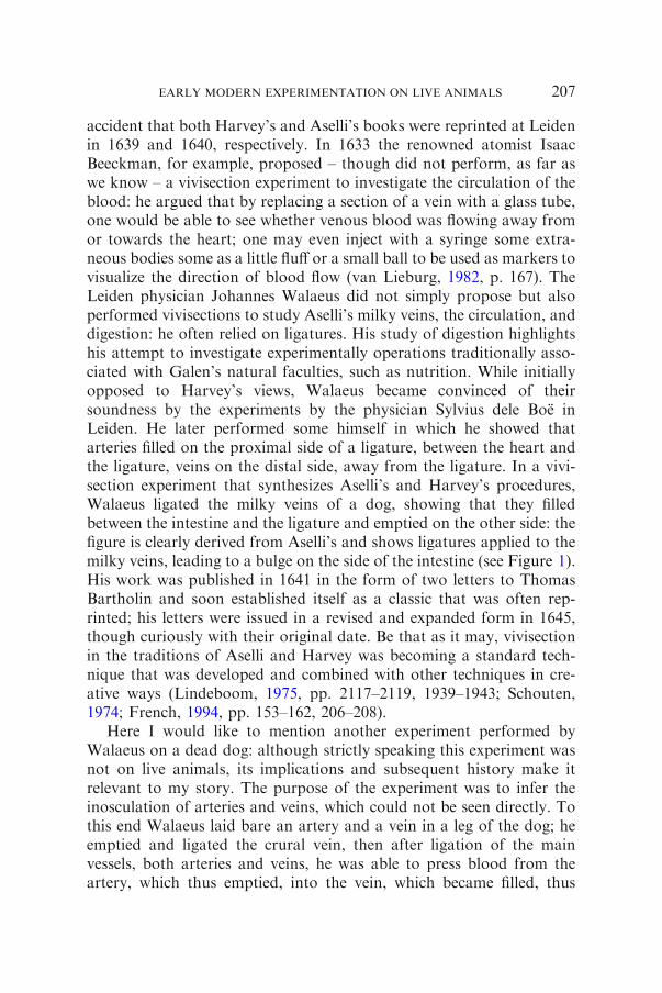
accident that both Harvey’s and Aselli’s books were reprinted at Leidenin 1639 and 1640, respectively. In 1633 the renowned atomist IsaacBeeckman, for example, proposed – though did not perform, as far aswe know – a vivisection experiment to investigate the circulation of theblood: he argued that by replacing a section of a vein with a glass tube,one would be able to see whether venous blood was flowing away fromor towards the heart; one may even inject with a syringe some extra-neous bodies some as a little fluff or a small ball to be used as markers tovisualize the direction of blood flow (van Lieburg, 1982, p. 167). TheLeiden physician Johannes Walaeus did not simply propose but alsoperformed vivisections to study Aselli’s milky veins, the circulation, anddigestion: he often relied on ligatures. His study of digestion highlightshis attempt to investigate experimentally operations traditionally asso-ciated with Galen’s natural faculties, such as nutrition. While initiallyopposed to Harvey’s views, Walaeus became convinced of theirsoundness by the experiments by the physician Sylvius dele Boe inLeiden. He later performed some himself in which he showed thatarteries filled on the proximal side of a ligature, between the heart andthe ligature, veins on the distal side, away from the ligature. In a vivi-section experiment that synthesizes Aselli’s and Harvey’s procedures,Walaeus ligated the milky veins of a dog, showing that they filledbetween the intestine and the ligature and emptied on the other side: thefigure is clearly derived from Aselli’s and shows ligatures applied to themilky veins, leading to a bulge on the side of the intestine (see Figure 1).His work was published in 1641 in the form of two letters to ThomasBartholin and soon established itself as a classic that was often rep-rinted; his letters were issued in a revised and expanded form in 1645,though curiously with their original date. Be that as it may, vivisectionin the traditions of Aselli and Harvey was becoming a standard tech-nique that was developed and combined with other techniques in cre-ative ways (Lindeboom, 1975, pp. 2117–2119, 1939–1943; Schouten,1974; French, 1994, pp. 153–162, 206–208).
Here I would like to mention another experiment performed byWalaeus on a dead dog: although strictly speaking this experiment wasnot on live animals, its implications and subsequent history make itrelevant to my story. The purpose of the experiment was to infer theinosculation of arteries and veins, which could not be seen directly. Tothis end Walaeus laid bare an artery and a vein in a leg of the dog; heemptied and ligated the crural vein, then after ligation of the mainvessels, both arteries and veins, he was able to press blood from theartery, which thus emptied, into the vein, which became filled, thus
EARLY MODERN EXPERIMENTATION ON LIVE ANIMALS 207

providing visual evidence in support of his claim (Schouten, 1974, pp.262, 271; Lindeboom, 1975, p. 281).
In 1650, in the aftermath of Walaeus’s letters, William Harvey tooperformed an experiment on a dead animal, the cadaver of a throttledman: as he wrote to the Hamburg physician Paul Marquard Schlegel,Harvey wished to refute Jean Riolan the Younger’s denial of the pul-monary transit, a debated point since Colombo’s times (see the essay byAllen Shotwell in this issue). Colombo had denied that venous bloodseeped through pores in the septum of the heart and argued instead thatbloodmoved from the right ventricle to the lungs, and then back to the leftventricle.Having ligated the vena arteriosa (pulmonary artery), the arteriavenosa (pulmonary vein), and the aorta, Harvey fastened an ox bladder toa tube, as was usually done in clysters, and injected warm water into thevena cava; the usage of warm water was presumably a precaution againstthe objection that the pores in the septum of the heart had closed because
Figure 1. Johannes Walaeus, ligated milky veins, from Thomas Bartholin, Instituti-ones anatomicae (Leiden, 1645). By courtesy of the Lilly Library, Indiana University,Bloomington
DOMENICO BERTOLONI MELI208

of lack of heat. While the right ventricle filled with water, not a dropreached the left ventricle, thus showing that there were no pores in theseptum.Having released those ligatures, Harvey inserted the tube into thevena arteriosa and ligated it between the tube and the heart, to preventwater from returning to the right ventricle. On pressing the bladder, thistime water came out from the left ventricle of the heart, thus revealing aneasy passage through the lungs to the arteria venosa.7
There are similarities between Walaeus’s experiment on the dead dogand Harvey’s second experiment on the throttled man: both sought toprovide evidence for the passage of blood through the flesh and lungs,though Walaeus was more explicit about the existence of anastomoses.
Jean Pecquet: Tracing Ephemeral and Subversive Structures
The experiments performed in Paris by Jean Pecquet while still amedical student and published in the 1651 Experimenta nova anatomicashare several features with Aselli’s. Both worked on related structureshaving started vivisections aimed at investigating motions. As Pecquetput it, the main difference between dead and live animals is motion,whose chief seat is the heart; therefore he set out to perform vivisectionsof dogs in order to study the heart (Pecquet, 1653, p. 7). On noticing amilky fluid from the vena cava, Pecquet at first thought it was pus, butupon reflection he realized that this was unlikely because the animalappeared very vigorous: by closer inspection and tasting he concludedthat what he had found was chyle. Aselli’s findings were widely knownby then and Pecquet was also well acquainted with the topic throughconversation with Pierre Gassendi, who had told him how Nicolas-Claude Fabri de Peiresc had arranged for a criminal to be executed afteran abundant meal, in order to find the milky veins in man. With furtherinvestigations and vivisections of animals about 4 h after they hadeaten, Pecquet found that chyle reached the subclavian vein in thevicinity of the heart through a vessel departing from a previouslyunknown receptacle located between the kidneys; chyle reached thereceptacle through a network of vessels he compared to a spider web(Pecquet, 1653, pp. 29, 34–35; Gassendi, 1641, book five). His workextended over a three-year period and involved over one hundred
7 Harvey, 1993, pp. 140–145, at 140–141, from the letter to Schlegel dated London,26 March 1651; French, 1994, pp. 279–285; Cole, 1917–1921, pp. 290–291; French,1985, p. 54, deals with injections using a syringe in order to study the passageways of
fetal anatomy.
EARLY MODERN EXPERIMENTATION ON LIVE ANIMALS 209

vivisections of many different types of animals. Ligatures engorged thechyle vessels with fluid, enabling him to trace their path and to study thedirection of flow (Pecquet, 1653, pp. 15–16, 32). Pecquet had traced anentirely new way through which chyle entered the bloodstream: not viathe liver, which according to Galen and Aselli received chyle andtransformed it into venous blood, but directly from the intestine,bypassing the liver. The largest internal organ was thus deprived ofits primary role of sanguification. Thus retrospectively Aselli’s workappeared problematic in that he had seen new structures but interpretedthem through Galenic eyes—or perhaps he had waited too long after theanimal’s meal and had detected additional minor vessels of the lym-phatic system as opposed to the main ones seen by Pecquet. Timing thevivisection after the last meal was becoming increasingly important.There is another aspect to Pecquet’s research: the French anatomistargued that the motion of chyle occurred purely mechanically throughpressure, without any need for attraction, on the example of a numberof phenomena observed in experiments on the air and its elasticity thatwere being debated at the time in the wake of Torricelli’s barometricexperiments (Bertoloni Meli, 2008, pp. 670–677).
Once the anatomists knew what to look for and where to look,subsequent researches could be carried out in special circumstances alsoon dead animals with the help of injections; the initial stimulus to thisarea, however, came from vivisections. Vivisections continued to rep-resent an important research tool in two related areas: on the one hand,the anatomist and physician Richard Lower investigated the purpose ofthe thoracic duct by resection, showing that the animal died even if itwas fed normally because food could not reach the blood stream. On theother hand, the thoracic duct was seen as part of an entirely new sys-tem—the lymphatics—that became more evident at a slightly longerinterval after the animal’s last meal in the investigations by ThomasBartholin and Olaus Rudbeck. Conscious efforts to find the receptacleof the chyle, thoracic duct, and lymphatic system in humans highlightthe widespread awareness that anatomical features may vary fromspecies to species and results could not be automatically generalized(Frank, 1980, pp. 197, 201, 209; Eales, 1974, pp. 280–282; Kooijmans,2010, pp. 56–59; Bertoloni Meli, 2011c).
Marcello Malpighi: Magnifying the Invisible
Marcello Malpighi received his training in dissections and vivisectionsat Bologna in mid-century when he was still an Aristotelian and
DOMENICO BERTOLONI MELI210

continued to perform vivisections until the end of his life in 1694. Heclaimed that the initial stimulus came from Harvey’s discovery of thecirculation and other recent findings, in all probability Aselli’s andPecquet’s. While at Pisa between 1656 and 1659 under the philosophicaltutelage of Giovanni Alfonso Borelli, Malpighi converted to the newcorpuscular and mechanical philosophy; his reliance on vivisectionirrespective of his philosophical allegiance highlights that this techniquecrossed philosophical boundaries. It was at Pisa, Borelli recounts in Demotu animalium, that he performed a brutal vivisection experiment byinserting first a finger and then a thermometrum into the viscera and theheart of a live stag, thus proving against traditional doctrines andDescartes that the heart was not hotter than the rest of the body. Theidea of investigating the heat of body parts through direct experience invivisection was not new: Colombo had argued that blood in the leftventricle was hotter than in the right ventricle by inserting a finger inthem. Borelli’s usage of an instrument, rather than relying on directsensory experience, is noteworthy here: he argued that the temperaturewas 40�, or the temperature of a hot summer day.8
In 1661 Malpighi performed his most famous vivisection, when heobserved with a microscope the contrary motion of blood in the arteriesand veins of the lungs of a frog: in this case vivisection combined with anew instrument enabled him to witness motion, arguably the mostimportant motion in the body, that of blood. It would be impossible toprovide a characterization of Malpighi’s vivisection experiments in ashort paper. His treatise De viscerum structura alone, dated 1666, con-tains many reports of vivisections relating to several organs. In the firstessay on the liver, for example, De hepate, Malpighi mentions severalexperiments seeking to refute the views of those like Sylvius dele Boeand Jacobus de Back, who had argued that bile is formed in the gallbladder and moves to the liver. Relying on ligatures of the neck of thegall bladder and the coledochus—the bile vessel to the intestine—,Malpighi refuted these views. In this case his experiments follow Har-vey’s study of directionality; indeed, he even tried to show that he couldnot push the bile back towards the liver, much like Harvey had shownthat venous blood could not be pushed back away from the heart. Inanother ‘‘vivisection’’ experiment clearly inspired by Harvey, Malpighiremoved the bark from the trunk of a tree and applied a tight ligature:he then kept the tree under observation and noticed an enlargementabove the ligature, thus showing that in the outer vessels sap flows
8 Borelli, 1680–1681, vol. 2, prop. 96; French, 1999, pp. 194, 209; Siraisi, 1990, p. 104;
Bertoloni Meli, 2011b, p. 46; Manzoni, 2008.
EARLY MODERN EXPERIMENTATION ON LIVE ANIMALS 211

downwards. In this remarkable case Malpighi relied on experimentaland observation techniques drawn from anatomy and natural historycombined.9
The experiment I wish to focus on concerns the kidneys. Malpighi’sdiscovery of the glomerules by means of injections and microscopy pro-vides the context: by injecting the emulgent artery with ink mixed withspirit of wine, and then removing the renal membrane and cutting thekidney longitudinally, one seeswith the help of amicroscope the glands orglomerules where urine is filtered, hanging like apples from an apple tree.This was a new anatomical feature that had not been previously observedand that localized the exact site where filtration occurred. In line withviews developed through the 1660s by the Danish anatomist NicholasSteno, Malpighi thought all secretions to occur in glands through filtra-tion (Bertoloni Meli, 2011b, Chapters 4 and 6; Cunningham, 1996).Malpighi tried desperately to grasp the connection among the vesselsinvolved inside the glomerules by means of a vivisection experiment herepeatedmany times. He ligated the renal veins and ureter of a dog, in thehope of increasing the size of the kidney’s microstructures. The animalsurvived a long time—Malpighi does not state how long—and when thekidney was examined again, it was found filled with blood. But even thistechnique failed to reveal the microstructures and connections Malpighiwas after (Bertoloni Meli, 2011b, pp. 123–124). I have chosen this vivi-section experiment because it captures a key aspect of Malpighi’s inves-tigations in many areas and over several decades: the attempt to makevisible and therefore understandable the mechanism of separation ofseveral fluids in the body. In this instanceMalpighi used vivisectionwith adouble ligature in order tomagnify the glomerule, or the site of separationof urine he had previously identified, and to understand its mode ofoperation: whereas the microscope magnifies the objects through refrac-tion, Malpighi sought to enlarge them for real.
Richard Lower: Preventing Motion and Locating Color Change
The mid-1660s witnessed a sustained body of research on respirationcarried out at Cambridge, Oxford, and the Royal Society in London.I shall focus on a number of vivisection experiments performed by theOxford anatomist and physician Lower, with the assistance of RobertHooke, curator of experiments at the Royal Society, seeking to prove a
9 Bertoloni Meli, 2011b, pp. 114–117 and 253. The entire book provides a more
detailed study of Malpighi’s vivisection experiments. Holmes, 1993, pp. 314–315.
DOMENICO BERTOLONI MELI212

chemical role for respiration against the so-called mechanical view ofrespiration put forward by Borelli and defended by Malpighi, WalterNeedham, and others: according to them, the purpose of respirationwas to mix properly all the components of blood, including chyle. Inorder for this mixing to occur, it was thought that the motion of thelungs was required. Additionally, Lower wished to determine whetherblood changed color from dark to bright red in the heart or the lungs. Inan initial vivisection experiment reported in the 1665 Vindicatio, Lowerhad found blood in the pulmonary vein, after it had passed through thelungs, to be dark or venous—probably because the lungs had collapsedand contained no air. Colombo had performed a similar vivisection inthe previous century but his aim was to determine whether the pul-monary vein contains blood or smoky waste, an easier task thandetermining the precise color and nature of blood. Lower’s task wasespecially challenging, so much so that even one of the leading experts inthis area—as Lower unquestionably was—had trouble and had torecant his views shortly thereafter (Frank, 1980, pp. 188–192; Lower,1669, p. 167; Colombo, 1559, p. 224; Bertoloni Meli, 2011a).
Initial vivisection experiments relied on insufflation, blowing air eitherwith a straw into the heart through the thoracic duct, or with a pair ofbellows into the lungs, to study the role of air in respiration, the motionof the heart, and the life of the animal. Insufflation experiments with apair of bellows had been performed by Galen since antiquity—as Harveyreminded readers of De motu cordis—and were to be lampooned byJonathan Swift in Gulliver’s Travels (Harvey, 1993, p. 13, from De motucordis, 1628; Bertoloni Meli, 2011b, p. 45; Steintrager, 2004, pp. 66–67).In 1668 Lower andHooke performed a striking experiment with two pairof bellows instead of one. They opened the chest of a dog, cut the tra-chea, and attached the two pairs of bellows blowing alternately so as toproduce a constant air flux. Air could escape the lungs that had beenpunctured at the opposite end, thus the animal could be kept alive for along time without any motion in its lungs. This experiment presentsparadoxical and counterintuitive features: traditionally vivisection wasused in order to investigate motions, including those associated withrespiration. In this case, however, Lower and Hooke used vivisection inorder to keep an organ—the lungs—still: only in this way could theyinvestigate whether motion of the lungs was a necessary component ofrespiration, in mixing blood, or rather fresh air was all that was needed.Thus the purely mechanical view of respiration was refuted in favor of achemical one according to which respiration was seen as analogous to aburning flame (Lower, 1669, pp. 170–171; Frank, 1980, Chapter 8).
EARLY MODERN EXPERIMENTATION ON LIVE ANIMALS 213

Some of these vivisection experiments appeared exceedingly crueland many spectators expressed their unease and discomfort at wit-nessing or performing them. In light of Descartes’s claims that theanimals were automata without a soul, those feelings had a philo-sophical significance too (Frank, 1980, pp. 159–160, 201; Maehle andTrohler, 1987; Maehle, 1990).
In order to investigate the site of the change of color of blood, Lowerperformed other experiments: he opened the chest of a live dog, cut thetrachea and corked it; then he opened the cervical artery, after the bloodhad passed through not only the lungs but also the heart, and foundthat the blood was venous. This showed that if no air enters the lungs,no change of color occurs in the blood even after it passes through theheart, thus refuting those who attributed change of color of blood to aferment or heat in the heart.
Finally, Lower performed the last experiment, one I can no longercall a vivisection because the dog was strangled. Soon after the animaldied, while keeping its lungs inflated with the two pairs of bellows andletting the air out through the incision in the lungs, venous blood wasinjected into the vena cava; one can gain a sense of the complexity ofthis setup by considering that at least three people must have beeninvolved at the same time, two operating the two pair of bellows andone injecting blood. The venous blood went through the right ventricle,the pulmonary artery, and the lungs, coming out bright red from thepulmonary vein, as if it had been drawn from the artery of a livinganimal, says Lower. Thus color change in blood from dark to bright redoccurred not in the heart because of the heart’s heat—or indeed of anyvital flame or property, since the animal was dead—but in the lungspurely as a result of fresh air. Similarly, color change in blood frombright red to dark was not due to lack of heat, since the blood goingthrough the dead animal and collected in a dish clearly was not heatedand yet it had not turned dark or venous (Lower, 1669, pp. 165–166;Frank, 1980, pp. 214–215).
The experiment on the dead dog had precedents in the ones carriedout by Walaeus and Harvey and described in the letters to Bartholinand Schlegel. Since Harvey had performed his experiment in front ofseveral colleagues and his letter to Schlegel was known to the physicianand anatomist George Ent, a friend of Harvey’s who remained active atthe Royal Society for several decades, it is possible that Lower andHooke may have known about it and may have been inspired by it. Butwhereas Walaeus was investigating the existence of inosculationsbetween arteries and veins and Harvey the plumbing of the heart and
DOMENICO BERTOLONI MELI214

lungs in order to deny the existence of intra-ventricular pores, Lowerwas doing something different. Respiration had been traditionallyassociated with life and motion: this is why it had been investigatedthrough vivisection since antiquity. Performing a vivisection in order tokeep an organ still was not the only paradox in their respirationexperiments: Lower enacted respiration in a dead animal by blowing airthrough its lungs, thus implicitly showing that one of the key operationsassociated with life involved only chemical and mechanical processes.
Reinier de Graaf and Johann Conrad Brunner: The Role of Pancreatic
Juice
The young Delft physician and anatomist Reinier de Graaf performedone of the most celebrated yet problematic vivisection experiments ofthe seventeenth century. De Graaf was a student of the Leiden professorof medicine Franciscus Sylvius dele Boe, who had made the chemicalanalysis of the fluids associated with digestion a central area of hisresearch. Another of his students, Steno, in writing on salivary glandsand secretion, for example, provided a brief chemical characterizationof saliva. Sylvius’s understanding of chemical processes relied on thedichotomy acid-alkali; as to digestion in particular, he sought to attri-bute acid and alkaline properties to the fluids at play, such as pancreaticjuice and bile: their coming together would produce effervescence,enabling digestion. Following the discovery of the pancreatic duct byJohann Georg Wirsung two decades earlier, pancreatic juice was a keypiece of the puzzle and while Sylvius’s theories predicted that it wouldbe acid, direct evidence was lacking (Ragland, 2008, pp. 624–631).
Therefore de Graaf set out to collect a sufficient amount of pancre-atic juice as to allow assaying. Whereas saliva is readily available andbile can be easily collected from the gall bladder, pancreatic juice isproduced in such small quantities as to render assaying arduous. Forthis reason de Graaf devised an elaborate contraption to collect pan-creatic juice from a live dog; in a stunning vivisection experiment hemanaged to insert in the pancreatic duct of a dog a quill leading to acontainer attached to the dog’s belly, where the fluid slowly collected. Ittook de Graaf six attempts in which many things went wrong before hecould collect enough juice to allow assaying—by tasting. His medicaldissertation under Sylvius’s direction, Disputatio medica de natura & ususucci pancreatici was turned into a small treatise in the same year inwhich it was defended (1664) and went through two further editions
EARLY MODERN EXPERIMENTATION ON LIVE ANIMALS 215

with important additions in 1666 and 1671. Overall De Graaf foundpancreatic juice to be acidic as his mentor had expected. In this casevivisection was required to collect a sufficient quantity of an elusive butimportant fluid; it was not the process of secretion, as in Malpighi’scase, that concerned de Graaf, but the nature of the fluid. One mayargue that vivisection in this case was merely enabling de Graaf tocollect an elusive juice, but in fact matters became more complex whenanatomists, following the failure to observe effervescence between bileand pancreatic juice in a flask, debated whether chemical reactionsoccurred in the intestine inside the body exactly as they do outside: inthis case some unspecified feature inside the body—possibly heat—mayhave enabled a reaction that was not occurring outside (Ragland, 2008,pp. 660–662; Lindeboom, 1975).
Following de Graaf’s experiments, others too sought to collect pan-creatic juice and assay it by tasting, finding contradictory results. Even-tually, its alleged key role in digestion was challenged by anothervivisection experiment coming from an entirely different tradition. In the1683Experimenta nova circa pancreas, Brunner reported having removedmost of the pancreas from several dogs without noticing serious adversereactions, except possibly increased urination; Brunner stated that he hadlearnt the procedures necessary for vivisection operations, such as dealingwith wounds, from a surgeon, thus highlighting the valuable lessonsoffered by surgical practices. Brunner’s vivisections posed a seriouschallenge to de Graaf’s views. The tradition of vivisection by excisingorganswas not new: in order to refute theAristotelian claim that the voicecame from the heart, for example, RealdoColombo had ligated themajorvessels of the heart and removed it from a dog, which continued to barkfor a few moments before dying (see also the essay by Allen Shotwell inthis issue). Others too in the seventeenth century performed excision oforgans in vivisection experiments involving vastly different technicalskills in order to investigate the consequences on the animal; in a sense,the old procedure of ligating or cutting the recurrent nerves to silence ananimal and Lower’s more recent resection of the thoracic duct in dogs,leading to their starvation, share common features with Brunner’s work.Brunner stated that he was inspired to perform the excision of the pan-creas—or most of it—from recent reports of splenectomies, since thepancreas had no more vascular connections than the spleen. But whereasexcising the heart led to a swift death, excising the pancreas and spleenrequired careful observation of the animal’s behavior over severalmonthsin order to ascertain any differences in its behavior (French, 1999,pp. 208–209; Webster, 1971; Bertoloni Meli, 2011b, pp. 156–158).
DOMENICO BERTOLONI MELI216

Johann Jakob Wepfer: Tracing the Poison’s Paths
In 1679 the Swiss physician and anatomist Johann Jakob Wepferpublished a remarkable work by many standards, Cicutae aquaticaehistoria et noxae. Wepfer reported in tragic detail a case of hemlockpoisoning of young children dating from 1670, resulting in twofatalities. There is no indication that Wepfer performed postmortemson the deceased children. In order to investigate the effects of poi-soning and the path the poison took inside the body, however, hesubjected the digestive system to renewed study and described for thefirst time a number of glands (Maehle, 1987). In addition, Wepferundertook a number of tests on animals, such as dogs, wolves, andothers, which he poisoned with hemlock and other toxic plants andminerals. His book is so rich in themes and problems that a com-prehensive analysis of its contents must fall outside the scope of thispaper. Here I shall outline some of his vivisection experiments: headministered his poisons to the animals orally rather than throughintravenous injections—a technique that had gained great notoriety inthe 1660s—and sought to trace the poison’s path inside their bodiesby examining its effects on the lymphatic system, the circulatorysystem and the heart, and the animal as a whole by examining itsbehavior. He advocated vivisection or at least immediate dissectionafter the animal’s death as the best method for tracing the poison’seffects. Some of Wepfer’s descriptions of the effects of poisoning arequite chilling in their detached observation and recording of theanimal’s final suffering, as in a natural history fashion. His followers,such as his son-in-law Johann Conrad Brunner, a Swiss anatomistand physician whom we will encounter below, pursued his researchesby injecting poisons intravenously (Wepfer, 1679, preface and p. 299;Maehle, 1987).
Wepfer’s vivisection experiments are of interest for his study of themovements of the digestive system and for study of the effects ofpoisoning. He relied on an eclectic combination of notions ofdisease—Galenic, Helmontian, chemical, and mechanical—and offeredthoughtful comments on how to conceptualize it. His work of the effectsof poisoning adds another striking dimension to the vast field of earlymodern vivisections, that of the study of pathology and cause of death;despite the obvious suffering inflicted on animals, the potential medicalapplications of his investigations were especially prominent in his con-cern with understanding and hopefully offsetting a poison that hadrecently killed young children.
EARLY MODERN EXPERIMENTATION ON LIVE ANIMALS 217

Anton Nuck and Anton de Heide: Explaining Processes
The Dutch retained a leading role in anatomical research despite theapparent refutation of de Graaf’s work on the pancreas. Anton Nucktaught anatomy and surgery at The Hague and, from 1687, medicineand anatomy at Leiden. In addition to his medical degree, Nuck was askilful surgeon who published a book on surgical operations (Linde-boom, 1984, pp. 1442–1445; Nuck, 1692; French, 1999, pp. 206–210).No doubt, his surgical skills came in handy in his vivisection experi-ments. I am going to mention briefly three experiments performed anddiscussed by Nuck, on the operation of glands, the location and natureof fecundation, and the origin of bladder stones.
Nuck was the heir of the tradition of Sylvius dele Boe and NicholasSteno: his first publication dealt primarily with a new salivary duct andthe chemical analysis of saliva. There were different ways to approachthe study of glands: by structural analysis, involving both the micro-scope and injections of different fluids; by chemical analysis of theirsecretions; by vivisection experiments, involving ligatures for example;and by remarkable case histories and pathology. Nuck relied on acreative combination of all these methods. In his study of saliva, forexample, De ductu salivali novo (Leiden, 1685) he reported the opinionaccording to which secretion occurs by means of a ferment provided bya nerve and proceeded to test the claim by ligating the nerve to thesalivary gland: saliva was still secreted, though in smaller quantities.Ligating the efferent vein, however, led to increased salivation, thussupporting the view that saliva originates from arterial blood and ner-vous action determined which proportion of arterial blood turned intosaliva and which became venous blood. In this case Nuck’s purpose wasneither finding structures nor chemical composition—as for Malpighiand de Graaf—but the mode of operation of glands: we have thusencountered three different purposes for studying glands through vivi-section.10
In his 1692 treatise of Glands, Adenographia, Nuck reported anumber of striking vivisection experiments only tangentially related toglands but remarkable in their own right. In one of them he ligated theleft uterine horn in a bitch 3 days after copulation; on the 21 day afterthe operation he dissected her and noticed two fetuses between the
10 Nuck’s De ductu salivali novo was republished with additions as Sialographia et
ductuum aquosorum anatome nova, also in Le Clerc and Manget, 1699, vol. 2, pp. 797,808b, 809a; Steno, 1910, vol. 1, pp. 25–26, 35–36, 38–39; vol. 2, pp. 100–101. Steno tooperformed resection and ligation experiments of the nerves and blood vessels connected
to muscles to study their contractions.
DOMENICO BERTOLONI MELI218

ligature and the ovary: thus the vivisection experiment enabled him tolocate the site where fecundation occurred. In his view his findingshowed that fecundation occurs through an ‘‘aura seminalis’’ ratherthan male semen directly, which he believed could not reach so high.In the other experiment he investigated the origin of bladder stones,suspecting that they formed through the accretion and incrustation ofsuccessive layers. He opened the bladder of a dog and inserted awooden globule in it; the dog survived the operation and lived happilyfor several weeks; after this time he reopened the bladder and found itcovered with incrustations, thus proving his point and also showingthe origin of the celebrated bezoar stone—a calculus found in thestomach or intestine of some animals—that were once believed to beantidotes to poison (Nuck, Adenographia, in Le Clerc and Manget,1699, vol. 2, pp. 838b, 839b–840a; Lindeboom, 1975, pp. 290–291;Needham, 1959
2
, p. 144).Much like Nuck, Anton de Heide too was a physician with surgical
skills who published an important contribution on the anatomy ofmussels, Anatomy mytuli (Amsterdam, 1684). In a series of Observati-ones attached to his treatise, de Heide discussed a variety of themes,including the regeneration of bones; to this end he relied on a number offrogs, broke their hind legs, and then followed the process of repair on adaily basis. His procedure resembled studies of the formation of thechick in the egg by Harvey and others: in the studies on generation eggswere opened at successive intervals to investigate growth stages, whereasin the studies on regeneration frogs were dissected at successive intervalsto investigate how far the bone had been repaired. Other relevant par-allels can be found in the regeneration experiments on the tails of lizardsor the claws of lobsters carried out by Robert Boyle in the late 1650s. DeHeide traced the process of regeneration from blood effusion after1 day, to a harder lamina after 5 days, to cartilage after 27, and to solidbone after 4 months; he concluded that the new bone originated fromextravasated blood. Although such experiments were to prove crucial inlater debates between mechanical and vitalist views, de Heide eschewedphilosophical discussions (de Heide, 1684, pp. 123–126; Lindeboom,1984, pp. 807–808; Frank, 1980, pp. 140–141; de Moulin, 1988, p. 104).
Stephen Hales and the Behavior of Fluids
In the first decades of the eighteenth century the clergyman and naturalphilosopher Stephen Hales carried out an extensive series of investiga-tions on many aspects of plant and animal physiology, especially to do
EARLY MODERN EXPERIMENTATION ON LIVE ANIMALS 219

with the behavior of fluids. Despite the fact that his treatment of ani-mals seems especially gruesome, Hales was careful to inform his readersthat the animals he was using for his experiments were often diseased orabout to be culled; at one point he also claimed that he had stopped hisresearches because of the ‘‘disagreeableness of anatomical dissections.’’Although he was not a professional anatomist, Hales was aware ofobvious problems related to vivisection procedures, as when he dis-cussed and measured differences in the heartbeat rate of a horse when itis not in pain or terrified and under vivisection. He focused withpainstaking detail on the absorption and transpiration of water inplants and the pressure of sap and blood in plants and animals, pro-viding extensive numerical tables: his experiments seemed inspired byRobert Boyle, who had studied the pressure of fluids in differentexperimental settings using similar devices, and the iatromathematicaltradition then flourishing in Britain. Already in Vegetable Staticks of1727, he provided details of his animal experiments, which were laterexpanded in the 1733 Haemastaticks. A feel for the question addressedby Hales can be gained from the following quotation11:
Which force [of the rising sap] is near five times greater than theforce of the blood in the great crural artery of a Horse; seven timesgreater than the force of the blood in the like artery of a Dog; andeight times greater than the blood’s force in the same artery of afallow Doe: which different forces I found by tying those severalanimals down alive upon their backs; and then laying open thegreat left crural artery, where it first enters the thigh. I fixed to it (bymeans of two brass pipes, which run one into the other) a glass tubeof above ten feet long, and 1/8th of an inch diameter in bore: Inwhich tube the blood of one horse rose eight feet, three inches, andthe blood of another Horse eight feet nine inches. The blood of alittle Dog six feet and half high: In a large Spaniel seven feet high.The blood of the fallow Doe mounted five feet seven inches.
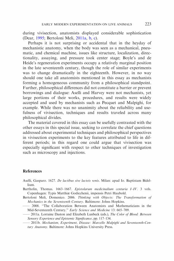
Figure 2 shows a plant set against a wall, with three s-shaped tubesattached at different points of the trunk. By means of mercury, Halesmeasured the changing pressure of sap along the trunk: his experimentalset-up shows a striking resemblance to that of Bernardino Ramazzini inhis investigation of the wells around Modena, a work well known inEngland at the time. In both cases the device measured differentialpressure along a fluid conduit (Bertoloni Meli, 2006, pp. 187–189). Theanalogy with Boyle’s and Ramazzini’s works highlights that in his
11 Hales, 1733, Introduction and pp. xviii, 2, 10, 13; quotation from Hales, 1969, p. 61.
DOMENICO BERTOLONI MELI220

experiments on plants and live animals Hales saw his subjects ashydraulic apparatus.
Conclusion
My overview of the complexity and richness of early modern experi-mentation on live animals shows that anatomists, surgeons, and physi-cians used vivisections on many grounds in a way that perhaps has notbeen fully appreciated either by historians of anatomy and physiology orby scholars of experimental practices. The fact that vivisection datesfrom antiquity may have led to the misleading impression that the earlymodern period offered little that was new. I hope that my brief study willcontribute to dispelling this myth and to showing the range of techniquesand purposes for which vivisection was used. Take de Graaf, forexample: in the early nineteenth century his experiments were deemedimpossible to reproduce by Francois Magendie; when Claude Bernardsucceeded in replicating them he was so impressed that he dedicated oneof his works to de Graaf. Yet one would look in vain for an account andanalysis of de Graaf’s work and of most other anatomical experi-ments—with the exception of Harvey’s—in standard accounts of earlymodern experimentation (Ragland, 2008, pp. 616–617).
Vivisection experiments raise a number of issues about experimen-tation on live animals. Traditionally vivisection was used primarily to
Figure 2. Stephen Hales, sap pressure in plants, from Vegetable Staticks (London,
1727). By courtesy of the Wellcome Library, London
EARLY MODERN EXPERIMENTATION ON LIVE ANIMALS 221

study motions, especially those related to the heartbeat and respira-tion—or the emotional reaction of a bitch in seeing her puppies, as inthe examples of Vesalius and Colombo discussed by Allen Shotwell.While Walaeus, Harvey, and Lower focused precisely on thosemotions too, they also studied both blood flow and respiration itself indead animals. Aselli, Pecquet, and Malpighi – at least in the exampleswe have seen – relied on vivisection primarily to investigate structures,while de Graaf was unique in the group I selected in focusing oncollecting a sufficient amount of pancreatic juice to enable chemicalassaying by tasting. Studies of directionality by Harvey, Walaeus,Malpighi, and Lower; the effects of poisoning investigated by Wepfer;the purpose and mode of operation of organs through excision andligation explored by Brunner and Nuck; the exact localization ofprocesses sought by Nuck; the regeneration of body parts investigatedby Boyle and de Heide; and the presence of fluid pressure in thevessels invoked by Aselli, Walaeus, Pecquet, and Hales; all necessitatethe animal to be alive.
Vivisection raises key questions at the intersection between practicesof active intervention and observation: on the one hand, vivisectionappears as the archetypal interventionist experimental technique; on theother, some experiments were associated with careful observation, attimes resembling practices of natural history, as with Malpighi’sobservation of the ligated tree trunk or Brunner’s observation of theeffects of excising the pancreas, both involving several months at least.Perhaps few examples could emphasize more convincingly that obser-vation intersects a variety of other techniques of investigation, includinga quintessentially interventionist one like vivisection (Daston andLunbeck, 2011).
Other problems concern the variability across species: since humanvivisection was not allowed, in order to draw conclusions on humansubjects it was necessary to generalize from animals, as we have seen inthe examples of the brain, milky veins, the lymphatics, and boneregeneration, thus raising the questions of the uniformity of nature anddifferences across species. These themes were debated at the time andare relevant to current debates on early modern experimentation, posinga broader set of concerns than those typical of the physical sciences.Early modern anatomists were very much aware not only of animalsuffering but also of the problematic nature of vivisection, whichdramatically altered the object of inquiry: from Harvey’s concern withthe transparent shrimps in the Thames and the reed in the arteryexperiment to Hales’s measurement of different rates of heartbeat
DOMENICO BERTOLONI MELI222
during vivisection, anatomists displayed considerable sophistication(Dear, 1995; Bertoloni Meli, 2011a, b, c).
Perhaps it is not surprising or accidental that in the heyday ofmechanistic anatomy, when the body was seen as a mechanical, pneu-matic, and chemical machine, issues like structure, localization, direc-tionality, assaying, and pressure took center stage; Boyle’s and deHeide’s regeneration experiments occupy a relatively marginal positionin the late seventeenth century, though the role of similar experimentswas to change dramatically in the eighteenth. However, in no wayshould one take all anatomists mentioned in this essay as mechanistsforming a homogeneous community from a philosophical standpoint.Further, philosophical differences did not constitute a barrier or preventborrowings and dialogue: Aselli and Harvey were not mechanists, yetlarge portions of their works, procedures, and results were widelyaccepted and used by mechanists such as Pecquet and Malpighi, forexample. While there was no unanimity about the reliability and use-fulness of vivisection, techniques and results traveled across manyphilosophical divides.
The material covered in this essay can be usefully contrasted with theother essays in this special issue, seeking to correlate the chief questionsaddressed about experimental techniques and philosophical perspectivesin vivisection experiments to the key features attributed to life in dif-ferent periods; in this regard one could argue that vivisection wasespecially significant with respect to other techniques of investigationsuch as microscopy and injections.
References
Aselli, Gasparo. 1627. De lactibus sive lacteis venis. Milan: apud Io. Baptistam Bidel-
lium.Bartholin, Thomas. 1663–1667. Epistolarum medicinalium centuria I–IV. 3 vols.
Copenhagen: Typis Matthiæ Godicchenii, impensis Petri Haubold.Bertoloni Meli, Domenico. 2006. Thinking with Objects: The Transformation of
Mechanics in the Seventeenth Century. Baltimore: Johns Hopkins.—— 2008. ‘‘The Collaboration Between Anatomists and Mathematicians in the
Mid-Seventeenth Century.’’ Early Science and Medicine 13: 665–709.
—— 2011a. Lorraine Daston and Elizabeth Lunbeck (eds.), The Color of Blood: BetweenSensory Experience and Epistemic Significance, pp. 117–134.
—— 2011b. Mechanism, Experiment, Disease: Marcello Malpighi and Seventeenth-Cen-
tury Anatomy. Baltimore: Johns Hopkins University Press.
EARLY MODERN EXPERIMENTATION ON LIVE ANIMALS 223

—— 2011c. ‘‘Reliability and Generalization in Early Modern Anatomy.’’ In MariaTeresa Monti (ed.), La Tradizione galileiana e lo sperimentalismo naturalistico d’eta
moderna. Florence: Olschki, pp. 1–26.Biagioli, Mario. 2006. Galileo’s Instruments of Credit. Chicago: University of Chicago
Press.
Borelli, Giovanni Alfonso. 1680–1681. De motu animalium. 2 vols. Rome: Angeli Ber-nabo.
Bylebyl, Jerome J. 1982. ‘‘Boyle and Harvey on the Valves in the Veins.’’ Bulletin of the
History of Medicine 56: 351–367.—— 1985. Andrew Wear, Roger K. French, and Iain M. Lonie (eds.), ‘‘Disputation and
Description in the Renaissance Pulse Controversy’’, pp. 223–245.
Carlino, Andrea. 1999. Books of the Body. Anatomical Ritual and Renaissance Learning.Translated by John and Anne C. Tedeschi. Chicago: University of Chicago Press.
Cole, Francis J. 1917–1921. ‘‘The History of Anatomical Injections.’’ C. Singer (ed.),Studies in the History and Method of Science. Oxford: Clarendon Press, Vol. 2, pp.
285–343.Colombo, Realdo. 1559. De re anatomica libri XV. Venice: Nicolaus Bevilacqua.Cook, Harold J. 2007. Matters of Exchange Commerce, Medicine, and Science in the
Dutch Golden Age. New Haven: Yale University Press.Cunningham, Andrew. 1985. Andrew Wear, Roger K. French, and Iain M. Lonie (eds.),
Fabricius and the ‘Aristotle Project’ in Anatomical Teaching and Research at Padua,
pp. 195–222.—— 1996. ‘‘The Historical Work of Wharton’s Work on Glands.’’ Thomas Wharton
(ed.), Adenographia, XXVII–LII. Oxford: Clarendon Press.—— 1997. The Anatomical Renaissance: The Resurrection of the Anatomical Projects of
the Ancients. Aldershot: Ashgate.Daston, Lorraine and Elizabeth Lunbeck (eds.). 2011. Histories of Scientific Observa-
tions. Chicago: University of Chicago Press.
de Heide, Anton. 1684. Anatome mytuli. Amsterdam: apud Janssonio Waesbergios.de Moulin, Daniel. 1988. A History of Surgery with Emphasis on the Netherlands.
Dordrecht: Martinus Nijhoff.
Dear, Peter. 1995. Discipline and Experience. The Mathematical Way in the ScientificRevolution. Chicago: University of Chicago Press.
Eales, Nellie B. 1974. ‘‘The History of the Lymphatic System, with Special Reference to
the Hunter-Monro Controversy.’’ Journal of the History of Medicine and AlliedSciences 29: 280–294.
Ekholm, Karin J. 2010. Generation and its Problems: Harvey, Highmore and TheirContemporaries. Ph.D. Thesis, Indiana University, Bloomington.
Eriksson, Ruben. 1959. Andreas Vesalius’ First Public Anatomy at Bologna. 1540. AnEyewitness Report by Baldasar Heseler. Uppsala and Stockholm: Almqvist & Wik-sell.
Fabrizi, Gerolamo. 1600. De formato foetu. Venice: Per Franciscum Bolzettam.Fournier, Marian. 1996. The Fabric of Life. Microscopy in the Seventeenth Century.
Baltimore: Johns Hopkins University Press.
Frank, Robert G. Jr. 1980. Harvey and the Oxford Physiologists. Berkeley: University ofCalifornia Press.
French, Roger K. 1985. Andrew Wear, Roger K. French, and Iain M. Lonie (eds.),Berengario da Carpi and the Use of Commentary in Anatomical Teaching, pp. 42–74.
DOMENICO BERTOLONI MELI224

—— 1994. William Harvey’s Natural Philosophy. Cambridge: Cambridge UniversityPress.
—— 1999. Dissection and Vivisection in the European Renaissance. Aldershot: Ashgate.Gassendi, Pierre. 1641. Viri illustris Nicolai Claudii de Peireesc, senatoris aqvisextiensis
vita. Paris: Sebastian Cramoisy.
Guerrini, Anita. 1989. ‘‘The Ethics of Animal Experimentation in Seventeenth-CenturyEngland.’’ Journal of the History of Ideas 50: 391–407.
—— 2003. Experimenting with Humans and Animals: From Galen to Animal Rights.
Baltimore: Johns Hopkins University Press.—— 2006. ‘‘Alexander Monro Primus and the Moral Theatre of Anatomy.’’ The
Eighteenth Century 47: 1–18.
Hales, Stephen. 1733. Statical Essays: Containing Hæmastaticks. London: W. Innis andR. Manby.
—— 1969. Vegetable Staticks. London: Macdonald; New York: American Elsevier(originally published London: W. and J. Innys, and T. Woodward, 1727).
Harvey, William. 1993. The Circulation of the Blood and Other Writings. Translation byKenneth J. Franklin with introduction by Andrew Wear. London: Everyman.
Holmes, Frederic L. 1993. ‘‘The Old Martyr of Science: The Frog in Experimental
Physiology.’’ Journal of the History of Biology 26: 311–328.Kooijmans, Luuc Webb. 2010. Death Defied: The Anatomy Lessons of Frederik Ruysch.
Boston, MA: Brill.
Lawrence, Christopher. 1987. Rupke (ed.), Cinema Verite?: The Image of WilliamHarvey’s Experiments in 1928, pp. 295–313.
Le Clerc, Daniel, and Manget, Jean-Jacques (eds.). 1699. Bibliotheca anatomica. 2 Vols.2nd ed., Geneva: Johan. Anthon. Chouet & David Ritter.
Lindeboom, Gerrit A. 1975. ‘‘Dog and Frog: Physiological Experiments at Leidenduring the Seventeenth Century.’’ Scheurleer Lunsingh, H. Theodoor, MeyjesPosthumus, and H.M. Guillame (eds.), Leiden University in the Seventeenth Century.
Leiden: E. J. Brill, pp. 279–294.—— 1984. Dutch Medical Biography. Amsterdam: Rodopi.Lower, Richard. 1669. Tractatus de corde. London: James Allestry.
Maehle, Andreas-Holger. 1987. Johann Jakob Wepfer (1620–1695) als Toxikologe.Aarau: Sauerlander.
—— 1990. ‘‘Literary Responses to Animal Experimentation in Seventeenth- and Eigh-
teenth-Century Britain.’’ Medical History 34: 27–51.Maehle, Andreas-Holger, and Trohler, Ulrich. 1987. Rupke (ed.), Animal Experimen-
tation from Antiquity to the End of the Eighteenth Century: Attitudes and Arguments,pp. 14–47.
Manzoni, Tullio, 2008. ‘‘Cuore caldo o cervello freddo? Antiche controversie allanascita delle neuroscienze’’. Marco Piccolino (ed.), Neuroscienze controverse. Torino:Bollati Boringhieri, pp. 3–48.
[Morgagni, Giovanni Battista]. 1705. Horatius de Florianis, Epistola, qua plus cento &qinquaginta errors ostenduntur. Luca Terranova, Altera epistola in illud idem argu-mentum. Rome: Typis Ioannis Francisci Buagni.
Needham, Joseph. 19592. A History of Embryology. New York: Abelard-Schuman.Nuck, Anton. 1692. Operationes & experimenta chirurgica. Leiden: Apud Cornelium
Boutesteyn.Oster, Malcolm R. 1989. ‘‘The ‘Beame of Divinity’: Animal Suffering in the Early
Thought of Robert Boyle.’’ British Journal for the History of Science. 22: 151–180.
EARLY MODERN EXPERIMENTATION ON LIVE ANIMALS 225

Park, Katharine. 1994. ‘‘The Criminal and Saintly Body: Autopsy and Dissection inRenaissance Italy.’’ Renaissance Quarterly 47: 1–33.
Pecquet, Jean. 1653. New Anatomical Experiments. London: Octoavian Pulleyn,translation of Experimenta nova anatomica. Paris: Sebastian and Gabriel Cramoisy,1651.
Ragland, Evan R. 2008. ‘‘Experimenting with Chemical Bodies: Reinier de Graaf’sInvestigations of the Pancreas.’’ Early Science and Medicine 13: 615–664.
Ruestow, Edward G. 1996. The Microscope in the Dutch Republic. Cambridge: Cam-
bridge University Press.Rupke, Nicolaas A. (ed.). 1987. Vivisection in Historical Perspective. London & New
York: Croom Helm.
Schouten, Jan. 1974. ‘‘Johannes Walaeus (1604–1649) and His Experiments on theCirculation of the Blood.’’ Journal of the History of Medicine 29: 259–279.
Shank, Michael H. 1985. ‘‘From Galen’s Ureters to Harvey’s Veins.’’ Journal of theHistory of Biology 18: 331–355.
Siraisi, Nancy G. 1990. Medieval & Early Renaissance Medicine. An Introduction toKnowledge and Practice. Chicago: University of Chicago Press.
Steintrager, Jmes A. 2004. Cruel Delight: Enlightenment Culture and the Inhuman.
Bloomington, IN: Indiana University Press.Steno, Nicolaus. 1910. Opera philosophica. 2 vols. Copenhagen: Vilhelm Tryde.van Lieburg, Martin J. 1982. ‘‘Isaac Beeckman and his Diary-Notes on William Har-
vey’s Theory on Bloodcirculation (1633–1634).’’ Janus 69: 161–183.von Staden, Heinrich. 1989. Herophilus: The Art of Medicine in Early Alexandria.
Cambridge: Cambridge University Press.Wear, Andrew, French, Roger K., French, Lonie, and Iain, M. (eds.). 1985. The
Medical Renaissance of the Sixteenth Century. Cambridge: Cambridge UniversityPress.
Webster, Charles. 1971. ‘‘The Helmontian George Thomson and William Harvey: The
Revival and Application of Splenectomy to Physiological Research.’’ Medical His-tory 15: 154–167.
Wepfer, Johann Jakob. 1679. Cicutae aquaticae historia et noxae. Basel: Joh. Rodolp-
hum Konig.Wilson, Catherine. 1995. The Invisible World. Early Modern Philosophy and the Inven-
tion of the Microscope. Princeton: Princeton University Press.
Wolfe, Charles. Forthcoming. ‘‘Why was There no Controversy over Life in the Sci-entific Revolution?’’ Vicor Boantza and Marcelo Dascal (eds.), Controversies in theScientific Revolution. Amsterdam: John Benjamins.
DOMENICO BERTOLONI MELI226