early pulpal involvement in an unusual case of dens in dente. case report
TRANSCRIPT
Early pulpal involvement in an unusual case of dens in dente. Case report Mustafa Turker, DDS, PhD" inci Karaca, DDS, PhDt
Key words: Extreme dens in dente, dens invaginatus, tooth within a tooth, dilated compo- site odontome, case report.
Abstract Dens in dente (dens invaginatus, tooth within a tooth, dilated composite odontome) is an abnormal tooth form which occurs most frequently in the permanent maxillary lateral incisor region. Dens in dente may occur, however, in any tooth in the dental arch, although these other forms are comparatively rare. It may appear within both the coronal part of the tooth and the root, although coronal forms are more common. In this paper, a case of extreme dens in dente with pulpal involvement at an early stage of eruption is presented.
(Received for publication February 1992. Accepted July 1992.)
Introduction Dens in dente is a developmental malformation
resulting from an alteration in the normal growth pattern of the dental papilla of a tooth.'-4 The reported incidence of dens in dente ranges from 0.04 per cent to 10 per cent.5 It has been determined that the permanent maxillary lateral incisors are the most frequently involved teeth, with the maxillary central incisors following as the second most common area of involvement; other teeth affected, in order of fre- quency, are maxluary premolars, canines, and molars!
*Professor and Chairman, Department of Oral and Maxillofacial Surgery, Gazi University, Ankara, Turkey. ?Assistant Professor, Department of Oral and Maxillofacial Surgery, Gazi University, Ankara, Turkey.
Theories of histogenesis of dens in dente have centred on the abnormal proliferation' or retardation' of the enamel organ. Kronfeld* attributed it to a retardation in the growth of a portion of the enamel that occurs while the surrounding dental tissue continues to grow normally. According to Atkinson: formation of dens in dente results from local stimuli to the tooth germ. Swanson and McCarthy" hypothesized that the proliferation of the enamel organ cells caused an ingrowth of the enamel organ apically into the dental papilla.
Different classifications have been reported to describe this anomaly. Depending on the site of the invagination, some authors classify it as a coronal type, a radicular type or a combination double dens in dente.1,2#11.12 A nother classification includes a conical crown with a dilated root, a dilated root with a normal crown, a dilated root with a dilated crown, and a combination of dilated crown and r00t.I~
The invagination may be slight, commencing at the lingual pit and being little more than a deep lingual cleft, or it may be more extensive, intruding into the crown of the tooth causing displacement of the coronal pulp. In exaggerated forms it may invade deeply into the root. In the more severe forms, the tooth usually has a conical crown with the foramen caecum located at or near the tip of the tooth.14
The purpose of this article is to report a case of an extreme dens in dente in the left maxillary lateral inci- sor with pulpal involvement of the tooth as it was just erupting into the oral cavity. This is believed to be the first such case reported in the dental literature.
Case report An 11 year old boy was first seen at the Oral and
Maxillofacial Surgery Department, Faculty of Den- tistry, Ankara University with a chief complaint of swelling and pain above the region of the upper left
Australian Dental Journal 1993;38(6):439-41. 439

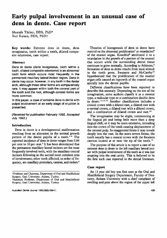
Fig. 1.-Incisal tip of permanent maxillary left lateral incisor appears only as a point. Note a reddish-coloured swelling of the upper left vestibular fold area.
Fig. 2.-Permanent maxillary left lateral incisor with conical crown and dilated root diagnosed as extreme dens in dente.
central and lateral incisors. There were no signifi- cant medical history or extraoral findings.
The intraoral examination showed that the permanent maxillary central incisors had erupted. The deciduous maxillary lateral incisor on the right was present. The deciduous maxillary lateral on the left had been extracted. The incisal tip of the
permanent maxillary lateral incisor on the left appeared only as a point. The intraoral soft tissues were normal except for the presence of a reddish coloured swelling on the upper left vestibular fold area suggesting paradontal pathosis (Fig. 1). A vitality test to an electric pulp tester was negative. Panoramic radiographs revealed that the right
440 Australian Dental Journal 1993;38:6.

permanent maxillary lateral incisor was impacted. On the left, there was an unusually shaped tooth resembling an inverted pear. The invagination was dilated in the root forming an ampulla-like cavity, which was very large (Fig. 2).
Morphologically, the tooth was classified as an extreme dens in dente with a conical crown and dilated root.
Because of difficulty in negotiating the root canal successfully, endodontic therapy was not considered as a treatment of choice. The involved tooth was extracted after initial management with antibiotics and the lesion was curetted. The tooth and curetted specimen were sent to the Pathology Department for further evaluation. The pathology report confirmed a diagnosis of an extreme dens in dente of the tooth in chronic granulation tissue.
Discussion The crown and root morphology in this case of
extreme dens in dente was in accordance with a previous report which stated that the most common dens in dente type was that of a conical crown with a dilated root, found in 48.5 per cent of all cases." Dens in dente may be limited to the coronal part of the tooth or may invade deeply into root, although crown invaginations are more common~l.2,11.12.15.16 In the case presented in this paper the invagination invaded the entire root.
A characteristic clinical feature of dens in dente is the almost invariable infection and subsequent necrosis of the pulp which occurs shortly after the tooth has e r u ~ t e d . ' ~ Kronfeld,8 Kitchin," and Hitchin and McHugh18 reported that teeth with this anomaly may undergo rapid pulp injury, once they erupt into the oral cavity because of bacterial penetration through channels that extend form the base of the invagination into the pulp. Both Peri- apical and periodontal abscesses have been reported in teeth with dens in dente.'3.19-22 The invaginated teeth tend to become pulpally involved, even though they are intact, because of communication between the oral environment and the pulp cavity via accessory openings.z3
In the current case, the extreme dens in dente in the left maxillary lateral incisor was found to undergo rapid pulp necrosis and periapical infec- tion in spite of being seen in the oral cavity as only a point. Such a rapid progression of the pulpal and apical pathology is believed to be the first case in the dental literature. In the reported case, conven- tional root canal therapy and/or a surgical endo-
dontic technique were not procedures of choice, because of a severe degree of dilatation of the root and therefore the difficulty of removal of the in- vaginated structures.
References 1. Oehlers FAC. Dens invaginatus: I. Variations of the invagi-
nation process and assodated anterior crown forms. Oral Surg Oral Med Oral Pathol 1957;101204-18.
2. Oehlers FAC. Dens invaginatus: 11. Associated posterior crown forms and pathogenesis. Ibid. 1302-16.
3. Oehlen FAC. The radicular variety of dens invaginatus. Oral Surg Oral Med Oral Pathol 1958;11:1251-60.
4. Hallett GEM. Incidence, nature and clinical significance of palatal invagination in maxillary incisor teeth. Proc Roy Soc Med 1953;46:491-9.
5. Rotstein I, Stabholz A, Freidmen S. Endodontic therapy for dens invaginatus in a maxillary second molar, Oral Surg Oral Med Oral Pathol 1987;63:237-40.
6. Pindborg JJ. Pathology of the dental hard tissues. Philadel- phia: WB Saunders, 1970:47-61.
7. Rushton MA. A collection of dilated composite odontomes. Br Dent J 1937:63:65-86.
8. Kronfeld R. Dens in dente. J Dent Res 1934;14:49-66. 9. Atkinson SR. The permanent maxillary lateral incisor. Am
10. Swanson WF, McCarthy FM. Bilateral dens in dente. J Dent
11. Ulmansky M, Hermel J. Double dens in dente. J Am Dent
12. Bhatt AP, Dholaika HM. Radicular variety of double dens invaginatus. Oral Surg Oral Med Oral Path01 1975;39:284-7.
13. Binstein E, Shteyer A. Dilated type of dens invaginatus in the permanent dentition: report of a case and review of the literature. J Dent Child 1976;43:410-3.
14. Beynon AD. Developing dens invaginatus (Dens in dente). Br Dent J 1982;153:255-60.
15. Munro D. Dens in dente. Br Dent J 1952;92:92-3. 16. Amos ER. Incidence ofthe small dens in dente. J Am Dent
17. Kitchin PC. Dens in dente. J Dent Res 1935;15:117-21. 18. Hitchin AD, McHugh WD. Three coronal invaginations
in a dilated composite odontome. Br Dent J 1954;97:90-2. 19. Conklin WW. Double bilateral dens invaginatus in the maxil-
lary incisor region. Oral Surg Oral Med Oral Pathol
20. Ulmansky M, Hermel J. Double dens in dente in a single tooth. Report of a case and radiologic study of the incidence of small dens in dente. Oral Surg Oral Med Oral Pathol
21. Burzynski NJ. Gemination and dens in dente. Oral Surg Oral Med Oral Pathol 1973;36:760-1.
22. Chawla HS, Tewari A. A study of four rare cases of dens invaginatus. J Oral Med 1977;32:4-8.
23. Kaufman AY, Kaffe I, Littner MM. Vitality preservation of an anomalous maxillary central incisor after endodontic therapy. Oral Surg Oral Med Oral Pathol 1984;57:668-72.
J Onhod 1943;29:685-98.
Res 1947;26:167-71.
ASSX 1955;51:31-3.
ASSW 1955;51:31-3.
1975;39:949-52.
1964;17:92-7.
Address for correspon&nce/reprints: Dr Inci Karaca,
GU Dis Hek Fakiiltesi, Aiiz, Dis, Gene Hastaliklari ve Cerrahisi Anabilim Dali,
8 Cadde, Emek, Ankara, Turkey.
Australian Dental Journal 1993;38:6. 441