effect of self-management education versus quadriceps
TRANSCRIPT

HUMAN MOVEMENT
64
EffEcT Of sElf-MANAgEMENT EdUcATiON VErsUs qUAdricEps sTrENgTHENiNg ExErcisEs ON pAiN ANd fUNcTiON iN pATiENTs wiTH kNEE OsTEOArTHriTis
OlUfUNMilAyO r. EgwU1, OlUsOlA O. AyANNiyi2, BABATUNdE O.A. AdEgOkE2, OlAdApO M. OlAgBEgi3, OMOyEMi O. OgwUMikE2, AdEsOlA c. OdOlE2
1 Department of Physiotherapy, Barau Dikko Teaching Hospital, Kaduna, Nigeria2 Department of Physiotherapy, College of Medicine, University of Ibadan, Ibadan, Nigeria3 Department of Physiotherapy, School of Health Sciences, University of KwaZulu-Natal, Durban, South Africa
ABSTrACTPurpose. To investigate and compare the effects of 6-week self-management education (SME) and quadriceps strengthening exercises (QSE) on pain and disability in individuals with knee osteoarthritis (OA).Methods. A total of 79 (13 males, 66 females) consecutive patients with knee OA were randomised into SME and QSE groups. The SME group were taught modules of self-management once a week for 6 weeks, while the QSE group had supervised QSE thrice a week for 6 weeks. Pain intensity and physical function were assessed with the Visual Analogue Scale and Ibadan Knee/Hip Osteoarthritis Outcome Measure (IKHOAM) at baseline, week 6, and at the follow-up time points of the 1st, 2nd
and 3rd months.Results. The effects of both interventions were comparable (p > 0.005) during intervention and follow-up. During intervention, in both groups, pain intensity significantly decreased (p < 0.001, effect size = 0.603) and IKHOAM scores improved (p < 0.001, effect size = 0.540). There were significant time by group interaction effects during follow-up as pain intensity (p < 0.001, effect size = 0.085) did not change in the QSE group but further decreased in the SME group, IKHOAM scores (p = 0.005, effect size = 0.053) remained the same in the SME group while it decreased in the QSE group with respect to the end of intervention (6th week).Conclusions. Supervised QSE and SME are both effective in reducing pain and disability in knee OA but improvements in the outcomes are better sustained with SME.Key words: knee osteoarthritis, self-management education, quadriceps strengthening exercises
original paperDOI: https://doi.org/10.5114/hm.2018.76081
2018; 19(3): 64– 74
Correspondence address: Dr Oladapo Michael Olagbegi, Department of Physiotherapy, School of Health Sciences, University of KwaZulu-Natal, Westville Campus, Durban, 4001, South Africa, e-mail: [email protected]
received: August 18, 2017Accepted for publication: January 29, 2018
Citation: Egwu Or, Ayanniyi OO, Adegoke BOA, Olagbegi OM, Ogwumike OO, Odole AC. Effect of self-management educa-tion versus quadriceps strengthening exercises on pain and function in patients with knee osteoarthritis. Hum Mov. 2018; 19(3):64–74; doi: https://doi.org/10.5114/hm.2018.76081.
© University School of Physical Education in Wroclaw
Introduction
Osteoarthritis (OA) is a major musculoskeletal challenge characterised by slow degradation of carti-lage, pain, and increasing chronic disability [1]. It is a frequent cause of morbidity, functional limitations, and loss of autonomy in the second half of human life [1, 2]. Internationally, OA affects about 24% of the general population, with symptomatic OA observed in 9.6% of men and 18% of women aged 60 years and above; the disease also occurs in about 80% of people aged over 65 years in high-income countries [3].
The majority of patients with OA are managed in primary care, and the prevalence of knee OA is such that simple interventions which are effective in the community setting are necessary [4]. There is no known modality for reversing the progression of knee OA; it can only be managed by ameliorating its symptoms [4]. Treatment of OA is aimed at reducing pain and dis-ability to improve function and quality of life [5–7]. International treatment guidelines have recommend-ed a combination of pharmacological and non-phar-macological therapies [8, 9]; surgical intervention is only indicated when the aforementioned approaches
Language editing of the scientific articles accepted for publication and ensuring the participation of foreign reviewers in the evaluation of publications in the Human Movement journal – tasks financed under the agreement No. 656/P-DUN/2018 by the Minister of Science and Higher Education allocated to the activities of disseminating science

O.r. Egwu et al., Self-management in osteoarthritis
HUMAN MOVEMENT
65Human Movement, Vol. 19, No 3, 2018
humanmovement.pl
fail [10]. However, drug treatments such as nonsteroidal anti-inflammatory drugs (NSAID), aimed at relieving pain, are often limited by gastrointestinal side effects [11]; cyclooxygenase-2 (COX-2) inhibitors, used to sal-vage these side effects, are also associated with adverse cardiovascular incidents such as myocardial infarc-tion [12]. Moreover, little or no evidence supports dis-ease-modifying OA drugs such as glucosamine and chondroitin, which intensifies interest in non-phar-macologic treatments for OA [13, 14].
Non-pharmacological approaches to knee OA man-agement include education, self-management, exercise, appliances (walking sticks, insoles, and knee bracing), acupuncture, telephone contact, physical therapy, and weight reduction [8–10]. McAlindon et al. [9], in a sys-tematic review, identified modalities such as self-man-agement education (SME), strengthening exercises, aerobic/fitness exercises (land- or water-based), and weight management as core aspects of managing OA. It was also suggested that these modalities could easily be adapted for self-care, considering the chronic na-ture of knee OA [9].
Chronic knee OA reportedly leads to marked weak-ening of the quadriceps femoris muscle, which func-tionally act as extensor and stabilizer of the knee [15]. Quadriceps muscle weakness results in its declined ability to reduce load across the knee joint, which in-creases joint stress; it may thus play a role in the aetiol-ogy and progression of knee OA [16, 17]. Therefore, ageing and atrophy of this muscle largely contribute to knee pain and functional impairment in knee OA [16]. A recent systematic review by Fransen et al. [18] concluded that land-based therapeutic exercises gener-ally provided benefits in terms of reduced knee pain and improved physical function and quality of life among people with knee OA. Another systematic re-view, by Lange et al. [5], specifically provides strong evidence in support of quadriceps strengthening as a modality for reducing pain and disability in pa-tients with knee OA. This indicates that well-planned quadriceps strengthening exercises (QSE) for pa-tients with knee OA can achieve pain relief and func-tional recovery of the muscle [19].
SME has become a popular component of manage-ment in several chronic conditions including arthritis and has been listed alongside therapeutic exercises as the mainstay of non-pharmacologic treatment of knee OA [5, 7, 20]. It is aimed at achieving more than just the provision of information to increase knowledge, which makes it different from the traditional patient education programmes. The purpose of self-manage-ment programmes is to change health behaviour and
health status by teaching patients to identify and solve problems, set goals, and plan actions [21]. A SME pro-gramme specific for knee OA was found to be effective in reducing pain and disability, as well as improving the health-related quality of life in patients with knee OA [22, 23].
A meta-analysis [24] concluded that exercise needs to be performed 3 times a week for optimal benefits. In spite of these benefits, exercise seems more expensive and demanding in terms of hospital attendance (to the patient) and health care resources (to the government) than SME. Studies comparing the effects of SME and supervised QSE are rather scarce. However, compar-ison of the 2 modalities may provide useful evidence on their relative efficacy in order to give an informed appraisal of the choice of therapy for patients with knee OA, especially in developing nations with limited resources. This study was therefore designed to com-pare and evaluate the effects of SME and QSE pro-grammes on pain and physical function in individuals with knee OA as well as their short-term carry-over results. It was hypothesised that there would be no difference in the effects of 6-week SME and QSE, or 3-month carry-over results of the 2 interventions.
Material and methods
Participants
The study was a single-blind randomized controlled trial involving patients with knee OA. All participants were volunteer male or female individuals with varying levels of knee OA severity [9, 10], attending the Outpa-tient Physiotherapy Clinic at Barau Dikko Teaching Hospital (BDTH), Kaduna, Nigeria between March 2013 and February 2015. They were diagnosed on the basis of clinical symptoms or radiographic assessment of their knee joints by orthopaedic surgeons. The sub-jects satisfied the American College of rheumatology criteria for clinical classification of knee OA, i.e. pain in the knee for most days of the prior month, crepitation on active joint motion, morning stiffness less than 30 minutes in duration, patient’s age 38 years or above, and bony enlargement of the knee on examination [25]. All the participants received standard medical treat-ment for knee OA.
Volunteers with knee OA who also had severe/un-controlled comorbidities (such as hypertension, diabe-tes, kidney failure, psychiatric disorders) or a neurologi-cal condition affecting the lower limb (such as post-stroke muscle weakness or poliomyelitis), as well as those un-able to walk were excluded from the study [26, 27].

O.r. Egwu et al., Self-management in osteoarthritis
HUMAN MOVEMENT
66Human Movement, Vol. 19, No 3, 2018
humanmovement.pl
The total of 79 participants (66 females, 13 males) were randomized to 2 groups by having them pick a wrapped paper with A or B letter with the tag num-ber between 1 and 100 (computer-generated random numbers) written on it from an opaque envelope. Participants who picked A were assigned to the SME group (n = 42) and those who picked B were assigned to the QSE group (n = 37).
Overall, 51 subjects (64.6%) had bilateral knee OA, while 15 (19.0%) had left knee OA and 13 (16.4%) had right knee OA. Sixty-nine participants (87.34%) (37 SME, 32 QSE) concluded the protocol. Among the 10 patients who did not finish the protocol, 5 (4 SME, 1 QSE) did not give relevant reasons when contacted on the telephone; 2 drop-outs (QSE) were lost because of logistic problems such as inflexibility of time and the venue of research being far from their residence; 2 (QSE) moved out of town; 1 (QSE) was hospitalised
for an unknown ailment. The flowchart of the partici-pants’ recruitment and progression in the protocol is presented in Figure 1.
Pain intensity assessment
The participants were asked to identify the activ-ity of daily living (ADL) that caused them most pain. Then, the Visual Analogue Scale (VAS) was used to assess the pain felt while performing the activity. The participant was then instructed to mark the point on the VAS that corresponded to the intensity of pain felt while performing the activity [28]. The point marked was measured with a mathematical ruler and re-corded in centimetres as the participants’ pain inten-sity. The validated Hausa version of VAS [29] was ad-ministered to the indigenous participants who could not understand the English language.
SME – self-management education, QSE – quadriceps strengthening exercise
Figure 1. Flow chart of the study

O.r. Egwu et al., Self-management in osteoarthritis
HUMAN MOVEMENT
67Human Movement, Vol. 19, No 3, 2018
humanmovement.pl
Physical function assessment
The first section of the Ibadan Knee/Hip Osteoar-thritis Outcome Measure (IKHOAM) was administered by a group-blinded assessor who also implemented the clinician’s section, after which the participants per-formed the required physical tasks. The IKHOAM is a Nigerian culture- and environment-friendly clinical tool developed at the University of Ibadan, Nigeria for outcome assessment in the care of patients with hip or knee OA [30]. The instrument includes 3 parts, 33 items. Parts 1 and 2 are patient-reported, while part 3 is rated by the clinician. IKHOAM has been shown to demonstrate good validity and responsiveness [30, 31]. The validated Hausa version of IKHOAM [32] was administered among illiterate participants who only understood the local language. Scores were computed as follows:
– The degree of difficulty and the nature of assis-tance required to carry out each of the 25 items in part 1 were scored between 0 and 4 each. This gave a maximum obtainable score of 200 (4 × 25 for de-gree of difficulty and 4 × 25 for nature of assistance).
– The extent of restriction in performing the 3 items in part 2 were scored between 0 and 3 each, giving the maximum obtainable score of 9 (3 × 3 for extent of restriction).
– Part 3 contains 5 items with the maximum ob-tainable score of 23 (5 for 250 walk test, 4 for squat test, 5 for one-leg stance test, 4 for stairs climbing test, and 5 for balance test).
Therefore, the maximum obtainable score on IKHOAM is 232 (200 + 9 + 23) [30].
The score for a participant was calculated in per-centage as:
participant’s score
maximum obtainable score × 100 [30].
Low and high scores on IKHOAM imply low and high levels of physical functioning, respectively [30].
Pain intensity and physical function were assessed at baseline, in week 6, and after the 1st, 2nd, and 3rd month of follow-up.
Intervention
Self-management education group
The weekly education was conducted over a 6-week period, thereby enabling participants to incorporate and consolidate information learned from week to week.
Each weekly session comprised 2 topics and practical demonstration sessions of strengthening exercise, pre-vention of falls, joint protection, or thermal therapy, in line with the topic of the week. The class session lasted for 90–120 minutes. In addition to the weekly sessions, the participants were also given an infor-mation booklet on knee OA (published by Arthritis research UK); the Hausa translated version was pro-vided to those who did not understand English. The group size was 4–8 subjects, depending on recruit-ment and randomization. Each group attended the class once a week for 6 weeks. Each participant was iden-tified by a tag number in order to monitor attendance and to ensure consistency of assessments. Self-man-agement constructs were employed to promote behav-ioural changes that were aimed at optimizing the pa-tients’ health status. Goal setting and the development of strategies to achieve these goals in the long term were emphasized in the class. A lesson note was de-veloped from the outline used by Coleman et al. [23]. A physiotherapist whose first language was Hausa was always present in class to explain the lesson for the benefit of participants who did not understand the English language.
Modules of SME adopted from Coleman et al. [23] and delivered to the participants in the SME group were focused on pathophysiology of OA, pain manage-ment strategies (cognitive, pharmaceutical, and physi-cal), joint protection, fitness/exercise, correct use of analgesia/medications, balance, falls prevention, pro-prioception, team approach to health care, nutritional/weight control, self-management skills, and specific measurable achievable realistic and time-framed goals.
Quadriceps strengthening exercise group
The participants in this group engaged in individ-ual QSE 3 times weekly. They were instructed not to modify their ADLs and not to take part in additional forms of physical activity while the study lasted. The subjects performed the following exercises:
– Open chain quadriceps setting. The participant lay in supine position while contracting the quadriceps muscle of the affected lower extremity by drawing up the patella and maintaining the knee in extension. The contraction was held for a count of 10, then relaxed. This was repeated 10 times [33]. The exercise was car-ried out by participants throughout the duration of the study [28].
– Closed chain quadriceps setting. The participant sat on a chair with their back supported, knee extended, and heel on the floor. They then pressed the heel against

O.r. Egwu et al., Self-management in osteoarthritis
HUMAN MOVEMENT
68Human Movement, Vol. 19, No 3, 2018
humanmovement.pl
the floor and the thigh against the seat of the chair. The position was held for a count of 10, after which the subject relaxed. This was done 10 times [33]. This exercise was carried out by the subjects throughout the duration of the study [28].
– Straight leg raising (SLr). The participant lay in supine position, contracting the quadriceps and lift-ing the lower extremity up to achieve about 45° hip flexion while maintaining the knee in extension. This position was held for a count of 10, then the limb was lowered. This is repeated 10 times. The contralateral knee and hip were flexed to about 45° to avoid undue stress on the lower back [33]. The subjects carried out this exercise during the first 2 weeks of the study and commenced SLr with weight from the 3rd week [28].
– Mini-squats. While standing, the participant bent both knees by about 30–60°, while maintaining the trunk upright. This position was maintained for a count of 10, relaxed, and repeated 10 times [33]. The sub-jects carried out this exercise during the first 2 weeks of the study and commenced mini-squats with weight from the 3rd week [28]. They had barbells placed across their shoulders for mini-squats with weight [33].
Exercise progression. The participants started with a weight equivalent to their 10 repetition maximum (10-rM) and progressed with a newly determined 10-rM at the beginning of each week [32].
Determination of 10-RM. A plastic weight (W) cor-responding to a certain rM of the participant was ran-domly selected and attached via the DeLorme’s boot to the participant’s foot. After a warm-up with a set of 10 lifts with a lighter weight, using the method described by Olagbegi et al. [27], the subjects lifted the weight r times until volitional exhaustion. The participants’ 1-rM was determined with Brzycki predictive equation:
1-rM = W / (1.0278–0.0278 × r) [27]
After one hour, 10-rM was determined by making the participants lift 75% of the determined 1-rM to vo-litional exhaustion [34]. When the subjects lifted the weight more than 10 times, the resistance was increased. If they stopped before 10 repetitions, the resistance was reduced to manage 10 lifts.
Data analysis
The data were analysed with the Statistica software package, version 13 (StatSoft, Inc, Tulsa, USA). The minimum sample size of 64 was calculated with the sample size formula by Macfarlane [35], with the effect size of 0.5 and 80% power. The effect size of 0.5 was
determined in an initial pilot study with VAS as the primary outcome measure. The outcomes were analysed in accordance with the intention-to-treat principle. Information for all 79 participants was included in the analysis by carrying the last available score forward.
Descriptive statistics of mean, standard deviation, and percentages were used to summarise the data. The groups’ demographic variables were compared with the independent t-test. With the use of treatment groups (SME and QSE) and time (baseline; week 6; 1st, 2nd, and 3rd month of follow-up) as between-group and with-in-group factors, respectively, a two-way repeated meas-ures ANOVA was separately computed for the analysis of time, time by group interaction, and between-group effects in the intervention and follow-up phases and the entire study period. Post-hoc analysis was performed with Tukey’s multiple comparison test. Cohen’s rule of thumb (0.01 – small, 0.06 – moderate, 0.14 – large) was applied to categorise effect sizes (partial eta-squared), while the level of significance was set at p < 0.05.
Ethical approvalThe research related to human use has been com-
plied with all the relevant national regulations and institutional policies, has followed the tenets of the Dec-laration of Helsinki, and has been approved by the Health research Ethics Committee of the University of Ibadan and University College Hospital (reg. No: NHrEC/05/01/2008a). The permission of the manage-ment of BDTH in Kaduna, Nigeria, was also obtained.
Informed consentInformed consent has been obtained from all indi-
viduals included in this study.
Results
Table 1 shows the demographic characteristics of the groups; there was no significant difference between the groups regarding their demographic characteristics.
The analyses of time, between-group, and time by group interaction effects for the intervention and fol-low-up phases and the entire duration of the study are presented in Table 2. The 2 interventions (SME and QSE) were not significantly different in their effects on pain intensity or IKHOAM scores in any of the 3 phases. During the intervention phase, there were no significant time by group interaction effects on any of the 2 outcomes; however, there were significant time effects (pain intensity: p < 0.001, effect size = 0.603; IKHOAM score: p < 0.001, effect size = 0.540) as pain intensity was reduced while IKHOAM score improved in the similar manner for both groups.

O.r. Egwu et al., Self-management in osteoarthritis
HUMAN MOVEMENT
69Human Movement, Vol. 19, No 3, 2018
humanmovement.pl
During follow-up, there were significant effects of time (pain intensity: p = 0.010, effect size = 0.048; IKHOAM score: p < 0.001, effect size = 0.169) and time by group interaction (pain intensity: p < 0.001, effect size = 0.085; IKHOAM score: p = 0.005, effect size = 0.053) for both outcomes, though with small effect sizes. Pain intensity did not change in the QSE group but further decreased in the SME group as com-pared with the end point of the intervention phase (6th week). IKHOAM scores remained the same in the SME group and decreased in the QSE group as compared with the end point of the intervention phase (6th week).
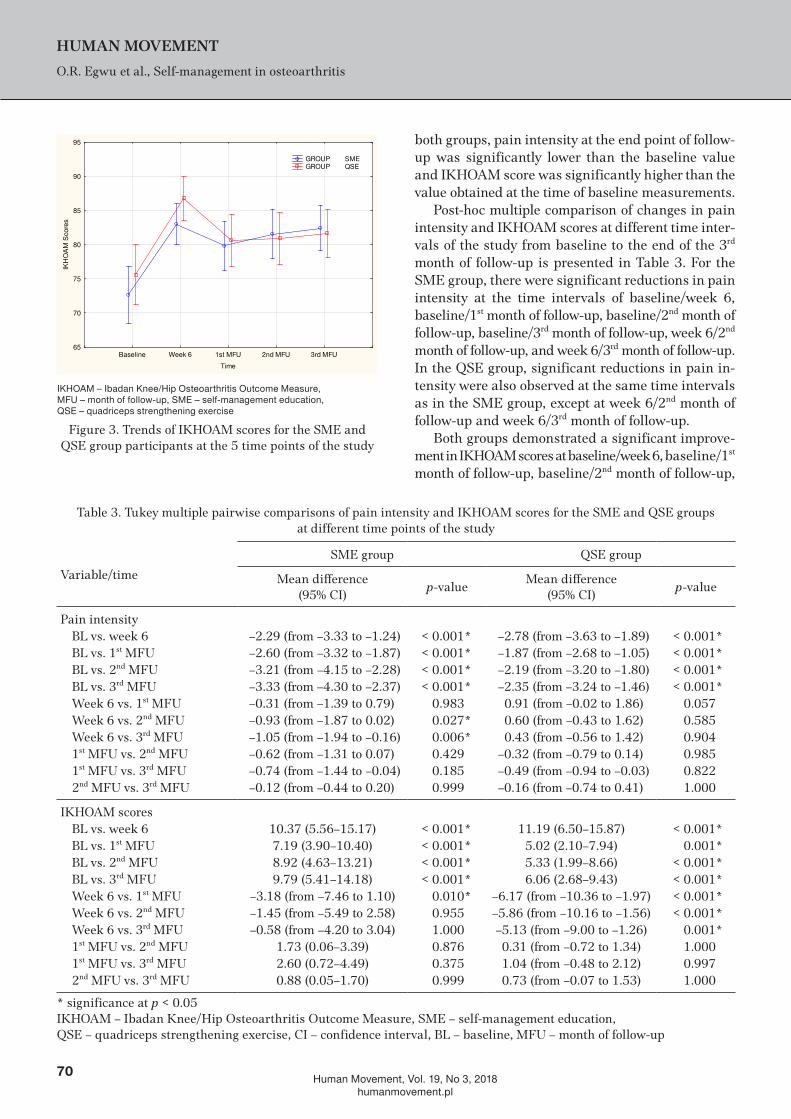
The analysis of the entire study duration indicated significant effects of time (pain intensity: p < 0.001, effect size = 0.468; IKHOAM score: p < 0.001, effect size = 0.379) and time by group interaction (pain in-tensity: p < 0.001, effect size = 0.067; IKHOAM score: p = 0.014, effect size = 0.040) for both measures. Fig-ures 2 and 3 illustrate time by group interaction effects and the trends of pain intensity and IKHOAM scores, respectively, across the 5 time points of the study. In
Table 1. Comparison of the participants’ demographics and baseline clinical variables
Variable
Treatment groups
t p-valueSME (n = 42) (mean ± SD)
QSE (n = 37)(mean ± SD)
Age (years) 53.86 ± 10.40 50.11 ± 10.20 1.615 0.110Height (m) 1.62 ± 0.07 1.62 ± 0.07 –0.310 0.611Weight (kg) 80.0 ± 16.98 81.0 ± 15.79 –0.286 0.776BMI (kg/m2) 31.02 ± 6.95 31.00 ± 5.91 0.012 0.990
SME – self-management education, QSE – quadriceps strengthening exercise, BMI – body mass index
Table 2. Analysis of time, between-group, and time × group interaction effects on pain intensity
and IKHOAM scores
Variable/effectMean square
F p-valueEffect size
Intervention phase
Pain intensityTime 252.769 116.952 < 0.001* 0.603Between-group 13.402 2.963 0.089 0.037Time × group 2.440 1.129 0.291 0.014
IKHOAM scoresTime 4569.200 90.321 < 0.001* 0.540Between-group 445.300 1.960 0.166 0.025Time × group 6.500 0.129 0.720 0.002
Follow-up phase
Pain intensityTime 5.459 3.841 0.010* 0.048Between-group 3.991 0.345 0.559 0.004Time × group 10.117 7.118 < 0.001* 0.085
IKHOAM scoresTime 317.000 15.627 < 0.001* 0.169Between-group 49.000 0.114 0.737 0.001Time × group 88.000 4.328 0.005* 0.053
Entire study period
Pain intensityTime 108.618 67.748 < 0.001* 0.468Between-group 1.262 0.112 0.739 0.001Time × group 8.821 5.502 < 0.001* 0.067
IKHOAM scoresTime 1240 47.010 < 0.001* 0.379Between-group 147 0.261 0.611 0.003Time × group 84 3.197 0.014* 0.040
* significance at p < 0.05IKHOAM – Ibadan Knee/Hip Osteoarthritis Outcome Measure
MFU – month of follow-up, SME – self-management education, QSE – quadriceps strengthening exercise
Figure 2. Trends of pain intensity for the SME and QSE group participants at the 5 time points of the study
Baseline Week 6 1st MFU 2nd MFU 3rd MFU
Time
2,0
2,5
3,0
3,5
4,0
4,5
5,0
5,5
6,0
6,5
7,0
7,5
Pai
n In
tens
ity
GROUP SME GROUP QSE
O.r. Egwu et al., Self-management in osteoarthritis
HUMAN MOVEMENT
70Human Movement, Vol. 19, No 3, 2018
humanmovement.pl
both groups, pain intensity at the end point of follow-up was significantly lower than the baseline value and IKHOAM score was significantly higher than the value obtained at the time of baseline measurements.
Post-hoc multiple comparison of changes in pain intensity and IKHOAM scores at different time inter-vals of the study from baseline to the end of the 3rd month of follow-up is presented in Table 3. For the SME group, there were significant reductions in pain intensity at the time intervals of baseline/week 6, baseline/1st month of follow-up, baseline/2nd month of follow-up, baseline/3rd month of follow-up, week 6/2nd month of follow-up, and week 6/3rd month of follow-up. In the QSE group, significant reductions in pain in-tensity were also observed at the same time intervals as in the SME group, except at week 6/2nd month of follow-up and week 6/3rd month of follow-up.
Both groups demonstrated a significant improve-ment in IKHOAM scores at baseline/week 6, baseline/1st month of follow-up, baseline/2nd month of follow-up,
IKHOAM – Ibadan Knee/Hip Osteoarthritis Outcome Measure, MFU – month of follow-up, SME – self-management education, QSE – quadriceps strengthening exercise
Figure 3. Trends of IKHOAM scores for the SME and QSE group participants at the 5 time points of the study
Baseline Week 6 1st MFU 2nd MFU 3rd MFU
Time
65
70
75
80
85
90
95
IKH
OA
M S
core
s
GROUP SME GROUP QSE
Table 3. Tukey multiple pairwise comparisons of pain intensity and IKHOAM scores for the SME and QSE groups at different time points of the study
Variable/time
SME group QSE group
Mean difference(95% CI)
p-valueMean difference
(95% CI)p-value
Pain intensityBL vs. week 6 –2.29 (from –3.33 to –1.24) < 0.001* –2.78 (from –3.63 to –1.89) < 0.001*BL vs. 1st MFU –2.60 (from –3.32 to –1.87) < 0.001* –1.87 (from –2.68 to –1.05) < 0.001*BL vs. 2nd MFU –3.21 (from –4.15 to –2.28) < 0.001* –2.19 (from –3.20 to –1.80) < 0.001*BL vs. 3rd MFU –3.33 (from –4.30 to –2.37) < 0.001* –2.35 (from –3.24 to –1.46) < 0.001*Week 6 vs. 1st MFU –0.31 (from –1.39 to 0.79) 0.983 0.91 (from –0.02 to 1.86) 0.057Week 6 vs. 2nd MFU –0.93 (from –1.87 to 0.02) 0.027* 0.60 (from –0.43 to 1.62) 0.585Week 6 vs. 3rd MFU –1.05 (from –1.94 to –0.16) 0.006* 0.43 (from –0.56 to 1.42) 0.9041st MFU vs. 2nd MFU –0.62 (from –1.31 to 0.07) 0.429 –0.32 (from –0.79 to 0.14) 0.9851st MFU vs. 3rd MFU –0.74 (from –1.44 to –0.04) 0.185 –0.49 (from –0.94 to –0.03) 0.8222nd MFU vs. 3rd MFU –0.12 (from –0.44 to 0.20) 0.999 –0.16 (from –0.74 to 0.41) 1.000
IKHOAM scoresBL vs. week 6 10.37 (5.56–15.17) < 0.001* 11.19 (6.50–15.87) < 0.001*BL vs. 1st MFU 7.19 (3.90–10.40) < 0.001* 5.02 (2.10–7.94) 0.001*BL vs. 2nd MFU 8.92 (4.63–13.21) < 0.001* 5.33 (1.99–8.66) < 0.001*BL vs. 3rd MFU 9.79 (5.41–14.18) < 0.001* 6.06 (2.68–9.43) < 0.001*Week 6 vs. 1st MFU –3.18 (from –7.46 to 1.10) 0.010* –6.17 (from –10.36 to –1.97) < 0.001*Week 6 vs. 2nd MFU –1.45 (from –5.49 to 2.58) 0.955 –5.86 (from –10.16 to –1.56) < 0.001*Week 6 vs. 3rd MFU –0.58 (from –4.20 to 3.04) 1.000 –5.13 (from –9.00 to –1.26) 0.001*1st MFU vs. 2nd MFU 1.73 (0.06–3.39) 0.876 0.31 (from –0.72 to 1.34) 1.0001st MFU vs. 3rd MFU 2.60 (0.72–4.49) 0.375 1.04 (from –0.48 to 2.12) 0.9972nd MFU vs. 3rd MFU 0.88 (0.05–1.70) 0.999 0.73 (from –0.07 to 1.53) 1.000
* significance at p < 0.05IKHOAM – Ibadan Knee/Hip Osteoarthritis Outcome Measure, SME – self-management education, QSE – quadriceps strengthening exercise, CI – confidence interval, BL – baseline, MFU – month of follow-up

O.r. Egwu et al., Self-management in osteoarthritis
HUMAN MOVEMENT
71Human Movement, Vol. 19, No 3, 2018
humanmovement.pl
baseline/3rd month of follow-up, week 6/2nd month of follow-up, and week 6/3rd month of follow-up time intervals. The QSE group significantly deteriorated in IKHOAM scores at week 6/1st month of follow-up, week 6/2nd month of follow-up, and week 6/3rd month of follow-up time intervals; significant deterioration in IKHOAM score for SME group was only observed at the week 6/1st month of follow-up time interval.
Discussion
The majority (83.54%) of the participants in the study were female. This suggests that knee OA is more prevalent among women than men, which is consist-ent with previous reports in literature [23, 36]. Most of the subjects were also obese, with body mass index (BMI) > 30; this appears understandable because high BMI has been associated with incidence and progres-sion of knee OA [37].
The major aim of the study was to compare the ef-fect of a 6-week SME and QSE intervention programmes on pain and physical function outcomes and their car-ry-over results in a 3-month follow-up. The 2 groups were not significantly different with regard to their pain and function outcomes at the end of the 6-week intervention. This suggests that the SME group partici-pants achieved pain reduction and consequent func-tional improvement comparable with those of QSE par-ticipants, who implemented a supervised strengthening exercise programme. This may also imply that SME subjects carried out the demonstrated exercises and complied with the given instructions regarding ade-quate dosing and progression. Exercise has been linked to reduction in pain and improvement in function; however, it is not enough to advise a patient to exer-cise – it is important that the patient understands the exercise to be carried out and performs it correctly for optimal benefits [4, 38].
Although the SME and QSE interventions had sta-tistically comparable effects on pain intensity and physi-cal function during the follow-up phase of the study, the relationship between time and intervention de-pends on the values of pain intensity and IKHOAM scores, as the effects of time and group interaction were significant for both outcomes. Findings from Figures 2 and 3 suggest that pain outcome improvements achieved through SME during intervention were better sustained during the follow-up period. The participants in the SME group are likely to continue what they were taught from the educational module since they observed im-provement without having to visit hospital frequently. Similar results were reported in previous studies [8, 9].
The observed increase in mean pain intensity among the QSE participants between the end of week 6 and the end of the 1st month of follow-up (mean difference: 0.91) further suggests that the benefits of QSE are not sustainable. It is probable that a large proportion of QSE subjects did not exercise at home after the in-tervention; SME patients continued to demonstrate im-provement in pain outcome, though with a small effect size. roddy et al. [4], in a systematic review of home-based QSE and aerobic walking exercises, noted that adherence to exercise was a key predictor of response, and encouraging patients with knee OA to maintain exercise programmes beyond a supervised period of instruction was a major challenge. That opinion seems supported by the results of our study.
The 2 treatment programmes showed comparable significant effects on IKHOAM scores during the in-tervention phase of the study. This finding may be attrib-uted to some similarities in the nature of both inter-ventions. In the first instance, both interventions had a significant time effect on pain as observed in this study. Both included components that have been shown to directly impact reduction of pain, the leading com-plaint in individuals with OA and the one that mostly drives health care use [39]. Pain reduction has been proved directly related with reduced disability and, hence, improvement in physical function [17, 23]. Connelly et al. [39], in a recent cross-sectional survey of 197 individuals with OA, observed a strong negative correlation between physical functioning and pain.
The QSE participants, however, demonstrated in-consistent changes in physical function (IKHOAM scores) during the follow-up phase of the study. They had a markedly significant reduction (mean differ-ence: –6.17) in their functional score at the end of the 1st month post-intervention, which appears in line with the trend of the aforementioned changes in pain outcome. Findings from clinical trials have shown that supervised QSE improves strength [27, 40], which in turn enhances knee joint stability and function [16, 17]. The result of the present study suggests that QSE par-ticipants were losing this clinical benefit one month post-intervention. It is, however, plausible that a num-ber of the SME participants augmented the stability of their knees with braces and supports, since their usage was taught as part of the modules. This could be the-orized to have enhanced knee joint stability, contrib-uting to sustained function among the SME group.
Our finding with regard to physical function is con-sistent with the work of McKnight et al. [36], who did not observe significant differences in the effects of strength training, SME, and a combination on self-reported and

O.r. Egwu et al., Self-management in osteoarthritis
HUMAN MOVEMENT
72Human Movement, Vol. 19, No 3, 2018
humanmovement.pl
performance-based physical function among individ-uals with early OA. Both studies are different in terms of the duration of supervised exercise (6 weeks vs. 9 months), outcome measures (IKHOAM vs. WOMAC), and exercise components (isometric and dynamic quadriceps strengthening vs. combination of isotonic, range of motion, stretching, and balance). Having sim-ilar results from both studies suggests that SME may confer short-term benefits regarding improvement of function for patients with knee OA.
Limitations of the study
This study is not without limitations. Analysing the effects of medical treatment that the participants received during intervention could have been helpful in interpreting the obtained results. Assessors were not blinded to the participant’s intervention alloca-tion, which could threaten the internal validity of the study. However, assessment-related bias was mini-mised by ensuring data recording by a neutral research assistant. Lack of a control group with knee OA un-dergoing no intervention is another limitation of the study. Including a control group would have shown the real treatment effects by eliminating any placebo ef-fects produced by the intervention groups. We could not recruit controls, though, because the initial pilot study revealed that patients with knee OA attending the Physiotherapy Clinic at BDTH, Kaduna would not consent to stopping exercise for over 4 months. Se-verity of OA was also not categorized.
Conclusions
SME and supervised QSE have comparable short-term benefits on pain and function in knee OA, but post-treatment outcome improvements appear better sustained with SME. Physiotherapists are encour-aged to use the SME approach for reducing pain and disability in patients with knee OA. Incorporating phys-iotherapist-supervised QSE in a once-weekly SME session may enhance the effect of SME on pain and physical function. This has the potential benefit of re-ducing patient load in physiotherapy facilities and the financial burden of the patient because of reduced number of hospital visits.
Disclosure statementNo author has any financial interest or received any
financial benefit from this research.
Conflict of interestThe authors state no conflict of interest.
References1. Brandt KD, Dieppe P, radin EL, Metrics P. Etiopatho-
genesis of osteoarthritis. rheum Dis Clin North Am. 2008;34(3):531–559; doi: 10.1016/j.rdc.2008.05.011.
2. Çubukçu D, Sarsan A, Alkan H. relationships between pain, function and radiographic findings in osteoar-thritis of the knee: a cross-sectional study. Arthritis. 2012;984060; doi: 10.1155/2012/984060.
3. Osteoarthritis research Society International (QAr-SI). Osteoarthritis: a serious disease. 2016.
4. roddy E, Zhang W, Doherty M, Arden NK, Barlow J, Birrell F, et al. Evidence-based recommendations for the role of exercise in the management of osteoarthritis of the hip or knee – the MOVE consensus. rheuma-tology. 2005;44(1):67–73; doi: 10.1093/rheumatology/keh399.
5. Lange AK, Vanwanseele B, Fiatarone Singh MA. Strength training for treatment of osteoarthritis of the knee: a systematic review. Arthritis rheum. 2008;59(10):1488– 1494; doi: 10.1002/art.24118.
6. Chaipinyo K, Karoonsupcharoen O. No difference be-tween home-based strength training and home-based balance training on pain in patients with knee osteo-arthritis: a randomised trial. Aust J Physiother. 2009; 55(1):25–30; doi: 10.1016/S0004-9514(09)70057-1.
7. Bezalel T, Carmeli E, Katz-Leurer M. The effect of a group education programme on pain and function through knowledge acquisition and home-based ex-ercise among patients with knee osteoarthritis: a par-allel randomized single-blind clinical trial. Physiother-apy. 2010;96(2):137–143; doi: 10.1016/j.physio.2009. 09.009.
8. Zhang W, Moskowitz rW, Nuki G, Abramson S, Alt-man rD, Arden N, et al. OArSI recommendations for the management of hip and knee osteoarthritis, part I: critical appraisal of existing treatment guidelines and systematic review of current research evidence. Osteo-arthritis Cartilage. 2007;15(9):981–1000; doi: 10.1016/ j.joca.2007.06.014.
9. McAlindon TE, Bannuru rr, Sullivan MC, Arden NK, Berenbaum F, Bierma-Zeinstra SM, et al. OArSI guidelines for the non-surgical management of knee osteoarthritis. Osteoarthritis Cartilage. 2014;22(3):363– 388; doi: 10.1016/j.joca.2014.01.003.
10. Jordan KM, Arden NK, Doherty M, Bannwarth B, Bi-jlsma JWJ, Dieppe P, et al. EULAr recommendations 2003: an evidence based approach to the management of knee osteoarthritis: report of a task force of the Standing Committee for International Clinical Studies Including Therapeutic Trials (ESCISIT). Ann rheum Dis. 2003;62(12):1145–1155; doi: 10.1136/ard.2003.011742.
11. Hippisley-Cox J, Coupland C, Logan r. risk of adverse gastrointestinal outcomes in patients taking cyclo-oxygenase-2 inhibitors or conventional non-steroidal anti-inflammatory drugs: population based nested case-control analysis. BMJ. 2005;331(7528):1310–1316; doi: 10.1136/bmj.331.7528.1310.

O.r. Egwu et al., Self-management in osteoarthritis
HUMAN MOVEMENT
73Human Movement, Vol. 19, No 3, 2018
humanmovement.pl
12. Hippisley-Cox J, Coupland C. risk of myocardial in-farction in patients taking cyclo-oxygenase-2 inhibitors or conventional non-steroidal anti-inflammatory drugs: population based nested case-control analysis. BMJ. 2005;330(7504):1366–1369; doi: 10.1136/bmj.330.7504. 1366.
13. Thomas KS, Muir Kr, Doherty M, Jones AC, O’reilly SC, Bassey EJ. Home based exercise programme for knee pain and knee osteoarthritis: randomised con-trolled trial. BMJ. 2002;325(7367):752–755.
14. Farr JN, Going SB, McKnight PE, Kasle S, Cussler EC, Cornett M. Progressive resistance training im-proves overall physical activity levels in patients with early osteoarthritis of the knee: a randomized controlled trial. Phys Ther. 2010;90(3):356–366; doi: 10.2522/ptj.20090041.
15. Hurley MV. The role of muscle weakness in the patho-genesis of osteoarthritis. rheum Dis Clin North Am. 1999;25(2):283–298; doi: 10.1016/S0889-857X(05) 70068-5.
16. O’reilly SC, Muir Kr, Doherty M. Effectiveness of home exercise on pain and disability from osteoarthritis of the knee: a randomised controlled trial. Ann rheum Dis. 1999;58(1):15–19; doi: 10.1136/ard.58.1.15.
17. Anwer S, Alghadir A. Effect of isometric quadriceps exercise on muscle strength, pain, and function in pa-tients with knee osteoarthritis: a randomized controlled study. J Phys Ther Sci. 2014;26(5):745–748; doi: 10.1589/jpts.26.745.
18. Fransen M, McConnell S, Harmer Ar, Van der Esch M, Simic M, Bennell KL. Exercise for osteoarthritis of the knee: a Cochrane systematic review. Br J Sports Med. 2015;49(24):1554–1557; doi: 10.1136/bjsports-2015- 095424.
19. Imoto AM, Peccin MS, Trevisani VFM. Quadriceps strengthening exercises are effective in improving pain, function and quality of life in patients with osteoar-thritis of the knee. Acta Orthop Bras. 2012;20(3):174–179; doi: 10.1590/S1413-78522012000300008.
20. Kawasaki T, Kurosawa H, Ikeda H, Takazawa Y, Ishi-jima M, Kutoba M, et al. Therapeutic home exercise versus intraarticular hyaluronate injection for osteo-arthritis of the knee: 6-month prospective randomized open-labeled trial. J Orthop Sci. 2009;14(2):182–191; doi: 10.1007/s00776-008-1312-9.
21. Lorig K. Partnerships between expert patient and physi-cian. Lancet. 2002;359(9309):814–815; doi: 10.1016/ S0140-6736(02)07959-X.
22. Coleman S, Briffa K, Conroy H, Prince r, Carroll G, McQuade J. Short and medium-term effects of an edu-cation self-management program for individuals with osteoarthritis of the knee, designed and delivered by health professionals: a quality assurance study. BMC Musculoskelet Disord. 2008;9:117; doi: 10.1186/1471-2474-9-117.
23. Coleman S, Briffa NK, Carroll G, Inderjeeth C, Cook N, McQuade J. A randomised controlled trial of a self-man-
agement education program for osteoarthritis of the knee delivered by health professionals. Arthritis res Ther. 2012;14(1):r21; doi: 10.1186/ar3703.
24. Juhl C, Christensen r, roos EM, Zhang W, Lund H. Impact of exercise type and dose on pain and disability in knee osteoarthritis: a systematic review and meta-re-gression analysis of randomized controlled trials. Ar-thritis rheumatol. 2014;66(3):622–636; doi: 10.1002/art.38290.
25. Symmons D, Mathers C, Pfleger B. Global burden of osteoarthritis in the year 2000. Global Burden Dis. 2000; 2002(4):1–26.
26. Silva LE, Valim V, Pessanha AP, Oliveira LM, My-amoto S, Jones A, et al. Hydrotherapy versus conven-tional land-based exercise for the management of pa-tients with osteoarthritis of the knee: a randomized clinical trial. Phys Ther. 2008;88(1):12–21; doi: 10.2522/ ptj.20060040.
27. Olagbegi OM, Adegoke BO, Odole AC. Effectiveness of three modes of kinetic-chain exercises on quadri-ceps muscle strength and thigh girth among individuals with knee osteoarthritis. Arch Physiother. 2017;7:9; doi: 10.1186/s40945-017-0036-6.
28. Olagbegi OM, Adegoke BOA, Odole AC. Effectiveness of combined chain exercises on pain and function in patients with knee osteoarthritis. Bangladesh J Med Sci. 2016;15(2):178–188; doi: 10.3329/bjms.v15i2.24808.
29. Odole AC, Akinpelu AO. Translation and alternate forms reliability of the Visual Analogue Scale in the three major Nigerian languages. Internet J Allied Health Sci Pract. 2009;7(3):1–7.
30. Akinpelu AO, Odole AC, Adegoke BOA, Adeniyi AF. Development and initial validation of the Ibadan Knee/Hip Osteoarthritis Outcome Measure. S Afr J Physi-other. 2007;63(2):3–8; doi: 10.4102/sajp.v63i2.127.
31. Odole AC, Akinpelu AO. Translation of the Ibadan Knee/Hip Osteoarthritis Outcome Measure into Igbo language. Afr J Biomed res. 2010;13(3):175–181.
32. Odole AC, Akinpelu AO. Validity and internal con-sistency of a Hausa version of the Ibadan Knee/Hip Osteoarthritis Outcome Measure. Health Qual Life Outcomes. 2008;6:86; doi: 10.1186/1477-7525-6-86.
33. Kisner C, Colby LA. Therapeutic exercise: foundations and techniques. Philadelphia: F.A. Davis Company; 2007.
34. Aarskog r, Wisnes A, Wilhelmsen K, Skogen A, Bjor-dal JM. Comparison of two resistance training proto-cols, 6rM versus 12rM, to increase the 1rM in healthy young adults. A single-blind, randomized controlled trial. Physiother res Int. 2012;17(3):179–186; doi: 10.1002/pri.527.
35. Macfarlane TV. Sample size determination for research projects. J Orthod. 2003;30(2):99–100; doi: 10.1093/ortho/30.2.99.
36. McKnight PE, Kasle S, Going S, Villaneuva I, Cornett M, Farr J, et al. A comparison of strength training, self-management and the combination for early osteoar-

O.r. Egwu et al., Self-management in osteoarthritis
HUMAN MOVEMENT
74Human Movement, Vol. 19, No 3, 2018
humanmovement.pl
thritis of the knee. Arthritis Care res. 2010;62(1):45–53; doi: 10.1002/acr.20013.
37. Zeni JA, Snyder-Mackler L. Most patients gain weight in the 2 years after total knee arthroplasty: comparison to a healthy control group. Osteoarthritis Cartilage. 2010;18(4):510–514; doi: 10.1016/j.joca.2009.12.005.
38. Cottrell E, roddy E, Foster NE. The attitudes, beliefs and behaviours of GPs regarding exercise for chronic knee pain: a systematic review. BMC Fam Pract. 2010; 11:4; doi: 10.1186/1471-2296-11-4.
39. Connelly AE, Tucker AJ, Kott LS, Wright AJ, Duncan AM. Modifiable lifestyle factors are associated with lower pain levels in adults with knee osteoarthritis. Pain res Manag. 2015;20(5):241–248; doi: 10.1155/ 2015/389084.
40. Jan MH, Lin CH, Lin YF, Lin JJ, Lin DH. Effects of weight-bearing versus nonweight-bearing exercise on function, walking speed, and position sense in partici-pants with knee osteoarthritis: a randomized controlled trial. Arch Phys Med rehabil. 2009;90(6):897–904; doi: 10.1016/j.apmr.2008.11.018.