effective photothermal chemotherapy using - cancer research
TRANSCRIPT

Therapeutics, Targets, and Chemical Biology
Effective Photothermal Chemotherapy Using Doxorubicin-Loaded Gold Nanospheres That Target EphB4Receptors in Tumors
Jian You1, Rui Zhang2, Chiyi Xiong2, Meng Zhong2, Maritess Melancon2, Sanjay Gupta3, Alpa M. Nick4,6,Anil K. Sood4,5,6, and Chun Li2,6
AbstractPhotothermal ablation (PTA) is an emerging technique that uses near-infrared (NIR) laser light–generated heat
to destroy tumor cells. However, complete tumor eradication by PTA therapy alone is difficult becauseheterogeneous heat distribution can lead to sublethal thermal dose in some areas of the tumor. SuccessfulPTA therapy requires selective delivery of photothermal conducting nanoparticles to mediate effective PTA oftumor cells, and the ability to combine PTA with other therapy modalities. Here, we synthesized multifunctionaldoxorubicin (DOX)-loaded hollow gold nanospheres (DOX@HAuNS) that target EphB4, a member of the Ephfamily of receptor tyrosine kinases overexpressed on the cell membrane of multiple tumors and angiogenic bloodvessels. Increased uptake of targeted nanoparticles T-DOX@HAuNSwas observed in three EphB4-positive tumorsboth in vitro and in vivo. In vivo release of DOX fromDOX@HAuNS, triggered by NIR laser, was confirmed by dual-radiotracer technique. Treatment with T-DOX@HAuNS followed by NIR laser irradiation resulted in significantlydecreased tumor growth when compared with treatments with nontargeted DOX@HAuNS plus laser or HAuNSplus laser. The tumors in 6 of the 8 mice treated with T-DOX@HAuNS plus laser regressed completely with onlyresidual scar tissue by 22 days following injection, and none of the treatment groups experienced a loss in bodyweight. Together, our findings show that concerted chemo-photothermal therapy with a single nanodevicecapable of mediating simultaneous PTA and local drug release may have promise as a new anticancer therapy.Cancer Res; 72(18); 4777–86. �2012 AACR.
IntroductionPhotothermal ablation (PTA) therapy is a recently developed
technique that uses near-infrared (NIR) laser light–generatedheat to destroy tumor cells. PTA has gained popularity recentlybecause a specific amount of photoenergy is delivered directlyinto the tumor mass without causing systemic effects, thuspromising minimally invasive intervention as an alternative tosurgery (1, 2). However, PTA therapy alone is unlikely to kill alltumor cells because the heat distribution is nonuniform,especially in areas peripheral to large blood vessels where heatcan be rapidly dissipated by circulating blood. To improve theefficacy and tumor selectivity of laser-induced PTA, light-
absorbing photothermal conducting nanoparticles are intro-duced. In principle, NIR laser–modulated photothermal effectscan not only enable PTA of tumor cells but also trigger releaseof anticancer agents. Such a multimodal approach, whichpermits simultaneous PTA therapy and chemotherapy, shouldprovide an opportunity for complete eradication of tumor cells.
Hollow gold nanospheres (HAuNS) are novel gold nanos-tructures ideally suited for PTA because of their unique com-bination of small size (30–50 nm), biocompatibility, and strongand tunable absorption in the entire NIR region (3, 4). Owing totheir hollow interior, exceptionally high doxorubicin (DOX)payload to HAuNS (up to 60% by weight, 4-fold of the payloadachievable with solid gold nanoparticles of the similar size)could be achieved with HAuNS (5). To maximize the thera-peutic efficacy of DOX-loaded HAuNS (DOX@HAuNS), it isessential that DOX@HAuNS are selectively delivered to thetumor. The Eph receptors constitute the largest known familyof receptor tyrosine kinases and have been reported to controlvarious pathological processes, including tumor progressionand angiogenesis, chronic pain following tissue damage, inhi-bition of nerve regeneration after spinal cord injury, andhuman congenital malformations (6–15). Overexpression ofEphB4 has also been observed in numerous tumor types,including prostate, breast, bladder, lung, colon, gastric, andovarian cancers (6–8, 14–18). Knocking down EphB4 can resultin inhibition of tumor growth, induction of apoptosis, and
Authors' Affiliations: 1College of Pharmaceutical Sciences, ZhejiangUniversity, Hangzhou, PRChina; Departments of 2Experimental DiagnosticImaging, 3Diagnostic Radiology, 4Gynecologic Oncology, and 5CancerBiology, 6Center for RNA Interference and Non-Coding RNA, The Univer-sity of Texas MD Anderson Cancer Center, Houston, Texas
Note: Supplementary data for this article are available at Cancer ResearchOnline (http://cancerres.aacrjournals.org/).
Corresponding Author: Chun Li, Department of Experimental DiagnosticImaging, Unit 59, The University of Texas MD Anderson Cancer Center,Houston, TX 77030. Phone: 713-792-5182; Fax: 713-794-5456; E-mail:[email protected]
doi: 10.1158/0008-5472.CAN-12-1003
�2012 American Association for Cancer Research.
CancerResearch
www.aacrjournals.org 4777
on April 4, 2019. © 2012 American Association for Cancer Research. cancerres.aacrjournals.org Downloaded from
Published OnlineFirst August 3, 2012; DOI: 10.1158/0008-5472.CAN-12-1003
reduction in tumor vascularity (18). Therefore, EphB4 is aparticularly promising target for tumor-specific delivery ofDOX@HAuNS.
We have recently reported the first micro-positron emissiontomography (PET) imaging agent for detection of EphB4 incancer cells (19). This PET imaging agent was developed on thebasis of a 14-mer peptide identified by phage display technol-ogy (15). The parent peptide TNYLFSPNGPIARAW (designatedas TNYL-RAW) and its derivatives displayed high bindingaffinity to EphB4, with an equilibrium dissociation constant(Kd) values of 1.98 to 23 nmol/L (19). Cyclic peptide c(TNYL-RAW) is a second-generation EphB4-binding antagonist withimproved plasma stability and high-receptor binding affinity.Here, we show for the first time that using c(TNYL-RAW) as ahoming ligand, DOX@HAuNS were selectively targeted toEphB4-positive tumors, and that concerted chemo-photother-mal therapy mediated by EphB4-targeting DOX@HAuNSinduced remarkable antitumor efficacy with reduced systemictoxicity. Our results support the concept of integrating mul-tiple functions into a single nanodevice to mediate simulta-neous PTA and local drug release.
Materials and MethodsSynthesis of SH-PEG-c(TNYL-RAW)
The heterofunctional PEG linker SATA-PEG-CONHS con-taining a protected sulfohydryl group (S-acetylthioacetate,SATA) and NHS activated ester on each end of the PEG chainwas first synthesized. Briefly, NH2-PEG-COOH (0.5 g, 0.25mmol) and 1.2 equivalent N-succinimidyl-S-acetylthioacetate(SATA) were dissolved in 2 mL anhydrous DCM, and then 3equivalents of dry DIPEA was added dropwise. The reactionsolution was stirred at room temperature overnight. After allorganic solvent was removed under a vacuum, the crudeproduct was purified by gel filtration with PD-10 column. Thecollected aqueous solution was lyophilized to yield SATA-PEG-COOH, which was further activated with 1-ethyl-3-[3-dimethy-laminopropyl]carbodiimide (EDC) and NHS to make SATA-PEG-CONHS. c(TNYL-RAW) was synthesized on solid support(Supplementary Information). The peptide was conjugated toSATA-PEG5000-CONHS in a 1:1 molar ratio in DCM to obtainSATA-PEG-c(TNYL-RAW). The product was purified by PD-10column and then treated with 0.5MNH2OH to obtain SH-PEG-c(TNYL-RAW) before conjugation with HAuNS.
Synthesis of DOX-loaded, c(TNYL-RAW)–conjugatedHAuNS
HAuNS were synthesized according to a previously reportedmethod (5). Briefly, cobalt nanoparticles were first synthesiz-ed by deoxygenating deionized water containing 4.5 mL of1 mol/L sodium borohydride, 2.8 mL of 0.1 mol/L sodiumcitrate, and 1.0 mL of 0.4 mol/L cobalt chloride. After chlor-oauric acid was added into the solution containing cobaltnanoparticles, the cobalt immediately reduced the gold ionsonto the surface of the nanoparticles and was simultaneouslyoxidized to cobalt oxide. Any remaining cobalt was furtheroxidizedby air, resulting in thefinal product,HAuNS. The size ofthe HAuNSwas determined using dynamic light scattering on aBrookhaven 90Plus particle size analyzer. The UV-visible spec-
tra were recorded on a Beckman Coulter DU-800 UV-visiblespectrometer. The morphology of the HAuNS was examinedusing a JEM 1010 transmission electron microscope (JEOL).
To obtain c(TNYL-RAW)-conjugated HAuNS, SH-PEG-c(TNYL-RAW) (50 nmol) was mixed with 1.0 mL HAuNS solu-tion [200 optical density (OD), 5.0 mg/mL] and stirred over-night. Then, SH-PEG (3 mmol) was added to the solution. Afteranother overnight stirring, c(TNYL-RAW)-conjugated HAuNSwas purified on a PD-10 column.
For DOX loading, free DOX (5mg) inwater (5mL)was addedto c(TNYL-RAW)-HAuNS (40 OD) in 5mL of 2.8mmol/L citratesolution, and the mixture was stirred at room temperature for24 hours. The resulting DOX-loaded c(TNYL-RAW)-HAuNS (T-DOX@HAuNS) were purified by 3 repeated centrifugation andwashing steps.
Cell uptake of HAuNSCells were transferred and cultured onto 20-mmglass cover-
slips in a 24-well plate and allowed to grow for 2 days. Themedium was replaced with 1 mL of fresh culture mediumcontaining T-DOX@HAuNS. After incubation for 2 hours, cellnuclei were stained with 40, 6-diamidino-2-phenylindole. Thecell monolayer on the coverslip was removed, repeatedlyrinsed with PBS, and then mounted for microscopic exami-nation. The cellularfluorescence and dark-field light scatteringimages were examined under a Zeiss Axio Observer Z1 fluo-rescence microscope equipped with a dark-field condenser.
Quantitative cellular uptake was determined using 111In-labeled nanoparticles. Hey and A549 cells (�1 � 106/dish)were cultured in 60 �15 mm2 dishes. Cells were incubat-ed with 111In-labeled T-DOX@HAuNS or 111In-labeled DOX@HAuNS (1.5 mCi/dish) for 3 hours at room temperature inculture medium lacking FBS and phenol red. For the block-ing experiments, cells were incubated with free c(TNYL-RAW) (1.0 mmol/L) for 0.5 hours and then with 111In-labeledT-DOX@HAuNS or 111In-labeled DOX@HAuNS under thesame conditions. Cells were rinsed 5 times with PBS (pH7.4). After the PBS was removed, the cells were scraped offthe dish, suspended in PBS, and the radioactivity of the cellsuspension was then measured with a gamma counter.Protein concentration in cell suspension was quantifiedusing the Bio-Rad protein assay kit. The data are expressedas radioactivity (dpm/mg protein).
CytotoxicityCytotoxicity wasmeasured using anMTT assay according to
the manufacturer's suggested procedures. EphB4-positiveA2780 cells and EphB4-negative A549 cells were exposed tofree DOX, DOX@HAuNS, or T-DOX@-HAuNS for 72 hours. Thedata are expressed as the percentage of surviving cells and arereported as the mean values of 3 measurements.
Pharmacokinetics and biodistributionAll animal studies were carried out under Institutional
Animal Care and Use Committee–approved protocols. Forthe pharmacokinetic analysis, 8 healthy female Swiss mice(22–25 g; Charles River Laboratories) for each group wereeach injected intravenously with a dose of either 111In-labeled
You et al.
Cancer Res; 72(18) September 15, 2012 Cancer Research4778
on April 4, 2019. © 2012 American Association for Cancer Research. cancerres.aacrjournals.org Downloaded from
Published OnlineFirst August 3, 2012; DOI: 10.1158/0008-5472.CAN-12-1003

T-DOX@-HAuNS or 111In-labeled DOX@HAuNS [both 20 mCi,1.0 mg/mL (40 OD) in 0.2 mL citrate–buffered solution]. Atpredetermined intervals up to 48 hours, blood samples (10 mL)were taken from the tail vein, and the radioactivity of eachsample was measured with a gamma counter. The percentageof the injected dose per gram of blood (%ID/g) was calculated.The blood pharmacokinetic parameters for the radiotracerwere analyzed using a noncompartmental model with Win-Nonlin 5.0.1 software (Pharsight).To investigate the in vivo distribution of T-DOX@-HAuNS,
we intravenously injected female nude mice (Harlan) bear-ing 6 to 8 mm subcutaneous A2780, MDA-MB-231, or Heytumors with 111In-labeled T-DOX@-HAuNS or 111In-labeledDOX@HAuNS (20 mCi/mouse in 0.2 mL). For the blockingstudies, the mice were intravenously injected with a mixtureof 111In-labeled T-DOX@-HAuNS (20 mCi/mouse in 0.2 mL)and an excess of free c(TNYL-RAW) (0.3 mmol). Mice werekilled at 24 hours after injection. Various tissues, includingtumors, were collected and weighted. The radioactivity foreach sample was measured with a gamma counter. Uptake ofnanoparticles in various tissues was calculated as %ID/g.
Single-photon emission computed tomography imagingMice bearing subcutaneous Hey tumors were intravenously
injected with 111In-labeled T-DOX@HAuNS or DOX@HAuNS[8.0 mCi/kg, 0.525 mg/mL (25 OD)]. For the blocking studies,mice were injected intravenously with a mixture of 111In-labeled DOX@c(TNYL-RAW)-HAuNS and free c(TNYL-RAW)(0.3 mmol). The mice were placed in a prone position andanesthetized with 0.5% to 2.0% isoflurane gas (Iso-Thesia) inoxygen. Single-photon emission computed tomography(SPECT) images were generated at 24 hours after injection.After imaging, the mice were killed and their tumors wereremoved. The tumors were snap-frozen and cut into 5-mmslices that were then used for autoradiography analysis on aFujifilm FLA-5100 imaging system.
NIR laser–triggered DOX release in vivoFor measuring the photothermal effect of T-DOX@HAuNS,
mice bearing Hey tumors were injected intravenously withsaline (5.0 mL/kg) or T-DOX@-HAuNS (5.0 mL/kg of 50 ODHAuNS). At 24 hours after injection, the tumor was irradiatedwith an NIR laser (3W/cm2) for 5 minutes (Diomed 15 Plus)through the skin surface. Temperature was measured with 2thermocouples inserted into the tumor. Care was taken toensure that the thermocouple was not directly exposed to thelaser beam.The DOX release mediated by the photothermal effect in the
Hey tumors was studied using 111In- and 3H-labeled T-DOX@-HAuNS, in which the HAuNS were labeled with 111In andthe DOX with 3H. Tumors were irradiated by NIR laser light(3 W/cm2 for 5 minutes) at 1 hour after intratumoral injectionof the dual-labeled nanoparticles (3H: 10 mCi; 111In: 20 mCi) intothe center of the tumor. The mice were killed 5 minutes afterlaser irradiation, and the tumors were removed, snap-frozen,and sliced into 10-mm sections. Injected tumors that did notreceive NIR laser treatment were similarly prepared and usedas controls.
The radioactive signals from 111In and 3H were detectedusing a Fujifilm FLA-5100 imaging system. Briefly, the sectionsof the tumors were exposed to phosphorous screen film (an SRimaging plate) for 15 minutes at �10�C, and the 111In auto-radiograph was obtained by scanning the film. After the 111Inwas completely decayed (stored at �80�C for 5 weeks), thesame sections were exposed to phosphorous screen film (a TRimaging plate) for 3 days at�10�C, and the 3H autoradiographwas obtained by scanning the film. Concurrently, controlspecimens from tumors that were not laser treated weresubjected to the same procedures. The autoradiographic dis-tribution of 111In-HAuNS and 3H-DOX was compared by over-laying the 2 autoradiograms.
Antitumor activity in vivoHey tumors were generated by subcutaneous injection of
Hey cells (5.0 � 106 cells/mouse). When the mean tumorvolume reached approximately 200 mm3, mice were dividedinto 4 groups consisting of 6 to 8 mice each. Mice in groups 1through 4 were injected intravenously with saline (n ¼ 6, 5.0mL/kg), HAuNS (n ¼ 6, 5.0 mL/kg of 1.25 mg HAuNS/mLsolution), DOX@HAuNS (n ¼ 7, 10 mg equivalent DOX/kg, 5.0mL/kg of 1.25 mg HAuNS/mL solution), and T-DOX@HAuNS(n¼ 8, 10mg equivalent DOX/kg, 5.0mL/kg of 1.25mgHAuNS/mL solution), respectively. All mice received NIR laser ir-radiation through the skin surface at 24 hours postinjection(2.0 W/cm2 for 3 minutes). The tumor dimensions weremeasured with a caliper, and the tumor volume was calculatedaccording to the following equation: volume¼ (tumor length)� (tumor width)2/2. At the end of the experiment (when thetumor size reached >1500mm3 or 22 days after initial injection,whichever came first), mice were killed by CO2 asphyxiation,and the tumors were collected and weighed. Parts of thetumors were fixed in formalin and cut into 5-mm slices forhematoxylin and eosin staining. Body weight was measuredweekly to assess systemic toxicity.
StatisticsPharmacokinetic data were analyzed using noncompart-
mental WinNonlin method. Statistical analysis was con-ducted using the SYSTAT program. P values were obtainedusing the 2-sample t test. For cellular uptake, biodistribu-tion, and antitumor activity data, statistic analyses wereconducted by ANOVA, with P < 0.05 considered to bestatistically significant.
ResultsSynthesis and characterization of EphB4-targetedT-DOX@HAuNS
Before carrying out efficacy studies, we conducted a numberof experiments to fully characterize the targeted DOX@HAuNSsystem for in vivo delivery. The average diameter of the HAuNSwas 36.8 � 1.6 nm, as determined by dynamic light scattering.Transmission electron microscopy (TEM) confirmed that theHAuNS consisted of a thin gold shell (�4 nm thickness) with ahollow interior. The extinction spectrum showed that theplasma resonance peak for HAuNS was approximately 800 nm(Supplementary Fig. S1).
Concerted Chemo-Photothermal Therapy Targeting EphB4
www.aacrjournals.org Cancer Res; 72(18) September 15, 2012 4779
on April 4, 2019. © 2012 American Association for Cancer Research. cancerres.aacrjournals.org Downloaded from
Published OnlineFirst August 3, 2012; DOI: 10.1158/0008-5472.CAN-12-1003

The targeting ligand cyclic peptide c(TNYL-RAW) is a sec-ond-generation EphB4–binding antagonist. The peptide hadKd of 4.4 nmol/L as determined by surface plasmon resonancesensorgram (Supplementary Fig. S2A). No degradation of 64Cu-labeled c(TNYL-RAW) was observed by high-performanceliquid chromatography after incubation of the peptide inmouse plasma over a period of 24 hours, whereas 64Cu-labeledlinear TNYL-RAW was degraded as soon as 2 hours afterincubation (Supplementary Fig. S2B). c(TNYL-RAW) waslinked to SATA-PEG-NHS through an activated ester. Afterdeprotection of the SH group, SH-PEG-c(TNYL-RAW) wasconjugated to HAuNS in an aqueous solution via S-Au bonding(Fig. 1). The amount of c(TNYL-RAW) conjugated to theHAuNS was determined by quantitative amino acid analysisafter complete dissolution of c(TNYL-RAW)-conjugated
HAuNS. The conjugation efficiency was 13.7% and there wereabout 880 molecules of c(TNYL-RAW) on each HAuNS nano-particle. DOX was readily loaded into c(TNYL-RAW)-conju-gated HAuNS using a previously reported method to give T-DOX@HAuNS (5). DOX loading efficiency was over 90%, andDOX content was 30% (w/w).
In vitro uptake in cancer cellsNext, we evaluated the selectivity of uptake of c(TNYL-
RAW)-conjugated DOX@HAuNS in EphB4-positive tumorcells. Western blotting indicated high expression levels ofEphB4 receptor in MBA-MD-231, A2780, and Hey cells, butonly low expression level in A549 cells (Supplementary Fig.S3A). Immunostaining using anti-EphB4 antibody confirmedstrong EphB4 signals from A2780, MDA-MB-231, and Hey cellsbut weak signal from A549 cells (Supplementary Fig. S3B). Onthe basis of these findings, we selected the Hey tumor forsubsequent efficacy studies.
Figure 2A shows representative photomicrographs of fluo-rescence and dark-field images of Hey cells incubated with T-DOX@HAuNS. The nanoparticles were readily taken up bythe tumor cells. The fluorescence signal from the DOXwas colocalized with the signal from the HAuNS, indicatingthat the DOX remained associated with the HAuNS afterT-DOX@HAuNSwere internalized. Under the same conditions,significantlymore T-DOX@HAuNSwas internalized in the cellswith high EphB4 receptor expression (Hey) than in the cellswith lowEphB4 receptor expression (A549;P<0.05, Fig. 2B). Theaddition of free c(TNYL-RAW) peptide into the culture medi-um (Hey cells) induced a significant decrease in the cellularuptake of the nanoparticles (P < 0.005) but did not cause achange in uptake in the A549 cells that had low EphB4 receptor
PEG-c(TNYL-RAW
SATA-PEG-CONHSc(TNYL-RAW) SATA-PEG-c(TNYL-RAW)
HS-PEG-c(TNYL-RAW)0.5 mol/L NH2OH
)
PEG
HAuNS c(TNYL-RAW)-conjugated HAuNS
SH-PEG-c(TNYL-RAW)
SH-PEG-OMe
Figure 1. Reaction scheme for the synthesis of SH-PEG-c(TNYL-RAW)and its conjugation to HAuNS.
ADark-field DAPIDOX Overlap
B
0
5
10
15
20
25
30
35
40
45
A549Hey
Ra
dio
activity/p
rote
in(d
pm
/µg
)
BlockingBlocking
***
Figure 2. Cellular uptake of T-DOX@HAuNS. A, representativephotomicrographs of Hey cellsafter incubation with T-DOX@HAuNS for 2 hours. Thescattering signal from the HAuNSwas visualized using a dark-fieldcondenser (green), and redfluorescence was from DOX. Cellnuclei were counterstained with 40,6-diamidino-2-phenylindole (blue).Bar, 20 mm. B, quantitative cellularuptake of nanoparticles in Hey andA549 cells after 3 hours incubationwith 111In-labeled T-DOX@HAuNSand T-DOX@HAuNS plus free c(TNYL-RAW) (blocking).��, P < 0.005; �, P < 0.05.
You et al.
Cancer Res; 72(18) September 15, 2012 Cancer Research4780
on April 4, 2019. © 2012 American Association for Cancer Research. cancerres.aacrjournals.org Downloaded from
Published OnlineFirst August 3, 2012; DOI: 10.1158/0008-5472.CAN-12-1003

expression (Fig. 2B). These data indicate that T-DOX@HAuNSwas taken up by EphB4-positive cells via receptor-mediatedendocytosis.
CytotoxicityFor cells with low EphB4 expression (A549), the differ-
ence in cytotoxicity between T-DOX@HAuNS and nontar-geted DOX@HAuNS was minimal, demonstrating theimportance of EphB4-mediated uptake (Supplementary Fig.S4A). Free DOX exhibited higher toxicity in A549 cells thanboth T-DOX@HAuNS and DOX@HAuNS. The lower cyto-toxic potency of T-DOX@HAuNS and DOX@HAuNS can beattributed to relatively stable complexes formed betweenDOX and HAuNS and delayed DOX release inside cells.However, significantly higher toxicity was shown forT-DOX@HAuNS in A2780 cells than either free DOX orDOX@HAuNS (Supplementary Fig. S4B). The higher cellularcytotoxicity with targeted nanoparticles can be attributedto the increased uptake of T-DOX@HAuNS in the targetcells.
Pharmacokinetics, biodistribution, and SPECT imagingFigure 3A shows the mean blood activity time profile
of 111In-labeled T-DOX@HAuNS and DOX@HAuNS. Thepharmacokinetic parameters are summarized in Table 1.DOX@HAuNS had moderately higher area under the blooddrug concentration–time curve extrapolated to infinite time(AUC0-¥, 322.3%IDh/mL� 72%IDh/mL) thanT-DOX@HAuNS
(242.8%ID h/mL � 46%ID h/mL, P ¼ 0.046). However, therewas no significant difference between the 2 HAuNS formula-tions in any other pharmacokinetic parameters (P � 0.05),suggesting that the conjugation of c(TNYL-RAW) toHAuNS did not significantly change the pharmacokineticproperties of the nanoparticles. Both types of nanoparticles(T-DOX@HAuNS and DOX@HAuNS) were almost completelyeliminated from the blood at 48 hours after injection. Figure 3Bshows the biodistribution of T-DOX@HAuNS, T-DOX@HAuNSwith blocking by free c(TNYL-RAW), and DOX@HAuNS innude mice at 24 hours after injection. Most nanoparticleswere taken up by the liver, spleen, and kidney. Interestingly,significantly less T-DOX@HAuNS than DOX@HAuNS accumu-lated in the liver (P ¼ 0.0009) and the spleen (P ¼ 0.0006).Conversely, T-DOX@HAuNS had significantly higher uptake inthe kidney than DOX@HAuNS did (P ¼ 0.004). With theexception of kidney uptake, coinjection with an excess of c(TNYL-RAW) did not affect the biodistribution pattern of T-DOX@HAuNS. c(TNYL-RAW) blocking reduced the kidneyuptake of T-DOX@HAuNS from 17.2%ID/g to 12.9%ID/g(P ¼ 0.02). This finding may be reflective of EphB4 expressionin venous endothelium of the kidney (20).
To show efficiency of tumor targeting, we next carriedout biodistribution studies in tumor-bearing mice.T-DOX@HAuNS displayed significantly higher accumulationthan nontargeted DOX@HAuNS in tumors with high EphB4receptor expression, with 3.0-, 2.1-, and 1.6-foldmean increasesin Hey (P ¼ 0.0016), A2780 (P ¼ 0.0005), and MDA-MB-231
Figure 3. T-DOX@HAuNSpharmacokinetics, biodistribution,and tumor uptake. A, activity–timeprofiles of 111In-labeled T-DOX@HAuNS and DOX@HAuNS inSwiss mice. The data are expressedas percentage of the injected doseper gram of blood (%ID/g) and arepresented as mean � SD (n ¼ 8). B,biodistribution of 111In-labeled T-DOX@HAuNS, T-DOX@HAuNS withblocking, and DOX@HAuNS in nudemice at 24 hours after injection. C,comparison of nanoparticle uptake inHey, A2780, and MDA-MB-231tumors at 24 hours postinjection. Thedata are expressed as percentage ofthe injected dose per gram of tissue(%ID/g) and are presented asmean�SD (n ¼ 6). ��, P < 0.005; �, P < 0.05.
A B
C
0
5
10
15
20
25
30
35
0 6 12 18 24 30 36 42 48
%ID
/g
%ID
/g
Time (h)
T-DOX@HAuNS
DOX@HAuNS
0
1
2
3
4
5
T-D
OX
@H
AuN
S
T-D
OX
@H
AuN
S(b
lockin
g)
DO
X@
HA
uN
S
ID %
/Tu
mo
r (g
)
0
1
2
3
4
5
T-D
OX
@H
AuN
S
T-D
OX
@H
AuN
S(b
lockin
g)
DO
X@
HA
uN
S
ID %
/tu
mo
r (g
)
****
0
1
2
3
4
5
T-D
OX
@H
AuN
S
T-D
OX
@H
AuN
S(B
lockin
g)
DO
X@
HA
uN
S
ID%
/Tu
mo
r (g
)
Hey A2780 MDA-MB231
***
**
0
5
10
15
20
25 T-DOX@HAuNS
T-DOX@HAuNS (Blocking)
DOX@HAuNS
Concerted Chemo-Photothermal Therapy Targeting EphB4
www.aacrjournals.org Cancer Res; 72(18) September 15, 2012 4781
on April 4, 2019. © 2012 American Association for Cancer Research. cancerres.aacrjournals.org Downloaded from
Published OnlineFirst August 3, 2012; DOI: 10.1158/0008-5472.CAN-12-1003

tumors (P ¼ 0.019), respectively (Fig. 3C). Furthermore, theaddition of free c(TNYL-RAW) peptide significantly reducedthe uptake of T-DOX@HAuNS in these tumors, with 2.3-, 1.5-,and 2.0-fold decreases, respectively (Fig. 3C). These resultsindicated that T-DOX@HAuNSwas actively targeted to tumorsexpressing EphB4.
SPECT images showed significant blood activity in the liverand spleen for both 111In-labeled T-DOX@HAuNS andDOX@HAuNS (Fig. 4). By 24 hours after injection, accumula-tion of T-DOX@HAuNS in the tumorwas clearly visualized (Fig.4A). The uptake of T-DOX@HAuNS in the Hey tumors wasblocked by an excess of free c(TNYL-RAW) (Fig. 4B). Similarly,tumor uptake of then nontargeted DOX@HAuNS was barelyvisible (Fig. 4C). Autoradiographs of all 3 tumors (Hey, A2780,and MDA-MB-231) showed stronger 111In radioactivity signalswith T-DOX@HAuNS than with T-DOX@HAuNS plus free c(TNYL-RAW) or with DOX@HAuNS (Fig. 4D).
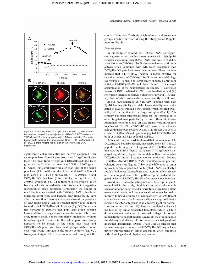
In vivo antitumor activityThe temperature measured by the thermocouple within
the tumor reached approximately 53�C after 5 minutes ofNIR laser exposure on the tumor surface at an output powerof 3 W/cm2 in mice injected with T-DOX@HAuNS (Fig. 5A).No change in temperature was noted under the same con-ditions in the tumors of control mice. Dual-tracer autora-diography showed that immediately after NIR laser irradi-ation, 3H-DOX was released and dispersed into the areasurrounding the site where T-DOX@HAuNS was introduced.Conversely, 3H-DOX was mostly colocalized with 111In-HAuNS in mice that did not undergo NIR laser treatment(Fig. 5B).
Figure 6A shows the Hey tumor growth curves afterintravenous injections of saline, HAuNS [5.0 mL/kg of 1.25mg HAuNS/mL saline (50 OD), no DOX, no targeting],DOX@HAuNS (10 mg equivalent DOX/kg, 5.0 mL/kg of1.25 mg HAuNS/mL), and T-DOX@HAuNS (10 mg equivalentDOX/kg, 5.0 mL/kg of 1.25 mg HAuNS/mL). Mice in eachgroup received NIR laser treatment (2.0 W/cm2 for 3 min-utes) 24 hours after injection. The mice in the saline plus
laser group were killed on day 9 after injection because mostof the tumors in this group were approximately 1500 mm3 atthat time. Mice in the other 3 groups were killed on day22. Treatment with T-DOX@HAuNS plus laser showed
Table 1. Pharmacokinetic parameters of DOX@HAuNS and T-DOX@HAuNS after intravenous injection infemale Swiss mice
PK parameters DOX@HAuNS T-DOX@HAuNS P
Elimination half-life, h 11.9 � 3.8 9.75 � 1.7 0.229Cmax, %ID/mL 28.9 � 4.9 30.8 � 3.7 0.459AUC0–¥, %ID h/mL 322.3 � 72 242.8 � 46 0.046CL, mL/h 0.323 � 0.068 0.426 � 0.092 0.050Vd, mL 5.30 � 0.81 5.85 � 0.69 0.239Vss, mL 4.94 � 0.70 5.25 � 0.61 0.439MRT, h 16.0 � 5.0 12.6 � 1.8 0.137
NOTE: ANOVA showed no differences between the 2 HAuNS formulations.Abbreviations: AUC0–¥, area under the blood drug concentration–time curve extrapolated to infinite time; Cmax, maximum bloodconcentration; CL, systemic clearance; MRT, mean residence time; Vd, volume of distribution; Vss, volume of distribution at steadystate.
Hey
MDA-
MB-231
A2780
D
T-DOX@HAuNS DOX@HAuNS
250
100
T-DOX@HAuNS +
c(TNYL-RAW)
A
Max
Min
CB
Figure 4. microSPECT/CT imaging and autoradiography of 111In-labeledT-DOX@HAuNS and DOX@HAuNS. A–C, representative SPECT imagesof nudemicebearingHey tumors at 24 hours after intravenous injection of111In-labeled T-DOX@HAuNS, 111In-labeled T-DOX@HAuNS plus free c(TNYL-RAW), and 111In-labeled DOX@HAuNS. Arrows indicate tumors.D, representative autoradiographs of slices sectioned from Hey, A2780,and MDA-MB-231 tumors at 24 hours after injection. Stronger signalswere visualized for tumors imaged after the injection of 111In-labeled T-DOX@HAuNS than of T-DOX@HAuNS plus free c(TNYL-HAuNS) orDOX@HAuNS.
You et al.
Cancer Res; 72(18) September 15, 2012 Cancer Research4782
on April 4, 2019. © 2012 American Association for Cancer Research. cancerres.aacrjournals.org Downloaded from
Published OnlineFirst August 3, 2012; DOI: 10.1158/0008-5472.CAN-12-1003
significantly enhanced antitumor activity compared withsaline plus laser, HAuNS plus laser, and DOX@HAuNS pluslaser. The mean tumor weight in T-DOX@HAuNS plus lasergroup on day 22 after treatment was 0.0038 � 0.0007 g (n ¼8), which was significantly smaller than that of the salineplus laser (1.2 � 0.51 g on day 9, n ¼ 6; P<0.0001), HAuNSplus laser (1.5 � 0.34 g on day 22, n ¼ 6; P<0.0001), andDOX@HAuNS plus laser (0.36 � 0.24 g on day 22, n ¼ 7;P<0.001) groups (Fig. 6B). The tumors in this group of micebecame whitish immediately after treatment, suggestingdisruption of blood perfusion. Remarkably, the tumors in6 of the 8 mice treated with T-DOX@HAuNS plus laserregressed completely and became scar tissue by 22 daysafter the injection. Histologic analysis showed the presenceof scar tissue and a lack of residual tumor cells in micetreated with T-DOX@HAuNS plus laser (Fig. 6C). Tumors inthe nontargeted DOX@HAuNS plus laser group becameloose and discrete, suggesting damage to tumor cells. How-ever, tumors could not be completely eradicated withouttargeting ligand. Tumors in the saline plus laser groupappeared to be intact. In the saline plus laser andDOX@HAuNS plus laser treatment groups, viable tumorcells were found throughout the tumor volumes (Fig. 6C).No apparent signs of toxicity were observed throughout the
course of the study. The body weight of mice in all treatmentgroups steadily increased during the study period (Supple-mentary Fig. S5).
DiscussionIn this study, we showed that T-DOX@HAuNS had signifi-
cantly greater cytotoxic effects in tumor cells with high EphB4receptor expression than DOX@HAuNS and free DOX did invitro. Moreover, T-DOX@HAuNS showed enhanced antitumoractivity when combined with NIR laser irradiation thanDOX@HAuNS plus laser treatment in vivo. These findingsindicate that c(TNYL-RAW) peptide is highly effective forselective delivery of T-DOX@HAuNS to tumors with highexpression of EphB4. The significantly enhanced antitumoractivity of T-DOX@HAuNS could be attributed to (i) increasedaccumulation of the nanoparticles in tumors, (ii) controlledrelease of DOX mediated by NIR laser irradiation, and (iii)synergistic interaction between chemotherapy and PTA ther-apy, both of which were activated concurrently by NIR laser.
In our nanoconstruct, c(TNYL-RAW) peptide with highEphB4 binding affinity and high plasma stability was conju-gated to HAuNS through a PEG linker, which ensured avail-ability of the peptide to the target receptor (Fig. 1). Thisstrategy has been successfully used for the formulation ofother targeted nanoparticles by us and others (4, 21–24).Additional monofunctional SH-PEG chains were introducedtogether with SH-PEG-c(TNYL-RAW) to ensure that all avail-able gold surface was covered by PEG. This process was used tocreate DOX@HAuNS and ligand-conjugated T-DOX@HAuNS,both of which had high colloidal stability.
Both in vitro and in vivo data showed that tumor uptake of T-DOX@HAuNS could be partially blocked by free c(TNYL-RAW)peptide, confirming that cell uptake of T-DOX@HAuNS wasmediated by EphB4 (Figs. 2–4). In vivo, T-DOX@HAuNS dis-played significantly higher accumulation than nontargetedDOX@HAuNS in all 3 tumor models evaluated. BecauseDOX@HAuNS and T-DOX@HAuNS exhibited similar pharma-cokinetic behaviors (Fig. 3A, Table 1), the difference in tumoruptake between targeted and nontargeted HAuNS is unlikely aresult of enhanced permeability and retention effect. Hence,our data support successful EphB4 receptor–mediated tar-geted delivery of T-DOX@HAuNS after intravenous injection.
In addition to active targeting mediated by receptor ligandsexemplified in this study, physiologic and physical methodssuch as tumor priming, vascular disruption, degradation of theextracellular matrix, and vessel normalization may be used toimprove tumor distribution of nanoparticles (25–29). Recentstudies have shown that losartan, a clinically approved angio-tensin II receptor antagonist, is an effective agent for normal-izing tumor vasculature (29). Losartan inhibited collagen Iproduction by tumor-associated fibroblasts, which led to adose-dependent reduction in stromal collagen in severalhuman tumor xenograftmodels. As a result, the drug enhancedthe delivery and efficacy of intravenously injected pegylatedliposomal doxorubicin (Doxil). These studies suggest thattargeted nanoparticles such as T-DOX@HAuNS may achievefurther improvement in tumor deposition when combinedwith physiological and physical approaches.
111In-HAuNS 3H-DOX Overlap
With NIR laser
Without NIR laser
A
B
20
25
30
35
40
45
50
55
60
0 100 200 300 400 500
Tem
pera
ture
(ºC
)
Time (s)
Laser off
Laser on
HAuNS
SalineX
—
Figure 5. In vivo release of DOX upon NIR treatment. A, NIR-inducedtemperature change in tumors injectedwith HAuNS. B, DOX release fromT-DOX@HAuNS in tumors treated with NIR laser irradiation. The whitebroken circle indicates the tumor outline. Green (111In-HAuNS) and red(3H-DOX) signals indicate the location of the HAuNS and DOX,respectively.
Concerted Chemo-Photothermal Therapy Targeting EphB4
www.aacrjournals.org Cancer Res; 72(18) September 15, 2012 4783
on April 4, 2019. © 2012 American Association for Cancer Research. cancerres.aacrjournals.org Downloaded from
Published OnlineFirst August 3, 2012; DOI: 10.1158/0008-5472.CAN-12-1003

The temperature in tumors of mice received intravenousinjection of T-DOX@HAuNS reached approximately 53�C after5 minutes of continuous wave NIR laser exposure at 3 W/cm2
(Fig. 5A). This temperature is sufficient for causing irreversibledamage to cancer cells (30). As expected, there was no tem-perature change in the tumors of mice that did not receive thenanoparticle injection followed by NIR irradiation. Therefore,T-DOX@HAuNS medicated efficient photothermal effect. Wehave previously shown that in aqueous solution, the release ofDOX from DOX@HAuNS could be activated by NIR laserirradiation (5). To show that DOX could be released in vivoupon NIR irradiation, we used an autoradiographic techniquewith dual-radiotracer labeling. 3H undergoes beta decay with a12.3-year half-life, releasing 18.6 keV of energy in the process.On the other hand, 111In emits gamma radiation with a half-life
of 2.81 days at much higher energy (171 and 245 KeV). Takingadvantage of these differences in the decay properties between3H and 111In, we were able to separate signals fromHAuNS andDOX in the tumors. Thus, immediately after NIR laser irradi-ation at 3 W/cm2, 3H-DOX was dissociated from 111In-HAuNS,whereas in the control tumor not exposed to NIR laser irra-diation, signals from 3H-DOX colocalized perfectly with 111In-HAuNS (Fig. 6B). These data confirm that free DOX wasreleased from T-DOX@HAuNS and then diffused away fromthe nanoparticles when tumors were exposed to the NIR laser.
The antitumor efficacy of free DOX, DOX@HAuNS,DOX@HAuNS plus NIR laser was investigated against MDA-MB-231 tumors in our previous work (31). There was nosignificant difference in tumor size between groups treatedwith DOX (15 mg/kg, 1 injection) and DOX@HAuNS (15 mg
A
B
0
0.5
1
1.5
2
Tu
mo
r w
eig
ht
(g)
T-DOX@HAUNS
+ laser
DOX@HAUNS
+ laser
HAUNS + laser
Saline + laser
0
500
1,000
1,500
0 5 10 15 20 25
Tu
mo
r vo
lum
e (
mm
3)
Time (d)
Saline + laser
HAuNS + laser
DOX@HAuNS + laser
T-DOX@HAuNS + laser
Sal
ine
+ la
ser (
9th
day)
HAuN
S +
lase
r
DO
X@
HAuN
S +
lase
r
T-DO
X@
HAuN
S +
lase
r
C DOX@HAuNS
+ laserSalineT-DOX@HAuNS
+ laser
20 µm
100 µm100 µm
20 µm 20 µm
500 µm
Figure 6. Antitumor activity ofvarious treatments against Heytumors. A, tumor growth curves formice treated with saline plus laser(n ¼ 6), HAuNS plus laser (n ¼ 6),DOX@HAuNSplus laser (n¼7), andT-DOX@HAuNS plus laser (n ¼ 8).NIR laser irradiation (2.0 W/cm2 for3 minutes) was commenced at 24hours after injection. B, averagetumor weights (left) andphotographs (right) of tumors fromdifferent treatment groups. Tumorswere removed on day 22 for allgroups except the saline plus lasergroup, in which tumors wereremoved on day 9. Six tumors in theT-DOX@HAuNS group regressedcompletely by day 22 aftertreatment, and only the scar tissuesat the site of tumor inoculationwereremoved and photographed (bluecircle, right). C, representativephotomicrographs of hematoxylinand eosin–stained slides from scartissue from a mouse treated withT-DOX@HAuNS plus laser ortumors from mice treated withsaline or DOX@HAuNS plus laseron day 22 after treatment.
You et al.
Cancer Res; 72(18) September 15, 2012 Cancer Research4784
on April 4, 2019. © 2012 American Association for Cancer Research. cancerres.aacrjournals.org Downloaded from
Published OnlineFirst August 3, 2012; DOI: 10.1158/0008-5472.CAN-12-1003

equivalent DOX/kg per injection, 2 injections). However,DOX@HAuNS plus NIR laser (15 mg equivalent DOX/kg perinjection, 2 injections) reduced tumor volume significantlymore than the other 2 treatments (31). In these studies, freeDOX at a single dose of 15 mg/kg caused serious systemictoxicity and cardiotoxicity. Therefore, we did not use free DOXas a control in the current study. Instead, we used nontargetedDOX@HAuNS as a control and compared the antitumor stud-ies of T-DOX@HAuNS against ovarian Hey tumors with that ofDOX@HAuNS. Combined T-DOX@HAuNS plus laser treat-ments showed significantly better antitumor activity thancombined HAuNS plus laser (PTA only) and nontargetedDOX@HAuNS plus laser treatments (Fig. 6).Significantly, this higher antitumor activity was achieved
without causing significant systemic toxicity, as indicated bycontinued increase in body weight in mice in all treatmentgroups (Supplementary Fig. S5). In our previous work, wecompared the toxicity of free DOX, liposomal DOX, andDOX@HAuNS (31). DOX@HAuNS after a single dose at 60 mgequivalent DOX/kg had no cardiotoxicity compared withliposomal DOX (2 doses at a total dose of 30 mg DOX/kg)and free DOX (single dose of 15 mg/kg). In the heart, 100% ofboth liposomal DOX- and free DOX–treated mice had avacuolar cardiomyopathy. However, for mice treated withDOX@HAuNS, the histopathologic features in the heart weresimilar to those observed in the saline-treated controlmice andno abnormal features were observed. Because DOX@HAuNSand T-DOX@HAuNS had similar pharmacokinetic propertiesand there was no significant different in heart accumulationbetween the 2 agents, we expect that T-DOX@HAuNSwill display toxicity profile similar to that of [email protected] studies are required to document both short- and long-term systemic toxicity of T-DOX@HAuNS after intravenousinjection.Our study has several limitations. First, in vivo release of
DOX upon laser irradiation was conducted after intratumoralinjection, which may not accurately reflect DOX distributionthatwill be seen after systemic administration, followed byNIRtreatment. However, the sole purpose of this experimentwas toexamine whether DOX release can be triggered by NIR laserirradiation under in vivo conditions, which was confirmed inthe current study. Second, we did not analyze the distributionof DOX when DOX@HAuNS and T-DOX@HAuNS weregiven intravenously. Detailed DOX biodistribution in compar-ison to HAuNS after systemic administration of nontargetedDOX@HAuNS has been studied and published in our previouswork using a dual-radiotracer technique (31). It was found thatDOX@HAuNS was stable in the blood during the first 6 hoursafter injection. By 24 hours after injection, there was disparityin biodistribution pattern between DOX and HAuNS, indicat-ing gradual release of DOX from DOX@HAuNS.
A potential limitation of the proposed concurrent chemo-photothermal therapy strategy is penetration depth of NIRlight. The current state-of-the-art enables NIR light to pene-trate up to 1 to 2 cm in soft tissue (32).With the newer 10- to 40-mm long quartz laser fibers andwater-cooled laser applicationsheaths, ablative areas of 50 to 80mm have been achieved (33–35). Deeper reach is also possible with various minimallyinvasive interventional techniques, such as endoscopic, intra-vascular, and percutaneous approaches. Currently, laser beamis delivered through laser fibers inserted into the tumors, as inthe case of primary and metastatic liver cancer (33, 35, 36) andlung metastases (37). In our previous work, we evaluated thetriggered release of paciltaxel via NIR laser irradiation and itsantitumor efficacy by hepatic arterial administration ofHAuNS- and paclitaxel-loaded microspheres into rabbits withliver carcinoma in situ (38). The results showed that NIR laserirradiation after the microspheres administration results inintratumoral heating. NIR laser irradiation can trigger releaseof paclitaxel from the microspheres, and can potentiallyincrease the efficacy of treatment after hepatic arterial deliveryof the microspheres in liver tumors. Thus, we believe thatchemo-photothermal therapy approach entailed in this work isbest suited for unresectable, locally advanced disease thatultimately causes patient death. It is envisioned that chemo-PTA therapy may also be used in conjunction with surgery,especially used in postsurgery setting to eradicate residualtumor cells. In such as case, the laser beam can be convenientlydelivered through surface exposure.
Disclosure of Potential Conflict of InterestNo potential conflicts of interest were disclosed.
Authors' ContributionsConception and design: J. You, C. Xiong, M. Zhong, S. Gupta, A.K. Sood, C. LiDevelopment of methodology: J. You, C. Xiong, S. Gupta, C. LiAcquisition of data (provided animals, acquired and managed patients,provided facilities, etc.): J. You, M. Melancon, C. LiAnalysis and interpretation of data (e.g., statistical analysis, biostatistics,computational analysis): J. You, C. Xiong, S. Gupta, A.K. Sood, C. LiWriting, review, and/or revision of the manuscript: J. You, M. Melancon, S.Gupta, A.M. Nick, A.K. Sood, C. LiAdministrative, technical, or material support (i.e., reporting or orga-nizing data, constructing databases): M. Zhong, M. Melancon, A.K. Sood
Grant SupportThis work was supported in part by NIH grants RC2 GM092599, Center of
Cancer Nanotechnology Excellence grant (U54 CA151668), the John S. DunnFoundation, and the National Nature Science Foundation of PR China(81001411). The authors thank the NCI Cancer Center Support Grant CA016672,which supports MD Anderson's Small Animal Imaging Facility and TEM CoreFacility.
The costs of publication of this article were defrayed in part by the payment ofpage charges. This article must therefore be hereby marked advertisement inaccordance with 18 U.S.C. Section 1734 solely to indicate this fact.
Received March 21, 2012; revised May 8, 2012; accepted June 1, 2012;published OnlineFirst August 3, 2012.
References1. Bardhan R, Lal S, Joshi A, Halas NJ. Theranostic nanoshells: from
probe design to imaging and treatment of cancer. Acc Chem Res2011;44:936–46.
2. MelanconM, LuW, Li C. Gold-basedmagneto/optical nanostructures:challenges for in vivo applications in cancer diagnostics and therapy.Mater Res Bull 2009;34:415–21.
Concerted Chemo-Photothermal Therapy Targeting EphB4
www.aacrjournals.org Cancer Res; 72(18) September 15, 2012 4785
on April 4, 2019. © 2012 American Association for Cancer Research. cancerres.aacrjournals.org Downloaded from
Published OnlineFirst August 3, 2012; DOI: 10.1158/0008-5472.CAN-12-1003

3. MelanconMP, LuW, Yang Z, ZhangR, Cheng Z, Elliot AM, et al. In vitroand in vivo targeting of hollow gold nanoshells directed at epidermalgrowth factor receptor for photothermal ablation therapy. Mol CancerTher 2008;7:1730–9.
4. LuW, Xiong C, Zhang G, Huang Q, Zhang R, Zhang JZ, et al. Targetedphotothermal ablation of murine melanomas with melanocyte-stimu-lating hormone analog-conjugated hollow gold nanospheres. ClinCancer Res 2009;15:876–86.
5. You J, Zhang G, Li C. Exceptionally high payload of doxorubicin inhollowgold nanospheres for near-infrared light-triggered drug release.ACS Nano 2010;4:1033–41.
6. Pasquale EB. Eph receptor signalling casts a wide net on cell behav-iour. Nat Rev Mol Cell Biol 2005;6:462–75.
7. Dodelet VC, Pasquale EB. Eph receptors and ephrin ligands: embryo-genesis to tumorigenesis. Oncogene 2000;19:5614–9.
8. Nakamoto M, Bergemann AD. Diverse roles for the Eph family ofreceptor tyrosine kinases in carcinogenesis. Microsc Res Tech2002;59:58–67.
9. Walker-Daniels J, Hess AR, Hendrix MJ, Kinch MS. Differential reg-ulation of EphA2 in normal and malignant cells. Am J Pathol 2003;162:1037–42.
10. Brantley-Sieders DM, Caughron J, Hicks D, Pozzi A, Ruiz JC, Chen J.EphA2 receptor tyrosine kinase regulates endothelial cell migrationand vascular assembly through phosphoinositide 3-kinase-mediatedRac1 GTPase activation. J Cell Sci 2004;117:2037–49.
11. Noren NK, Lu M, Freeman AL, Koolpe M, Pasquale EB. InterplaybetweenEphB4on tumor cells and vascular ephrin-B2 regulates tumorgrowth. Proc Natl Acad Sci U S A 2004;101:5583–8.
12. Battaglia AA, Sehayek K, Grist J, McMahon SB, Gavazzi I. EphBreceptors and ephrin-B ligands regulate spinal sensory connectivityand modulate pain processing. Nat Neurosci 2003;6:339–40.
13. Goldshmit Y, Galea MP, Wise G, Bartlett PF, Turnley AM. Axonalregeneration and lack of astrocytic gliosis in EphA4-deficient mice.J Neurosci 2004;24:10064–73.
14. XiaG, Kumar SR, Stein JP, Singh J, Krasnoperov V, ZhuS, et al. EphB4receptor tyrosine kinase is expressed in bladder cancer and providessignals for cell survival. Oncogene 2006;25:769–80.
15. Koolpe M, Burgess R, Dail M, Pasquale EB. EphB receptor-bindingpeptides identified by phage display enable design of an antagonistwith Ephrin-like affinity. J Biol Chem 2005;280:17301–11.
16. Erber R, Eichelsbacher U, Powajbo V, Korn T, Djonov V, Lin JH, et al.EphB4 controls blood vascular morphogenesis during postnatalangiogenesis. EMBO J 2006;25:628–41.
17. Kumar SR, Scehnet JS, Ley EJ, Singh J, Krasnoperov V, Liu R, et al.Preferential induction of EphB4 over EphB2 and its implication incolorectal cancer progression. Cancer Res 2009;69:3736–45.
18. Kumar SR, Singh J, Xia GB, Krasnoperov V, Hassanieh L, Ley EJ, et al.Receptor tyrosine kinase EphB4 is a survival factor in breast cancer.Am J Pathol 2006;169:279–93.
19. Xiong C, Huang M, Zhang R, Song S, Lu W, Flores L II, et al. In vivosmall-animal PET/CT of EphB4 receptors using 64Cu-labeled peptide.J Nucl Med 2011;52:241–8.
20. Ozgur E, Heidenreich A, Dagtekin O, Engelmann U, Bloch W. Distri-bution of EphB4 and EphrinB2 in normal and malignant urogenitaltissue. Urol Oncol 2011;29:78–84.
21. Wang X, Qian X, Beitler JJ, Chen ZG, Khuri FR, Lewis MM, et al.Detection of circulating tumor cells in human peripheral blood usingsurface-enhanced Raman scattering nanoparticles. Cancer Res2011;71:1526–32.
22. Davis ME, Zuckerman JE, Choi CH, Seligson D, Tolcher A, Alabi CA,et al. Evidence of RNAi in humans from systemically administeredsiRNA via targeted nanoparticles. Nature 2010;464:1067–70.
23. Petros RA, DeSimone JM. Strategies in the design of nanoparticles fortherapeutic applications. Nat Rev Drug Discov 2010;9:615–27.
24. Lu W, Zhang G, Zhang R, Flores LG II, Huang Q, Gelovani JG, et al.Tumor site-specific silencing of NF-kappa B p65 by targeted hollowgold nanosphere-mediated photothermal transfection. Cancer Res2010;70:3177–88.
25. Lu D, Wientjes MG, Lu Z, Au JL. Tumor priming enhances delivery andefficacy of nanomedicines. J Pharmacol Exp Ther 2007;322:80–8.
26. Melancon MP, Elliott A, Ji X, Shetty A, Yang Z, Tian M, et al. Ther-anostics with multifunctional magnetic gold nanoshells: photothermaltherapy and t2� magnetic resonance imaging. Invest Radiol 2011;46:132–40.
27. Eikenes L, Tari M, Tufto I, Bruland OS, de Lange Davies C. Hyaluron-idase induces a transcapillary pressure gradient and improves thedistribution and uptake of liposomal doxorubicin (Caelyx) in humanosteosarcoma xenografts. Br J Cancer 2005;93:81–8.
28. Eikenes L, Bruland OS, Brekken C, Davies Cde L. Collagenaseincreases the transcapillary pressure gradient and improves theuptakeand distribution of monoclonal antibodies in human osteosarcomaxenografts. Cancer Res 2004;64:4768–73.
29. Diop-FrimpongB,ChauhanVP, KraneS, Boucher Y, Jain RK. Losartaninhibits collagen I synthesis and improves the distribution and efficacyof nanotherapeutics in tumors. Proc Natl Acad Sci U S A 2011;108:2909–14.
30. Melancon MP, Zhou M, Li C. Cancer theranostics with near-infraredlight-activatable multimodal nanoparticles. Acc Chem Res 2011;44:947–56.
31. You J, Zhang R, Zhang G, Zhong M, Liu Y, Van Pelt CS, et al.Photothermal-chemotherapy with doxorubicin-loaded hollow goldnanospheres: a platform for near-infrared light-trigged drug release.J Control Release 2012;158:319–28.
32. Gu Y, Chen WR, Xia M, Jeong SW, Liu H. Effect of photothermaltherapy on breast tumor vascular contents: noninvasive monitoringby near-infrared spectroscopy. Photochem Photobiol 2005;81:1002–9.
33. Gough-Palmer AL, Gedroyc WM. Laser ablation of hepatocellularcarcinoma—a review. World J Gastroenterol 2008;14:7170–4.
34. Muralidharan V, Malcontenti-Wilson C, Christophi C. Interstitial laserhyperthermia for colorectal liver metastases: the effect of thermalsensitization and the use of a cylindrical diffuser tip on tumor necrosis.J Clin Laser Med Surg 2002;20:189–96.
35. Vogl TJ, Straub R, Zangos S, Mack MG, Eichler K. MR-guided laser-induced thermotherapy (LITT) of liver tumours: experimental andclinical data. Int J Hyperthermia 2004;20:713–24.
36. Vogl TJ, Straub R, Eichler K, Sollner O, Mack MG. Colorectalcarcinoma metastases in liver: laser-induced interstitial thermother-apy—local tumor control rate and survival data. Radiology 2004;230:450–8.
37. Rosenberg C, Puls R, Hegenscheid K, Kuehn J, Bollman T,WesterholtA, et al. Laser ablation of metastatic lesions of the lung: long-termoutcome. Am J Roentgenol 2009;192:785–92.
38. Gupta S, Stafford RJ, Javadi S, Ozkan E, Ensor JE, Wright KC, et al.Effects of near-infrared laser irradiation of biodegradable micro-spheres containing hollow gold nanospheres and paclitaxel adminis-tered intraarterially in a rabbit liver tumor model. J Vasc Interv Radiol2012;23:553–61.
You et al.
Cancer Res; 72(18) September 15, 2012 Cancer Research4786
on April 4, 2019. © 2012 American Association for Cancer Research. cancerres.aacrjournals.org Downloaded from
Published OnlineFirst August 3, 2012; DOI: 10.1158/0008-5472.CAN-12-1003

2012;72:4777-4786. Published OnlineFirst August 3, 2012.Cancer Res Jian You, Rui Zhang, Chiyi Xiong, et al. Gold Nanospheres That Target EphB4 Receptors in TumorsEffective Photothermal Chemotherapy Using Doxorubicin-Loaded
Updated version
10.1158/0008-5472.CAN-12-1003doi:
Access the most recent version of this article at:
Material
Supplementary
http://cancerres.aacrjournals.org/content/suppl/2012/09/14/0008-5472.CAN-12-1003.DC1
Access the most recent supplemental material at:
Cited articles
http://cancerres.aacrjournals.org/content/72/18/4777.full#ref-list-1
This article cites 38 articles, 13 of which you can access for free at:
Citing articles
http://cancerres.aacrjournals.org/content/72/18/4777.full#related-urls
This article has been cited by 2 HighWire-hosted articles. Access the articles at:
E-mail alerts related to this article or journal.Sign up to receive free email-alerts
Subscriptions
Reprints and
To order reprints of this article or to subscribe to the journal, contact the AACR Publications Department at
Permissions
Rightslink site. Click on "Request Permissions" which will take you to the Copyright Clearance Center's (CCC)
.http://cancerres.aacrjournals.org/content/72/18/4777To request permission to re-use all or part of this article, use this link
on April 4, 2019. © 2012 American Association for Cancer Research. cancerres.aacrjournals.org Downloaded from
Published OnlineFirst August 3, 2012; DOI: 10.1158/0008-5472.CAN-12-1003