with combined photothermal-chemotherapy mtx nps for
TRANSCRIPT

Page 1/23
Hierarchical Drug Release Designed Au @PDA-PEG-MTX NPs for Targeted Delivery to Breast Cancerwith Combined Photothermal-chemotherapyWen Li
Beijing University of Chinese MedicineZhiwen Cao
Beijing University of Chinese MedicineLiuchunyang Yu
Beijing University of Chinese MedicineQingcai Huang
Beijing University of Chinese MedicineDongjie Zhu
Beijing University of Chinese MedicineCheng Lu
China Academy of Chinese Medical SciencesAiping Lu
Hong Kong Baptist UniversityYuanyan Liu ( [email protected] )
Beijing University of Chinese Medicine https://orcid.org/0000-0003-3840-5292
Research
Keywords: Au @PDA-PEG-MTX NPs, Targeted for Breast cancer, Fluorescence imaging, Hierarchical DrugRelease, Combined photothermal-chemotherapy
Posted Date: January 27th, 2021
DOI: https://doi.org/10.21203/rs.3.rs-152625/v1
License: This work is licensed under a Creative Commons Attribution 4.0 International License. Read Full License

Page 2/23
AbstractBreast cancer (BC) is the frequently diagnosed cancer and one of the deadliest causes of cancer-relateddeath with a severe survival rate. Methotrexate (MTX) is an anti-tumor drug used in the treatment of BC.The poor dispersion in water and toxic side effects limit its clinical application. Gold nanoparticles(AuNPs), due to their speci�c structures and unique biological and physiochemical properties, haveemerged as attractive candidates as vehicles for tumor targeting, bio-imaging and therapy. An innovativenano drug-loading system (Au @PDA-PEG-MTX NPs) was prepared for targeted treatment of BC. Au@PDA-PEG-MTX NPs under near infra-red region (NIR) irradiation showed effective photothermal therapyagainst MDA-MB-231 human BC cells growth in vitro by inducing apoptosis through triggering reactiveoxygen species (ROS) overproduction and generating excessive heat. In vivo studies revealed that Au@PDA-PEG-MTX NPs under NIR irradiation showed deep penetration and cancer-targeted �uorescenceimaging application and strong photothermal therapy against BC xenograft growth in vivo by inductionof apoptosis. Analysis of histopathology, cellular uptake, cytotoxicity assay, apoptosis experimentindicated that Au @PDA-PEG-MTX NPs had a good therapeutic effect with high biocompatibility and lessside effect. This Au NPs drug-loading system achieved speci�c BC targeting ability by surface decorationof MTX, NIR laser irradiation for �uorescence imaging and combined photothermal-chemotherapy as wellas pH- and NIR- triggered hierarchical drug release.
IntroductionBreast cancer (BC) is one of the most frequently observed malignant diseases among women with highmortality and economic burden[1, 2]. On account of its complicated etiology, poor response and severityside-effect to chemotherapy, both safe and e�cacious are considered as the central challenge associateddeaths[3]. The main aim in the �ght against BC is developing effective therapeutic plans with low toxicityand high speci�city to eliminate tumors[4]. However, presently used BC treatment approaches, such assurgery, chemotherapy and radiotherapy, cause diverse side effects to patients and these measures alonedo not seem to achieve this aim[5]. The combination of chemotherapy drugs and the gold nanoparticles(AuNPs) drug carrier system with photothermal property can provide a promising platform forintracellular delivery of various anti-BC drugs and synergistic therapy[6, 7].
NPs designed for cancer treatment targeting can deliver chemotherapeutic drugs to speci�c cancer cellswhile reducing the exposure of normal healthy cells, so that larger doses of drugs can be delivered to thetumor site to achieve high-targeting and low-toxicity therapeutic effects.[8]. AuNPs are one type ofinorganic cargo with versatile surfaces for multi-functionalization and high surface area-to-volume ratiofor drug loading and superior optical properties for bioimaging and even photothermal properties fortherapy, but toxicity and low biocompatibility of AuNPs are still challenges that cannot be ignored[9].When adjusted to an appropriate size and shape, such as 15 nm size of the AuNPs have longer plasmacirculation time [10, 11], the small-sized of which can be used to reduce the toxicity of AuNPs itself as aninorganic material.

Page 3/23
Increasingly, AuNPs have attracted great attention during the past decade due to their facile synthesisand surface functionalization[12], which revealed high-performance of photothermal conversion capacityin the near infra-red region (NIR) area without harmful side effects in biological systems[13-15]. AuNPshave the characteristics of tumor NIR imaging and good stability[16]. In addition, the photothermal effectalso produces excessive heat and reactive oxygen species (ROS) that destroys cancer cells[17]. To makeAuNPs into perfect drug carriers, modi�cation should be performed for targeted delivery and controlledrelease[18]. The strong adhesion of polydopamine (PDA) is conducive to its deposition in AuNPs, whichcan improve the drug loading and endow biocompatibility and the ability of AuNPs to enter mammaliancells[19]. Moreover, PDA shell can prevent the leakage of loading drugs during delivery, while achieve anon-demand drug release in the targeted location, such as NIR stimuli responsive drug release under hightemperature or acidic conditions[19-21]. PEG can also improve the ability of cells to take up goldnanocomposite and prolong the cycle times in plasma [22-24]. In addition, drugs linked to PEG will bereleased under acidic conditions, which can lay the foundation for hierarchical drug release system[25].
Methotrexate (MTX), a folic acid analog is a highly potent antagonist on folate pathway to induce cellapoptosis, who is widely used for the treatment of rheumatoid arthritis and acute leukemia [26]. Due to itsspecial characteristic, many experimental researches tend to explore its treatment for cancer[27]. MTX asa folic acid analog can inhibit dihydrofolate reductase (DHFR), an enzyme that helps producetetrahydrofolate and its by-products, both of which are essential for the growth of tumor cells[28, 29].Folate receptor (FR) is a glycosyl phosphatidylinositol (GPI)-linked protein has low expression in normaltissues, but is over-expressed in certain malignant cells such as BC[30]. Except for therapeutic purpose, asa folate analog, MTX exhibits high a�nity to FR that can be targeted to deliver to BC cells. However, aliketo most conventional chemotherapy drugs, the main challenge of single-agent chemotherapy is theaccumulated toxicity in the body accompanied with low speci�city and poor therapeutic e�cacy[31].Therefore, in recent years, the combination of chemo-therapeutics and nano-materials against BC hasbecome an important therapeutic regimen [32]. Here, equipped MTX with AuNPs are not only as acombined photothermal-chemotherapeutic drug, but can be precisely identi�ed as targeted ligand bywhom are highly expressed on the BC surface.
In this study, Au @PDA-PEG-MTX NPs were designed for targeted delivery and hierarchical release incombination of photothermal and chemotherapy on BC with improved e�ciency and low toxicity. In thisinnovative nano-drug loading system, AuNPs not just play roles as a drug carrier, even when modi�edwith PDA and PEG exhibit synergistic photothermal therapeutic e�cacy, bioimaging and hierarchicalrelease stimulated with NIR and acidic conditions.
MethodsReagents. The following chemicals were obtained from commercial sources and were used withoutfurther puri�cation: Chloroauric acid,99.95%, from Innochem. Dopamine hydrochloride,1-ethyl-3-(3-dimethylaminopropyl) carbodiimide hydrochloride (EDCI), N-hydroxy succinimide (NHS) and Dimethylsulfoxide (DMSO) were purchased from Sigma-Aldrich, USA. All aqueous solutions used in experiments

Page 4/23
were deionized water (18.2 MΩ.cm) obtained from a Milli-Q water puri�cation system. Fetal bovine serum(FBS) and DMEM/HIGH Glucose medium were purchased from Gibco, United States origin. Penicillin-Streptomycin (penicillin 100 U/ml, and streptomycin 100 mg/ml) were obtained from BI. Human breastcancer MDA-MB-231 cells were obtained from BNCC. All the antibodies were purchased from the Abcam.
Preparation of Au @PDA-PEG-MTX NPs
Preparation of AuNPs
AuNPs (15 nm in diameter) were synthesized according to the trisodium citrate reduction methodreported by Frens[33]. Before all reactions started, the reaction vessel was thoroughly washed with freshlyprepared aqua regia (HNO3 / HCl = 1:3) and washed with double distilled water 3 times. First add 1 mL of1% chloroauric acid solution to 100 mL of ultrapure water, and quickly boil it. Add 4 mL of 1% sodiumcitrate solution immediately. When the color of the solution changes from light yellow to wine red, goldnanoparticles AuNP with an average diameter of 15 nm will be formed and stored under dark conditionsat 4°C. The average size of AuNP was estimated by transmission electron microscope (TEM, HT7700,Tokyo, Japan, Hitachi).
Preparation of Au @PDA NPs Bioconjugates
Disperse the previously synthesized AuNPs in 50 mL pH 8.5 Tris-HCl buffer, and the solutionconcentration is 2 mg/mL at this time. In the dark, this solution is subjected to strong magnetic stirringovernight after 5mL (2mg/mL) of the prepared dopamine solution added dropwise to the solution. Ablack solution is obtained, which is centrifuged at 12,000 rpm at 4°C for 10 minutes; the supernatant isdiscarded, and the black precipitate is collected, and washed with deionized water repeatedly for 3 times.
Preparation of Au @PDA-PEG NPs
Disperse the synthesized Au @PDA NPs complex in 50 mL of deionized water. At this time, theconcentration of the dispersion solution was 2 mg/ml. In the dark, added NH2-PEG-SH (2k) (2 mg/mL)dropwise to the solution and stirred it magnetically overnight. Then, it was centrifuged at 12,000 rpm at4°C for 10 minutes to obtain Au @PDA-PEG NP, which was washed repeatedly with deionized water for 3times[34].
Preparation of Au @PDA-PEG-MTX NPs
In order to modify the folate receptor targeting drug MTX on the synthesized Au @PDA-PEG NPs, 8 mgMTX, 4 mg EDCI and 2.8 mg NHS were added to 50 mL of Au @PDA-PEG NPs aqueous solution undermagnetic stirring. After 3 hours of magnetic stirring in the dark, centrifuge at high speed at 4°C (12,000rpm, 10 min), save the supernatant, and wash the precipitate with deionized water 3 times. Theprecipitate was Au @PDA-PEG-MTX NPs bioconjugate.
Preparation of Au @PDA/FITC-PEG-MTX NPs

Page 5/23
The synthesized Au @PDA-PEG-MTX NPs bioconjugate was re-dispersed in 2 mL deionized water (2mg/mL). In the dark state, magnetically stir continuously for 12 hours at room temperature, and add 50 μlof DMSO solution containing 10% (w/w) FITC �uorescent agent. The resulting mixture solution was thencentrifuged at 4°C (12,000 rpm, 10 minutes), and the precipitate obtained after centrifugation waswashed with deionized water three times. The �nal product was Au @PDA / FITC-PEG-MTX NPs.
Drug loading study of Au @PDA-PEG-MTX NPs
For MTX loading, UV-vis spectroscopy was used to determine the absorbance of the washed drug, theabsorbance of the supernatant solution and the absorbance strength of the initial drug. The MTX loadinge�ciency was as follows:
AW was absorbance of the drug after washing, AS was absorbance of supernatant and ADrug wasabsorbance intensity of initial drug[30].
In vitro MTX release pattern from Au @PDA-PEG-MTX NPs Au @ The PDA-PEG-MTX NPs bioconjugatewas dissolved in 10 mL phosphate buffer solution (20 mM) with pH 5.4 and 7.4, and the solution systemwas placed in a ready-to-use dialysis bag with a molecular weight cut-off of 1 kda. At the same time, addprotease (1 mg/ml) to the phosphate buffer to cleave the amide bond between MTX and PEG[35]. At thespeci�ed time point, 500μl of the dialyzed solution was collected to obtain the released MTX, and theabsorbance was measured at 305 nm by ultraviolet-visible spectroscopy (UV-vis, PerkinElmer, Singapore)[36].
Characterization of Au @PDA-PEG-MTX NPs and its intermediate products
A UV-Vis spectrometer was used to record the UV-Vis absorption spectrum of the synthesized goldnanoparticles (15nm). A vacuum Fourier transform infrared spectrometer (FT-IR, America, Thermo FisherScienti�c) was used to record the Fourier transform infrared spectrum of MTX-PEG. The morphology ofAuNPs, Au @PDA NPs and Au @PDA-PEG-MTX NPs were recorded by TEM. The Brookhaven Zeta PALSinstrument was used to record the dynamic light scattering (DLS) and zeta potential of AuNPs, Au @PDANPs, Au @PDA-PEG NPs and Au @PDA-PEG-MTX NPs.
Biological experiments
Cell culture and in vitro cytotoxicity assay
The human BC MDA-MB-231 cells were cultured in a high glucose medium containing 10% FBS, and theparameters of the incubator were set to 37°C and 5% CO2[37]. The MDA-MB-231 cells were digested withtrypsin containing EDTA, and the MDA-MB-231 cells were digested and seeded on a transparent 96-well

Page 6/23
plate. The cell density of each well was 1.8×10⁵ cells. Incubate overnight in an incubator to allow cells toattach to 96-well plates. After adding 2.5-30 µg/mL Au @PDA-PEG-MTX NPs for 12 h, the cells weretreated with or without 808 nm NIR irradiation (200mW/cm2) for a total of 20 minutes. CCK-8 was addedto measure and evaluate cell viability. Use a microplate reader (Tecan M200 PRO) to measure absorbanceat 450nm wavelength. The cell survival rate (%) was calculated as (average absorbance value of thetreatment group/average absorbance value of the control group) ×100%. Repeat the measurement 3times.
In vitro cellular uptake ofAu @PDA-PEG-MTX NPs
The uptake of Au @PDA / FITC-PEG-MTX NPs by MDA-MB-231 cells was recorded by a confocal laserscanning microscope. Digest the cells with trypsin, inoculate 1 ml of MDA-MB-231 cells (1×10⁵) into aglass-bottom cell culture dish, and incubate in an incubator for 24 h. Remove the original medium andwash the cells with PBS. Use nanocomposite Au @PDA/FITC-PEG-MTX with a concentration of 15μg/ml,20μg/ml, 25μg/ml to replace this medium. Incubate in an incubator for 4 hours to fully absorb the drug.Wash the cells with PBS three times and �x them with 4% cell �xation solution (1 ml/well) at 4°C for 30minutes. The cells were washed 3 times with PBS, and then stained with DAPI solution (2 ml/well) for 5min to ensure coloration of the nuclei. After washing the cells with PBS, confocal laser scanning imagingwas performed.
Measurement of ROS generation
MDA-MB-231 cells were seeded in 6-well plates (105 cells/well) and divided into 3 groups of blank cells,Au @PDA-PEG-MTX NPs and NIR + Au @PDA-PEG NPs. Incubate for 24 hours in a cell culture incubator.The NIR + Au @PDA-PEG NPs group used 808nm NIR laser radiation to treat the cells. The DCFH-DAprobe was diluted 1:1000 in serum-free medium, and the medium containing the DCFH-DA probe wasadded to each well to cover the cells, and then incubated in an incubator for 30 minutes. Then, wash thecells with PBS and use a laser confocal microscope to set the excitation wavelength to 488nm and theemission wavelength to 525nm to check the generation of ROS.
Western blot analysis
The cells were treated with MTX, Au @PDA-PEG-MTX NPs and NIR+Au @PDA-PEG-MTX NPs and proteinwas extracted, and the total protein was quanti�ed using the BCA kit. Western blotting was used toanalyze the effects of MTX, Au @PDA-PEG-MTX NPs and NIR+Au @PDA-PEG-MTX NPs on the expressionof Bcl-2, Bax, Caspase 7 and Caspase 9 proteins involved in the apoptotic pathway.
In vivo biodistribution
BALB/c female nude mice were administered intravenous tail injections with Au @PDA-PEG-MTX NPs.The �uorescence images of the nude mice were detected at 0, 2, 6, 12, 24 and 48 h after injection usingthe IVIS Spectrum in vivo �uorescence imaging system.

Page 7/23
In vivo anticancer effects
BALB/c female nude mice were purchased from Beijing Vital River Laboratory Animal Technology Co.,Ltd. and bred in a sterile environment (SPF). The tumor model was established by subcutaneouslyinjecting MDA-MB-231 cells (106 cells in 100 µL) into the armpit of nude mice. After 16 days ofinoculation, the tumor volume reached 90mm3. The mice were randomly divided into four groups, eachwith 6 mice, namely the saline control group, the MTX treatment group, the Au @PDA-PEG-MTX NPstreatment group and the NIR+Au @PDA-PEG-MTX NPs treatment group. The mice were administered byintravenous tail injection, and the weight of the mice was recorded in real time. After 19 days oftreatment, the mice were sacri�ced and the tumor mass was surgically removed to measure the volumeand weighed. The tumor inhibitory rate was as follows:
Wc was the average tumor weight of the control group, Wo was the average tumor weight of averagetumor weight of operation group.
Safety evaluation of Au @PDA-PEG-MTXNPs
In order to evaluate the toxicity of Au @PDA-PEG-MTX NP and NIR+Au @PDA-PEG-MTX NP during in vivotreatment, healthy ICR mice were set as the control group, and ICR mice were sacri�ced 30 days afteradministration. The important organs of mice (liver, spleen, kidney, heart and lung) were collected andstained with H&E to observe histopathological changes.
Statistical analysis
All data and images were from three independent experiments. Data were expressed as mean ± SD.Statistical analysis was performed by Prism graph pad 8.0, and then Tukey's post-test was performed.*P<0.05 vs. blank, **P<0.01 vs. blank.
Results And DiscussionCharacterization of Au @PDA-PEG-MTX NPs and its intermediate products
In the TEM (Figure 3), the uniform size of AuNPs prepared by sodium citrate reduction method wasobserved, and the diameter was about 15 nm. Dopamine polymerized spontaneously in an alkalineenvironment and adsorbed on the surface of AuNPs to form polydopamine. The shape of Au @PDA NPswas shown in Figure 3. In order to improve the dispersibility and biocompatibility of AuNPs in aqueoussolutions, NH2-PEG-SH (2k) was introduced, which successfully connected with Au @PDA NPs throughthe Michael addition reaction between sulfhydryl groups and PDA. Finally, the targeted drug MTX wasconnected to Au @PDA-PEG NPs through the condensation reaction of the carboxyl group and the amino

Page 8/23
group of NH2-PEG(2k)-SH to form an amide bond. AuNPs, Au @PDA NPs, Au @PDA-PEG NPs, Au @PDA-PEG-MTX NPs all showed excellent dispersibility in solution, as shown in Figure 2D for the measurementresults of dynamic light scattering (DLS). The �nal particle size of the synthesized Au @PDA-PEG-MTXNPs was about 150nm. From Au NP to Au @PDA-PEG-MTX NP, the zeta potential of each step in thesynthesis process changed, indicating that the modi�cation was successfully carried out in each step ofthe process (Figure 2C).
AuNPs were one of the commonly used inorganic nanocarriers. Under the irradiation of near infrared light,they would generate heat and reactive oxygen to promote cell apoptosis.15nm-sized gold nanoparticleswere experimentally proven to be less toxic than other-sized nanoparticles, which owed better tissuepenetration and would not be recognized and cleared in the blood circulation [23]. PDA had low toxicityand was easily soluble in water, which could improve the toxicity and biocompatibility of the goldnanocomposite. In addition, PDA absorbed in the near-infrared light region, and would disintegrate itselfunder high temperature or acidic conditions to cause drug release, becoming a potential photothermaltherapeutic. Experiments had proved that PEG can be used to functionalize Au and modify the surface ofAu to facilitate further drug loading and reduce toxicity. Moreover, PEG could improve the ability of cellsto take up gold nanocomposite. MTX as a commonly used anti-BC drug causes certain toxicity to normaltissues in the process of metabolism due to the production of various metabolites, which was also atumor targeting ligand. The Au @PDA-PEG-MTX NPs could have good biocompatibility, dispersion andtumor targeting.
Drug loading and release
The MTX loading e�ciency was determined by comparing the absorbance of the supernatant obtainedby centrifuging the drug-loaded particles with the absorbance of the initial drug in the drug-loadingexperiment. MTX was an anti-BC drug with strong side effects. In this study, the carboxyl group of MTXwas coupled with amine functionalized polyethylene glycol to reduce toxicity and improve the effect ofanti-BC. The data showed that when the ratio of drug to particles was 3:1, the loading e�ciency of MTXwas 36.21%.
In order to evaluate the MTX release ability of Au @PDA-PEG-MTX NP in the normal physiological (pH7.4) environment and tumor lysosomal (pH 5.5) microenvironment in the body, UV spectrophotometrywas used to determine the MTX release capacity at pH 7.4 and pH 5.5. PBS buffer was used as asimulated body �uid. Figure 4 showed the release of MTX at pH 5.4 and pH 7.4 after 48 hours. Therelease of MTX from Au @PDA-PEG-MTX NPs was pH-dependent. At pH 5.5, MTX was released rapidly,with a cumulative release of 64.83% within 48 hours. This release might be attributable to the hydrolysisof the amide bond connected to MTX to release a large amount of drug under the acidic conditions of thesimulated lysosome. At pH 7.4 the release of MTX after 48 hours was 28.97%, and a small amount ofMTX release was probably due to the fact that the amide bond was relatively stable under normalphysiological conditions. In addition, the release of MTX from Au @PDA-PEG-MTX NPs treated with NIRlaser irradiation was also studied. Using 808nm NIR laser to irradiate Au @PDA-PEG-MTX NPs solution,

Page 9/23
the release percentage of MTX reached 81.56% after 48 hours, which was much higher than the releaseof MTX without NIR laser irradiation under the same conditions (64.83%). We speculated that in additionto being bonded to the gold nanocomposite by means of amide bonds, MTX could also be adsorbed onthe surface of the gold nanocomposite through its adhesion to dopamine.
Cellular uptake of Au @PDA-PEG-MTX NPs
The degree to which the nanomedicine was taken up by the cells would affect the delivery andtherapeutic effect of the drug. In order to facilitate the observation of the uptake of the synthesizednanocomposite in the cell, Au @PDA-PEG-MTX NP was labeled with FITC. After MDA-MB-231 cells wereincubated with Au @ PDA / FITC-PEG-MTX NPs, confocal laser scanning imaging showed strong�uorescence signals of Au @ PDA / FITC-PEG-MTX NPs in the cytoplasm.
The result suggested that the Au @PDA-PEG-MTX NPs can be e�ciently internalized by the human BCMDA-MB-231 cells. In addition, the result also showed that under different dosages, different �uorescenceintensities represented different cell uptake capabilities. The results showed that the drug had thestrongest �uorescence intensity at 25μg/ml, indicating that the drug at this concentration had a strongcell uptake ability. (Figure 6)
ROS detection in MDA-MB-231 cells
Photothermal therapy could cause the production of ROS. In recent years, studies on ROS had found thatROS can achieve the purpose of treatment by accelerating tumor cell death, which was considered to bethe main anti-cancer mechanism of photothermal therapy. Therefore, a laser confocal microscope wasused to detect the generation of ROS in cells treated with NIR+Au @PDA-PEG-MTX NPs.
As shown in Figure 7, the green �uorescence of Au @PDA-PEG-MTX NPs under NIR treatment wasenhanced compared to the blank cells in the control group and Au @PDA-PEG-MTX NPs. It shows that Au@PDA-PEG-MTX NPs under NIR treatment will generate more ROS. The results showed that Au @PDA-PEG-MTX NPs under NIR irradiation caused overproduction of ROS.
The expression of apoptotic-related proteins in MDA-MB-231 Cells Apoptosis referred to the orderly andautonomous death of cells controlled by genes in order to maintain a stable internal environment[38].Mitochondria were the control center of cell life activities. It was not only the center of cell respiratorychain and oxidative phosphorylation, but also the center of apoptosis regulation. Caspase played anessential role in the process of apoptosis[39]. It was reported that the release of cytochrome C frommitochondria was a key step in cell apoptosis. The cytochrome C released into the cytoplasm promotedthe activation of Caspase-9, and then activated Caspase-9 could activate the downstream Caspase-3,inducing cell apoptosis[40]. As shown in the Figure 8, cells treated with Au @PDA-PEG-MTX NPs with NIRlaser irradiation, Au @PDA-PEG-MTX NPs alone or MTX alone showed protein expression trends. Anincrease in the expression of Caspase-3 and Caspase-9 were observed in MDA-MB-231 cells following

Page 10/23
treatment with Au @PDA-PEG-MTX NPs with NIR laser irradiation group compared with group Au @PDA-PEG-MTX NPs alone or MTX alone groups.
The Bcl-2 family was the main role of anti-apoptosis. They were key regulators of mitochondrialpathways. Overexpression of anti-apoptotic Bcl-2 and decreased expression of pro-apoptotic Bax werecommon in many human cancers. Bax was the �rst member of the pro-apoptotic family discovered, andit was mainly located in the cytoplasm of normal cells. Bax was up-regulated after being stimulated byapoptosis and transferred to the mitochondria, directly or indirectly forming pores in the mitochondria,causing the release of cytochrome C. Accordingly, the expression of the anti-apoptotic protein Bcl-2 wasdown-regulated. As shown in the Figure 8, in the NIR+Au @PDA-PEG-MTX NPs group, compared with theAu @PDA-PEG-MTX NPs alone and the MTX alone group, the expression of Bax increased and theexpression of Bcl-2 decreased.
In summary, NIR+Au @PDA-PEG-MTX NPs was a better way to treat BC.
In vivo distribution and anticancer activity
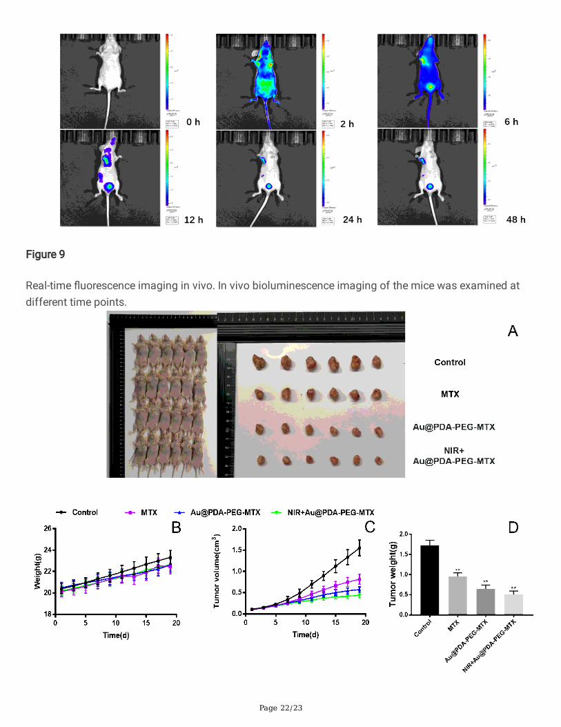
As shown in the Figure 9, after injection of Au @PDA-PEG-MTX NPs into the body through the tail vein, themice exposed to NIR showed obvious bio�uorescence in the tumor and surrounding areas. After 6 hours,the �uorescent signal was detected in the tumor area as early as possible, and the complete aggregationto the tumor site was �nally completed at 12 hours, which con�rmed the tumor-targeted imaging of Au@PDA-PEG-MTX NPs in vivo.
The anti-BC activity in vivo was evaluated in BALB/c nude mice bearing MDA-MB-231 cancer xenografts.The BC tumor volume of mice in the control group (normal saline) increased by about 15 times. After 19days of treatment, the tumor weight of BC treated with MTX alone was reduced by 44.68%. The inhibitionrate of BC tumors in the Au @PDA-PEG-MTX NPs group under NIR irradiation was 70.21%, which wasmuch higher than that when MTX was used alone.
Photos of BALB/c nude mice and BC solid tumors were arranged in the Figure 10 A. The results showedthat tail vein injection of MTX alone could slightly suppress the volume of BC tumors. However, after Au@PDA-PEG-MTX NPs combined with near-infrared radiation treatment, the BC tumor volume wassigni�cantly suppressed. Statistical analysis of BC tumor volume (Figure 10 B), body weight changes ofmice (Figure 10 C) and BC tumor weight (Figure10 D) further con�rmed this in vivo growth inhibitoryeffect.
Safety evaluation in vitro
Side effect of the Au @PDA-PEG-MTX NPs and NIR+Au @PDA-PEG-MTX NPs were examined to evaluateits safety. H&E staining was performed for histopathological changes. As shown in Figure 11, in thecontrol group, cardiomyocytes were arranged neatly, with abundant cytoplasm, intact membrane andclearly visible nucleus, with no or occasional in�ammatory cell in�ltration. The Au @PDA-PEG-MTX NPsand NIR+Au @PDA-PEG-MTX NPs groups were similar to the control group. In the control group, the

Page 11/23
number of hepatocytes was very abundant, the hepatic cords were neatly arranged, clear, and complete,without obvious abnormalities, the blood vessels were round, there was little in�ammation around theliver, and the liver lobules were rarely in�amed. In the Au @PDA-PEG-MTX NPs group, hepatocytes wereslightly swollen and deformed, hepatocytes had many fat droplets, liver lobules were degenerated, livercords were arranged irregularly, and liver lobules were in�amed. The NIR+Au @PDA-PEG-MTX NPs groupwas similar to the control group. In the control group, the glomerulus volume was roughly normal, the sizewas relatively uniform, the glomerular basement membrane was roughly intact, the tubular epithelial cellswere arranged neatly, and the tubular and interstitial-interstitial structures were acceptable. The volumeand size of the glomeruli in the Au @PDA-PEG-MTX NPs group were inconsistent. The cells in theglomeruli increased more than normal, the extracellular matrix increased more than normal, and thearrangement of renal tubular epithelial cells was irregular. The structure of the renal tubules was unclear.The NIR+Au @PDA-PEG-MTX NPs group was similar to the control group. In the control group and the twoexperimental groups, the lung tissue structure was relatively clear, the entire alveolar structure wasrelatively complete, the thickness of the alveolar wall was relatively normal, and the degree of bronchialstenosis was relatively light. Alveolar epithelial cells, eosinophils and lymphocytes rarely in�ltrate thealveolar cavity, and the congestion sites in the alveoli and alveolar septum were signi�cantly reduced. Inthe control group, the spleen had a clear structure, red and white pulps were neatly distributed, and nosinus congestion was seen. The structure of splenic nodules was clear. The structural characteristics ofthe two experimental groups were similar to those of the control group.
These results indicated that the Au @ PDA-PEG-MTX NPs group and NIR+Au @ PDA-PEG-MTX NPs hadsimilar histological characteristics compared with the control group. Except for the slight liver and kidneydamage in the Au @ PDA-PEG-MTX NPs group, no signi�cant damage or in�ammation was observed inall groups.
ConclusionA novel nanoplatform based on hierarchical drug release for chemo-photothermal treatment on BC wasdesigned, which showed excellent antitumor e�cacy and low toxicity. As a drug carrier, AuNPs showgood biocompatibility, stability and drug release triggered by NIR laser irradiation/pH. When the Au@PDA-PEG-MTX NPs were taken up by tumor cells, MTX was released through amide bond cleavage inthe speci�c acidic microenvironment (pH 5.5) of the lysosome to achieve the �rst step of chemotherapy.Subsequently, NIR laser irradiation caused the AuNPs to generate heat, and the MTX adsorbed on thesurface of dopamine was released in a second step. The synthesized nano drug-carrying systemgenerates heat as well as a large amount of ROS, realizing chemotherapy, photothermal andphotodynamic multiple therapy to treat BC. The as-synthesized NPs promoted circulation and targeteddelivery of the drug accompanied with bioimaging. We believe that the combination of multiple therapiesto treat cancer is a promising strategy that will accelerate the further development of the �eld ofoncology. In future research, detailed mechanism in the delivery process and action forms will be pursuedin-depth for clinical purpose.

Page 12/23
DeclarationsEthics approval and consent to participate
All animal experiments were conducted under the Ethical and Regulatory Guidelines for AnimalExperiments de�ned by Institute of Basic Theory, China Academy of Chinese Medical Sciences (LicenseNumber: SCXK (Beijing) 2016-0011, SYXK (Beijing) 2017-0033).
Consent for publication
All authors agreed to submit this manuscript.
Availability of data and materials
All data generated or analyzed during this study are included in this published article.
Competing Interests
The authors have declared that no competing interest exists.
Funding
This work was supported by the National Science Foundation of China (Project No. 81573569 and81873009) and National Science and Technology Major Project (2018ZX10101001-005-003 and2018ZX10101-001-005-004). All special thanks for the long-term subsidy mechanism from the Ministryof Finance and the Ministry of Education of PRC for BUCM.
Authors' contributions
WL and YYL designed the project. LCYY, QCH and DJZ were involved in the discussion. CL and APLdirected the experiment. WL and ZWC performed the experiments. WL analyzed the data. WL and YYLwrote the manuscript. All authors read and approved the �nal manuscript.
Acknowledgments
Not applicable.
Authors' information
1School of Chinese Materia Medica, Beijing University of Chinese Medicine, Beijing 100029, China.2Institute of Basic Research in Clinical Medicine, China Academy of Chinese Medical Sciences, Beijing100700, China. 3School of Chinese Medicine, Hong Kong Baptist University, Kowloon, Hongkong, China.
References

Page 13/23
[1] Adak A, Unal YC, Yucel S, Vural Z, Turan FB, Yalcin-Ozuysal O, et al. Connexin 32 induces pro-tumorigenic features in MCF10A normal breast cells and MDA-MB-231 metastatic breast cancer cells.Biochimica et biophysica acta Molecular cell research 2020;1867:118851.
[2] Xiong K, Zhang Y, Wen Q, Luo J, Lu Y, Wu Z, et al. Co-delivery of paclitaxel and curcumin bybiodegradable polymeric nanoparticles for breast cancer chemotherapy. International journal ofpharmaceutics 2020;589:119875.
[3] Kim J, Shim M, Yang S, Moon Y, Song S, Choi J, et al. Combination of cancer-speci�c prodrugnanoparticle with Bcl-2 inhibitor to overcome acquired drug resistance. Journal of controlled release :o�cial journal of the Controlled Release Society 2020.
[4] Venetis K, Invernizzi M, Sajjadi E, Curigliano G, Fusco N. Cellular immunotherapy in breast cancer: Thequest for consistent biomarkers. Cancer treatment reviews 2020;90:102089.
[5] Nunnery SE, Mayer IA. Targeting the PI3K/AKT/mTOR Pathway in Hormone-Positive Breast Cancer.Drugs 2020.
[6] Tabassam Q, Mehmood T, Raza A, Ullah A, Saeed F, Anjum F. Synthesis, Characterization and Anti-Cancer Therapeutic Potential of Withanolide-A with 20nm sAuNPs Conjugates Against SKBR3 BreastCancer Cell Line. International journal of nanomedicine 2020;15:6649-58.
[7] Zhu Y, Yang L, Xu J, Yang X, Luan P, Cui Q, et al. Discovery of the anti-angiogenesis effect ofeltrombopag in breast cancer through targeting of HuR protein. Acta pharmaceutica Sinica B2020;10:1414-25.
[8] He H, Liu L, Zhang S, Zheng M, Ma A, Chen Z, et al. Smart gold nanocages for mild heat-triggered drugrelease and breaking chemoresistance. Journal of controlled release : o�cial journal of the ControlledRelease Society 2020;323:387-97.
[9] Wang W, Li D, Zhang Y, Zhang W, Ma P, Wang X, et al. One-pot synthesis of hyaluronic acid-coated goldnanoparticles as SERS substrate for the determination of hyaluronidase activity. Mikrochimica acta2020;187:604.
[10] Bhatia E, Banerjee R. Hybrid silver-gold nanoparticles suppress drug resistant polymicrobial bio�lmformation and intracellular infection. Journal of materials chemistry B 2020;8:4890-8.
[11] Bai X, Wang Y, Song Z, Feng Y, Chen Y, Zhang D, et al. The Basic Properties of Gold Nanoparticles andtheir Applications in Tumor Diagnosis and Treatment. International journal of molecular sciences2020;21.
[12] Chandrasekaran R, Madheswaran T, Tharmalingam N, Bose R, Park H, Ha D. Labeling and trackingcells with gold nanoparticles. Drug discovery today 2020.

Page 14/23
[13] Wang J, Zhang Y, Jin N, Mao C, Yang M. Protein-Induced Gold Nanoparticle Assembly for Improvingthe Photothermal Effect in Cancer Therapy. ACS applied materials & interfaces 2019;11:11136-43.
[14] Zheng T, Wang W, Wu F, Zhang M, Shen J, Sun Y. Zwitterionic Polymer-Gated Au@TiO2 Core-ShellNanoparticles for Imaging-Guided Combined Cancer Therapy. Theranostics 2019;9:5035-48.
[15] Hou Z, Wang Z, Liu R, Li H, Zhang Z, Su T, et al. The effect of phospho-peptide on the stability of goldnanoparticles and drug delivery. Journal of Nanobiotechnology 2019;17.
[16] Amouzadeh Tabrizi M, Shamsipur M, Saber R, Sarkar S. Isolation of HL-60 cancer cells from thehuman serum sample using MnO2-PEI/Ni/Au/aptamer as a novel nanomotor and electrochemicaldetermination of thereof by aptamer/gold nanoparticles-poly(3,4-ethylene dioxythiophene) modi�ed GCelectrode. Biosensors & bioelectronics 2018;110:141-6.
[17] Malaikolundhan H, Mookkan G, Krishnamoorthi G, Matheswaran N, Alsawalha M, Veeraraghavan V,et al. Albizia lebbeckAnticarcinogenic effect of gold nanoparticles synthesized from on HCT-116 coloncancer cell lines. Arti�cial cells, nanomedicine, and biotechnology 2020;48:1206-13.
[18] Zheng Y, Zhang J, Zhang R, Luo Z, Wang C, Shi S. Gold nano particles synthesized from Magnoliao�cinalis and anticancer activity in A549 lung cancer cells. Arti�cial cells, nanomedicine, andbiotechnology 2019;47:3101-9.
[19] Scarano S, Palladino P, Pascale E, Brittoli A, Minunni M. Colorimetric determination of p-nitrophenolby using ELISA microwells modi�ed with an adhesive polydopamine nano�lm containing catalyticallyactive gold nanoparticles. Mikrochimica acta 2019;186:146.
[20] Sy KHS, Ho LWC, Lau WCY, Ko H, Choi CHJ. Morphological Diversity, Protein Adsorption, and CellularUptake of Polydopamine-Coated Gold Nanoparticles. Langmuir : the ACS journal of surfaces and colloids2018;34:14033-45.
[21] Cai S, Yan J, Xiong H, Xing H, Liu Y, Liu S, et al. Aptamer-Functionalized Molybdenum Disul�deNanosheets for Tumor Cell Targeting and Lysosomal Acidic Environment/NIR Laser Responsive DrugDelivery to Realize Synergetic Chemo-Photothermal Therapeutic Effects. International journal ofpharmaceutics 2020:119948.
[22] Mao W, Kim HS, Son YJ, Kim SR, Yoo HS. Doxorubicin encapsulated clicked gold nanoparticleclusters exhibiting tumor-speci�c disassembly for enhanced tumor localization and computerizedtomographic imaging. Journal of controlled release : o�cial journal of the Controlled Release Society2018;269:52-62.
[23] Feito MJ, Diez-Orejas R, Cicuendez M, Casarrubios L, Rojo JM, Portoles MT. Characterization of M1and M2 polarization phenotypes in peritoneal macrophages after treatment with graphene oxidenanosheets. Colloids and surfaces B, Biointerfaces 2019;176:96-105.

Page 15/23
[24] Liu R, An Y, Jia W, Wang Y, Wu Y, Zhen Y, et al. Macrophage-mimic shape changeable nanomedicineretained in tumor for multimodal therapy of breast cancer. Journal of Controlled Release 2020;321:589-601.
[25] He Y, Cong C, Li X, Zhu R, Li A, Zhao S, et al. Nano-drug System Based on Hierarchical Drug Releasefor Deep Localized/Systematic Cascade Tumor Therapy Stimulating Antitumor Immune Responses.Theranostics 2019;9:2897-909.
[26] Murawala P, Tirmale A, Shiras A, Prasad BL. In situ synthesized BSA capped gold nanoparticles:effective carrier of anticancer drug methotrexate to MCF-7 breast cancer cells. Materials science &engineering C, Materials for biological applications 2014;34:158-67.
[27] Ong Y, Bañobre-López M, Costa Lima S, Reis S. A multifunctional nanomedicine platform for co-delivery of methotrexate and mild hyperthermia towards breast cancer therapy. Materials science &engineering C, Materials for biological applications 2020;116:111255.
[28] Ong YS, Banobre-Lopez M, Costa Lima SA, Reis S. A multifunctional nanomedicine platform for co-delivery of methotrexate and mild hyperthermia towards breast cancer therapy. Materials science &engineering C, Materials for biological applications 2020;116:111255.
[29] Ali EMM, Elashkar AA, El-Kassas HY, Salim EI. Methotrexate loaded on magnetite iron nanoparticlescoated with chitosan: Biosynthesis, characterization, and impact on human breast cancer MCF-7 cell line.International journal of biological macromolecules 2018;120:1170-80.
[30] Dutta B, Nema A, Shetake NG, Gupta J, Barick KC, Lawande MA, et al. Glutamic acid-coated Fe3O4nanoparticles for tumor-targeted imaging and therapeutics. Materials science & engineering C, Materialsfor biological applications 2020;112:110915.
[31] Chen TW, Jan IS, Chang DY, Lin CH, Chen IC, Chen HM, et al. Systemic treatment of breast cancer withleptomeningeal metastases using bevacizumab, etoposide and cisplatin (BEEP regimen) signi�cantlyimproves overall survival. Journal of neuro-oncology 2020;148:165-72.
[32] Wang C, Vazquez-Gonzalez M, Fadeev M, Sohn YS, Nechushtai R, Willner I. Thermoplasmonic-Triggered Release of Loads from DNA-Modi�ed Hydrogel Microcapsules Functionalized with AuNanoparticles or Au Nanorods. Small 2020;16:e2000880.
[33] Zhang X, Feng Y, Duan S, Su L, Zhang J, He F. Mycobacterium tuberculosis strain H37RvElectrochemical Sensor Mediated by Aptamer and AuNPs-DNA. ACS sensors 2019;4:849-55.
[34] Liu P, Wang Y, Liu Y, Tan F, Li J, Li N. S-nitrosothiols loaded mini-sized Au@silica nanorod elicitscollagen depletion and mitochondrial damage in solid tumor treatment. Theranostics 2020;10:6774-89.
[35] Sargazi A, Kamali N, Shiri F, Heidari Majd M. Hyaluronic acid/polyethylene glycol nanoparticles forcontrolled delivery of mitoxantrone. Arti�cial cells, nanomedicine, and biotechnology 2018;46:500-9.

Page 16/23
[36] Wu C, Tong Y, Wang P, Wang D, Wu S, Zhang J. Identi�cation of impurities in methotrexate drugsubstances using high-performance liquid chromatography coupled with a photodiode array detector andFourier transform ion cyclotron resonance mass spectrometry. Rapid communications in massspectrometry : RCM 2013;27:971-8.
[37] De AK, Muthiyan R, Mondal S, Mahanta N, Bhattacharya D, Ponraj P, et al. A Natural QuinazolineDerivative from Marine Sponge Hyrtios erectus Induces Apoptosis of Breast Cancer Cells via ROSProduction and Intrinsic or Extrinsic Apoptosis Pathways. Marine drugs 2019;17.
[38] Zhang Y, Hai Y, Miao Y, Qi X, Xue W, Luo Y, et al. The toxicity mechanism of different sized ironnanoparticles on human breast cancer (MCF7) cells. Food chemistry 2020;341:128263.
[39] Szoka L, Palka J. Capsaicin up-regulates pro-apoptotic activity of thiazolidinediones in glioblastomacell line. Biomedicine & pharmacotherapy = Biomedecine & pharmacotherapie 2020;132:110741.
[40] Liu X, Ma R, Yi B, Riker A, Xi Y. MicroRNAs are involved in the development and progression of gastriccancer. Acta pharmacologica Sinica 2020.
Figures
Figure 1

Page 17/23
Process diagram for Synthesis of nanoparticles, targeting tumor tissues in vivo, releasing drugs andpathways that cause apoptosis.
Figure 2
(A)Ultraviolet spectrum of synthesized 15nm AuNPs; (B)FT-IR spectra of the PEG-MTX; (C)Trend of Au, Au@PDA, Au @PDA-PEG-MTX zeta potential; (D) Size distribution of Au,Au @PDA,Au @PDA-PEG-MTX.

Page 18/23
Figure 3
From left to right were the morphological characteristics of the synthesized Au, Au @PDA, Au @PDA-PEG-MTX under the TEM.
Figure 4

Page 19/23
(A) In vitro drug release experiment, Au @PDA-PEG-MTX placed in the simulated tumor acid environmentpH 5.5, MTX released by NIR irradiation with 808nm and MTX released by nanometers not irradiated by808nm NIR; (B) Au @PDA-PEG-MTX placed in the simulated normal physiological environment pH 7.4,release of MTX by nanometers.
Figure 5
The cytotoxicity of MTX, Au @PDA-PEG-MTX and NIR+Au @PDA-PEG-MTX against MDA-MB-231 cells.

Page 20/23
Figure 6
CLSM images of MDA-MB-231cells incubated with 15μg/ml, 20μg/ml, 25μg/ml Au @PDA-PEG-MTX.

Page 21/23
Figure 7
ROS generation in MDA-MB-231 cells with blank, Au @PDA-PEG-MTX, NIR + Au @PDA-PEG-MTX.
Figure 8
Expression of Cas-9, Cas-3, Bal-2, Bax protein in MTX MDA-MB-231 cells with MTX, Au @PDA-PEG-MTXand NIR+Au @PDA-PEG-MTX. *p < 0.05, **p < 0.01.
Page 22/23
Figure 9
Real-time �uorescence imaging in vivo. In vivo bioluminescence imaging of the mice was examined atdifferent time points.

Page 23/23
Figure 10
In vivo therapeutic effects. (A) Images of tumors; (B) Changes of BALB/c nude mice weight; (C) Changesof tumor volume; (D) Tumor weight measured in the nineteenth day. *p < 0.05, **p < 0.01.
Figure 11
Histological analyses by H&E staining of heart, liver, spleen, lung, and kidney in BALB/c mice that weretreated with 0.9% NaCl, Au @PDA-PEG-MTX, and NIR+Au @PDA-PEG-MTX.
Supplementary Files
This is a list of supplementary �les associated with this preprint. Click to download.
GraphicalAbstract.docx