effects of alloying elements (mn, co, al, w, sn, b, c and s...
TRANSCRIPT

Acta Biomaterialia 7 (2011) 1407–1420
Contents lists available at ScienceDirect
Acta Biomaterialia
journal homepage: www.elsevier .com/locate /actabiomat
Effects of alloying elements (Mn, Co, Al, W, Sn, B, C and S) on biodegradabilityand in vitro biocompatibility of pure iron
B. Liu a, Y.F. Zheng a,b,⇑a State Key Laboratory for Turbulence and Complex System and Department of Advanced Materials and Nanotechnology, College of Engineering, PekingUniversity, Beijing 100871, People’s Republic of Chinab Center for Biomedical Materials and Tissue Engineering, Academy for Advanced Interdisciplinary Studies, Peking University, Beijing 100871, People’s Republic of China
a r t i c l e i n f o a b s t r a c t
Article history:Received 25 June 2010Received in revised form 23 September2010Accepted 1 November 2010Available online 4 November 2010
Keywords:IronBiodegradable metalCorrosionCytotoxicityHemocompatibility
1742-7061/$ - see front matter � 2010 Acta Materialdoi:10.1016/j.actbio.2010.11.001
⇑ Corresponding author at: State Key LaboratorySystem and Department of Advanced Materials andEngineering, Peking University, Beijing 100871, People+86 10 6276 7411.
E-mail address: [email protected] (Y.F. Zheng).
Pure iron was determined to be a valid candidate material for biodegradable metallic stents in recent ani-mal tests; however, a much faster degradation rate in physiological environments was desired. C, Mn, Si,P, S, B, Cr, Ni, Pb, Mo, Al, Ti, Cu, Co, V and W are common alloying elements in industrial steels, with Cr, Ni,Mo, Cu, Ti, V and Si being acknowledged as beneficial in enhancing the corrosion resistance of iron. Thepurpose of the present work (using Fe–X binary alloy models) is to explore the effect of the remainingalloying elements (Mn, Co, Al, W, B, C and S) and one detrimental impurity element Sn on the biodegrad-ability and biocompatibility of pure iron by scanning electron microscopy, X-ray diffraction, metallo-graphic observation, tensile testing, microhardness testing, electrochemical testing, static (for6 months) and dynamic (for 1 month with various dissolved oxygen concentrations) immersion testing,cytotoxicity testing, hemolysis and platelet adhesion testing. The results showed that the addition of allalloying elements except for Sn improved the mechanical properties of iron after rolling. Localized cor-rosion of Fe–X binary alloys was observed in both static and dynamic immersion tests. Except for theFe–Mn alloy, which showed a significant decrease in corrosion rate, the other Fe–X binary alloy corrosionrates were close to that of pure iron. It was found that compared with pure iron all Fe–X binary alloysdecreased the viability of the L929 cell line, none of experimental alloying elements significantly reducedthe viability of vascular smooth muscle cells and all the elements except for Mn increased the viability ofthe ECV304 cell line. The hemolysis percentage of all Fe-X binary alloy models were less than 5%, and nosign of thrombogenicity was observed. In vitro corrosion and the biological behavior of these Fe–X binaryalloys are discussed and a corresponding mechanism of corrosion of Fe–X binary alloys in Hank’s solutionproposed. As a concluding remark, Co, W, C and S are recommended as alloying elements for biodegrad-able iron-based biomaterials.
� 2010 Acta Materialia Inc. Published by Elsevier Ltd. All rights reserved.
1. Introduction
Biodegradable materials have been considered as promisingcandidates for ideal stents because they can provide short-termvessel scaffolding and avoid long-term clinical problems, such asin-stent restenosis, late stent thrombosis, and the need for pro-longed anti-platelet therapy [1,2]. Theoretically, an ideal biode-gradable stent should demonstrate a compromise betweendegradation and mechanical integrity: specifically, it would bethe best that a stent material maintains its integrity for the first6–12 months but be totally degraded after 12–24 months [2].
ia Inc. Published by Elsevier Ltd. A
for Turbulence and ComplexNanotechnology, College of
’s Republic of China. Tel./fax:
Nowadays, Mg-based [3–7] and Fe-based [8–12] alloys are twoclasses of widely researched biodegradable metallic stent materi-als. Although the interest in Mg-based alloys has been increasing,poor mechanical properties compared with 316L stainless steel,fast degradation rate in physiological environments and significanthydrogen evolution during the corrosion process might limit theirfuture biomedical application.
Recent results of in vivo animal tests [13–15] showed iron to bea suitable metal for the production of biodegradable stents withoutsignificant obstruction of the stented vessel due to inflammation,neointimal proliferation, or thrombotic events, but a faster degra-dation rate is desirable since in the previous works large portionsof the biodegradable iron stent remained intact after 1 year. Asproposed by Schinhammer et al. [12], the design strategy for Fe-based alloys should aim at both an increase in the degradation rateand an improvement in the mechanical properties by modifyingthe chemical composition and microstructural characteristics.
ll rights reserved.
1408 B. Liu, Y.F. Zheng / Acta Biomaterialia 7 (2011) 1407–1420
Some pioneering work had been done on the following threeaspects.
(i) Alloying. Hermawan et al. [8,11] developed Fe–Mn alloyswith higher corrosion rates than that of pure iron and thesebinary alloys showed low inhibition of fibroblast cell meta-bolic activity in cell viability tests.
(ii) Surface modification. Zhu et al. [10] prepared Fe–O thin filmson a pure iron surface by plasma immersion ion implanta-tion and deposition, which effectively improved both thecorrosion resistance and biocompatibility.
(iii) Novel fabrication methods. Moravej et al. [16] fabricated pureiron foils with a fine grain microstructure, suitable mechan-ical properties, and moderate corrosion rate by an electro-forming technique. Electroformed pure iron showed fasterdegradation than pure iron fabricated by a casting techniqueand did not result in a decrease in metabolic activity whenexposed to primary rat smooth muscle cells [17].
As is well known, C, Mn, Si, P, S, B, Cr, Ni, Pb, Mo, Al, Ti, Cu, Co, V,and W are common alloying elements in industrial steels[18] (http://www.materialsengineer.com/E-Alloying-Steels.htm).Among these elements: (i) C is the major alloying element in steelsand has different effects on the corrosion properties under differ-ent circumstances; (ii) alloying elements Cr, Ni, Mo, Cu, Ti, V, andSi are used in stainless steels and are acknowledged as being ben-eficial in enhancing corrosion resistance; (iii) a consensus has notyet been reached on the influence of the elements Mn, B, Al, Co,and W the on corrosion properties after alloying with iron; (iv)the elements S and P are normally controlled at low levels becauseof their adverse effects on ductility and toughness; and (v) the ele-ment Pb is not only nephrotoxic but also toxic to the hematopoieticand nervous systems [19]. For the industrial application of Fe-based alloys a fundamental criterion is to improve the corrosionresistance of iron under various working conditions. To theauthors’ knowledge no consideration of how to increase the corro-sion rate of iron by alloying has been made from the viewpoint of
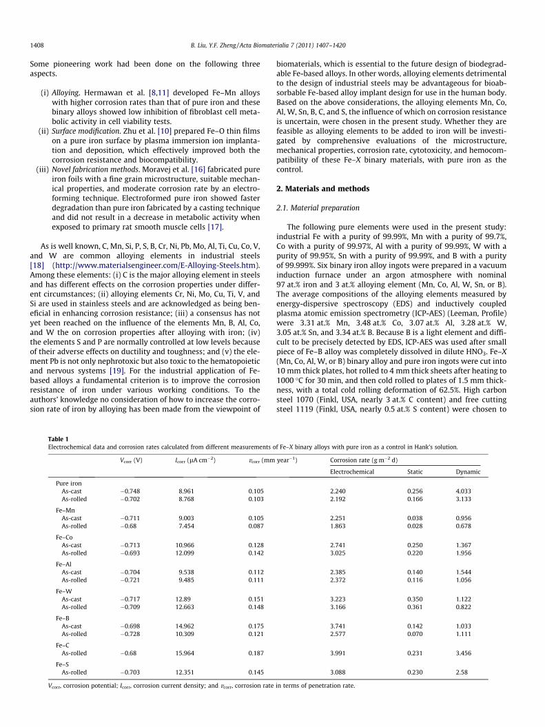
Table 1Electrochemical data and corrosion rates calculated from different measurements o
Vcorr (V) Icorr (lA cm�2) vcorr (mm
Pure ironAs-cast �0.748 8.961 0.105As-rolled �0.702 8.768 0.103
Fe–MnAs-cast �0.711 9.003 0.105As-rolled �0.68 7.454 0.087
Fe–CoAs-cast �0.713 10.966 0.128As-rolled �0.693 12.099 0.142
Fe–AlAs-cast �0.704 9.538 0.112As-rolled �0.721 9.485 0.111
Fe–WAs-cast �0.717 12.89 0.151As-rolled �0.709 12.663 0.148
Fe–BAs-cast �0.698 14.962 0.175As-rolled �0.728 10.309 0.121
Fe–CAs-rolled �0.68 15.964 0.187
Fe–SAs-rolled �0.703 12.351 0.145
Vcorr, corrosion potential; Icorr, corrosion current density; and vcorr, corrosion rate
biomaterials, which is essential to the future design of biodegrad-able Fe-based alloys. In other words, alloying elements detrimentalto the design of industrial steels may be advantageous for bioab-sorbable Fe-based alloy implant design for use in the human body.Based on the above considerations, the alloying elements Mn, Co,Al, W, Sn, B, C, and S, the influence of which on corrosion resistanceis uncertain, were chosen in the present study. Whether they arefeasible as alloying elements to be added to iron will be investi-gated by comprehensive evaluations of the microstructure,mechanical properties, corrosion rate, cytotoxicity, and hemocom-patibility of these Fe–X binary materials, with pure iron as thecontrol.
2. Materials and methods
2.1. Material preparation
The following pure elements were used in the present study:industrial Fe with a purity of 99.99%, Mn with a purity of 99.7%,Co with a purity of 99.97%, Al with a purity of 99.99%, W with apurity of 99.95%, Sn with a purity of 99.99%, and B with a purityof 99.999%. Six binary iron alloy ingots were prepared in a vacuuminduction furnace under an argon atmosphere with nominal97 at.% iron and 3 at.% alloying element (Mn, Co, Al, W, Sn, or B).The average compositions of the alloying elements measured byenergy-dispersive spectroscopy (EDS) and inductively coupledplasma atomic emission spectrometry (ICP-AES) (Leeman, Profile)were 3.31 at.% Mn, 3.48 at.% Co, 3.07 at.% Al, 3.28 at.% W,3.05 at.% Sn, and 3.34 at.% B. Because B is a light element and diffi-cult to be precisely detected by EDS, ICP-AES was used after smallpiece of Fe–B alloy was completely dissolved in dilute HNO3. Fe–X(Mn, Co, Al, W, or B) binary alloy and pure iron ingots were cut into10 mm thick plates, hot rolled to 4 mm thick sheets after heating to1000 �C for 30 min, and then cold rolled to plates of 1.5 mm thick-ness, with a total cold rolling deformation of 62.5%. High carbonsteel 1070 (Finkl, USA, nearly 3 at.% C content) and free cuttingsteel 1119 (Finkl, USA, nearly 0.5 at.% S content) were chosen to
f Fe–X binary alloys with pure iron as a control in Hank’s solution.
year�1) Corrosion rate (g m�2 d)
Electrochemical Static Dynamic
2.240 0.256 4.0332.192 0.166 3.133
2.251 0.038 0.9561.863 0.028 0.678
2.741 0.250 1.3673.025 0.220 1.956
2.385 0.140 1.5442.372 0.116 1.056
3.223 0.350 1.1223.166 0.361 0.822
3.741 0.142 1.0332.577 0.070 1.111
3.991 0.231 3.456
3.088 0.230 2.58
in terms of penetration rate.
B. Liu, Y.F. Zheng / Acta Biomaterialia 7 (2011) 1407–1420 1409
investigate the effects of the alloying elements C and S on iron.They were classified in the as-rolled group. Stainless steel 316L(Goodfellow, USA, cold rolled condition) was used as the reference.Specimens were cut into square plates (10 � 10 � 1.5 mm) for cor-rosion, cytotoxicity and hemocompatibility tests. Each specimenwas mechanically polished up to 2000 grit, ultrasonically cleanedin absolute ethanol, and then dried in the open air. For cytotoxicitytesting the specimens were further sterilized with ultraviolet radi-ation for at least 2 h.
2.2. Microstructural characterization
The microstructure of the experimental specimens was ob-served by optical microscopy (OM) (Olympus BX51 M) after etch-ing with a 4% HNO3/alcohol solution. X-ray diffraction (XRD)(Rigaku DMAX 2400) using Cu Ka radiation was employed to iden-tify the constituent phases of pure iron and the Fe–X binary alloys.
2.3. Mechanical test
The mechanical properties of pure iron and the Fe–X binary al-loys were determined by tensile testing according to ASTM-E8-04[20] (gage length 26 mm, length of grip section 24 mm, width8 mm, radius of fillet 6 mm, thickness 2.5 mm) and microhardnesstests. The tensile tests were carried out at a displacement rate of1 mm min�1 with an Instron 3365 universal test machine. An aver-age of three measurements was taken for each group. Microhard-ness measurements were taken with a Vickers indenter appliedto the specimens at a load of 1 kgf and dwell time of 10 s using amicrohardness tester (Shimadzu HMV-2T). An average of at leastfive measurements was taken for each group.
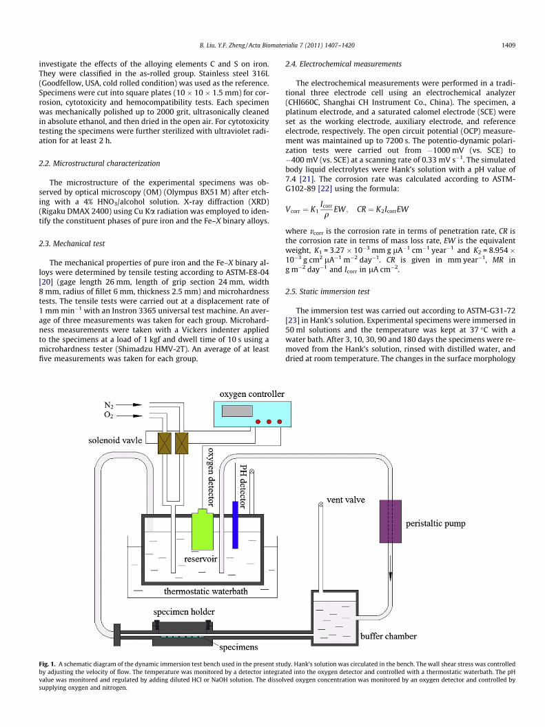
Fig. 1. A schematic diagram of the dynamic immersion test bench used in the present stuby adjusting the velocity of flow. The temperature was monitored by a detector integratvalue was monitored and regulated by adding diluted HCl or NaOH solution. The dissolsupplying oxygen and nitrogen.
2.4. Electrochemical measurements
The electrochemical measurements were performed in a tradi-tional three electrode cell using an electrochemical analyzer(CHI660C, Shanghai CH Instrument Co., China). The specimen, aplatinum electrode, and a saturated calomel electrode (SCE) wereset as the working electrode, auxiliary electrode, and referenceelectrode, respectively. The open circuit potential (OCP) measure-ment was maintained up to 7200 s. The potentio-dynamic polari-zation tests were carried out from �1000 mV (vs. SCE) to�400 mV (vs. SCE) at a scanning rate of 0.33 mV s�1. The simulatedbody liquid electrolytes were Hank’s solution with a pH value of7.4 [21]. The corrosion rate was calculated according to ASTM-G102-89 [22] using the formula:
Vcorr ¼ K1Icorr
qEW ; CR ¼ K2IcorrEW
where vcorr is the corrosion rate in terms of penetration rate, CR isthe corrosion rate in terms of mass loss rate, EW is the equivalentweight, K1 = 3.27 � 10�3 mm g lA�1 cm�1 year�1 and K2 = 8.954 �10�3 g cm2 lA�1 m�2 day�1. CR is given in mm year�1, MR ing m�2 day�1 and Icorr in lA cm�2.
2.5. Static immersion test
The immersion test was carried out according to ASTM-G31-72[23] in Hank’s solution. Experimental specimens were immersed in50 ml solutions and the temperature was kept at 37 �C with awater bath. After 3, 10, 30, 90 and 180 days the specimens were re-moved from the Hank’s solution, rinsed with distilled water, anddried at room temperature. The changes in the surface morphology
dy. Hank’s solution was circulated in the bench. The wall shear stress was controlleded into the oxygen detector and controlled with a thermostatic waterbath. The pH
ved oxygen concentration was monitored by an oxygen detector and controlled by
1410 B. Liu, Y.F. Zheng / Acta Biomaterialia 7 (2011) 1407–1420
of the specimens after immersion were characterized by environ-mental scanning electron microscope (ESEM) (AMRAY-1910FE)equipped with an EDS attachment and image measuring apparatus(HLEO, VCAD-1010). The concentrations of various elements re-leased into solution were determined by ICP-AES (Leeman, Profile)after the corrosion products were completely dissolved in HNO3.An average of three measurements was taken for each group. Theunit of corrosion rate was unified to g m�2 day�1 for the compari-son in Table 1 according to the formula:
CR ¼ cVST
where CR is the corrosion rate, c is the released ion concentration, Vis the volume of immersion solution, S is the surface area of thespecimen and t is the time. CR is given in g m�2 day�1, c in g ml�1,V in ml, S in m2 and t in days.
2.6. Dynamic immersion test
Following the pioneering work of Levesque et al. [24], a dy-namic immersion test bench was designed to simulate the bloodflow inside the coronary artery, shown schematically in Fig. 1.Since oxygen is an important factor in the corrosion of iron in neu-tral medium the dissolved oxygen concentration in Hank’s solutionwas regulated to better simulate the blood environment. In thisbench the wall shear stress (0.68 Pa) [25], temperature (36.9–37.1 �C), pH value (7.35–7.45), and dissolved oxygen (2.8–
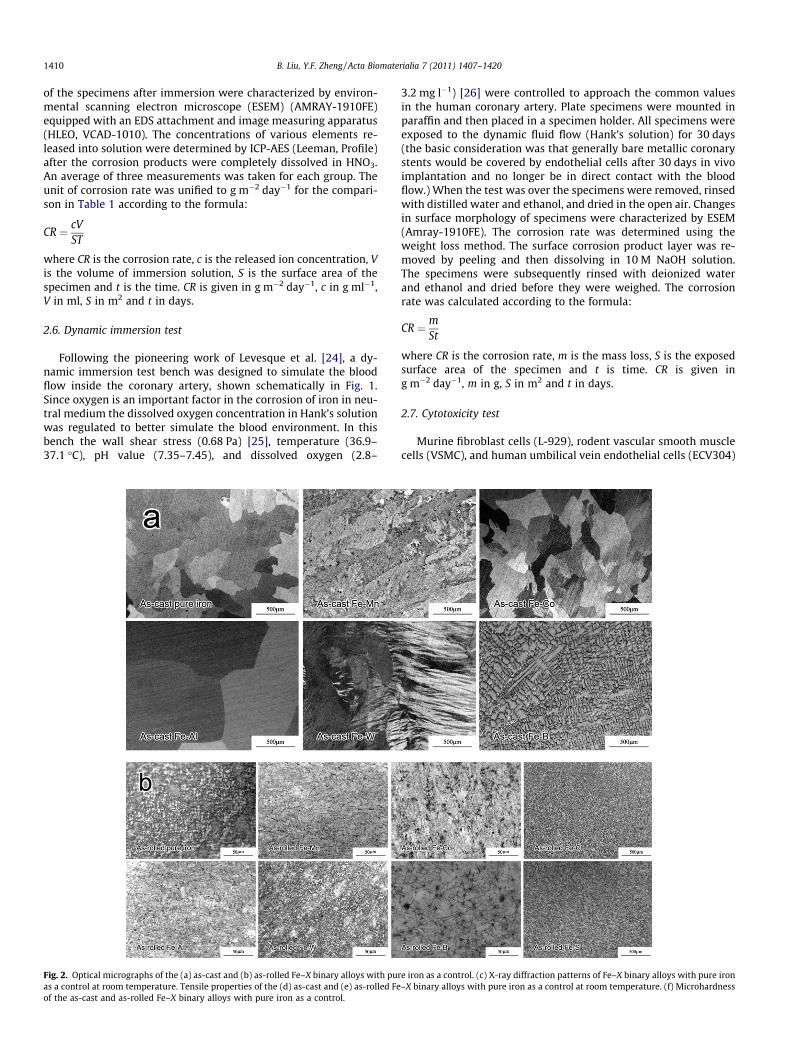
Fig. 2. Optical micrographs of the (a) as-cast and (b) as-rolled Fe–X binary alloys with puras a control at room temperature. Tensile properties of the (d) as-cast and (e) as-rolled Feof the as-cast and as-rolled Fe–X binary alloys with pure iron as a control.
3.2 mg l�1) [26] were controlled to approach the common valuesin the human coronary artery. Plate specimens were mounted inparaffin and then placed in a specimen holder. All specimens wereexposed to the dynamic fluid flow (Hank’s solution) for 30 days(the basic consideration was that generally bare metallic coronarystents would be covered by endothelial cells after 30 days in vivoimplantation and no longer be in direct contact with the bloodflow.) When the test was over the specimens were removed, rinsedwith distilled water and ethanol, and dried in the open air. Changesin surface morphology of specimens were characterized by ESEM(Amray-1910FE). The corrosion rate was determined using theweight loss method. The surface corrosion product layer was re-moved by peeling and then dissolving in 10 M NaOH solution.The specimens were subsequently rinsed with deionized waterand ethanol and dried before they were weighed. The corrosionrate was calculated according to the formula:
CR ¼ mSt
where CR is the corrosion rate, m is the mass loss, S is the exposedsurface area of the specimen and t is time. CR is given ing m�2 day�1, m in g, S in m2 and t in days.
2.7. Cytotoxicity test
Murine fibroblast cells (L-929), rodent vascular smooth musclecells (VSMC), and human umbilical vein endothelial cells (ECV304)
e iron as a control. (c) X-ray diffraction patterns of Fe–X binary alloys with pure iron–X binary alloys with pure iron as a control at room temperature. (f) Microhardness

B. Liu, Y.F. Zheng / Acta Biomaterialia 7 (2011) 1407–1420 1411
were cultured in the Dulbecco’s modified Eagle’s medium (DMEM),10% fetal bovine serum (FBS), 100 U ml�1 penicillin and100 lg ml�1 streptomycin at 37 �C in a humidified atmosphere of5% CO2. Following the instructions in ISO 10993-12, extractionmedium was prepared using DMEM serum-free medium with asurface area/extraction medium ratio 1 cm2 ml�1 in a humidifiedatmosphere with 5% CO2 at 37 �C for 72 h and the extraction med-ium was stored at 4 �C before the cytotoxicity test. The controlgroups involved the use of DMEM medium as a negative controland DMEM medium containing 10% dimethyl sulfoxide as a posi-tive control. Cells were incubated in 96-well cell culture plates at5 � 103 cells per 100 ll of medium in each well and incubatedfor 24 h to allow attachment. The medium was then replaced with100 ll of extraction medium. After incubating the cells in a humid-ified atmosphere with 5% CO2 at 37 �C for 1, 2, and 4 days the 96-well cell culture plates were observed under an optical microscope.After that, 10 ll of MTT were added to each well. The specimenswere incubated with MTT for 4 h at 37 �C and then 100 ll of forma-zan solubilization solution (10% sodium dodecyl sulfate in 0.01 MHCl) were added to each well for more than 10 h in the incubatorin a humidified atmosphere. The spectrophotometrical absorbanceof the specimens was measured with a microplate reader (Bio-Rad680) at 570 nm, with a reference wavelength of 630 nm. The con-centrations of iron and alloying element ions in the extractionmedium were also measured by ICP-AES (Leeman, Profile).
2.8. Hemolysis test
Healthy human blood from a volunteer containing sodium cit-rate (3.8 wt.%) at a ratio of 9:1 was taken and diluted with normal
Fig. 2 (cont
saline (4:5 ratio by volume). Pure iron and Fe–X binary alloy spec-imens were dipped in separate standard tubes containing 10 ml ofnormal saline that were previously incubated at 37 �C for 30 min.Then 0.2 ml of diluted blood was added to these standard tubesand the mixtures were incubated for 60 min at 37 �C. As a positivecontrol for hemolysis, 0.2 ml of blood was diluted in distilledwater, whereas saline diluted blood was added to an empty stan-dard tube which served as a negative control. After this periodspecimens were removed and all the tubes were centrifuged at800g for 5 min. The supernatant from each tube was transferredto a well in a 96-well plate where the absorbance was measuredwith a microplate reader (Bio-Rad 680) at 545 nm. Hemolysiswas calculated as follows:
Hemolysis ¼ OD ðtestÞ � OD ðnegative controlÞOD ðpositive controlÞ � OD ðnegative controlÞ � 100%
where OD is the optical density at 545 nm.
2.9. Platelet adhesion
Platelet-rich plasma (PRP) was prepared by centrifuging wholehuman blood from a volunteer at 200g for 15 min. The PRP coveredthe top surface of the experimental samples and was incubated at37 �C. After 1 h the specimens were gently rinsed with phosphate-buffered saline (PBS) to remove non-adherent platelets. They werethen fixed in 2.5% glutaraldehyde solutions for 1 h at room temper-ature, followed by dehydration in a gradient ethanol/distilledwater mixture from 50% to 100% in 10% increments for 10 min each
inued)

1412 B. Liu, Y.F. Zheng / Acta Biomaterialia 7 (2011) 1407–1420
and finally freeze-dried for 2 days. The surfaces of platelet-attached samples were observed by ESEM.
Fig. 3. Potentio-dynamic polarization curves of as-cast and as-rolled Fe–X binaryalloys immersed in Hank’s solution with pure iron as a control.
3. Results
3.1. Effects of alloying elements on the microstructure and mechanicalproperties of pure iron
Fig. 2a and b is a series of optical micrographs showing themicrostructure of various Fe–X binary alloys, with pure iron asthe control. For the as-cast condition the addition of B decreasesthe grain size from 400 to 100 lm, while the addition of Mn, Co,Al and W had no significant effect on grain size. For the as-rolledcondition the grain sizes of all the experimental Fe–X binary alloysand pure iron were about 10 lm. As a reference, the average grainsize of industrial steels 1070 and 1119 (as-rolled Fe–C and Fe–S al-loys) is about 30 lm. It can be seen from the XRD results in Fig. 2cthat -Fe is the main phase in all experimental Fe–X binary alloysand pure iron. Fig. 2d and e show the tensile properties of the as-cast and as-rolled Fe–X binary alloys, with pure iron as the control.(i) It can be seen that the Fe–Sn alloy exhibits poor mechanicalproperties because segregation of the alloying element Sn at grainboundaries leads to a deterioration in ductility [27,28]. (ii) Theaddition of various alloying elements has different influences onthe yield strength (YS), ultimate strength (US), and elongation ofpure iron in the as-cast group, but the differences between YSand US all increased after alloying. This is advantageous for theirfuture application as coronary stents, since the YS and US valuesof pure iron are close and fracture can easily occur during expan-sion [29]. (iii) The YS and US of all specimens are significantly im-proved and elongation is reduced after rolling. (iv) The addition ofMn, Co, Al, W, B, C, and S improves both the YS and US of pure ironin the as-rolled group. Fig. 2f illustrates the microhardness of theFe–X binary alloys and pure iron, with 316L SS as a reference.The microhardness of the Fe–X binary alloys are enhanced com-pared with pure iron, except for the as-cast Fe–Co alloy. After coldrolling deformation the microhardness of the Fe–X binary alloysand pure iron are comparable with that of 316L SS.
3.2. Effects of alloying elements on the corrosion behavior of pure iron
3.2.1. Electrochemical corrosion behaviorFig. 3 shows the potentio-dynamic polarization curves of as-
cast and as-rolled Fe–X binary alloy specimens immersed in Hank’ssolution, with pure iron as the control. The average electrochemi-cal parameters and corrosion rates are listed in Table 1. For the as-cast group the addition of various alloying elements increases thecorrosion potential of pure iron. For the as-rolled group addition ofthe elements Mn, Co and C is conducive to a higher corrosion po-tential, while addition of the elements Al, W, B and S results in alower corrosion potential. Except for the as-rolled Fe–Mn alloy,the addition of alloying elements increases the corrosion currentdensities compared with pure iron. However, the difference in cor-rosion current densities between the Fe–X binary alloys and pureiron is not significant. The corrosion rates of the Fe–X binary alloysand pure iron calculated from corrosion current densities are ofthe same order of magnitude. The corrosion rates of the Fe–X bin-ary alloys and pure iron decreased slightly after rolling, except forthe Fe–Co alloy. The as-rolled Fe–C alloy exhibits the most rapidcorrosion rate among the experimental alloys, judging from theelectrochemical data.
3.2.2. Static immersion corrosion behaviorAfter specimens had been immersed in Hank’s solution for
3 days most of the localized corrosion, with brown corrosion prod-
ucts, was observed at the edges of the specimens. After 30 days thesurfaces of only a very few specimens were completely covered bybrown corrosion products, which corresponded with higher re-leased ion concentrations. After 180 days the surfaces of more thanhalf of the specimens were completely covered, but locally brightand shining areas still existed on some specimens. The pH valuesof the Hank’s solution ranged from 7.25 to 8.48 after experimentalspecimens had been immersed for different periods. Fig. 4 showsthe surface morphology of the as-rolled Fe–W alloy after 180 daysimmersion, with typical localized corrosion features, however, thescratch caused by the mechanical polishing process can still beseen on the bright metal surface. In the brown areas where theFe–W alloy has already been corroded white CaP compounds areapparent on the cracked corrosion product layer, which demon-strates that CaP compounds are more easily formed on the cor-roded surface. A rough surface with a metallic luster is seen afterthe corrosion product layer is removed.
Fig. 5 shows the surface morphologies (the most serious corro-sion regions) of the Fe–X binary alloys after 180 days immersion inHank’s solution, with pure iron as the control. It can be seen that:(i) the corroded surface composition includes Fe, O, Ca, and P, as re-vealed by surface EDS analysis; (ii) the corrosion product layer isporous, non-compact and not tightly adhering to the original alloysurface. As a result of this the corrosion product layer easilyfalls away from the surface of the specimens, especially after
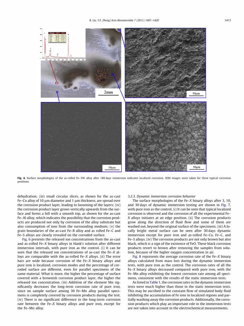
Fig. 4. Surface morphologies of the as-rolled Fe–3W alloy after 180 days immersion indicates localized corrosion. SEM images were taken for three typical corrosionpositions.
B. Liu, Y.F. Zheng / Acta Biomaterialia 7 (2011) 1407–1420 1413
dehydration; (iii) small circular slices, as shown for the as-castFe–Co alloy of 10 lm diameter and 1 lm thickness, are spread overthe corrosion product layer, leading to loosening of the layers; (iv)the corrosion product layer grows vertically upwards from the sur-face and forms a hill with a smooth top, as shown for the as-castFe–Al alloy, which indicates the possibility that the corrosion prod-ucts are produced not only by corrosion of the alloy substrate butalso consumption of ions from the surrounding medium; (v) thegrain boundaries of the as-cast Fe–B alloy and as rolled Fe–C andFe–S alloys are clearly revealed on the corroded surface.
Fig. 6 presents the released ion concentrations from the as-castand as-rolled Fe–X binary alloys in Hank’s solution after differentimmersion intervals, with pure iron as the control. (i) It can beseen that the released ion concentrations of as-cast the Fe–X al-loys are comparable with the as-rolled Fe–X alloys. (ii) The errorbars are wide because corrosion of the Fe–X binary alloys andpure iron is localized, corrosion modes and the percentage of cor-roded surface are different, even for parallel specimens of thesame material. What is more, the higher the percentage of surfacecovered with a brownish corrosion product layer, the higher thereleased ion concentration. (iii) Addition of the element Mn sig-nificantly decreases the long-term corrosion rate of pure iron,since no sample surface among 30 Fe–Mn alloy parallel speci-mens is completely covered by corrosion products during the test.(iv) There is no significant difference in the long-term corrosionrate between the Fe–X binary alloys and pure iron, except forthe Fe–Mn alloy.
3.2.3. Dynamic immersion corrosion behaviorThe surface morphologies of the Fe–X binary alloys after 3, 10,
and 30 days of dynamic immersion testing are shown in Fig. 7,with pure iron as the control. (i) It can be seen that typical localizedcorrosion is observed and the corrosion of all the experimental Fe–X alloys initiates at an edge position. (ii) The corrosion productsgrow along the direction of fluid flow and some of them arewashed out, beyond the original surface of the specimens. (iii) A lo-cally bright metal surface can be seen after 30 days dynamicimmersion except for pure iron and as-rolled Fe–Co, Fe–C, andFe–S alloys. (iv) The corrosion products are not only brown but alsoblack, which is a sign of the existence of FeO. These black corrosionproducts revert to brown after removing the samples from solu-tion, because of the higher oxygen concentration in air.
Fig. 8 represents the average corrosion rate of the Fe–X binaryalloys calculated from mass loss during the dynamic immersiontests, with pure iron as the control. The corrosion rates of all theFe–X binary alloys decreased compared with pure iron, with theFe–Mn alloy exhibiting the lowest corrosion rate among all speci-mens, consistent with the results of the static immersion tests.
As listed in Table 1, the corrosion rates in the dynamic immersiontests were much higher than those in the static immersion tests.This may be ascribed to the constant flow of simulated body fluidinhibiting the accumulation of Fe ions in localized regions and par-tially washing away the corrosion products. Additionally, the corro-sion products which play an important role in the immersion testsare not taken into account in the electrochemical measurements.
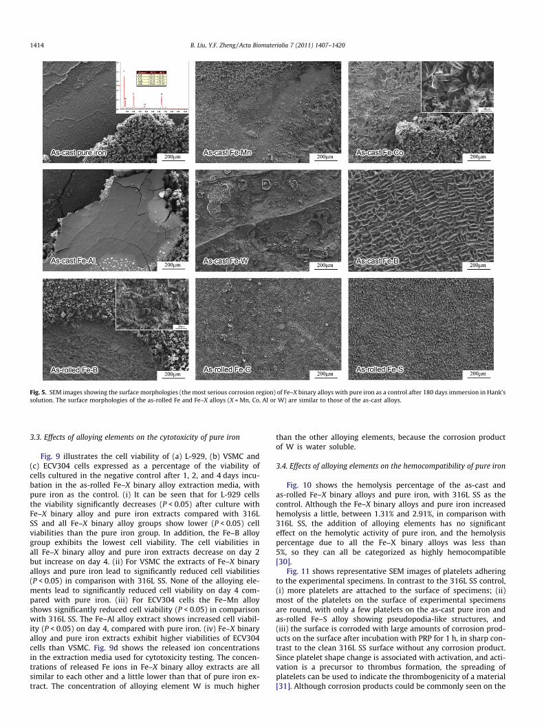
Fig. 5. SEM images showing the surface morphologies (the most serious corrosion region) of Fe–X binary alloys with pure iron as a control after 180 days immersion in Hank’ssolution. The surface morphologies of the as-rolled Fe and Fe–X alloys (X = Mn, Co, Al or W) are similar to those of the as-cast alloys.
1414 B. Liu, Y.F. Zheng / Acta Biomaterialia 7 (2011) 1407–1420
3.3. Effects of alloying elements on the cytotoxicity of pure iron
Fig. 9 illustrates the cell viability of (a) L-929, (b) VSMC and(c) ECV304 cells expressed as a percentage of the viability ofcells cultured in the negative control after 1, 2, and 4 days incu-bation in the as-rolled Fe–X binary alloy extraction media, withpure iron as the control. (i) It can be seen that for L-929 cellsthe viability significantly decreases (P < 0.05) after culture withFe–X binary alloy and pure iron extracts compared with 316LSS and all Fe–X binary alloy groups show lower (P < 0.05) cellviabilities than the pure iron group. In addition, the Fe–B alloygroup exhibits the lowest cell viability. The cell viabilities inall Fe–X binary alloy and pure iron extracts decrease on day 2but increase on day 4. (ii) For VSMC the extracts of Fe–X binaryalloys and pure iron lead to significantly reduced cell viabilities(P < 0.05) in comparison with 316L SS. None of the alloying ele-ments lead to significantly reduced cell viability on day 4 com-pared with pure iron. (iii) For ECV304 cells the Fe–Mn alloyshows significantly reduced cell viability (P < 0.05) in comparisonwith 316L SS. The Fe–Al alloy extract shows increased cell viabil-ity (P < 0.05) on day 4, compared with pure iron. (iv) Fe–X binaryalloy and pure iron extracts exhibit higher viabilities of ECV304cells than VSMC. Fig. 9d shows the released ion concentrationsin the extraction media used for cytotoxicity testing. The concen-trations of released Fe ions in Fe–X binary alloy extracts are allsimilar to each other and a little lower than that of pure iron ex-tract. The concentration of alloying element W is much higher
than the other alloying elements, because the corrosion productof W is water soluble.
3.4. Effects of alloying elements on the hemocompatibility of pure iron
Fig. 10 shows the hemolysis percentage of the as-cast andas-rolled Fe–X binary alloys and pure iron, with 316L SS as thecontrol. Although the Fe–X binary alloys and pure iron increasedhemolysis a little, between 1.31% and 2.91%, in comparison with316L SS, the addition of alloying elements has no significanteffect on the hemolytic activity of pure iron, and the hemolysispercentage due to all the Fe–X binary alloys was less than5%, so they can all be categorized as highly hemocompatible[30].
Fig. 11 shows representative SEM images of platelets adheringto the experimental specimens. In contrast to the 316L SS control,(i) more platelets are attached to the surface of specimens; (ii)most of the platelets on the surface of experimental specimensare round, with only a few platelets on the as-cast pure iron andas-rolled Fe–S alloy showing pseudopodia-like structures, and(iii) the surface is corroded with large amounts of corrosion prod-ucts on the surface after incubation with PRP for 1 h, in sharp con-trast to the clean 316L SS surface without any corrosion product.Since platelet shape change is associated with activation, and acti-vation is a precursor to thrombus formation, the spreading ofplatelets can be used to indicate the thrombogenicity of a material[31]. Although corrosion products could be commonly seen on the
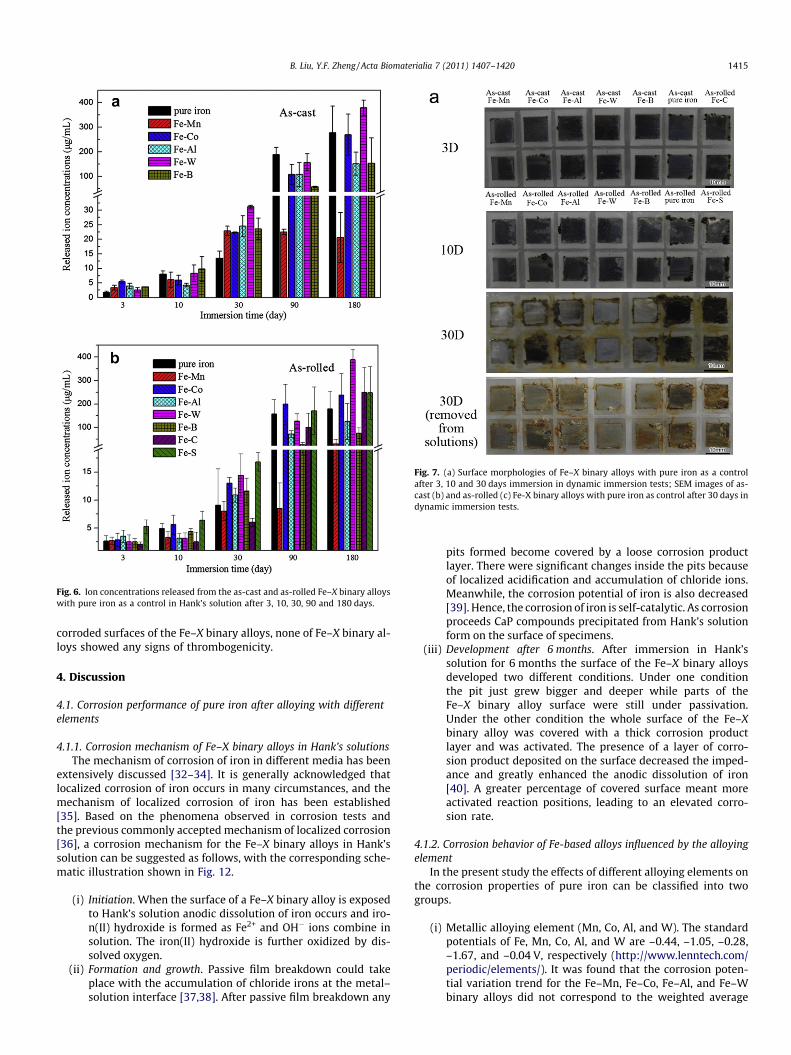
Fig. 6. Ion concentrations released from the as-cast and as-rolled Fe–X binary alloyswith pure iron as a control in Hank’s solution after 3, 10, 30, 90 and 180 days.
Fig. 7. (a) Surface morphologies of Fe–X binary alloys with pure iron as a controlafter 3, 10 and 30 days immersion in dynamic immersion tests; SEM images of as-cast (b) and as-rolled (c) Fe-X binary alloys with pure iron as control after 30 days indynamic immersion tests.
B. Liu, Y.F. Zheng / Acta Biomaterialia 7 (2011) 1407–1420 1415
corroded surfaces of the Fe–X binary alloys, none of Fe–X binary al-loys showed any signs of thrombogenicity.
4. Discussion
4.1. Corrosion performance of pure iron after alloying with differentelements
4.1.1. Corrosion mechanism of Fe–X binary alloys in Hank’s solutionsThe mechanism of corrosion of iron in different media has been
extensively discussed [32–34]. It is generally acknowledged thatlocalized corrosion of iron occurs in many circumstances, and themechanism of localized corrosion of iron has been established[35]. Based on the phenomena observed in corrosion tests andthe previous commonly accepted mechanism of localized corrosion[36], a corrosion mechanism for the Fe–X binary alloys in Hank’ssolution can be suggested as follows, with the corresponding sche-matic illustration shown in Fig. 12.
(i) Initiation. When the surface of a Fe–X binary alloy is exposedto Hank’s solution anodic dissolution of iron occurs and iro-n(II) hydroxide is formed as Fe2+ and OH� ions combine insolution. The iron(II) hydroxide is further oxidized by dis-solved oxygen.
(ii) Formation and growth. Passive film breakdown could takeplace with the accumulation of chloride irons at the metal–solution interface [37,38]. After passive film breakdown any
pits formed become covered by a loose corrosion productlayer. There were significant changes inside the pits becauseof localized acidification and accumulation of chloride ions.Meanwhile, the corrosion potential of iron is also decreased[39]. Hence, the corrosion of iron is self-catalytic. As corrosionproceeds CaP compounds precipitated from Hank’s solutionform on the surface of specimens.
(iii) Development after 6 months. After immersion in Hank’ssolution for 6 months the surface of the Fe–X binary alloysdeveloped two different conditions. Under one conditionthe pit just grew bigger and deeper while parts of theFe–X binary alloy surface were still under passivation.Under the other condition the whole surface of the Fe–Xbinary alloy was covered with a thick corrosion productlayer and was activated. The presence of a layer of corro-sion product deposited on the surface decreased the imped-ance and greatly enhanced the anodic dissolution of iron[40]. A greater percentage of covered surface meant moreactivated reaction positions, leading to an elevated corro-sion rate.
4.1.2. Corrosion behavior of Fe-based alloys influenced by the alloyingelement
In the present study the effects of different alloying elements onthe corrosion properties of pure iron can be classified into twogroups.
(i) Metallic alloying element (Mn, Co, Al, and W). The standardpotentials of Fe, Mn, Co, Al, and W are –0.44, –1.05, –0.28,–1.67, and –0.04 V, respectively (http://www.lenntech.com/periodic/elements/). It was found that the corrosion poten-tial variation trend for the Fe–Mn, Fe–Co, Fe–Al, and Fe–Wbinary alloys did not correspond to the weighted average

Fig. 7 (continued)
Fig. 8. Average corrosion rate of Fe–X binary alloys calculated from mass loss indynamic immersion tests with pure iron as a control.
1416 B. Liu, Y.F. Zheng / Acta Biomaterialia 7 (2011) 1407–1420
potential of their two component elements. The corrosioncurrent densities and corrosion rates of these alloys wereclose in terms of electrochemical properties.
(ii) Non-metallic alloying elements (B, C, and S). The solubility ofthe elements B, C, and S in Fe is less than 0.1 at.% at roomtemperature according to the binary phase diagrams, so sec-ond phase Fe2B, Fe3C, and FeS exists in these three Fe–X bin-ary alloys (although no diffraction peaks were observed inFig. 2). According to the corroded surface morphology afterthe corrosion products were removed, intergranular corro-sion could be seen, although localized corrosion was alsoobserved.
Based on the present results and previous works on steels, theeffect of alloying elements on the biodegradability of Fe-based al-loys could be described as follows.
(i) Slightly increased corrosion rate. According to the presentexperimental results Fe–X (X = Co, W, C, or S) binary alloysshow slightly increased corrosion rates in long-term immer-sion tests compared with pure iron. They were beneficial inthat they improved the corrosion rate of biodegradable iron.
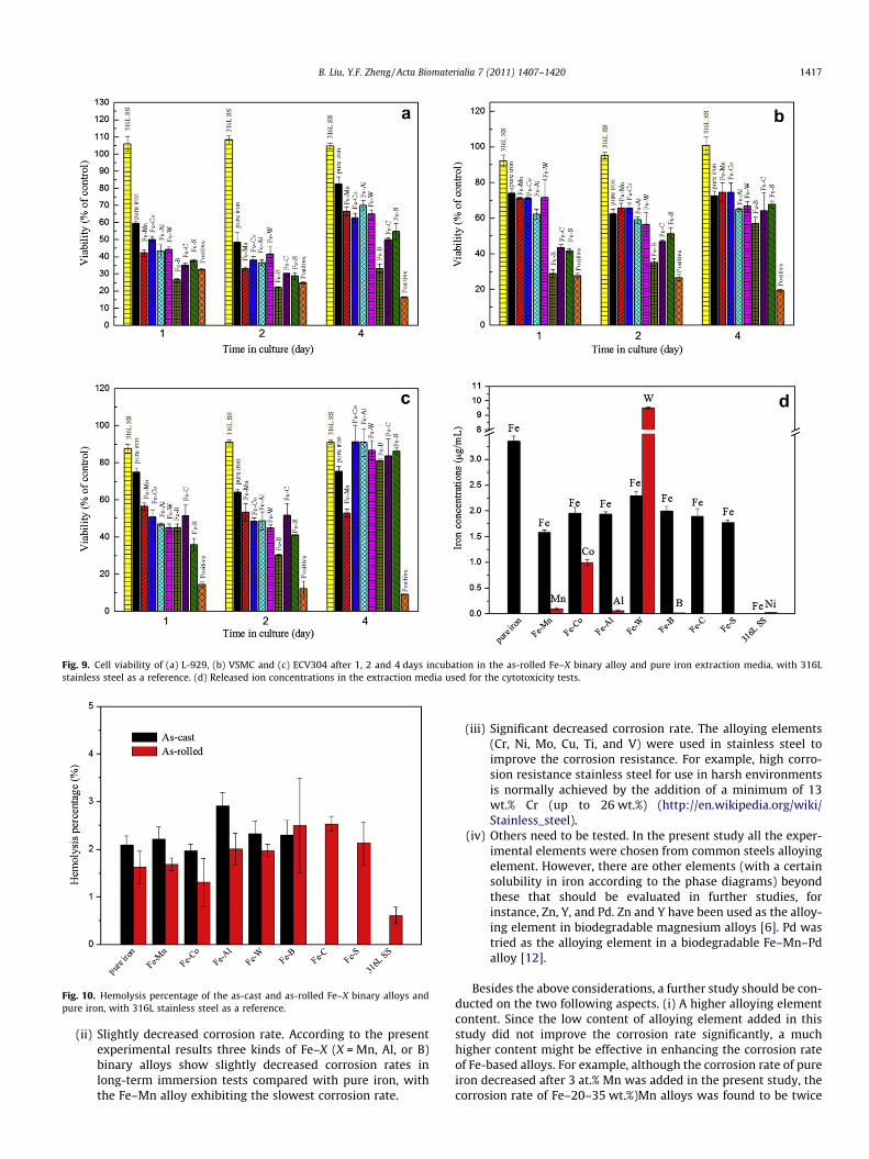
Fig. 9. Cell viability of (a) L-929, (b) VSMC and (c) ECV304 after 1, 2 and 4 days incubation in the as-rolled Fe–X binary alloy and pure iron extraction media, with 316Lstainless steel as a reference. (d) Released ion concentrations in the extraction media used for the cytotoxicity tests.
Fig. 10. Hemolysis percentage of the as-cast and as-rolled Fe–X binary alloys andpure iron, with 316L stainless steel as a reference.
B. Liu, Y.F. Zheng / Acta Biomaterialia 7 (2011) 1407–1420 1417
(ii) Slightly decreased corrosion rate. According to the presentexperimental results three kinds of Fe–X (X = Mn, Al, or B)binary alloys show slightly decreased corrosion rates inlong-term immersion tests compared with pure iron, withthe Fe–Mn alloy exhibiting the slowest corrosion rate.
(iii) Significant decreased corrosion rate. The alloying elements(Cr, Ni, Mo, Cu, Ti, and V) were used in stainless steel toimprove the corrosion resistance. For example, high corro-sion resistance stainless steel for use in harsh environmentsis normally achieved by the addition of a minimum of 13wt.% Cr (up to 26 wt.%) (http://en.wikipedia.org/wiki/Stainless_steel).
(iv) Others need to be tested. In the present study all the exper-imental elements were chosen from common steels alloyingelement. However, there are other elements (with a certainsolubility in iron according to the phase diagrams) beyondthese that should be evaluated in further studies, forinstance, Zn, Y, and Pd. Zn and Y have been used as the alloy-ing element in biodegradable magnesium alloys [6]. Pd wastried as the alloying element in a biodegradable Fe–Mn–Pdalloy [12].
Besides the above considerations, a further study should be con-ducted on the two following aspects. (i) A higher alloying elementcontent. Since the low content of alloying element added in thisstudy did not improve the corrosion rate significantly, a muchhigher content might be effective in enhancing the corrosion rateof Fe-based alloys. For example, although the corrosion rate of pureiron decreased after 3 at.% Mn was added in the present study, thecorrosion rate of Fe–20–35 wt.%)Mn alloys was found to be twice

Fig. 11. SEM images of platelets adhering to Fe–X binary alloys and pure iron specimens. The SEM images of the as-rolled Fe–X binary alloy models are similar to the identicalas-cast alloys.
Fig. 12. Schematic diagrams illustrating both the corrosion mechanism and ideal corrosion process of Fe-based biodegradable alloys in Hank’s solution.
1418 B. Liu, Y.F. Zheng / Acta Biomaterialia 7 (2011) 1407–1420
B. Liu, Y.F. Zheng / Acta Biomaterialia 7 (2011) 1407–1420 1419
as fast as pure iron in a work by Hermawan et al. [11]. (ii) Multi-component Fe-based alloys. More than two alloying elementsmight be considered to be added to iron to form alloys, introducingmore new intermetallic and thus causing microgalvanic corrosion.For example, the addition of Pd was reported to effectively enhancethe degradation rate of an Fe–Mn alloy [12].
4.1.3. Ideal control on the corrosion behavior of Fe-basedbiodegradable alloys
Theoretically, localized corrosion is undesirable in Fe-based cor-onary stents because it may result in local failure and an uncertaincorrosion rate. The localized corrosion of iron is more likely to oc-cur in a high chloride ion concentration solution [38] and in cre-vices [41]. Unfortunately, the chloride ion concentration in bloodplasma is extremely high and crevices formed between the stentand inner wall of the blood vessel after the stent is deployed. Inaddition, a non-uniform stress distribution at different pointsalong a coronary stent is a potential factor influencing the induc-tion of localized corrosion. To enhance general corrosion in theblood environment two kinds of improvements might be helpful,as shown in the ideal corrosion process shown in Fig. 12. (i) Pitsshould be formed as much as possible during the formation andgrowth stages. As time goes on and the pits grow they will becomeconnected, forming a corroded surface. General corrosion can beachieved as all metals beneath are in the same corrosion productcovered condition. Decreasing the pitting potential or introducinga widespread secondary phase intermetallic network is possibleway to realize this ideal corrosion process. (ii) Pits grow in the hor-izontal direction. It is hard for pits to grow in this way because ofthe self-catalytic mechanism of iron corrosion. Increasing the cor-rosion potential of the corrosion products and the inhibition oflocalized acidification could be tried to solve this problem.
4.2. Biological performance of pure iron after alloying with differentelements
4.2.1. Cytotoxicity of Fe–X binary alloys with different cell line modelsMueller et al. [42] found that the smooth muscle cell growth
rate was reduced when soluble ferrous ions were added to the cellculture medium, which suggested that specific iron stent degrada-tion products could have beneficial effects on the control of neoin-tima proliferation. A significant decrease in cell proliferation ofsmooth muscle cells was also observed in a work by Moravejet al. [17]. For endothelial cells, as reported by Zhu et al. [9], ironions almost have no influence on the metabolic activity when theiron concentration is less than 50 lg ml�1. In the present studycells cultured with Fe–X binary alloy extracts showed higherviabilities of ECV304 cells than VSMC, a significant advantage forapplication as coronary stents. Although the endothelial cell viabil-ity in the presence of iron was comparable with 316L SS on day 4, aslightly reduced viability was observed over the first 2 days. Theviabilities of not only endothelial cells but also the other two kindsof cells were increased on day 4. This may be ascribed to the func-tions of the iron transport protein transferrin and the iron storageprotein ferritin [43]. As iron ions combined with transferrins andferritins the absolute iron ion concentration in the extract mediadecreased.
4.2.2. Cytotoxicity of Fe–X binary alloys influenced by ionconcentrations of alloying elements
For the dominant element Fe the half-maximal inhibitory con-centration (IC50) is 5420 lM l�1 [44]. As shown in Fig. 9, the ironion concentration in the extract media of the Fe–X binary alloysand pure iron varied from 28.47 to 60.16 lM l�1, which is muchlower than the IC50. The IC50 of Mn, Co, Al, and W for L929 cellsare 45.9, 81.2, 4180 and 622 lM l�1, respectively [44]. The alloying
element ion concentrations in the present extract media are 1.8,16.9, 2.4 and 51.6 lM l�1, below their corresponding IC50 values.Consistent with this, the viabilities of cells in these extract mediawere above 50%. As Hermawan et al. reported [11], Mn showed asmaller inhibitory effect than pure Mn when it was alloyed withiron to form Fe35Mn alloy. In a work by Peuster et al. [45], veryhigh (>50 lg ml�1) W concentrations were needed to produce localcytopathological effects on human pulmonary arterial endothelialand smooth muscle cells and human dermal fibroblasts. A Fe–B al-loy extract significantly decreased the cell viabilities of L929 cellsand VSMC compared with pure iron extract for a B concentrationof 1.8 lM l�1 in the present study. However, there was no sign oftoxic activity when NIH-3T3 fibroblasts were cultured with1 mM l�1 B2O3 extract [46].
4.2.3. Corrosion products of the present Fe–X binary alloys and ironmetabolism in the human body
Iron does not occur as free ions in body fluids or tissues. Only atiny fraction of the total body iron circulates in the plasma andother extracellular fluids, bound to transferrin. Transferrin in theplasma and extracellular fluids transports iron between the differ-ent cellular compartments [43]. Most iron loss is from the gastro-intestinal tract, in shed enterocytes, extravasated erythrocytes, andbiliary haem breakdown products. A smaller amount is lost in theurine and shed skin cells [47]. It is thought that iron can be re-moved from the body as ions, however, the main corrosion productof iron in biological environments is insoluble iron hydroxideprecipitate. Precipitates in in vivo tests are phagocytosed bymacrophages. Accumulations of iron-laden macrophages andmultinucleated giant cells range from a sparse isolated localizationto accumulation in clusters [15]. Although iron transportation inthe human body has been well researched, the interaction betweeniron hydroxide precipitate and macrophages is not clear yet. Theprocess of corrosion product precipitate excretion from the humanbody should be investigated in further studies.
5. Conclusions
The in vitro effects of the alloying elements Mn, Co, Al, W, Sn, B,C, and S on pure iron were investigated for the future design of newbiodegradable Fe-based alloys for use as coronary stents. The alloy-ing elements Mn, Co, W, B, C, and S were found to improve theyield and ultimate strength of iron in the as-rolled group and in-crease the difference between the yield strength and ultimatestrength of iron, whereas the alloying element Sn led to a severereduction in the mechanical properties. According to the resultsof electrochemical measurements, static and dynamic immersiontests, localized corrosion was the main mode of corrosion of pureiron and the Fe–X binary alloys. The corrosion rates of pure ironand the Fe–X binary alloys were of the same order of magnitude.The pure iron and Fe–X binary alloy extracts decreased the cellviabilities of L929 cells and VSMC compared with 316L SS butshowed no significant cytotoxicity to ECV304 cells, except for theFe–Mn alloy. Although there was a slight increase in percentagehemolysis for pure iron and the Fe–X binary alloys compared with316L SS, all hemolysis percentage was less than 5%. Most of theplatelets adherent on the surface of the Fe–X binary alloys wereround, with no sign of thrombogenicity. To sum up, the elementsCo, W, C, and S are suitable as alloying elements for iron bio-materials on a comprehensive consideration of the improvedmechanical properties, appropriate corrosion rates and good bio-compatibility. High content alloy additions and multi-componentiron alloys should be tried to improve corrosion performance inthe future.
1420 B. Liu, Y.F. Zheng / Acta Biomaterialia 7 (2011) 1407–1420
Acknowledgements
This work was supported by the National Natural Science Foun-dation of China (Grants Nos. 30670560 and 30770580), ResearchFunds for the Central Universities (PKUJC2009001) and the Pro-gram for New Century Excellent Talents in Universities (NCET-07-0033).
Appendix A. Figures with essential colour discrimination
Certain figure in this article, particularly Figures 1–10, are diffi-cult to interpret in black and white. The full colour images can befound in the on-line version, at doi:10.1016/j.actbio.2010.11.001.
References
[1] Erne P, Schier M, Resink TJ. The road to bioabsorbable stents: reaching clinicalreality? Cardiovasc Inter Rad 2006;29:11–6.
[2] Hermawan H, Dubé D, Mantovani D. Developments in metallic biodegradablestents. Acta Mater 2009;6: 1693–7.
[3] Witte F, Fischer J, Nellesen J, Crostack HA, Kaese V, Pisch A, et al. In vitro andin vivo corrosion measurements of magnesium alloys. Biomaterials2006;27:1013–8.
[4] Staiger MP, Pietak AM, Huadmai J, Dias G. Magnesium and its alloys asorthopedic biomaterials: a review. Biomaterials 2006;27:1728–34.
[5] Kannan MB, Raman RKS. In vitro degradation and mechanical integrity ofcalcium-containing magnesium alloys in modified-simulated body fluid.Biomaterials 2008;29:2306–14.
[6] Gu XN, Zheng YF, Cheng Y, Zhong SP, Xi TF. In vitro corrosion andbiocompatibility of binary magnesium alloys. Biomaterials 2009;30:484–98.
[7] Gu XN, Zheng YF, Zhong SP, Xi TF, Wang JQ, Wang WH. Corrosion of, andcellular responses to Mg–Zn–Ca bulk metallic glasses. Biomaterials2010;31:1093–103.
[8] Hermawan H, Alamdari H, Mantovani D, Dube D. Iron–manganese: new classof metallic degradable biomaterials prepared by powder metallurgy. PowderMetall 2008;51:38–45.
[9] Zhu SF, Huang N, Xu L, Zhang Y, Liu HQ, Sun H, et al. Biocompatibility of pureiron: in vitro assessment of degradation kinetics and cytotoxicity onendothelial cells. Mat Sci Eng C–Bio S 2009;29:1589–92.
[10] Zhu SF, Huang N, Xu L, Zhang Y, Liu HQ, Lei YF, et al. Biocompatibility of Fe–Ofilms synthesized by plasma immersion ion implantation and deposition. SurfCoat Tech 2009;203:1523–9.
[11] Hermawan H, Purnama A, Dube D, Couet J, Mantovani D. Fe–Mn alloys formetallic biodegradable stents: degradation and cell viability studies. ActaBiomater 2009;6:1852–60.
[12] Schinhammer M, Hänzi AC, Löffler JF, Uggowitzer PJ. Design strategy forbiodegradable Fe-based alloys for medical applications. Acta Biomater2009;6:1705–13.
[13] Peuster M, Hesse C, Schloo T, Fink C, Beerbaum P, von Schnakenburg C. Long-term biocompatibility of a corrodible peripheral iron stent in the porcinedescending aorta. Biomaterials 2006;27:4955–62.
[14] Waksman R, Pakala R, Baffour R, Seabron R, Hellinga D, Tio FO. Short-termeffects of biocorrodible iron stents in porcine coronary arteries. J Interv Cardiol2008;21:15–20.
[15] Peuster M, Wohlsein P, Brugmann M, Ehlerding M, Seidler K, Fink C, et al. Anovel approach to temporary stenting: degradable cardiovascular stentsproduced from corrodible metal – results 6–18 months after implantationinto New Zealand white rabbits. Heart 2001;86:563–9.
[16] Moravej M, Prima F, Fiset M, Mantovani D. Electroformed iron as newbiomaterial for degradable stents: development process and structure–properties relationship. Acta Biomater 2010;6:1726–53.
[17] Moravej M, Purnama A, Fiset M, Couet J, Mantovani D. Electroformed pure ironas a new biomaterial for degradable stents: in vitro degradation andpreliminary cell viability studies. Acta Biomater 2010;6:1843–51.
[18] Bringas JE. Handbook of comparative world steel standards. WestConshohocken (PA): ASTM International; 2004.
[19] Skerfving Staffan, Gerhardsson Lars, Schutz Andrejs, Stromberg U. Lead –biological monitoring of exposure and effects. J Trace Elem Exp Med1998;11:289–301.
[20] American Society for Testing and Materials. ASTM-E8-04, Standard testmethods for tension testing of metallic materials. In: Annual book of ASTMstandards. Philadelphia (PA): ASTM; 2004.
[21] Chang E, Lee TM. Effect of surface chemistries and characteristics of Ti6Al4V onthe Ca and P adsorption and ion dissolution in Hank’s ethylene diamine tetra-acetic acid solution. Biomaterials 2002;23:2917–25.
[22] American Society for Testing and Materials. ASTM-G102-89, Standard practicefor calculation for corrosion rates and related information fromelectrochemical measurements. In: Annual book of ASTM standards.Philadelphia (PA): ASTM; 1999.
[23] American Society for Testing and Materials. ASTM-G31-72, Standard practicefor laboratory immersion corrosion testing of metals. In: Annual book of ASTMstandards. Philadelphia (PA): ASTM; 2004.
[24] Levesque J, Hermawan H, Dube D, Mantovani D. Design of a pseudo-physiological test bench specific to the development of biodegradablemetallic biomaterials. Acta Biomater 2008;4:284–95.
[25] Doriot PA, Dorsaz PA, Dorsaz L, De Benedetti E, Chatelain P, Delafontaine P. In-vivo measurements of wall shear stress in human coronary arteries. CoronaryArtery Dis 2000;11:495–502.
[26] Morita M, Sasada T, Nomura I, Wei YQ, Tsukamoto Y. Influence of lowdissolved-oxygen concentration in body-fluid on corrosion fatigue behaviorsof implant metals. Ann Biomed Eng 1992;20:505–16.
[27] Pardee WJ, Robertson WM, James MR. Tin segregation at iron grain-boundariesmeasured by EXAFS. JoM J Min Met Mat S 1980;32:12.
[28] Yuan ZX, Jia J, Guo AM, Shen DD, Song SH. Cooling-induced tin segregation tograin boundaries in a low-carbon steel. Scripta Mater 2003;48:203–6.
[29] Mani G, Feldman MD, Patel D, Agrawal CM. Coronary stents: a materialsperspective. Biomaterials 2007;28:1689–710.
[30] Khan W, Kapoor M, Kumar N. Covalent attachment of proteins tofunctionalized polypyrrole-coated metallic surfaces for improvedbiocompatibility. Acta Biomater 2007;3:541–9.
[31] Armitage DA, Parker TL, Grant DM. Biocompatibility and hemocompatibility ofsurface-modified NiTi alloys. J Biomed Mater Res A 2003;66A:129–37.
[32] Wilson RE. The mechanism of the corrosion of iron. Ind Eng Chem1923;15:426–7.
[33] Rajagopalan KS, Srividyarajagopalan C, Venkatachari G. Kinetics andmechanism of corrosion of structural-steel and pure iron in salt-solutions. JElectrochem Soc 1977;124:C282.
[34] Iofa ZA. Mechanism of the effect of hydrogen-sulfide and inhibitors on ironcorrosion in acid-solutions. Prot Met+ 1980;16:220–5.
[35] Pickerin Hw, Frankent Rp. Mechanism of localized corrosion of iron andstainless-steel. 1. Electrochemical studies. J Electrochem Soc 1972;119:1297.
[36] Frankel GS, Sridhar N. Understanding localized corrosion. Mater Today2008;11:38–44.
[37] Alvarez MG, Galvele JR. The mechanism of pitting of high-purity iron in NaClsolutions. Corros Sci 1984;24:27–48.
[38] Laycock NJ, Newman RC. Localised dissolution kinetics, salt films and pittingpotentials. Corros Sci 1997;39:1771–90.
[39] Zhu YL, Qiu YB, Guo XP. Underscale corrosion behavior of carbon steel in aNaCl solution using a new occluded cavity cell for simulation. J ApplElectrochem 2009;39:1017–23.
[40] Meng GZ, Zhang C, Cheng YF. Effects of corrosion product deposit on thesubsequent cathodic and anodic reactions of X-70 steel in near-neutral pHsolution. Corros Sci 2008;50:3116–22.
[41] Wolfe RC, Pickering HW, Shaw BA. Microprobe study of pH during theinduction period preceding crevice corrosion. J Electrochem Soc2006;153:B25–32.
[42] Mueller PP, May T, Perz A, Hauser H, Peuster M. Control of smooth muscle cellproliferation by ferrous iron. Biomaterials 2006;27:2193–200.
[43] Crichton R. Inorganic biochemistry of iron metabolism. New York: John Wiley& Sons; 2001.
[44] Yamamoto A, Honma R, Sumita M. Cytotoxicity evaluation of 43 metal saltsusing murine fibroblasts and osteoblastic cells. J Biomed Mater Res1998;39:331–40.
[45] Peuster M, Fink C, von Schnakenburg C. Biocompatibility of corroding tungstencoils: in vitro assessment of degradation kinetics and cytotoxicity on humancells. Biomaterials 2003;24:4057–61.
[46] Hopp M, Rogaschewski S, Groth T. Testing the cytotoxicity of metal alloys usedas magnetic prosthetic devices. J Mater Sci-Mater M 2003;14:335–45.
[47] Reilly C. The nutritional trace metals. Oxford: Blackwell Publishing; 2004.