effects of black cohosh on estrogen biosynthesis in normal breast
TRANSCRIPT
A
OMutwaRmaCh©
K
1
s
f
s
0
Maturitas 57 (2007) 382–391
Effects of black cohosh on estrogen biosynthesisin normal breast tissue in vitro
Petra Stute a,∗, Thomas Nisslein b, Martin Gotte a, Axel Kamischke c,Ludwig Kiesel a, Walter Klockenbusch a
a Department of Obstetrics and Gynecology, University Clinic of Muenster, 48149 Muenster, Germanyb Schaper & Brummer GmbH & Co. KG, 38259 Salzgitter, Germany
c Department of Obstetrics and Gynecology, St. Barbara Clinic, Hamm, Germany
Received 21 November 2006; received in revised form 16 April 2007; accepted 20 April 2007
bstract
bjectives: To investigate the effect of black cohosh on the estrogen biosynthesis in the breast in vitro.ethods: Steroid sulfatase (STS) activity was studied in normal breast tissue obtained from pre- and postmenopausal women
ndergoing reduction mammoplasty. STS protein expression was studied by immunohistochemistry and western blotting. Breastissue was incubated in vitro without or with black cohosh (iCR) at concentrations ranging from 0.1 mg/ml to 1 ng/ml. STS activityas evaluated by incubating homogenized breast tissue with [3H]-estrone sulfate, separating the formed products, estrone (E1)
nd estradiol (E2), by thin layer chromatography and measuring the amounts of E1 and E2 by scintillation counting.esults: STS protein expression and enzymatic activity were detected in all specimens investigated. In all groups, significantlyore E1 than E2 was produced. Local estrogen formation was decreased in premenopausal breast tissue by treatment with iCR
t 0.1 mg/ml (p ≤ 0.05).onclusions: iCR decreases local estrogen formation in normal human breast tissue in vitro. This may contribute to the lack oformonal effects of black cohosh in breast tissue observed in previous studies.
2007 Elsevier Ireland Ltd. All rights reserved.
emosa;
eywords: Breast; Black cohosh; Actaea racemosa; Cimicifuga rac. Introduction
Postmenopausal hormone therapy (HT) has beeneen as a specific treatment for climacteric symptoms
∗ Corresponding author. Tel.: +49 251 83 48202;ax: +49 251 83 48267.
E-mail addresses: [email protected],[email protected] (P. Stute).
iihatrcd
378-5122/$ – see front matter © 2007 Elsevier Ireland Ltd. All rights reserdoi:10.1016/j.maturitas.2007.04.007
Sulfatase activity
n the short term and as a prevention of chronic diseasesn the long term [1–3]. However, several clinical trialsave indicated an increased risk of breast cancer (BC)ssociated with HT [4–8]. In recent years, extracts of
he rhizome of black cohosh (Actaea, syn. Cimicifugaacemosa) (CR) have been recognized as a rationalhoice for treatment and prevention of menopausalisorder [9,10]. So far, in vitro and in vivo studiesved.

uritas 5
seuohvtotettimav(hwbthcanrtsImdptofc
oCi
2
2
&
Tr4iepti0hTaESwaG
2
igSGSoMa5ipmsicto(tiogP
P. Stute et al. / Mat
uggest CR to be safe for the breast [11–16]. How-ver, the precise mode of action of CR is not yet fullynderstood. According to the concept of intracrinol-gy [17] suggesting that inactive hormones released byormone-producing organs are transported via bloodessels to their target tissues where enzymes secureheir conversion into active hormones, the amountf steroids locally available is more important thanheir serum concentration. The most biologically activestrogen in breast tissue is 17�-estradiol (E2). Breastissue and mammary cancer cells have been showno possess the enzymatic systems necessary for thentratumoral biosynthesis of estrogens from precursor
olecules circulating in plasma. Three main enzymesre important in this process: aromatase, which con-erts androgens to estrogens [18–20], E1S-sulfataseSTS) which hydrolizes E1S to E1 [20–28], and 17�-ydroxysteroid dehydrogenase type 1 (17�HSD-1)hich reduces E1 to E2 [29–32]. The activity of STS inreast tumors has been shown to be 10–500-fold higherhan aromatase activity [33–35]. In BC, STS mRNAas been shown to be an independent prognostic indi-ator in predicting shorter relapse free survival [36]nd to correlate positively with tumor size and lymphode metastasis [37]. Thus, STS seems to play a crucialole in local biosynthesis of estrogens in breast (cancer)issue. Estradiol and various progestogens have beenhown to influence STS in BC cells in vitro [38–44].n vitro, the effect of steroids on local estrogen for-ation in normal and malignant breast cells is rather
ue to changes in STS activity than in its mRNA androtein levels [45]. We recently demonstrated that long-erm HT alters local estrogen formation in breast tissuef postmenopausal cynomolgus monkeys (Macacaascicularis) and women depending on breast tissueomposition [46,47].
The aim of this study was to investigate the effectf an isopropanolic-aqueous (40%, v/v) extract ofR (iCR) on estrogen biosynthesis in breast tissue
n vitro.
. Methods
.1. Chemicals, reagents, and treatment
The iCR (B. Nr. 010720) was provided by SchaperBrummer GmbH & Co.KG. Salzgitter, Germany.
ifib
7 (2007) 382–391 383
he concentration of the extract was 100 mg/ml inelation to the dry residue. Alcohol concentration was0% (v/v). The final concentration of isopropanoln tissue homogenates during iCR treatment did notxceed 0.5% (v/v). The same concentration of iso-ropanol was present in the control samples. Breastissue homogenates were incubated without and withCR at a concentration range from 10 mg/ml to.1 �g/ml leading to a final concentration range in theomogenate from 0.1 mg/ml to 1 ng/ml, respectively.he radioactive labeled steroid [3H]-E1S (specificctivity 57.3 Ci/mmol), was purchased from Perkinlmer Life and Analytical Sciences (549 Albanytreet, Boston, MA, USA). Unlabeled E2 and E1ere obtained from Sigma. Unless stated otherwise,
ll chemicals were from Sigma–Aldrich (Deisenhofen,ermany).
.2. Subjects
E1S metabolism in normal breast tissue was studiedn five post- and five premenopausal women under-oing reduction mammoplasty (Department of Plasticurgery, Clinic Centrum Hohenzollernring, Muenster,ermany, Department of Gynecology and Obstetrics,t. Barbara Clinic, Hamm, Germany, and Departmentf Obstetrics and Gynecology, University Clinic ofuenster, Germany). The postmenopausal women’s
ge was in the range from 48 to 70 years (mean6 ± 7.7 years), and the premenopausal women’s agen the range from 31 to 57 years (mean 43 ± 9 years;= 0.05). None of the women received hormonal treat-ent prior to breast surgery. Immediately following
urgical removal, the tissue samples were shock frozenn liquid nitrogen and stored at −70 ◦C until thin layerhromatography for enzyme activity and western blot-ing for protein quantification were performed. Sincene postmenopausal woman was extremely overweightbody mass index (BMI) 50) she was excluded fromhe subset of postmenopausal women when STS activ-ty was evaluated. In addition, normal breast tissuebtained from another subset of women (n = 5) under-oing breast reduction mammoplasty (Department oflastic Surgery, Fachklinik Hornheide, Germany) was
mmediately fixed in 10% neutral phosphate-bufferedormalin for H&E staining and immunohistochem-stry. Histological evaluation was performed by aoard-specified pathologist (G. Edel, M.D., Ph.D.,

3 uritas 5
IMNtl
bp
2
mftfAiiDmdtraluhsMtAM
2
pw1jgbmTtbT
fTcN5f(nMBigaac(irU
2
ofiit1ni1oiiwtinope1Tg
84 P. Stute et al. / Mat
nstitute of Pathology at the Franziskus Hospital inuenster, Germany, Institute of Pathology Diebold,iemann & Schonfeld, Hamm, Germany). The breast
issue examined did not contain any malignantesions.
Women gave written informed consent to theirreast tissue being analyzed in addition to establishedathological analysis.
.3. Immunohistochemistry
Normal breast tissue obtained from a reductionammoplasty of a 49-year-old healthy woman was
ormalin-fixed and paraffin-embedded using standardechniques. Consecutive sections of 2–3 �m were cutrom the paraffin blocks, dewaxed and re-hydrated.ntigen retrieval was performed by hot water steaming
n citrate buffer (pH 6; 30 min). Following a block-ng step with Aurion BSAc solution (DAKO, Glostrup,enmark) for 30 min, sections were incubated with aonospecific rabbit-anti human STS-antiserum [48],
iluted 1:1000 in phosphate-buffered saline (PBS) con-aining 1% bovine serum albumine (BSA), or a controlabbit serum overnight at 4 ◦C. Endogenous peroxidasectivity was quenched with methanol/0.6% H2O2, fol-owed by three washes with PBS. STS was detectedsing the DAKO cytomation rabbit-EnVision plus-orseradish peroxidase (HRP) system and the AECubstrate (DAKO), followed by counterstaining with
ayer’s Hemalum (Merck, Darmstadt, Germany). Sec-ions were observed and documented using a Zeissxiovert 100 microscope equipped with an Axiophotrc camera.
.4. Western blotting
For Western blotting, breast tissue extracts of fivere- and five postmenopausal women were dilutedith SDS-sample buffer to a protein concentration of�g/�l. Twenty microliters of sample/lane were sub-
ected to electrophoresis on 12% SDS-polyacrylamideels and electro-transferred to nitrocellulose mem-ranes as previously described [50]. Subsequently, theembranes were blocked with 5% non-fat dry milk in
BS containing 1% (v/v) Tween 20 for 1 h at roomemperature, washed 3× 5 min with TBS and incu-ated with anti-STS-antiserum [48] diluted 1:1500 inBS containing 5% BSA and 1% (v/v) Tween at 4 ◦C
woTn
7 (2007) 382–391
or 16 h. The membrane was washed 3× 5 min withBS (1% Tween) and incubated with a peroxidase-onjugated goat-anti rabbit IgG antibody (Calbiochem,ottingham, UK) diluted 1:2000 in TBS containing% BSA and 1% (v/v) Tween at room temperatureor 1 h. The blot was washed 3× 5 min with TBS1% Tween), followed by an enhanced chemolumi-escence reaction (Super Signal, Pierce, Rockford,A) and exposition to ECL–hyperfilm (Amersham,raunschweig, Germany). To normalize for a load-
ng control, blot membranes were stripped with 0.2 Mlycine, pH 2.5 [50] and reprobed as describedbove, using a monoclonal mouse-anti tubulin primaryntibody (Sigma–Aldrich, dilution 1:4000) and HRP-onjugated goat-anti-mouse IgG secondary antibodiesCalbiochem, dilution 1:10,000), respectively. Scannedmages of the exposed films were analyzed densiomet-ically using the NIH Image J software (NIH, Bethesda,SA), normalizing STS expression to tubulin content.
.5. E1S-sulfatase (STS) activity assay
STS activity was measured as described previ-usly [45,46]. In brief, breast tissue (150 mg) wasnely minced with scissors and homogenized on ice,
n 2 ml 0.06 M Tris–HCl-buffer pH 7.0 (assay bufferhroughout the entire procedure) for approximatelymin at maximum speed using a Polytron homoge-izer. All incubations were carried out in duplicate. Thencubation mixtures consisted of 200 �l homogenate,9.39 pmole [3H]-E1S in 100 �l of buffer, and 100 �gf NADH and NADPH in 100 �l of buffer (totalncubation volume 400 �l). Simultaneous backgroundncubations using buffer instead of tissue homogenateere performed in order to correct for non-enzymatic
ransformation of the substrate. After 15 and 45 min ofncubation in air at 37 ◦C in a water bath, 200 �g ofon-radioactive E1 and 17�-E2 were added in 100 �lf ethanol together with 0.5 ml of 0.5 M sodium phos-hate buffer, pH 7.0. The unconjugated steroids werextracted with 2.0 ml toluene. After inverting the tubes00 times they were centrifuged at 1500 × g for 10 min.he lower aqueous phase was frozen in liquid nitro-en. Hundred microliters of the upper toluene phase
as removed and added to 100 �l of buffer and 3.0 mlf scintillation fluid to determine total hydrolysis.he remaining toluene phase was evaporated to dry-ess in nitrogen atmosphere and dissolved in 100 �l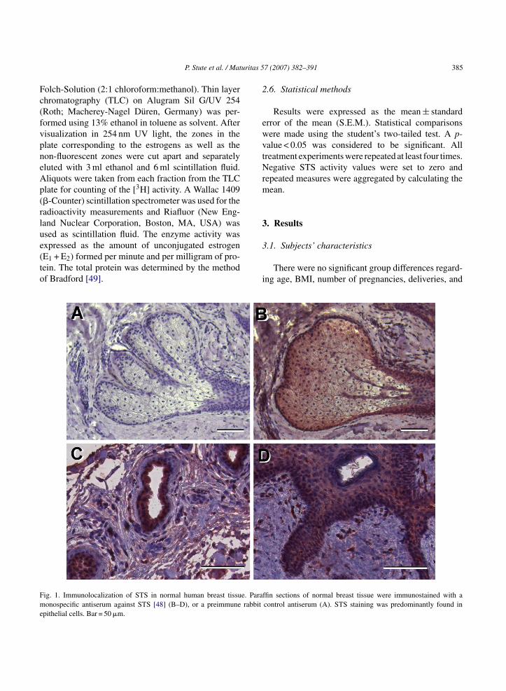
uritas 5
Fc(fvpneAp(rlue(to
2
ewvtNrm
3
Fme
P. Stute et al. / Mat
olch-Solution (2:1 chloroform:methanol). Thin layerhromatography (TLC) on Alugram Sil G/UV 254Roth; Macherey-Nagel Duren, Germany) was per-ormed using 13% ethanol in toluene as solvent. Afterisualization in 254 nm UV light, the zones in thelate corresponding to the estrogens as well as theon-fluorescent zones were cut apart and separatelyluted with 3 ml ethanol and 6 ml scintillation fluid.liquots were taken from each fraction from the TLClate for counting of the [3H] activity. A Wallac 1409�-Counter) scintillation spectrometer was used for theadioactivity measurements and Riafluor (New Eng-and Nuclear Corporation, Boston, MA, USA) wassed as scintillation fluid. The enzyme activity was
xpressed as the amount of unconjugated estrogenE1 + E2) formed per minute and per milligram of pro-ein. The total protein was determined by the methodf Bradford [49].3
i
ig. 1. Immunolocalization of STS in normal human breast tissue. Paraonospecific antiserum against STS [48] (B–D), or a preimmune rabbit
pithelial cells. Bar = 50 �m.
7 (2007) 382–391 385
.6. Statistical methods
Results were expressed as the mean ± standardrror of the mean (S.E.M.). Statistical comparisonsere made using the student’s two-tailed test. A p-alue < 0.05 was considered to be significant. Allreatment experiments were repeated at least four times.egative STS activity values were set to zero and
epeated measures were aggregated by calculating theean.
. Results
.1. Subjects’ characteristics
There were no significant group differences regard-ng age, BMI, number of pregnancies, deliveries, and
ffin sections of normal breast tissue were immunostained with acontrol antiserum (A). STS staining was predominantly found in

3 uritas 5
mrpppfipwofttwhrcass
3l
bfw[eHptwsiSaw6
3E
ttwb
awpppwrw1tsm
dt
3a
wct(0Sfbceab
pmsff
4
e
86 P. Stute et al. / Mat
iscarriages. Mean BMI was within the overweightange (postmenopausal women: 30 ± 11 kg/m2, andremenopausal women: 26 ± 4 kg/m2, respectively;= 0.53). Hysterectomy without ovarectomy had beenerformed in two of the five post-, and one of theve premenopausal women, respectively. None of theostmenopausal but two of the premenopausal womenere current smoker. The family history (first and sec-nd degree) referring to breast cancer was negativeor both groups. Breast cancer had occurred earlier inhe mammary gland which was now subject of reduc-ion mammoplasty in three of the five postmenopausalomen. In the premenopausal subset one in five womenad been treated for contralateral breast cancer prior toeduction mammoplasty. Reduction mammoplasty forosmetic reason only was performed in one in five post-nd four in five premenopausal women. Prior to breasturgery a mammogram was performed to prove lack ofuspicious lesions.
.2. E1S-sulfatase (STS) protein expression andocalization
To confirm the expression of STS in normalreast tissue, we performed immunohistochemistry onormalin-fixed, paraffin-embedded specimens using aell-characterized monospecific anti-STS-antiserum
48]. STS protein was predominantly localized inpithelial cells of lobuloalveolar and ductal tissue.owever, STS protein staining was also occasionallyresent in stromal cells (Fig. 1). To quantify STS pro-ein in the breast tissue samples studied, we performedestern blots of tissue extracts. A specific band corre-
ponding to the Mr of dimeric STS [51] was detectedn all breast tissues investigated (Fig. 2). Interestingly,TS protein expression was significantly increased bypproximately 30% in breast tissue of premenopausalomen, resulting in a signal overload (Fig. 2, lanes–10).
.3. Local E1 and E2 formation as indicative for1S-sulfatase (STS) activity in vitro
The activity of STS in kryoconserved normal breast
issue could be demonstrated. The enzyme effec-ively metabolized the radioactive substrate E1S. E1as the most abundant, labeled estrogen found inreast tissue after incubation with labeled E1S. Onitdb
7 (2007) 382–391
n average, the concentration of locally formed E1as about 18 (untreated postmenopausal women;= 0.08) and 53 (untreated premenopausal women;≤ 0.05) times higher than that of E2. In untreatedostmenopausal women, mean E1 and E2 formationas 9.4 ± 2.9 and 0.5 ± 0.2 fmol/(mg(protein) min),
espectively. In contrast, in untreated premenopausalomen, mean E1 and E2 formation was 55.5 ± 16.3 and.0 ± 0.5 fmol/(mg(protein) min), respectively. Nei-her E1 (p = 0.06) nor E2 (p = 0.4) formation wereignificantly different comparing pre- and post-enopausal women.Interestingly, total estrogen formation was
ecreased after 45 min of incubation when comparedo 15 min of incubation (n.s.).
.4. Influence of iCR on E1S-sulfatase (STS)ctivity in vitro
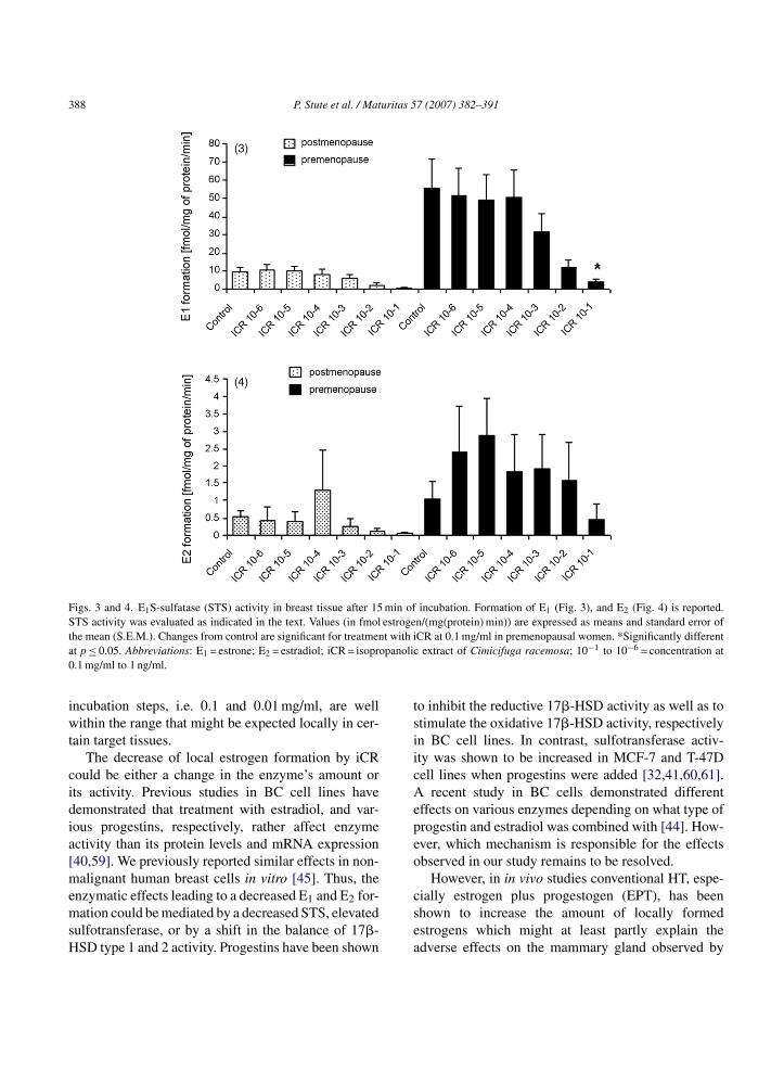
Both, E1 and E2 formation after 15 min of incubationere dose-dependently decreased by iCR treatment
ompared to controls regardless of menopausal sta-us. However, the reduction of total estrogen formationE1 + E2) was only significant for iCR treatment at.1 mg/ml in premenopausal women (Figs. 3 and 4).imilarly, after 45 min of incubation total estrogenormation (E1 + E2) was also significantly decreasedy treatment with iCR at 0.1 mg/ml (p ≤ 0.05) inomparison to controls (Fig. 5). E1, E2, and totalstrogen formation (E1 + E2) after treatment with iCRt 0.1 mg/ml to 1 ng/ml did not differ significantlyetween pre- and postmenopausal women.
Interestingly, the local estrogen formation of oneostmenopausal woman was measured in the pre-enopausal range. This woman had a BMI of 50
uggesting that the higher amount of body and breastat tissue might have an impact on the local estrogenormation the mammary gland.
. Discussion
To our knowledge we are the first to investigate theffect of iCR on STS activity in normal breast tissue
n vitro. Although the amount of epithelial tissue inhe mammary gland changes due to age and repro-uctive stage [52] STS activity has been shown toe present regardless of menopausal stage [53]. We
P. Stute et al. / Maturitas 57 (2007) 382–391 387
Fig. 2. Western blot analysis of STS protein expression in normal breast tissue from postmenopausal (1–5) and premenopausal (6–10) women.Twenty micrograms protein/lane were subjected to SDS-PAGE and Western blotting using a monospecific STS-antiserum [48]. (A) STS proteinexpression was detected in all breast tissue samples. High STS protein expression levels in specimens obtained from premenopausal womenr Densite n in pau
disiEwb
obcS4i
1[bBisotsr
esulted in a chemoluminescence signal overload (lanes 9–10). (B)xpression. The signal intensities of the STS and tubulin bands shownits, *p > 0.05 (n = 5).
emonstrated local estrogen formation being highern untreated pre- than in postmenopausal breast tis-ue supporting previous results [53]. Treatment withCR induced a dose-dependent decrease of both, E1 and2 formation in pre- and postmenopausal breast tissuehich was significant at 0.1 mg/ml in premenopausalreast tissue.
Regarding our enzyme activity assay, we focusedn rapid changes in desulfation. After 45 min of incu-ation STS activity was not significantly decreased in
omparison to 15 min of incubation. Various Km forTS activity have been reported by others ranging fromto 27 �M in human BC tissue [34,35,54–56], 5.8 �Mn dysplastic human breast tissue [52], and from 4.1 to
rsa
ometric analysis of STS protein expression normalized for tubulinnel A were quantified using NIH image J software. AU = arbitrary
8.2 �M in normal human breast tissue homogenates57]. Vmax for STS activity has been reported to rangeetween 0.8 and 125 �mol E1/(g(protein) h) in humanC tissue [35], and between 18.2 and 75.9 �mol E1/g
n normal breast tissue homogenates [57]. Since theubstrate concentration in our assay was in the physi-logical range, no further increase in STS activity waso be expected after 45 min of incubation, as a steadytate of biosynthesis of estrogens had obviously beeneached.
Supposing a 100% bioavailability, the human dailyecommended dose, i.e. 50 �l iCR, would result in aerum concentration of 1.7 �g/ml [58]. Therefore, thective doses of iCR as present in the tissue homogenate
388 P. Stute et al. / Maturitas 57 (2007) 382–391
Figs. 3 and 4. E1S-sulfatase (STS) activity in breast tissue after 15 min of incubation. Formation of E1 (Fig. 3), and E2 (Fig. 4) is reported.STS activity was evaluated as indicated in the text. Values (in fmol estrogen/(mg(protein) min)) are expressed as means and standard error oft t with ia panolic0
iwt
cidia[memsH
tsiicAepeo
he mean (S.E.M.). Changes from control are significant for treatment p ≤ 0.05. Abbreviations: E1 = estrone; E2 = estradiol; iCR = isopro.1 mg/ml to 1 ng/ml.
ncubation steps, i.e. 0.1 and 0.01 mg/ml, are wellithin the range that might be expected locally in cer-
ain target tissues.The decrease of local estrogen formation by iCR
ould be either a change in the enzyme’s amount orts activity. Previous studies in BC cell lines haveemonstrated that treatment with estradiol, and var-ous progestins, respectively, rather affect enzymectivity than its protein levels and mRNA expression40,59]. We previously reported similar effects in non-alignant human breast cells in vitro [45]. Thus, the
nzymatic effects leading to a decreased E1 and E2 for-ation could be mediated by a decreased STS, elevated
ulfotransferase, or by a shift in the balance of 17�-SD type 1 and 2 activity. Progestins have been shown
csea
CR at 0.1 mg/ml in premenopausal women. *Significantly differentextract of Cimicifuga racemosa; 10−1 to 10−6 = concentration at
o inhibit the reductive 17�-HSD activity as well as totimulate the oxidative 17�-HSD activity, respectivelyn BC cell lines. In contrast, sulfotransferase activ-ty was shown to be increased in MCF-7 and T-47Dell lines when progestins were added [32,41,60,61].
recent study in BC cells demonstrated differentffects on various enzymes depending on what type ofrogestin and estradiol was combined with [44]. How-ver, which mechanism is responsible for the effectsbserved in our study remains to be resolved.
However, in in vivo studies conventional HT, espe-
ially estrogen plus progestogen (EPT), has beenhown to increase the amount of locally formedstrogens which might at least partly explain thedverse effects on the mammary gland observed by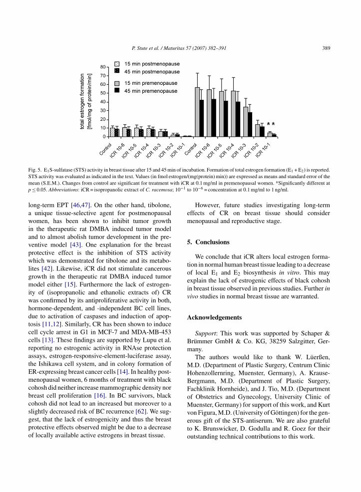
P. Stute et al. / Maturitas 57 (2007) 382–391 389
F in of incS trogen/m ith iCRp ; 10−1 t
lawiavpwlgmiwhdtccratEmcbcsgpo
em
5
toeiv
A
Bm
MHBFoM
ig. 5. E1S-sulfatase (STS) activity in breast tissue after 15 and 45 mTS activity was evaluated as indicated in the text. Values (in fmol esean (S.E.M.). Changes from control are significant for treatment w≤ 0.05. Abbreviations: iCR = isopropanolic extract of C. racemosa
ong-term EPT [46,47]. On the other hand, tibolone,unique tissue-selective agent for postmenopausalomen, has been shown to inhibit tumor growth
n the therapeutic rat DMBA induced tumor modelnd to almost abolish tumor development in the pre-entive model [43]. One explanation for the breastrotective effect is the inhibition of STS activityhich was demonstrated for tibolone and its metabo-
ites [42]. Likewise, iCR did not stimulate cancerousrowth in the therapeutic rat DMBA induced tumorodel either [15]. Furthermore the lack of estrogen-
ty of (isopropanolic and ethanolic extracts of) CRas confirmed by its antiproliferative activity in both,ormone-dependent, and -independent BC cell lines,ue to activation of caspases and induction of apop-osis [11,12]. Similarly, CR has been shown to induceell cycle arrest in G1 in MCF-7 and MDA-MB-453ells [13]. These findings are supported by Lupu et al.eporting no estrogenic activity in RNAse protectionssays, estrogen-responsive-element-luciferase assay,he Ishikawa cell system, and in colony formation ofR-expressing breast cancer cells [14]. In healthy post-enopausal women, 6 months of treatment with black
ohosh did neither increase mammographic density norreast cell proliferation [16]. In BC survivors, blackohosh did not lead to an increased but moreover to a
lightly decreased risk of BC recurrence [62]. We sug-est, that the lack of estrogenicity and thus the breastrotective effects observed might be due to a decreasef locally available active estrogens in breast tissue.veto
ubation. Formation of total estrogen formation (E1 + E2) is reported.(mg(protein) min)) are expressed as means and standard error of the
at 0.1 mg/ml in premenopausal women. *Significantly different ato 10−6 = concentration at 0.1 mg/ml to 1 ng/ml.
However, future studies investigating long-termffects of CR on breast tissue should considerenopausal and reproductive stage.
. Conclusions
We conclude that iCR alters local estrogen forma-ion in normal human breast tissue leading to a decreasef local E1 and E2 biosynthesis in vitro. This mayxplain the lack of estrogenic effects of black cohoshn breast tissue observed in previous studies. Further inivo studies in normal breast tissue are warranted.
cknowledgements
Support: This work was supported by Schaper &rummer GmbH & Co. KG, 38259 Salzgitter, Ger-any.The authors would like to thank W. Luerßen,
.D. (Department of Plastic Surgery, Centrum Clinicohenzollernring, Muenster, Germany), A. Krause-ergmann, M.D. (Department of Plastic Surgery,achklinik Hornheide), and J. Tio, M.D. (Departmentf Obstetrics and Gynecology, University Clinic ofuenster, Germany) for support of this work, and Kurt
on Figura, M.D. (University of Gottingen) for the gen-rous gift of the STS-antiserum. We are also gratefulo K. Brunswicker, D. Godulla and R. Goez for theirutstanding technical contributions to this work.

3 uritas 5
R
[
[
[
[
[
[
[
[
[
[
[
[
[
[
[
[
[
[
[
[
[
[
90 P. Stute et al. / Mat
eferences
[1] McKinney KA, Thompson W. A practical guide to prescribinghormone replacement therapy. Drugs 1998;56:49–57.
[2] Nelson HD, Humphrey LL, Nygren P, Teutsch SM, AllanJD. Postmenopausal hormone replacement therapy: scientificreview. JAMA 2002;288:872–81.
[3] North American Menopause Society. Treatment of menopause-associated vasomotor symptoms: position statement ofThe North American Menopause Society. Menopause2004;11(1):11–33.
[4] Collaborative Group on Hormonal Factors in Breast Cancer.Breast cancer and hormone replacement therapy: collaborativereanalysis of data from 51 epidemiological studies of 52,705women with breast cancer and 108,411 women without breastcancer. Lancet 1997;350:1047–59.
[5] Beral V. Breast cancer and hormone-replacement therapy in themillion women study. Lancet 2003;362:419–27.
[6] Chlebowski RT, Hendrix SL, Langer RD, et al. Influence ofestrogen plus progestin on breast cancer and mammography inhealthy postmenopausal women: the Women’s Health InitiativeRandomized Trial. J Am Med Assoc 2003;289:3243–53.
[7] Chen WY, Hankinson SE, Schnitt SJ, Rosner BA, Holmes MD,Colditz GA. Association of hormone replacement therapy toestrogen and progesterone receptor status in invasive breastcarcinoma. Cancer 2004;101:1490–500.
[8] Writing Group for the Women’s Health Initiative Investiga-tors. Risks and benefits of estrogen plus progestin in healthypostmenopausal women: principal results from the women’shealth initiative randomized controlled, trial. J Am Med Assoc2002;288:321–33.
[9] Liebermann S. A review of the effetiveness of Cimicifugaracemosa (Black Cohosh) for the symptoms of menopause. JWomen’s Health 1998;7:525–9.
10] Liske E. Therapeutic efficacy and safety of Cimicifuga race-mosa for gynecologic disorders. Adv Nat Ther 1998;15:45–53.
11] Hostanska K, Nisslein T, Freudenstein J, Reichling J, SallerR. Cimicifuga racemosa extract inhibits proliferation of estro-gen receptor-positive and negative human breast carcinomacell lines by induction of apoptosis. Breast Cancer Res Treat2004;84:151–60.
12] Hostanska K, Nisslein T, Freudenstein J, Reichling J, SallerR. Evaluation of cell death caused by triterpene glyco-sides and phenolic substances from Cimicifuga racemosaextract in human MCF-7 breast cancer cells. Biol Pharm Bull2004;27(12):1970–5.
13] Saxe Einbond L, Shimizu M, Xiao D, et al. Growth inhibitoryactivity of extracts and purified components of black cohoshon human breast cancer cells. Breast Cancer Res Treat2004;83:221–31.
14] Lupu R, Mehmi I, Atlas E, et al. Black cohosh, a menopausalremedy, does not have estrogenic activity and does not promote
breast cancer cell growth. Int J Oncol 2003;23(5):1407–12.15] Freudenstein J, Dasenbrock C, Nisslein T. Lack of promo-tion of estrogen-dependent mammary gland tumors in vivoby am isopropanolic Cimicifuga racemosa extract. Cancer Res2002;62:3448–52.
[
7 (2007) 382–391
16] Linden Hirschberg A, Edlund M, Svane G, Azavedo E, Skoog L,von Schoultz B. An isopropanolic extract of black cohosh doesnot increase mammographic breast density or breast cell prolif-eration in postmenopausal women. Menopause 2007;14(1).
17] Suzuki T, Miki Y, Nakamura Y, et al. Sex steroid-producingenzymes in human breast cancer. Endocr Relat Cancer2005;12:701–20.
18] Abul-Hajj YJ, Iverson R, Kiang DT. Aromatization of andro-gens by human breast cancer. Steroids 1979;33:205–22.
19] Lipton A, Santner SJ, Santen RJ, et al. Aromatase activ-ity in primary and metastatic human breast cancer. Cancer1987;59:779–82.
20] Perel E, Daniilescu D, Kharlip L, Blackstein M, Killinger DW.Steroid modulation of aromatase activity in human culturedbreast carcinoma cells. J Steroid Biochem 1988;29:393–9.
21] Dao TL, Hayes C, Libby PR. Steroid sulfatase activities inhuman breast tumors. Proc Soc Exp Biol Med 1974;146:381–4.
22] Vignon F, Terqui M, Westley B, Derocq D, Rochefort H.Effects of plasma estrogen sulfates in mammary cancer cells.Endocrinology 1980;106:1079–86.
23] Prost O, Turrel MO, Dahan N, Craveur C, Adessi GL. Estroneand dehydroepiandrosterone sulfatase activities and plasmaestrone sulfate levels in human breast carcinoma. Cancer Res1984;44:661–4.
24] Chapman O, Purohit A, Wang DY, Ghilchik MW, Reed MJ.Oestrone sulphatase activity in normal and malignant breasttissues: relationship with tumour location. Anticancer Res1995;15:1467–71.
25] Chetrite GS, Cortes-Prieto J, Philippe JC, Wright F, PasqualiniJR. Comparison of estrogen concentrations, estrone sulfataseand aromatase activities in normal, and in cancerous, humanbreast tissues. J Steroid Biochem Mol Biol 2000;72:23–7.
26] MacIndoe JH. The hydrolysis of estrone sulfate and dehy-droepiandrosterone sulfate by MCF-7 human breast cancercells. Endocrinology 1988;123:1281–7.
27] Pasqualini JR, Chetrite G, Nestour EL. Control and expres-sion of oestrone sulphatase activities in human breast cancer. JEndocrinol 1996;150(Suppl):S99–105.
28] Evans TR, Rowlands MG, Law M, Coombes RC. Intratumoraloestrone sulphatase activity as a prognostic marker in humanbreast carcinoma. Br J Cancer 1994;69:555–61.
29] Pasqualini JR. Role, control and expression of estrone sulfa-tase and 17 beta-hydroxysteroid dehydrogenase activities inhuman breast cancer. Zentralbl Gynakol 1997;119(Suppl 2):48–53.
30] Peltoketo H, Isomaa V, Maentausta O, Vihko R. Com-plete amino acid sequence of human placental 17beta-hydroxysteroid dehydrogenase deduced from cDNA. FEBSLett 1998;239:73–7.
31] Luu-The V, Labrie C, Zhao HF, et al. Characterization of cDNAsfor human estradiol 17beta-dehydrogenase and assignment ofthe gene to chromosome 17: evidence of two mRNA species
with distinct 5′-termini in human placenta. Mol Endocrinol1989;3:1301–9.32] Pasqualini JR, Chetrite G, Nguyen BL, et al. Estrone sulfate-sulfatase and 17 beta-hydroxysteroid dehydrogenase activities:a hypothesis for their role in the evolution of human breast

uritas 5
[
[
[
[
[
[
[
[
[
[
[
[
[
[
[
[
[
[
[
[
[
[
[
[
[
[
[
[
[
P. Stute et al. / Mat
cancer from hormone-dependence to hormone-independence. JSteroid Biochem Mol Biol 1995;53:407–12.
33] Pasqualini JR, Chetrite G, Blacker C, et al. Concentrations ofestrone, estradiol, and estrone sulfate and evaluation of sulfa-tase and aromatase activities in pre- and postmenopausal breastcancer patients. J Clin Endocrinol Metab 1996;81:1460–4.
34] Santen RJ, Leszczynski D, Tilson-Mallet N, et al. Enzymaticcontrol of estrogen production in human breast cancer: relativesignificance of aromatase versus sulfatase pathways. Ann NYAcad Sci 1986;464:126–37.
35] Santner SJ, Feil PD, Santen RJ. In situ estrogen production viathe estrone sulfatase pathway in breast tumors: relative impor-tance versus the aromatase pathway. J Clin Endocrinol Metab1984;59:29–33.
36] Utsumi T, Yoshimura N, Takeuchi S, et al. Steroid sulfataseexpression is an independent predictor of recurrence in humanbreast cancer. Cancer Res 1999;59:377–81.
37] Miyoshi Y, Ando A, Hasegawa S, et al. High expression ofsteroid sulfatase mRNA predicts poor prognosis in patientswith estrogen receptor-positive breast cancer. Clin Cancer Res2003;9:2288–93.
38] Pasqualini JR, Chetrite G. Paradoxical effect of estradiol: itcan block its own bioformation in human breast cancer cells. JSteroid Biochem Mol Biol 2001;78:21–4.
39] Pasqualini JR. Differential effects of progestins on breast tissueenzymes. Maturitas 2003;46(Suppl 1):S45–54.
40] Pasqualini JR, Caubel P, Friedman AJ, Philippe JC, ChetriteGS. Norelgestromin as selective estrogen enzyme modulatorin human breast cancer cell lines. Effect on sulfatase activity incomparison to medroxyprogesterone acetate. J Steroid BiochemMol Biol 2003;84:193–8.
41] Nguyen BL, Chetrite G, Pasqualini JR. Transformation ofestrone and estradiol in hormone-dependent and hormone-independent human breast cancer cells. Effects of theantiestrogen ICI 164, 384, danazol, and promegestone (R-5020). Breast Cancer Res Treat 1995;34:139–46.
42] Chetrite G, Kloosterboer HJ, Pasqualini JR. Effect of tibolone(ORG OD14) and its metabolites on estrone sulphatase activ-ity in mcf-7 and t-47d mammary cancer cells. Anticancer Res1997;17:135–40.
43] Kloosterboer HJ, Schoonen WG, Deckers GH, Klijn JG. Effectsof progestagens and org od14 in in vitro and in vivo tumormodels. J Steroid Biochem Mol Biol 1994;49:311–8.
44] Xu B, Kitawaki J, Koshiba H, et al. Differential effectsof progestogens, by type and regimen, on estrogen-metabolizing enzymes in human breast cancer cells. Maturitas2007;56(2):142–52.
45] Stute P, Gotte M, Kiesel L. Differential effect of hormone ther-
apy on E1S-sulfatase activity in non-malignant and cancerousbreast cells in vitro, Breast Cancer Res Treat, 2007, in press.46] Stute P, Register TC, Blair R, Cline JM. Effects of tibolone onestrogen biosynthesis in the mammary tissue of postmenopausalmonkeys. Menopause 2006;13:232–40.
[
7 (2007) 382–391 391
47] Stute P, Szuwart T, Schlueter M, Packeisen J, Kiesel L. Effectsof hormone therapy on estrogen biosynthesis in the mammarygland of postmenopausal women, submitted for publication.
48] Stein C, Hille A, Seidel J, et al. Cloning and expression ofhuman steroid-sulfatase. J Biol Chem 1989;264:13865–72.
49] Bradford MM. A rapid and sensitive method for the quantitationof microgram quantities of protein utilizing the principle ofprotein–dye binding. Anal Biochem 1976;72:248–54.
50] Sonntag B, Gotte M, Wulfing P, Schuring A, Kiesel L, GrebR. Metformin alters insulin signaling and viability of humangranulosa cells. Fertil Steril 2005;84(Suppl 2):1173–9.
51] Sugawara T, Nomura E, Hoshi N. Both N-terminal and C-terminal regions of steroid sulfatase are important for enzymeactivity. J Endocrinol 2006;188:365–74.
52] Russo J, Russo IH. Molecular basis of breast cancer. Preventionand treatment. 1st ed. Berlin: Springer; 2003.
53] Soderqvist G, Olsson H, Wilking N, von Schoultz B, CarlstromK. Metabolism of estrone sulfate by normal breast tissue: influ-ence of menopausal status and oral contraceptives. J SteroidBiochem Mol Biol 1994;48:221–4.
54] Hawkins RA, Thomson ML, Killen E. Oestrone sulphate,adipose tissue, and breast cancer. Breast Cancer Res Treat1985;6:75–87.
55] Tseng L, Mazella J, Lee LY, Stone ML. Estrogen sulfatase andestrogen sulfotransferase in human primary mammary carci-noma. J Steroid Biochem 1983;19:1413–7.
56] Hobkirk R. Steroid sulfotransferases and steroid sulfate sulfa-tases: characteristics and biological roles. Can J Biochem CellBiol 1985;63:1127–44.
57] Chatterton Jr RT, Geiger AS, Gann PH, Khan SA. Forma-tion of estrone and estradiol from estrone sulfate by normalbreast parenchymal tissue. J Steroid Biochem Mol Biol2003;86:159–66.
58] Osmers R, Friede M, Liske E, Schnitker J, Freudenstein J,Henneicke-von Zepelin H-H. Efficacy and safety of isopropano-lic black cohosh extract for climacteric symptoms. ObstetGynecol 2005;105:1074–83.
59] Evans TR, Rowlands MG, Luqmani YA, Chander SK, CoombesRC. Detection of breast cancer-associated estrone sulfatase inbreast cancer biopsies and cell lines using polymerase chainreaction. J Steroid Biochem Mol Biol 1993;46:195–201.
60] Chetrite GS, Kloosterboer HJ, Philippe JC, Pasqualini JR.Effects of Org OD14 (Livial) and its metabolites on 17 beta-hydroxysteroid dehydrogenase activity in hormone-dependentMCF-7 and T-47D breast cancer cells. Anticancer Res1999;19:261–7.
61] Chetrite GS, Pasqualini JR. The selective estrogen enzyme mod-ulator (SEEM) in breast cancer. J Steroid Biochem Mol Biol
2001;76:95–104.62] Henneicke-von Zepelin HH, Meden H, Kostev K, Schroder-Bernhardi D, Stammwitz U, Becher H. Isopropanolic blackcohosh extract and recurrence-free survival after breast cancer.Int J Clin Pharmacol Ther 2007;45(3):143–54.