effects of different microenvironmental conditions on the
TRANSCRIPT
Clemson UniversityTigerPrints
All Theses Theses
5-2012
EFFECTS OF DIFFERENTMICROENVIRONMENTAL CONDITIONSON THE GROWTH ANDDIFFERENTIATION OF DENTAL PULPSTEM CELLSMatthew CupelliClemson University, [email protected]
Follow this and additional works at: https://tigerprints.clemson.edu/all_theses
Part of the Biomedical Engineering and Bioengineering Commons
This Thesis is brought to you for free and open access by the Theses at TigerPrints. It has been accepted for inclusion in All Theses by an authorizedadministrator of TigerPrints. For more information, please contact [email protected].
Recommended CitationCupelli, Matthew, "EFFECTS OF DIFFERENT MICROENVIRONMENTAL CONDITIONS ON THE GROWTH ANDDIFFERENTIATION OF DENTAL PULP STEM CELLS" (2012). All Theses. 1355.https://tigerprints.clemson.edu/all_theses/1355
TITLE PAGE
EFFECTS OF DIFFERENT MICROENVIRONMENTAL CONDITIONS ON THE
GROWTH AND DIFFERENTIATION OF DENTAL PULP STEM CELLS
_______________________________________________
A Thesis
Presented to
The Graduate School of
Clemson University
_______________________________________________
In Partial Fulfillment
of the Requirements for the Degree
Master of Science
Bioengineering
_______________________________________________
By
Matthew David Cupelli
May 2012
________________________________________________
Accepted by:
Dr. Delphine Dean, Committee Chair
Dr. Marian Kennedy
Dr. Jiro Nagatomi

ii
ABSTRACT
Human teeth are very complex structures that are susceptible to many different
pathologies due to poor dental health. Currently, there are many restorative methods to
reestablish some of the function that teeth have, but the materials used in these methods
all have drawbacks and cannot fully mimic the native teeth. Tissue engineering research
groups have begun to explore regenerating bone or dental tissue using mesenchymal stem
cells derived from the bone marrow. However, our group focuses on regenerating dental
tissues using multipotent stem cells from dental pulp.
Dental pulp stem cells (DPSCs) have shown similarities to bone marrow stem
cells in in that they can differentiate into many cell types. Also, stem cells in general have
shown that differentiation can be induced with microenvironmental factors such as
growth factors and substrate properties. If enough is known about the cues that cells
receive that induces differentiation, tissues could be engineered using the constructs and
growth conditions necessary.
To determine the effect of substrate stiffness on human DPSCs, cells were placed
on polyacrylamide gels of varying stiffness and in varying growth factor conditions. The
cells were then observed with light and confocal microscopy, and the amount of alkaline
phosphatase (ALP) activity was measured. These tests gave an indication of growth and
differentiation. It was seen that the growth patterns were different on the gels than they
were on a glass control, but there was little difference between the two gels. Also, the
growth factors did not appear to have a significant contribution to differentiation.

iii
Much work has been done to determine the effects of mechanical compression
muscleoskeletal tissues, such as cartilage and bone. Dental tissues are also subject to
loading throughout the day. Therefore, it was hypothesized that if dental pulp stem cells
are compressed with pressure similar to that seen physiologically, it will induce
differentiation to a bone or tooth-like lineage.
To determine the effects of static compression on dental pulp stem cells, a custom
compressive device was fabricated. The device was tested for usability and it was deemed
acceptable for use. ALP assays were performed similar to the previous studies.
Preliminary results showed that that after only 1 day of culture time, the compression did
not have much of an effect on dental pulp stem cells, while it did have an effect on
osteoblasts. More work is to be done to determine the effects of compressive forces on
dental pulp stem cells.

iv
DEDICATION
This work is dedicated to my friends and family for their continued support. To
my parents, Richard and Anne, thank you for always pushing me to be the best and for
showing me the importance of education. To my fiancé, Emily, thank you for always
supporting me and showing me how proud you are of me. To my friends, thank you for
holding me accountable for having balance in my life.

v
ACKNOWLEDGEMENTS
I would like to thank my advisor, Dr. Delphine Dean, for mentoring me almost
from the first time I stepped on campus and for giving me the opportunity to research in
the Multiscale Bioelectromechanics Lab. Your support and encouragement enabled me to
complete a degree and grow as a person and a researcher. I would also like to thank my
committee members, Dr. Marian Kennedy and Dr. Jiro Nagatomi, for your continued
guidance and support.
I would like to acknowledge Mrs. Cassie Gregory for cell culture assistance, Ms.
Linda Jenkins for histological assistance, and Dr. Terri Bruce for imaging assistance. I
would like to thank my lab mates Dr. Scott Wood, Dr. Sandy Deitch, Ms. Laura Datko,
Mr. Will McCallister, and Ms. Amanda Farley for all of your help in any and all areas I
needed it. I would like to thank all of the undergraduates I have had the opportunity to
work with in creative inquiries or otherwise, thank you for being great students.
Finally, I would like to thank the Clemson Bioengineering Department for the
means and opportunity to further my education in bioengineering.

vi
TABLE OF CONTENTS
TITLE PAGE ....................................................................................................................... i
ABSTRACT ........................................................................................................................ ii
DEDICATION ................................................................................................................... iv
ACKNOWLEDGEMENTS ................................................................................................ v
LIST OF FIGURES ............................................................................................................ x
LIST OF TABLES ............................................................................................................ xii
CHAPTER 1: Oral Anatomy and Physiology .................................................................... 1
1.1 Oral Cavity Anatomy and Physiology ................................................................. 1
1.2 Tooth Anatomy and Physiology ................................................................................ 4
1.3 Dental Pathologies................................................................................................... 10
1.4 Natural Repair Mechanisms of Teeth...................................................................... 12
1.5 Comparisons Between Tooth and Bone .................................................................. 13
1.6 References ............................................................................................................... 15
CHAPTER 2: Restorative Dentistry ................................................................................. 18
2.1 Market for Restorative Dental Procedures .............................................................. 18
2.2 Current State of the Art ........................................................................................... 21
2.2.1 Fillings ............................................................................................................. 21
2.2.2 Crowns ............................................................................................................. 22

vii
2.3 Regenerative Medicine in Dentistry ........................................................................ 24
2.4 Dental Pulp Stem Cells ........................................................................................... 27
2.4.1 Characterization ............................................................................................... 27
2.4.2 Regenerative Medicine Applications ............................................................... 30
2.5 References ............................................................................................................... 32
CHAPTER 3: Determining the Effect of Substrate Stiffness on the Growth and
Differentiation of Dental Pulp Stem Cells ........................................................................ 38
3.1 Background ............................................................................................................. 38
3.1.1 Mechanical Properties of the Substrate............................................................ 38
3.1.2 Dental Pulp Stem Cells’ Differentiation Potential ........................................... 40
3.1.3 Comparison to Bone Marrow Stromal Cells .................................................... 41
3.1.4 Testing Methods............................................................................................... 42
3.2 Materials and Methods ............................................................................................ 43
3.2.1 Cell Acquisition and Isolation ......................................................................... 43
3.2.2 Cell Culturing................................................................................................... 44
3.2.3 Polyacrylamide Substrate Polymerization ....................................................... 46
3.2.4 Experimental Timeline..................................................................................... 47
3.2.5 Imaging ............................................................................................................ 48
3.2.6 ALP and BCA Assays ...................................................................................... 48

viii
3.3 Results ..................................................................................................................... 50
3.3.1 Basic Imaging .................................................................................................. 50
3.3.2 Immunocytochemistry Imaging ....................................................................... 54
3.3.3 ALP and BCA Assays ...................................................................................... 57
3.4 Discussion ............................................................................................................... 59
3.4.1 Basic Imaging .................................................................................................. 59
3.4.2 Immunocytochemistry Imaging ....................................................................... 60
3.4.3 ALP and BCA Assays ...................................................................................... 60
3.5 Conclusions ............................................................................................................. 61
3.6 Future Work ............................................................................................................ 62
3.7 References ............................................................................................................... 64
CHAPTER 4: The Effect of Static Compressive Stress on the Growth and Differentiation
of Dental Pulp Stem Cells ................................................................................................. 68
4.1 Introduction ............................................................................................................. 68
4.2 Experimental Design ............................................................................................... 70
4.2.1 Design of a Static Compression Chamber ....................................................... 70
4.2.2 Cell Acquisition and Isolation ......................................................................... 71
4.2.3 Culture and Compression Conditions .............................................................. 73
4.2.4 Testing Analysis............................................................................................... 75
ix
4.3 Results ..................................................................................................................... 76
4.3.1 Compression Chamber ..................................................................................... 76
4.3.2 ALP and BCA Assays ...................................................................................... 78
4.4 Discussion and Conclusions .................................................................................... 82
4.5 Future Work ............................................................................................................ 83
4.6 References ............................................................................................................... 85
CHAPTER 5: Conclusions ............................................................................................... 87
APPENDIX A: Polyacrylamide Gel Protocol .................................................................. 89
APPENDIX B: Confocal Staining Protocol ..................................................................... 92
APPENDIX C: BCA and ALP Assay Protocol ................................................................ 93
x
LIST OF FIGURES
Figure Page
Figure 1.1: Oral Anatomy…………………………………………………………………3
Figure 1.2: Histology of the Tooth………………………………………………….…….6
Figure 1.3 Gross Anatomy of the Tooth…………………………………………………..6
Figure 1.4: Deep Anatomy of the Tooth…………………………………………………..9
Figure 1.5: Shapes of Teeth……………………………………………………………10
Figure 1.6: Healthy vs. Infected Tooth…………………………………………………..11
Figure 2.1: Dental Market Analysis…………………………………………………..….20
Figure 2.2: Comparison of Dental Filling Materials………………………………….….22
Figure 2.3: Diagram of the Crowning Process…………………………………………...23
Figure 3.1: Polyacrylamide Chemical Reaction…………………………...…………….39
Figure 3.2: ALP Assay Reaction…………………………………………………..…….43
Figure 3.3: Experimental Conditions…………………………………………………….47
Figure 3.4: Experimental Timeline…………………………………………...………….47
Figure 3.5: Light Microscope Images of Cells…………………………...……….….50-54
Figure 3.6: Confocal Microscope Images of Cells……………...……………………….56
Figure 3.7: ALP Assay Data for Cells on Glass…………………………………...…….57
Figure 3.8: ALP Assay Data for Cells on 30kPa gel.…………………………...…….57
Figure 3.9: ALP Assay Data for Cells on 100kPa gel…………………………...…….58
Figure 4.1: Bottom View of Initial Compression Chamber Design………….........…….70
xi
Figure 4.2: Top View of Initial Compression Chamber Design…………...............…….71
Figure 4.3: PDMS Molds………………………………………….………….........…….73
Figure 4.4: Experimental Design Conditions.............................................................…....74
Figure 4.5: Views of the Fabricated Custom Compression Device.………….........…….77
Figure 4.6: Pressure Profile of the Compression Device………………………………...78
Figure 4.7: ALP Assay Data for DPSCs…………………………………………………79
Figure 4.8 ALP Assay Data for 7F2 Osteoblasts………………………………………...80

xii
LIST OF TABLES
Table Page
Table 1.1: Primary and Secondary Tooth Distribution…………………………...……….5
Table 3.1: Polyacrylamide Hydrogel Composition……………………………..……….46
Table 4.1: Physical Specifications of the Compression Device….…….…….........…….76
Table 4.2: Statistical Analysis Results…………………………………………………...81
1
CHAPTER 1: Oral Anatomy and Physiology
Almost every adult has dealt with some sort of dental pathology over time. With
the poor dental health and lack of natural restorative mechanisms in teeth, clinicians are
forced to replace or repair teeth in an attempt to restore the function. Replacement with
synthetic materials has been a long-established dental procedure, but these replacements
fail in time. At best, they can only afford some of the original functionality of the tooth.
Research is being done to assess the possibilities for stem cell tissue engineering in dental
applications to replace teeth with native dental material.
It is necessary to understand the biological and functional oral and tooth anatomy
to be able to reconstruct or repair any issues within the systems. This is important so that
a suitable replacement can be created. The following chapter gives an overview of oral
and tooth anatomy, pathologies within the systems, and natural repair mechanisms for the
pathologies. Also, a comparison between dental tissue and bone tissue will be discussed
for tissue engineering applications.
1.1 Oral Cavity Anatomy and Physiology
The oral cavity, or mouth, belongs in the digestive system as it is the entryway to
the gastrointestinal system. It begins the digestion process by mechanical and chemical
means and also facilitate speech and sensory reception. The oral cavity proper includes
organs such as the teeth, the gingiva, the tongue, the hard and soft palates, the uvula, the

2
tonsils and the salivary glands. Every structure in the oral cavity except the teeth is lined
by a mucous membrane.(Shier, Butler, & Lewis, 2007)
Teeth develop within the alveolar processes of the mandibulary arch (lower jaw)
and maxillary arch (upper jaw). The gingivae, or gums, cover the alveolar processes of
jawbones and surround the necks of the teeth. This covering is composed of dense,
irregular connective tissue with an overlying nonkeratinized stratified squamous
epithelium. The lips are also connected to the internal surfaces of the lips.(McKinley &
O'Loughlin, 2008) The tongue occupies the floor of the oral cavity and is an accessory
digestive organ. It is composed of mostly skeletal muscle fibers that run in several
directions to aid in mixing food particles during chewing and compressing them into a
bolus. A mucous membrane covers the tongue, and the superior surface of the tongue has
rough projections called papillae that function to provide friction and contain taste buds.
The hard and soft palates combine to form the upper barrier of the oral cavity to separate
it from the nasal cavity. The hard palate is part of the maxilla and functions to assist the
tongue in manipulating food particles, while the soft palate is composed mostly of
skeletal muscle and functions with the uvula to prevent food from entering the nasal
region. The uvula is a projection from the soft palate. The tonsils are organs of the
lymphatic system that act as a first line of defense against ingested antigens. They
monitor the ingested food and drink and can initiate an immune response if
necessary.(McKinley & O'Loughlin, 2008) The salivary glands can be divided into
different categories, but they all include serous cells, mucous cells, and ducts to perform
the function of secreting 1.0-1.5 liters of saliva per day. Saliva is 99.5% water and has

3
many functions. First, it begins chemical digestion with enzymes as well as moistening
food to make it easier to swallow. Also, the pH of saliva (6.5-7.5) and its buffering
capabilities shield teeth from acidic environments from foods. Saliva also acts as a
solvent for food particles to dissolve so that they may be tasted. Lastly, saliva lubricates
the mucous membranes throughout the oral cavity and cleanses the oral cavity structures
by maintaining a moist environment in the mouth.(Shier et al., 2007)
The structures of the oral cavity can be seen in Figure 1.1 below. The figure
includes the anatomical features discussed as well as a greater distinction of the teeth.
The anatomy and physiology of the teeth, also called the dentition, will be discussed
further in the next section. Overall, the oral cavity functions in many of the aspects of
early digestion utilizing the teeth and saliva to achieve that function.
Figure 1.1: Oral anatomy (Wikipedia, 2012a)
4
1.2 Tooth Anatomy and Physiology
During a lifetime, there are two sets of teeth in the human mouth, primary and
secondary. The primary (deciduous) set consists of 10 teeth in each jaw, and these teeth
are fewer in number and smaller than the teeth in the permanent dentition. The primary
teeth usually erupt through the gingivae at regular intervals between around six months to
two to three years of age. The teeth function until the child reaches the age range of six to
eight years and then begin getting replaced by the secondary (permanent) teeth in the
same order they appeared. From age 6 or 7 to about age 12, the dentition is usually mixed
between the primary and permanent teeth. The permanent set of teeth consists of 16 teeth
in each jaw, and these teeth are larger than the primary teeth. By age 12 or 13, most
humans have their full set of permanent teeth. The lack of tooth replacement or renewal
after age 12 shows the need for a suitable replacement. Table 1.1 below shows the types
and numbers of primary and secondary teeth.

5
Table 1.1: Primary and Secondary Tooth Distribution Summary
Primary Teeth (Deciduous) Secondary Teeth (Permanent)
Type Number Type Number
Incisor, Central 4 Incisor, Central 4
Incisor, Lateral 4 Incisor, Lateral 4
Cuspid (canine) 4 Cuspid (canine) 4
Bicuspid, First 4
Bicuspid, Second 4
Molar, First 4 Molar, First 4
Molar, Second 4 Molar, Second 4
Molar, Third 4
TOTAL 20 TOTAL 32
Teeth are the hardest structures in the body, and they develop within the alveolar
processes of the mandibular (lower) and maxillary (upper) bones. They are anchored to
the alveolar processes of the jaw by the periodontal ligaments. Each tooth consists of two
main sections, the crown and the root, which are joined by the neck. The root is
ensheathed with a hard material called cementum and is anchored to the alveolar process.
The root provides the vasculature to provide nutrient exchange for the cellular component
of the tooth. Also, the root contains the nerves that allow sensing in the tooth. The neck
of the tooth, marked by a narrowing of the tooth, marks the transition area from the root
to the crown. The crown is the exposed portion of the tooth that extends beyond the gum

6
and is active is mastication (chewing) for digestive purposes.(McKinley & O'Loughlin,
2008) Figures 1.2 and 1.3 show the anchoring of the tooth as well as the gross tooth
anatomy.
Figure 1.2: Histology of the tooth showing the (A) erupted tooth, (B) gingiva, (C) alveolar bone, and
(D) periodontal ligament. Note the distinct organization of the tooth and its surrounding structures.
(Wikipedia, 2012b)
Figure 1.3: Gross anatomy of a molar showing the root, neck, and crown.(Datko, 2011)

7
The crown of the tooth is layered in three sections: the enamel, dentin, and pulp. Enamel
is a glossy, white, a-cellular layer that consists mainly (94%) of inorganic calcium salts
and completely covers the crown of the tooth. The most abundant inorganic mineral is
hydroxyapatite, a calcium phosphate that almost makes up the majority of the inorganic
component in bone, dentin, and other osseous tissues. There is a small organic portion of
the enamel, mostly enamelin. If it is worn away or injured, enamel cannot be replaced.
This is because it is formed during a process called amelogenesis, and the cells that
perform that process are not found after the developmental stages of life. The main
function of the enamel is protection as it is the hardest biologic tissue in the body.
However, this makes it brittle and susceptible to fracture. The inorganic components give
the enamel these properties.(Avery, 2000; McKinley & O'Loughlin, 2008)
Moving medially from the enamel, one finds a transition region to the next layer
of the crown(dentin) called the dentinoenamel junction. Underneath the dentinoenamel
junction is the living, cellular tissue called dentin. Dentin makes up the primary mass of
the tooth, and it is very similar to bone. Dentin is composed of roughly 70% inorganic
minerals (mostly hydroxyapatite). The hydroxyapatite crystals in dentin are smaller than
those found in enamel. Also, instead of enamelin, the major organic component of dentin
is collagen, so dentin is less stiff and more elastic than enamel. This elasticity is due to
the structure and composition of dentin.
Dentin can be characterized as primary, secondary, or tertiary. Primary dentin is
the first type of dentin that develops near the dentinoenamel junction during
dentinogenesis. Secondary dentin develops more slowly than primary dentin, and makes

8
up most of the volume of dentin in the tooth. It doesn’t develop until after the crown has
reached clinical functionality. Dentin has both tubular and globular structures, but
secondary dentin contains mainly tubular components. The tubular structure allows for
fluid movement. Finally, tertiary (or reparative) dentin only is produced when an injury
occurs. Dentin is formed by odontoblasts during dentinogenesis. Upon completion of
dentinogenesis, odontoblasts can only be found along the dental pulp edge. Odontoblasts
are post-mitotic cells that secrete and mineralize dentin and are sensitive to heat and
mechanical stress. It is thought that dentin is formed as mineralized nodules that fuse
together to form the mass of dentin.(Hao et al., 1997; M. Liu et al., 2012; Magloire,
Couble, Thivichon-Prince, Maurin, & Bleicher, 2009)
The center of the tooth is a pulp cavity that contains dental pulp, a connective
tissue. The pulp houses blood vessels and nerves which are fed through tubular root
canals through an opening called the apical foramen. Also, the pulp contains most of the
cellular component of the tooth. As previously stated, odontoblasts live along the edge of
the pulp with connections to both the dentin and pulp. However, other cells types such as
fibroblasts and undifferentiated stem cells are also present in the pulp. The dental pulp
allows the tooth to communicate with the rest of the body via the system of nerves and
blood vessels mentioned above. This allows the body to respond to stimuli felt by the
teeth such as temperature or pressure. Also, cell damage stimuli can be received to spur a
restorative cellular response.(Avery, 2000)
The root of the tooth is similar to the crown in that it has three layers of tissue, but
the outer layer is not the same. Where the crown has enamel, the root has the cementum
9
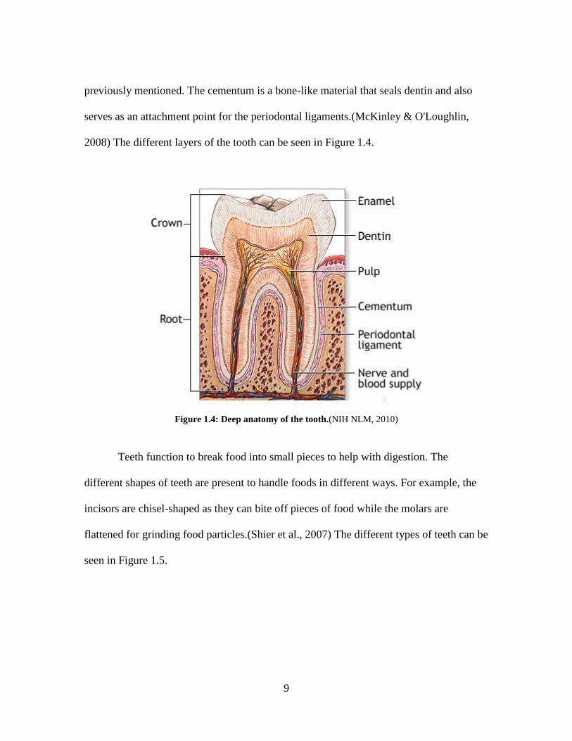
previously mentioned. The cementum is a bone-like material that seals dentin and also
serves as an attachment point for the periodontal ligaments.(McKinley & O'Loughlin,
2008) The different layers of the tooth can be seen in Figure 1.4.
Figure 1.4: Deep anatomy of the tooth.(NIH NLM, 2010)
Teeth function to break food into small pieces to help with digestion. The
different shapes of teeth are present to handle foods in different ways. For example, the
incisors are chisel-shaped as they can bite off pieces of food while the molars are
flattened for grinding food particles.(Shier et al., 2007) The different types of teeth can be
seen in Figure 1.5.

10
Figure 1.5: Shapes of the different types of teeth.(Web Dental Office, 2009)
1.3 Dental Pathologies
Tooth damage and eventual tooth loss can be caused by several things, but they
are most often associated with diseases of the gums (gingivitis) and the dental pulp
(endodontitis). Other common pathologies associated with teeth are periodontitis and
caries. All of the pathologies listed are preventable, but untreated conditions will
lead to teeth that cannot be easily saved.(Shier et al., 2007) As stated previously, teeth
have poor repair mechanisms, and permanent loss can affect digestive processes and
speech.
Gingivitis is one of the most common forms of dental disease, and it involves
inflammation of the gingiva, or gum. It is caused by the prolonged presence of plaque, a
sticky material composed of bacteria, mucus, and food debris that collects on teeth. If
plaque if allowed to remain on a tooth, it hardens and becomes trapped at the base of the
11
tooth transforming into tartar. Plaque and tartar irritate the gums as the bacteria produce
toxins that cause the gums to become infected, swollen, and tender. This effect on the
gums is labeled gingivitis. It can be prevented with regular tooth brushing and dental
cleaning. Gingivitis can be reversed, left untreated, it can become a more serious disease
called periodontitis.(Shafer, Hine, & Levy, 1974)
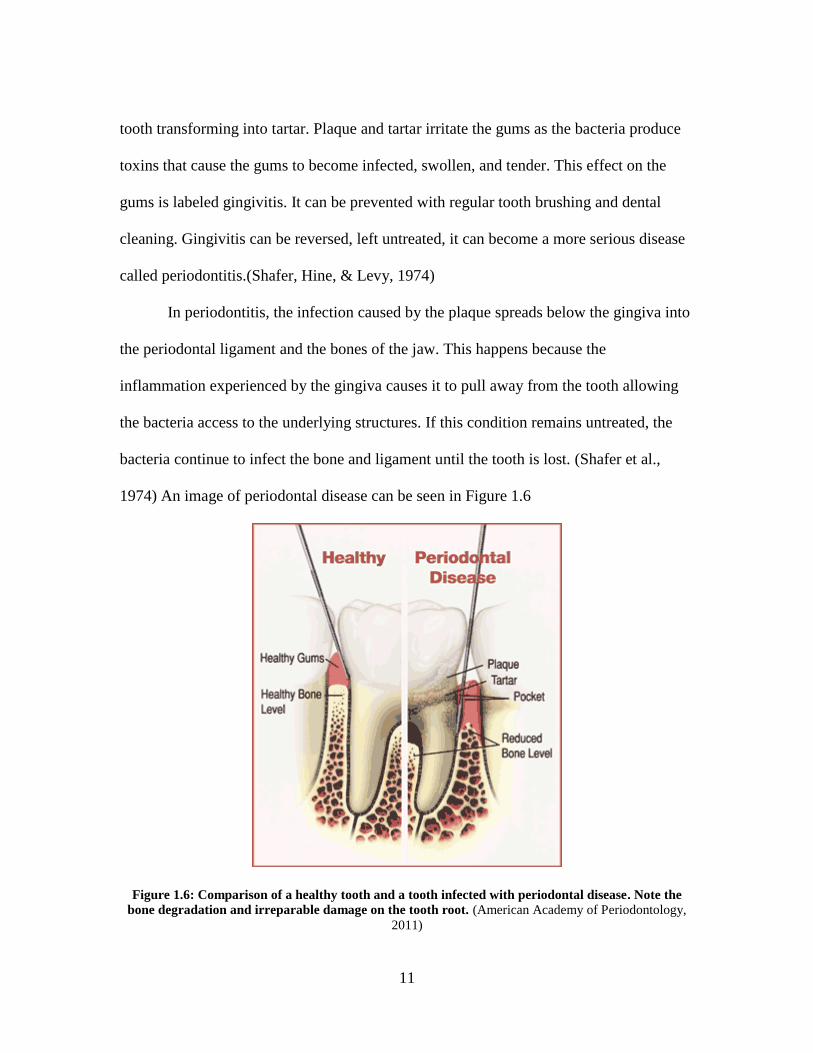
In periodontitis, the infection caused by the plaque spreads below the gingiva into
the periodontal ligament and the bones of the jaw. This happens because the
inflammation experienced by the gingiva causes it to pull away from the tooth allowing
the bacteria access to the underlying structures. If this condition remains untreated, the
bacteria continue to infect the bone and ligament until the tooth is lost. (Shafer et al.,
1974) An image of periodontal disease can be seen in Figure 1.6
Figure 1.6: Comparison of a healthy tooth and a tooth infected with periodontal disease. Note the
bone degradation and irreparable damage on the tooth root. (American Academy of Periodontology,
2011)
12
Tooth decay, also called dental caries or cavities, is the second most common
dental pathology in humans behind gingivitis. This condition is caused by the same
plaque and bacteria that cause gingivitis. Bacteria such as Actinomyces, Streptococcus
mutans, and Lactobacillus metabolize food lodged in between teeth at on the gum. The
metabolic by-products of these bacteria are acidic, and these acids can degrade the
enamel and later the dentin of the teeth. The bacteria also produce sticky substances to
ensure they stay in place to continue to cause damage. However, the bacteria and plaque
can be prevented and treated by normal brushing and dental visits. Also, the drinking of
fluorinated water is helpful in preventing dental caries as fluoride is incorporated into the
chemical structure of enamel to help strengthen it and protect it from acids and
decay.(Shier et al., 2007)
1.4 Natural Repair Mechanisms of Teeth
When a tooth is damaged, a limited amount of natural repair of the tooth can
occur. Dentin can be restored to some extent, but enamel cannot. This is because enamel
is produced by ameloblasts which under desmolysis (destruction and disintegration) after
enamel maturity. Therefore, if an injury causes damage to enamel, no biological repair
occurs. If the injury extends into the dentin, natural restoration of the dentin can occur if
there is at least 0.25mm of intact dentin remaining. The dentin that is formed in response
to injury is tertiary dentin as previously discussed. This dentin is unorganized in structure
and mechanically weak compared to primary or secondary dentin due to the fact it is

13
produced extremely rapidly. This is analogous to vasculature formation in cancer tumors
or scar tissue formation being disorganized and mechanically weak. If less than 0.25 mm
of dentin remain after injury, little tertiary dentin is formed and the injury will need to be
treated clinically. If the injury extends into the dental pulp, a majority of the dentin-
producing cells odontoblasts are most likely injured as they reside on the junction
between the dentin and pulp. The undifferentiated dental pulp stem cells migrate to edge
of the pulp tissue to take the place of the damaged odontoblasts.(H. Liu, Gronthos, & Shi,
2006; Yen & Yelick, 2011) This migration and differentiation leads to odontoblastoid
cells which produce tertiary dentin similar to the previous example. This dentin is very
rapidly deposited as its primary goal is to protect the pulp.(Murray & Garcia-Godoy,
2004; Shafer et al., 1974)
It should be noticed that the mechanisms for repair of dental tissue are, at the very
best, acceptable to protect the tooth from further damage. However, clinical methods of
treating dental tissue damage do not achieve a much better outcome. The methods of
dental repair will be discussed in the Chapter Two along with an extensive discussion
about dental pulp stem cells.
1.5 Comparisons Between Tooth and Bone
While they are similar in appearance, mechanical properties, and composition,
teeth are not considered to be bones and are not included in the skeletal system. Bone and
teeth are analogous in that they both have layers of tissue that have different
compositions and functions, but those layers are the not the same. Unlike teeth whose
14
outer layer is a hard enamel, bone’s outermost layer is either a cartilage layer or the
periosteum. Both are very different from enamel.(McKinley & O'Loughlin, 2008; Shafer
et al., 1974; Shier et al., 2007; Wikipedia, 2012b)
Bone and the dentin component of a tooth both have a majority of mineralized
tissue composed mostly of hydroxyapatite, but their organic components differ. For
example, there are multiple types of proteins that are found specifically in dental tissue
and not found in bones. Some of these proteins include enamelins, amelogenins, and
dentin sialophosphoproteins. Also, bone contains more collagen, so it is not as hard as
dentin. Comparisons have been drawn between bone marrow and dental pulp due to the
presence of the undifferentiated stem cells present in both. However, the tissues remain
very different, for bone marrow produces blood cells. Lastly, the regenerative capabilities
of bone and teeth are very contrasting. Bone, even if fully broken, can heal itself in a
matter of months. The dentin and enamel components of tooth have very limited
regeneration capabilities.(Shier et al., 2007)
15
1.6 References
American Academy of Periodontology. (2011). Types of gum disease. Retrieved 3/12,
2012, from http://www.perio.org/consumer/2a.html
Avery, J. K. (2000). In Steele P. F. (Ed.), Essentials of oral histology and embryology
(2nd ed.). St. Louis, MO: Mosby.
Datko, L. (2011). Effects of microenvironment on growth and differentiation of human
dental pulp stem cells. (Masters, Clemson University).
Hao, J., Shi, S., Niu, Z., Xun, Z., Yue, L., & Xiao, M. (1997). Mineralized nodule
formation by human dental papilla cells in culture. European Journal of Oral
Sciences, 105(4), 318-324.
Liu, H., Gronthos, S., & Shi, S. (2006). Dental pulp stem cells. Methods in Enzymology,
419, 99-113. doi:10.1016/S0076-6879(06)19005-9
Liu, M., Sun, Y., Liu, Y., Yuan, M., Zhang, Z., & Hu, W. (2012). Modulation of the
differentiation of dental pulp stem cells by different concentrations of beta-
glycerophosphate. Molecules (Basel, Switzerland), 17(2), 1219-1232.
doi:10.3390/molecules17021219
16
Magloire, H., Couble, M. L., Thivichon-Prince, B., Maurin, J. C., & Bleicher, F. (2009).
Odontoblast: A mechano-sensory cell. Journal of Experimental Zoology.Part B,
Molecular and Developmental Evolution, 312B(5), 416-424.
doi:10.1002/jez.b.21264
McKinley, M., & O'Loughlin, V. D. (2008). Human anatomy (2nd ed.). New York, NY:
McGraw-Hill.
Murray, P. E., & Garcia-Godoy, F. (2004). Stem cell responses in tooth regeneration.
Stem Cells and Development, 13(3), 255-262. doi:10.1089/154732804323099181
NIH NLM. (2010). Tooth anatomy. Retrieved March/1, 2012, from
http://www.nlm.nih.gov/medlineplus/ency/imagepages/1121.htm
Shafer, W. G., Hine, M. K., & Levy, B. M. (1974). A textbook of oral pathology (3rd
ed.). Philadelphia, PA: Saunders.
Shier, D., Butler, J., & Lewis, R. (2007). Hole's human anatomy and physiology (11th
ed.). New York, NY: McGraw-Hill.
Web Dental Office. (2009). Tooth morphology. Retrieved March/1, 2012, from
http://users.forthnet.gr/ath/abyss/dep1151.htm
Wikipedia. (2012a). Mouth assessment. Retrieved March/10, 2012, from
http://en.wikipedia.org/wiki/Mouth_assessment

17
Wikipedia. (2012b). Tooth development. Retrieved March/14, 2012, from
http://en.wikipedia.org/wiki/Tooth_development
Yen, A. H., & Yelick, P. C. (2011). Dental tissue regeneration - a mini-review.
Gerontology, 57(1), 85-94. doi:10.1159/000314530
18
CHAPTER 2: Restorative Dentistry
With the prevalence of dental pathologies and the lack of natural healing in teeth,
clinicians and scientists have been searching for a suitable solution to restore function
and appearance in teeth after damage. Synthetic materials have been the main choice, but
these materials often fail. Even when failure is not present, they can only provide some of
the functionality of the original tooth as the living component is removed. Research is
being done to assess the possibilities for stem cell tissue engineering in dental
applications, and the research will be discussed in the following chapters.
2.1 Market for Restorative Dental Procedures
The market for dental restoration procedures is not as high as it once was due to
better education and the fluoridation of water in many communities around the country.
In a 2004 statement, the US Surgeon General Richard H. Carmona, M.D. was quoted
saying:
Scientific studies have found that people living in communities
with fluoridated water have fewer cavities than those living where the
water is not fluoridated . . . An economic analysis has determined that in
most communities, every $1 invested in fluoridation saves $38 or more in
treatment costs. Fluoridation is the single most effective public health
measure to prevent tooth decay and improve oral health over a lifetime,
for both children and adults.(Carmona, 2004)

19
However, there are still many complications associated with oral health in the
United States today. A Gallup poll indicated that 34% of Americans did not visit a dentist
at all in the previous year.(Marcus, 2009) Also, according to the Nation Institute of
Dental and Craniofacial Research (NIDCR), 23% of children aged 2-11 have never been
to the dentist while 12% of adults aged 20-64 haven’t visited a dentist in over five
years.(NIDCR, 2011) This shows that the deficiency of oral health is significant for all
ages and gives an indication of the prevalence of dental pathologies. The lack of dental
care and other factors has led to a large number of dental caries and other oral health
conditions. Some of the NIDCR statistics are as follows:
21% of children ages 6-11 have had tooth decay (dental caries)
59% of adolescents ages 12 to 19 have had tooth decay, and 23% of adolescents
continue to have untreated decay.
92% of adults 20 to 64 have had tooth decay, and 23% of adults continue to have
untreated decay.
Adults have an average of 3.28 decayed or missing teeth and 13.65 decayed and
missing surfaces.
Approximately 5% of adults have no teeth.
8.52% of adults 20 to 64 have periodontal disease, and 5.08% have moderate or
severe periodontal disease.
It can be seen that dental restoration procedures have a large market even with the better
oral education and increase in fluoridation of water.(NIDCR, 2011)

20
The dental industry provides treatments to restore the form, function, and
appearance of teeth, and most of the research is focused on improving techniques,
equipment, and materials. This is because the constant numbers of restorative procedures
demand a high price tag for the best technologies. In the US, the dental market reached
$12.7 billion in 2010, and it is expected to increase to $14.4 billion by 2016. This money
is split between equipment/supplies and consumer dental care. The split along with
projections can be seen in Figure 2.1.
Figure 2.2: US Dental Market Analysis by Major Product Segment. This graph was reprinted
without permission. (Research, 2012)
Also, in global markets, the value of dental consumables and prosthetics was
$15.9 billion in 2010 and is projected to be $18.5 billion in 2015. The supplies, materials,
and prosthetics market accounts for the largest share of that at $9 billion, and it is driven
by the sales generated for crowns, bridges, and dental implants. In the materials market,
21
filling materials account for a majority of the sales totaling $5.8 in 2010. This shows that
both globally and in the United States, dental products are extensively used and can be
very profitable. Therefore, finding a better replacement dental material to supplant the
current materials would be a huge breakthrough in the dental health field.
2.2 Current State of the Art
2.2.1 Fillings
Fillings are a common treatment for tooth decay where the dentist removes
decayed sections of the tooth and restores the original tooth shape and function with a
synthetic material. This stops continued decay and also prevents further decay by closing
the spaces that bacteria can enter. The materials used for fillings include metals (gold,
amalgam (a mixture of silver, tin, zinc, copper, and mercury)), composites, and porcelain.
None of these restorative materials has the ideal set of properties, and dentists select the
filling type bases on the specific patient case. For example, gold fillings have an
extremely long life because they are durable and do not corrode, but gold has an
extremely high cost. Another consideration is appearance. Gold does not match the color
of teeth and is easily noticeable, so porcelain may be used as it is tooth-colored. Yet,
porcelain is brittle so shouldn’t be used for load-bearing surfaces, etc. There are many
other considerations to make before choosing a dental material.(Colgate, 2012; Dental
Association, ) Figure 2.2 shows the range of filling appearance when used to restore a
human tooth.

22
Figure 2.3: Comparison of Gold (left), Amalgam (top right), and composite material (bottom right).
Note the visual blending of the composite with natural tooth as opposed to the stark contrast in gold
and amalgam. These photos were reprinted without permission. (Heritage Dental Centre, 2009)
Amalgam is the most common filling material, but recent concerns of the safety
of amalgams due to their mercury content is leading government and health organizations
to recommend ceramic and composites in their place. Gold is still preferred, but the high
price of a gold filling leaves it out of reach for most consumers. Replacing amalgam with
composites and porcelain is acceptable in some applications, but amalgams are used in
load-bearing surfaces, such as molars, where composites and porcelain could fracture.
They are more suited to incisors and canines due to good wear resistance.(Colgate, 2012;
Dental Association, )
2.2.2 Crowns
Crowns are similar in materials to fillings, but the application is different. A
crown is simply a cap that goes on top of an existing tooth or dental implant. If a tooth is
too weak or damaged for a filling or even fractured, crowns can be used to protect what is
remaining of the tooth, specifically the dental pulp. The damaged tooth is reduced in size

23
and a mold is made of the remaining structure. Then, the fitted crown is applied and
cemented into place. Figure 2.3 shows this process. The materials used in crowns are
similar to fillings, but the preferred material is porcelain bonded to a metal shell because
it is both strong and attractive.(Colgate, 2012)
Figure 2.4: A diagram of the crowning process for dental restoration.(Dolce, 2012)
In both crowns and fillings, a discrepancy between the material properties of the
replacement material and the natural tooth can lead to failure. Almost all dental
restoration procedures need revisions at least once, while the average time frame of
functionality is 5-12 years for non-gold materials. Research is currently being done to
find a suitable replacement material that can mimic the tooth better in terms of strength
and resistance to damage. It is probable that the only suitable material would be the
natural dental material itself. Guiding stem cells with tissue engineering techniques could
be an ideal solution to solving this material shortage.

24
2.3 Regenerative Medicine in Dentistry
The terms tissue engineering and regenerative medicine have been used since the
1980’s, and in the early 1990’s, Langer and Vacanti defined tissue engineering as “an
interdisciplinary field that applies the principles of engineering and life sciences toward
the development of biological substitutes that restore, maintain or improve tissue
function.”(Ricci & Terracio, 2011) Tissue engineering is a very intriguing area of
research, and many research groups are focused on regenerative medicine applications.
The problems seen in dental restoration procedures are evident in every medical field-
materials that exactly mimic the body’s natural forms and functions are desired. Some
areas of the body are already being treated by tissue engineered constructs.
Dentistry has long tried to restore the functions of native teeth, and one source
argues that dentistry has always been at the forefront of regenerative medicine, even
before the term was used, as the goal of any treatment all along was to restore tissue and
organ function in teeth. However, the stem cell based tissue engineering approaches are
coming along as well. While a full tooth may never be grown (some argue that it
wouldn’t be useful even if it was possible), using stem cells to regenerate dental pulp to
save teeth. Also, stem cells could be used to regenerate the periodontal ligament and the
underlying alveolar bone as those are also damaged in oral diseases.(Ricci & Terracio,
2011)
The source of these stem cells remains an issue. While a lot of press has been
given to embryonic stem cells, studies have begun to show that they are susceptible to
forming cancerous tissues.(Li et al., 2011) Work is being done with induced pluripotent

25
stem cells (iPS cells) and also multipotent stromal cells. Some of these stem cells come
from human teeth while others come from bone marrow, adipose tissue, or adult
muscle.(Gimble, Grayson, Guilak, Lopez, & Vunjak-Novakovic, 2011; Gronthos et al.,
2002; Gronthos, 2011; Murray & Garcia-Godoy, 2004)
Multipotent stromal cells have shown promise in studies as many groups have
been able to regenerate almost all tissues in the body including bone, periodontal
ligament, cementum, and dentin in animals.(Shi et al., 2005; Yen & Sharpe, 2008; Yen &
Yelick, 2011)
The results of many studies are promising, but guiding the stem cells down a
specific path reproducibly can be difficult. It is known that cells during development
respond to numerous different and complex cues to ensure that each cell is directed down
the correct lineage. This process is not fully understood, but directed stem cell
commitment in the laboratory has been focused on exposing stem cells to soluble factors
in growth media (growth factors, cytokines, serum proteins, etc.) and properties of the
biomaterial substrate (surface energy, roughness, chemistry, elasticity,composition, etc.).
Alterations in these factors have been shown to change a cell’s adhesion, morphology,
and/or proliferation.(Engler, Sen, Sweeney, & Discher, 2006; Phillips, Petrie, Creighton,
& Garcia, 2010)
To understand and be able to manipulate odontogenic differentiation (dentin-
forming cells), osteogenic differentiation (bone-forming cells) can be used as a model as
they are similar and more work has been done on bone. Bone morphogenic proteins
(BMP) are cytokines that, as the name would suggest, induce differentiation down an

26
osseous lineage. Also, dexamethasone has been successful in the same goal.(Chaudhary,
Hofmeister, & Hruska, 2004; Jo, Lee, Suh, & Kim, 2007; Zhang, Walboomers, Shi, Fan,
& Jansen, 2006)
Also, RGD peptides, a cellular recognition molecule found in numerous ECM
proteins, have been used to promote proliferation and migration or differentiation in stem
cells. RGD peptides interact with the cells via integrin receptors and can be easily
conjugated to multiple types of scaffolds.(Comisar, Kazmers, Mooney, & Linderman,
2007; Moore, Lin, Gallant, & Becker, 2011) When paired with BMP and patterned on a
surface, differentiation down an osteogenic pathway was observed. This was important
because there was no supplemental osteogenic growth media present, so the
differentiation is believed to be from the signaling molecules attached to the substrate.
Also in this study, it was noted that early osteogenic differentiation and mineralization of
the bone marrow stem cells was strongly correlated with cell density, attachment and
proliferation.(Moore et al., 2011)
Knowing the effects of different molecules on cell guidance is important, but just
as important is understanding the effects of substrate matrix properties. In studies done by
Engler, et al and Tse, et al, the matrix mechanical properties were varied to determine the
mechano-transduction signaling pathways in stem cells. The resistance that a cell feels
when attempting to deform its substrate (in the body, the substrate is extracellular matrix)
is measured by the elastic modulus, E. It was found that mesenchymal stem cells were far
more responsive to the matrix elasticity than differentiated cells. It was also noted that
differentiation is enhanced on stiffer substrates (25-50 kPa). On less stiff substrates (0.1-

27
10 kPa), the cells were guided towards a soft tissue or neuron-like lineage.(Engler et al.,
2006; Tse & Engler, 2011)
Studies like the examples above begin to demonstrate the complexity of the
development of the human body. The mechanisms of formation and direction may never
fully be understood, but if enough information is gained about the factors that we do
understand, great strides can be made in tissue engineering.
2.4 Dental Pulp Stem Cells
The idea of tissue engineering for dental applications is to cultivate stem cells
with odontogenic induction signals to program the stem cells to adopt dental lineages
and, with the help of scaffold/extracellular matrix, to become part of a tooth. For dental
applications, it is intuitive that dental pulp stem cells should be the cell type studied as
they naturally have a connection to a dental lineage. Also, it is known that teeth contain
stem cells based on the natural repairing ability of dentin after injury suggesting that cells
in a fully developed tooth can still function as odontoblasts.(Yen & Sharpe, 2006; Yen &
Sharpe, 2008)
2.4.1 Characterization
Many cell sources are available within teeth. Human dental pulp stem cells
(DPSCs) have been derived from wisdom teeth (third molars) and have been shown to
form odontoblast-like cells that can produce dentin-like materials when cultured in

28
mineralization-enhancing conditions. Another source of stem cells is primary teeth that
have been exfoliated naturally during development around 6-10 years of age. These cells
are recognized as “stem cells from human exfoliated deciduous teeth” or SHED. The
cells isolated from both of these locations have shown multipotent differentiation,
expression of stem cell markers Stro-1 and CD146, dentin regeneration in vivo, and the
ability to form colonies in vitro. While these two populations of stem cells are similar,
they are considered different as SHED can produce dentin and bone, but not dentin-pulp
complexes like DPSCs are able to in immunocompromised mice.(Gronthos, Mankani,
Brahim, Robey, & Shi, 2000; Yen & Sharpe, 2008) Also, periodontal ligament stem cells
have been isolated from periodontal tissue, but the ligaments must be intact for this
isolation.(Liu, Gronthos, & Shi, 2006) These types of stem cells will all be referred to as
dental pulp stem cells for the purposes of this thesis. Having many sources for these stem
cells can be beneficial as a large number might be necessary for successful tissue
engineering.
The DPSCs mentioned can be characterized under the broad heading of
mesenchymal stem cells (MSCs). A mesenchymal stem cell is any undifferentiated cell
from the mesenchyme, a type of loose connective tissue derived from the mesoderm
during development. The first mesenchymal stem cells discovered and used were bone
marrow mesenchymal stem cells (BMMSCs), and they are still the gold-standard for
comparison of potency.(Huang, Gronthos, & Shi, 2009) When compared to bone marrow,
dental pulp contains stem cells that produce many of the same markers that indicate
multipotency: Stro-1, CD73, CD90, CD105, and CD146.(Gronthos et al., 2000; Gronthos
29
et al., 2002; Liu et al., 2006; Perry et al., 2008; Shi et al., 2005; Yen & Sharpe, 2008) In
addition to being similar to BMMSCs, the DPSCs have markers similar to osteogenic
cells as well: alkaline phosphatase, collagen I, osteonectin, osteopontin, osteocalcin, bone
sialoprotein, and matrix extracellular phosphaglycoprotein (MEPE). (Gronthos et al.,
2002)
In differentiation studies, DPSCs have been tested with different growth factors
known to induce guidance down certain lineages. For example, BMP and dexamethasone
for bone-formation as previously mentioned. In the studies, DPSCs were successfully
induced into differentiating down osteogenic, neurogenic, adipogenic, myogenic,
endothelial, and chondrogenic pathways.(d'Aquino et al., 2007; Laino et al., 2005) It was
noticed that osteogenic differentiation was the easiest to achieve which makes sense
intuitively. The DPSCs were able to form mineralized nodules in vitro after a few weeks
in a culture environment with even low levels of inductive growth factors.(Zhang et al.,
2005)
Dental Pulp Stem Cells can also be characterized based on factors not associated
with their stem cell potential. It has been found that DPSCs are highly proliferative (30-
50 times the rate of BMMSCs) and can survive cryopreservation well. These are
important traits for tissue engineering applications.(Huang et al., 2009; Li et al., 2011;
Zhang et al., 2006)
30
2.4.2 Regenerative Medicine Applications
As previously stated, dental structures can be formed using DPSCs that are
transplanted into immunocompromised mice, but the tissue is poorly organized and
multiple tooth-like structures form. This is an unacceptable end product for tissue
engineering or regenerative medicine applications. However, the concept that these cells
can form tooth-like structures is very promising.(Yen & Sharpe, 2008)
In the previous experiment, DPSCs were seeded onto a scaffold and transplanted
into the immunocompromised mice. This technique illustrates the traditional tissue
engineering theory to combine cells, osteogenic factors, and an engineered construct in a
bioreactor to promote cell growth in the form of the scaffold. The cell-scaffold
combination is then implanted into the patient.(Yen & Sharpe, 2008)
Another experiment combine the DPSCs with endothelial cells on the scaffold to
more closely mimic the environment that the DPSCs are in within the patient. Upon
implantation, only one odontogenic structure formed, which is desired to regenerate tooth
tissue.(Chai & Slavkin, 2003) One last recent experiment showed that using a gelatin
scaffold, DPSCs showed differentiation into osteoblasts in 14 days in culture. The cell-
scaffold construct was then implanted into nude mice, and the cells retained their
osteoblastic phenotype and began to form bone-like tissue. This group suggested that
DPSCs could be a suitable autologous seed cell type of bone tissue engineering due to the
following advantages: osteogenic differentiation potential, obtainability from a common
dental practice (wisdom tooth removal), high proliferation rates, and ease of culturing.
However, it is noted that a vascularized tissue-engineered bone material has not been

31
accomplished yet, and this step is key to complete integration into the host.(Li et al.,
2011)
Overall, dental pulp stem cells have a high potential for bone and tooth tissue
engineering applications, but that potential has not been reached yet. DPSCs have many
desirable characteristics that make them an attractive source for tissue engineering, and
they even seem to have advantages over cells derived from bone marrow, currently the
gold-standard. Research to explore the vast possibilities with DPSCs under different
conditions needs to be completed to have a better understanding of the mechanisms and
factors that affect DPSCs differentiation and growth.

32
2.5 References
Carmona, R. H. (2004). Statement on water fluoridation. Retrieved 3/14, 2012, from
http://www.nidcr.nih.gov/OralHealth/Topics/Fluoride/StatementWaterFluoridation.h
tm
Chai, Y., & Slavkin, H. C. (2003). Prospects for tooth regeneration in the 21st century: A
perspective. Microscopy Research and Technique, 60(5), 469-479.
doi:10.1002/jemt.10287
Chaudhary, L. R., Hofmeister, A. M., & Hruska, K. A. (2004). Differential growth factor
control of bone formation through osteoprogenitor differentiation. Bone, 34(3), 402-
411. doi:10.1016/j.bone.2003.11.014
Colgate. (2012). Dental procedures. Retrieved March/1, 2012, from
http://www.colgate.com/app/CP/US/EN/OC/Information/Articles/Oral-and-Dental-
Health-Basics/Checkups-and-Dental-Procedures.cvsp
Comisar, W. A., Kazmers, N. H., Mooney, D. J., & Linderman, J. J. (2007). Engineering
RGD nanopatterned hydrogels to control preosteoblast behavior: A combined
computational and experimental approach. Biomaterials, 28(30), 4409-4417.
doi:10.1016/j.biomaterials.2007.06.018
33
d'Aquino, R., Graziano, A., Sampaolesi, M., Laino, G., Pirozzi, G., De Rosa, A., et al.
(2007). Human postnatal dental pulp cells co-differentiate into osteoblasts and
endotheliocytes: A pivotal synergy leading to adult bone tissue formation. Cell
Death and Differentiation, 14(6), 1162-1171. doi:10.1038/sj.cdd.4402121
Dental Association, A. Dental materials. Retrieved February/20, 2012, from
http://www.ada.org/5164.aspx?currentTab=1
Dolce, V. (2012). Dental crowns and bridges. Retrieved 3/14, 2012, from
http://drvincentdolce.com/dental-crowns-and-bridges/
Engler, A. J., Sen, S., Sweeney, H. L., & Discher, D. E. (2006). Matrix elasticity directs
stem cell lineage specification. Cell, 126(4), 677-689. doi:10.1016/j.cell.2006.06.044
Gimble, J. M., Grayson, W., Guilak, F., Lopez, M. J., & Vunjak-Novakovic, G. (2011).
Adipose tissue as a stem cell source for musculoskeletal regeneration. Frontiers in
Bioscience (Scholar Edition), 3, 69-81.
Gronthos, S. (2011). The therapeutic potential of dental pulp cells: More than pulp
fiction? Cytotherapy, 13(10), 1162-1163. doi:10.3109/14653249.2011.623827;
10.3109/14653249.2011.623827
Gronthos, S., Brahim, J., Li, W., Fisher, L. W., Cherman, N., Boyde, A., et al. (2002).
Stem cell properties of human dental pulp stem cells. Journal of Dental Research,
81(8), 531-535.
34
Gronthos, S., Mankani, M., Brahim, J., Robey, P. G., & Shi, S. (2000). Postnatal human
dental pulp stem cells (DPSCs) in vitro and in vivo. Proceedings of the National
Academy of Sciences of the United States of America, 97(25), 13625-13630.
doi:10.1073/pnas.240309797
Heritage Dental Centre. (2009). Dental restorations. Retrieved 3/14, 2012, from
http://www.heritagedentalcentre.net/index.php?page_id=7
Huang, G. T., Gronthos, S., & Shi, S. (2009). Mesenchymal stem cells derived from
dental tissues vs. those from other sources: Their biology and role in regenerative
medicine. Journal of Dental Research, 88(9), 792-806.
doi:10.1177/0022034509340867
Jo, ,Inho, Lee, ,Jung, Suh, ,Hwal, & Kim, ,Hyongbum. (2007). Bone tissue engineering
using marrow stromal cells. Biotechnology and Bioprocess Engineering, (1), 48-53.
doi:10.1007/BF02931803
Laino, G., d'Aquino, R., Graziano, A., Lanza, V., Carinci, F., Naro, F., et al. (2005). A
new population of human adult dental pulp stem cells: A useful source of living
autologous fibrous bone tissue (LAB). Journal of Bone and Mineral Research : The
Official Journal of the American Society for Bone and Mineral Research, 20(8),
1394-1402. doi:10.1359/JBMR.050325

35
Li, J. H., Liu, D. Y., Zhang, F. M., Wang, F., Zhang, W. K., & Zhang, Z. T. (2011).
Human dental pulp stem cell is a promising autologous seed cell for bone tissue
engineering. Chinese Medical Journal, 124(23), 4022-4028.
Liu, H., Gronthos, S., & Shi, S. (2006). Dental pulp stem cells. Methods in Enzymology,
419, 99-113. doi:10.1016/S0076-6879(06)19005-9
Marcus, M. B. (2009, March 9). Many americans say they forgo routine dental care. USA
Today,
Moore, N. M., Lin, N. J., Gallant, N. D., & Becker, M. L. (2011). Synergistic
enhancement of human bone marrow stromal cell proliferation and osteogenic
differentiation on BMP-2-derived and RGD peptide concentration gradients. Acta
Biomaterialia, 7(5), 2091-2100. doi:10.1016/j.actbio.2011.01.019
Murray, P. E., & Garcia-Godoy, F. (2004). Stem cell responses in tooth regeneration.
Stem Cells and Development, 13(3), 255-262. doi:10.1089/154732804323099181
NIDCR. (2011). Dental, oral, and craniofacial data and statistics. Retrieved 3/14, 2012,
from http://www.nidcr.nih.gov/DataStatistics/
Perry, B. C., Zhou, D., Wu, X., Yang, F. C., Byers, M. A., Chu, T. M., et al. (2008).
Collection, cryopreservation, and characterization of human dental pulp-derived
mesenchymal stem cells for banking and clinical use. Tissue Engineering.Part C,
Methods, 14(2), 149-156. doi:10.1089/ten.tec.2008.0031

36
Phillips, J. E., Petrie, T. A., Creighton, F. P., & Garcia, A. J. (2010). Human
mesenchymal stem cell differentiation on self-assembled monolayers presenting
different surface chemistries. Acta Biomaterialia, 6(1), 12-20.
doi:10.1016/j.actbio.2009.07.023
Research, B. (2012). The dental market: Techniques, equipment & materials. Retrieved
March/1, 2012, from http://www.bccresearch.com/report/dental-market-techniques-
equipment-materials-hlc028d.html
Ricci, J. L., & Terracio, L. (2011). Where is dentistry in regenerative medicine?
International Dental Journal, 61 Suppl 1, 2-10. doi:10.1111/j.1875-
595X.2011.00024.x; 10.1111/j.1875-595X.2011.00024.x
Shi, S., Bartold, P. M., Miura, M., Seo, B. M., Robey, P. G., & Gronthos, S. (2005). The
efficacy of mesenchymal stem cells to regenerate and repair dental structures.
Orthodontics & Craniofacial Research, 8(3), 191-199. doi:10.1111/j.1601-
6343.2005.00331.x
Tse, J. R., & Engler, A. J. (2011). Stiffness gradients mimicking in vivo tissue variation
regulate mesenchymal stem cell fate. PloS One, 6(1), e15978.
doi:10.1371/journal.pone.0015978
Yen, A. H., & Sharpe, P. T. (2006). Regeneration of teeth using stem cell-based tissue
engineering. Expert Opinion on Biological Therapy, 6(1), 9-16.
doi:10.1517/14712598.6.1.9

37
Yen, A. H., & Sharpe, P. T. (2008). Stem cells and tooth tissue engineering. Cell and
Tissue Research, 331(1), 359-372. doi:10.1007/s00441-007-0467-6
Yen, A. H., & Yelick, P. C. (2011). Dental tissue regeneration - a mini-review.
Gerontology, 57(1), 85-94. doi:10.1159/000314530
Zhang, W., Walboomers, X. F., Shi, S., Fan, M., & Jansen, J. A. (2006). Multilineage
differentiation potential of stem cells derived from human dental pulp after
cryopreservation. Tissue Engineering, 12(10), 2813-2823.
doi:10.1089/ten.2006.12.2813
Zhang, W., Walboomers, X. F., Wolke, J. G., Bian, Z., Fan, M. W., & Jansen, J. A.
(2005). Differentiation ability of rat postnatal dental pulp cells in vitro. Tissue
Engineering, 11(3-4), 357-368. doi:10.1089/ten.2005.11.357

38
CHAPTER 3: Determining the Effect of Substrate Stiffness on the Growth
and Differentiation of Dental Pulp Stem Cells
This chapter will outline previous work completed by myself and my laboratory
mates. This work is similar to the work done by Engler, et al. but DPSCs are used instead
of bone-marrow derived stem cells. The cells were exposed to substrates with different
mechanical properties to observe the differences in growth patterns.
3.1 Background
3.1.1 Mechanical Properties of the Substrate
It is known that many different chemical, mechanical, and electrical cues can induce
differentiation and cell phenotypic changes. A well-documented cue that seems to have an
effect on almost all cells is substrate stiffness. For differentiated cells such as fibroblasts,
their anchorage dependency is better understood and documented. For example, softer
substrates induce different morphological changes than stiffer substrates due to the focal
adhesion interactions with the substrate. Multipotent stromal cells derived from bone marrow
have been shown to have an effect as well. Stem cells derived from dental pulp have not been
studied extensively in terms of the effect of matrix elasticity or stiffness.(Engler, Sen,
Sweeney, & Discher, 2006)
In previous work by Engler, et. al, polyacrylamide hydrogels were polymerized with
different ratios of acrylamide to acrylamide-bis concentrations according to a protocol from
Wang and Pelham.(Wang & Pelham, 1998) Polyacrylamide gels are easily tunable to have

39
different mechanical properties by changing the ratio of monomers in the polymer solution,
so they are often chosen as a substrate.(Tse & Engler, 2010) Figure 3.1 below diagrams the
polymerization of a polyacrylamide.
Figure 3.5: The chemical reaction to form a crosslinked polyacrylamide hydrogel.(Thermo Scientific,
2012)
Increasing the percentage of bis-polyacrylamide in the mixture will lead to a
greater degree of cross-linking during polymerization, leading to a gel with a greater
stiffness. Also, according to the Wang protocol, the glass surface that the gel is on top of
needs to be activated by an amino group (3-Aminopropyltriethoxysilane (APTES) so that
some of the acrylamide monomers bind to the surface. This step is done so that the gel
can be polymerized between two thin, flat glass plates to create a uniform thickness, and
the gel will remain adhered to only the bottom glass plate when the top plate is removed.
The last step to prepare for cell seeding is to attach ECM proteins such as collagen or
fibronectin to the surface of the gel to enhance cell attachment.(Engler et al., 2006; Tse &
Engler, 2010)

40
It should be noted that while polyacrylamide has valuable properties as a easily
manipulated substrate as far as stiffness is concerned, there are many questions about the
toxicity of polyacrylamide and its monomer, acrylamide. The FDA approves
polyacrylamide for many applications, and it has been suggested that polymerized and
crosslinked polyacrylamide is not toxic in low doses. However, the monomer acrylamide
has been shown to be a neurotoxin. Therefore, if any monomer is present within the
polymer or if the polymer is degraded in the body, toxicity will become an issue.(King &
Noss, 1989) So, for the experiments outlined below, polyacrylamide is suitable, but for
the translation to a clinical application a different substrate would need to be used.
3.1.2 Dental Pulp Stem Cells’ Differentiation Potential
As previously written, mesenchymal stem cells, as well as dental pulp stem cells,
have multipotent differentiation potential under the correct environmental conditions. As
the tissue desired is bone-like in form and function, the environments necessary to induce
osteogenic differentiation in bone marrow stem cells have been studied. The common
soluble growth factors used to induce osteogenic differentiation are dexamethasone,
BMPs (mostly BMP-2 and BMP-7), L-ascorbic acid 2-phosphate (Vitamin C), and β-
glycerophosphate.(Kadar et al., 2009; Laino et al., 2005; Zhang et al., 2005) Also, a stiff
substrate has been shown to induce osteogenesis.(Engler et al., 2006) As the growth
factors need to be constantly supplied to the tissue engineered construct, they are less
practical for translation to clinical applications. Therefore, if the substrate properties

41
could be adjusted to induce osteogenic differentiation, the cells would be able to receive
those signals post-implantation as well.
3.1.3 Comparison to Bone Marrow Stromal Cells
Most of the background information has been focused on bone marrow stromal
cells, as they are the most studied. However, the cells have shown differences to dental
pulp stem cells that should be addressed. First, dental pulp stem cells have been easily
differentiated into osteogenic and neurogenic cells, but not myogenic, adipogenic, or
chondrogenic. Differentiation has been successful towards most cell types, but it was
done with a very strong dose of growth factors and a long culture time. It is thought that
since nerves and bone-like tissue are present in/near the pulp tissue, those dental pulp
stem cells are predisposed to becoming those types of cells.(Zhang, Walboomers, Shi,
Fan, & Jansen, 2006)
Using the data from Engler, et. al showing the differentiation potential for bone
marrow stem cells gives an insight into how dental pulp stem cells should be expected to
react.(Engler et al., 2006) Also, the fact that dental pulp stem cells have been shown to
form a structure similar to the dentin-pulp complex, while hardly any other types of stem
cells have shown that, is important.(Gronthos, Mankani, Brahim, Robey, & Shi, 2000;
Shi et al., 2005) Combining the information, if the correct conditions could be created,
there is great promise for dental tissue regeneration.
42
3.1.4 Testing Methods
The first method used for testing the cells in this experiment was microscopy.
Both light microscopy and confocal fluorescence microscopy were performed. In
fluorescence microscopy, immunocytochemistry is used to stain the cells. First, the cells
are killed and fixed before they are permeabilized and incubated in an antibody solution
that attaches to the protein or molecule of interest. After that, the cells are incubated in
another antibody that is tagged with a fluorescent molecule, and that antibody only
attaches to the primary antibody. In this way, the protein or molecule of interest is
effectively fluorescently tagged and will show up in fluorescence imaging.
The second method to test cells was using alkaline phosphatase (ALP) and
bicinchoninic acid (BCA) assays to determine the specific activity of the enzyme alkaline
phosphatase, a common enzyme found in many cells. Elevated ALP levels indicates that
there could be active bone formation occurring as ALP is a byproduct
of osteoblast activity.(Duplomb, Dagouassat, Jourdon, & Heymann, 2007)
In these assays, a colormetric test is done to see how much ALP is present and also how
much total protein is present. The data is then normalized to find specific activity. The
alkaline phosphatase reaction is shown below in Figure 3.2. Note that the yellow color
produced by the product (p-Nitrophenolate) is measured to indicate the amount of ALP
present.

43
Figure 3.6: The ALP assay reaction. (Napier University, 2007)
3.2 Materials and Methods
3.2.1 Cell Acquisition and Isolation
Dental pulp was obtained from human impacted third molars (wisdom teeth)
during a common dental procedure by Dr. Satish Alapati. The dental pulp stem cells were
isolated from the pulp tissue using the protocols found in literature.(Huang, Chen, Lin,
Shieh, & Chan, 2008; Liu, Gronthos, & Shi, 2006)
First, the extracted teeth were bathed in sterile saline, and the external surfaces of
the tooth were cleaned and cleared of bacteria with washes of 70% ethanol and sterile
Phosphate Buffered Saline (PBS). Dental instruments were used to expose the pulp

44
cavity, and the pulp was removed and placed into α-Minimum Essential Medium (α-
MEM). The pulp was minced using a scalpel, and the remaining pieces were “digested”
using a 3mg/mL solution of collagenase type I and 4mg/mL solution of dispase for 30-45
minutes at 37°C. After digestion, the cells were centrifuged at 1200rpm for 10 minutes to
obtain a cell pellet. The supernatant was discarded, and the pellet was re-suspended in
mesenchymal stem cell growth medium. The cells then were passed through a 70µm cell
strainer to ensure only cells remained. The cells were then cultured in mesenchymal stem
cell growth medium at 37°C and 5% CO2.(Liu et al., 2006)
To purify the cell suspension to only contain DPSCs, it was incubated with
STRO-1 (mouse IgM), 3G5 (mouse IgM), or CC9 (mouse IgG2a) antibodies for 1 hour
on ice. After washing with 1% BSA/PBS, the cells were incubated with anti-mouse IgG-
conjugated Dynabeads for 1 hour at 4°C. The bead-positive cells were selected out using
a Dynal MPC-1 magnetic particle concentrator.(Liu et al., 2006) The cells were then
cultured as described below.
3.2.2 Cell Culturing
Two different types of cell culture growth media were used in these experiments.
The first was Mesenchymal Stem Cell Growth Medium (MSCGM; Lonza) which was
“developed for growing large numbers of mesenchymal stem cells without inducing
diferentiation.”(Lonza, 2012) Later in the experimental stages, MSCGM was formulated
by the research lab based on literature, and it contained α-MEM with 15% FBS, 2mM
GlutaMAX, 100 μM L-ascorbic acid 2-phosphate, 2.5μg/mL amphotericin B, 100 U/mL

45
penicillin/streptomycin. The second type of media was Osteogenic Differentiation
Medium (ODM). It was formulated similarly to the MSCGM, but the additional
components were 1.8 mM KH2PO4 and 10 nM dexamethasone.
All cells were allowed to grow in the MSCGM prior to seeding onto the
polyacrylamide gels, so they were able to remain stem cells until given a cue to
differentiate. The cells were counted, and it was determined that a cell density of 10,000
cells/hydrogel would be used. The cells were transferred to and cultured in standard 6-
well cell culture plates on top of the polyacrylamide gels. The appropriate amount of cell
solution was added to the gels, and the cells were allowed to adhere in an incubator for 30
minutes before media was added. Each well in the culture plate contained 2-3 mL of
media, and the media was removed and replaced with fresh media each 2-3 days. The day
when the experimental conditions were applied was defined as day 0, and the cells were
analyzed at time points of 0, 1, 3, and 5 days.
Another experimental condition was that half of the cells that were grown in the
ODM were also grown in the presence of hyrdroxyapatite (HA) particles. As stated in a
previous section, HA is the most common mineral component found in both bone and
tooth. The hydroxyapatite particles (MP Biomedicals) were characterized by a lab
member, William McCallister, and they had an average particle diameter of 40µm.
Before addition of 50µL of HA solution to the samples, the HA particles were diluted to
10mg/mL in de-ionized (DI) water, sterilized using sonication, and vortexed to prevent
aggregation.
46
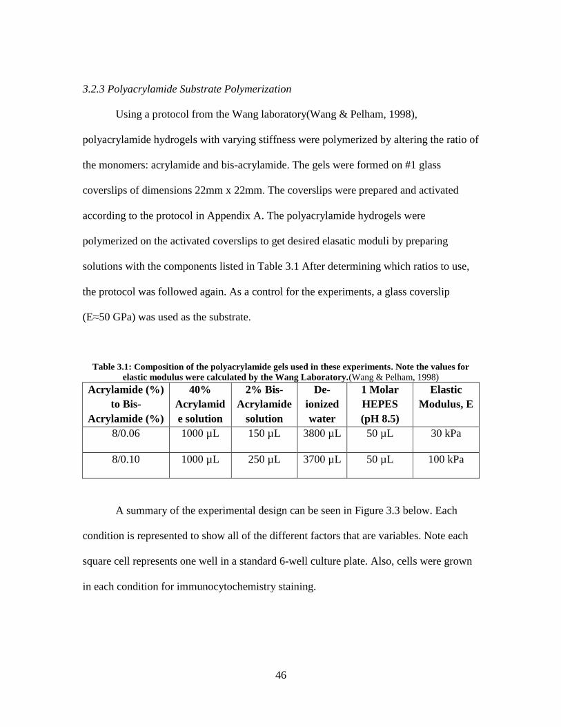
3.2.3 Polyacrylamide Substrate Polymerization
Using a protocol from the Wang laboratory(Wang & Pelham, 1998),
polyacrylamide hydrogels with varying stiffness were polymerized by altering the ratio of
the monomers: acrylamide and bis-acrylamide. The gels were formed on #1 glass
coverslips of dimensions 22mm x 22mm. The coverslips were prepared and activated
according to the protocol in Appendix A. The polyacrylamide hydrogels were
polymerized on the activated coverslips to get desired elasatic moduli by preparing
solutions with the components listed in Table 3.1 After determining which ratios to use,
the protocol was followed again. As a control for the experiments, a glass coverslip
(E≈50 GPa) was used as the substrate.
Table 3.1: Composition of the polyacrylamide gels used in these experiments. Note the values for
elastic modulus were calculated by the Wang Laboratory.(Wang & Pelham, 1998)
Acrylamide (%)
to Bis-
Acrylamide (%)
40%
Acrylamid
e solution
2% Bis-
Acrylamide
solution
De-
ionized
water
1 Molar
HEPES
(pH 8.5)
Elastic
Modulus, E
8/0.06 1000 µL 150 µL 3800 µL 50 µL 30 kPa
8/0.10 1000 µL 250 µL 3700 µL 50 µL 100 kPa
A summary of the experimental design can be seen in Figure 3.3 below. Each
condition is represented to show all of the different factors that are variables. Note each
square cell represents one well in a standard 6-well culture plate. Also, cells were grown
in each condition for immunocytochemistry staining.
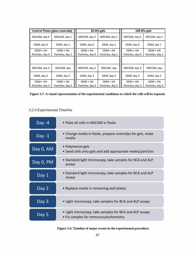
47
Figure 3.7: A visual representation of the experimental conditions to which the cells will be exposed.
3.2.4 Experimental Timeline
MSCGM, day 0 MSCGM, day 1 MSCGM, day 0 MSCGM, day 1 MSCGM, day 0 MSCGM, day 1
ODM, day 0 ODM, day 1 ODM, day 0 ODM, day 1 ODM, day 0 ODM, day 1
ODM + HA
Particles, day 0
ODM + HA
Particles, day 1
ODM + HA
Particles, day 0
ODM + HA
Particles, day 1
ODM + HA
Particles, day 0
ODM + HA
Particles, day 1
MSCGM, day 3 MSCGM, day MSCGM, day 3 MSCGM, day MSCGM, day 3 MSCGM, day
ODM, day 3 ODM, day 5 ODM, day 3 ODM, day 5 ODM, day 3 ODM, day 5
ODM + HA
Particles, day 3
ODM + HA
Particles, day 5
ODM + HA
Particles, day 3
ODM + HA
Particles, day 5
ODM + HA
Particles, day 3
ODM + HA
Particles, day 5
Control Plates (glass coverslip) 30 kPa gels 100 kPa gels
• Plate all cells in MSCGM in flasks Day -4
• Change media in flasks, prepare coverslips for gels, make media Day -1
• Polymerize gels
• Seed cells onto gels and add appropriate media/particles Day 0, AM
• Standard light microscopy, take samples for BCA and ALP assays Day 0, PM
• Standard light microscopy, take samples for BCA and ALP assays Day 1
• Replace media in remaining well plates Day 2
• Light microscopy, take samples for BCA and ALP assays Day 3
• Light microscopy, take samples for BCA and ALP assays
• Fix samples for immunocytochemistry Day 5
Figure 3.4: Timeline of major events in the experimental procedure.

48
3.2.5 Imaging
An Olympus IX71 light microscope was used to image the cells almost daily
during the experiments. Images were taken to observe morphological changes, and
objectives of 4X and 10X were used.
Cells from day 5 were fixed to preserve the cells as close to their natural state as
possible and to prevent decay. Fixing was achieved by washing the cells with PBS and
letting them sit in 4% paraformaldehyde for 10 minutes. After that time, the cells were
washed with PBS again and ready for immunocytochemistry for confocal microscopy.
The protocol found in Appendix B was used for the immunocytochemistry. A primary
antibody of mouse anti-osteopontin IgG1 (diluted 1:10 in 1% BSA/PBS) was used. This
antibody was developed by M. Solursh and A. Franzen and obtained from the
Developmental Studies Hybridoma Bank developed under the auspices of the NICHD
and maintained by The University of Iowa, Department of Biology, Iowa City, IA
52242.(Datko, 2011) The secondary antibody used in the immunocytochemistry was
AlexaFluor 647 donkey anti-mouse IgG (diluted 1:100 in 1% BSA/PBS). All other steps
in the protocol found in Appendix B were followed. Images were taken with the help of
Dr. Terri Bruce on a Zeiss LSM510 Laser Confocal Microscope.
3.2.6 ALP and BCA Assays
At time points on days 0, 1, 3, and 5, alkaline phosphatase (ALP) and
bicinchoninic acid (BCA) assays were performed. See Appendix C for the protocol steps
49
detailing how to obtain standard curves for the assays as well as experimental results and
principles of the methods. Note that a BCA Assay Kit (Thermo Scientific) was used.
Using the total protein values calculated from the BCA assay and the absorbance
values from the ALP assay, specific enzyme activity was calculated using the following
steps:
Using the ALP standard curve, the concentration of pNP formed by the reaction
for each sample was calculated in µg/mL.
That value was converted to µmol/mL formed by dividing by the molecular
weight of pNP which is 139g/mol.
The resulting value is equivalent to the amount of substrate (pNPP) consumed in
the reaction in units of µmol S/mL, where S stands for substrate consumed.
That value was then divided by 30 since the samples were allowed to incubate for
30 minutes, so the substrate consumed can be expressed in µmol S/mL/min.
To normalize the data for each sample, the resulting value was divided by the
total protein concentration for the corresponding sample calculated from the BCA
assay leaving the alkaline phosphatase specific activity with units of µmol S/mg
protein/min.

50
3.3 Results
3.3.1 Basic Imaging
A 10X objective (100X total magnification) was used for each culture condition at
most time points. Due to the large amount of pictures, representative images are shown
below in Figure 3.5 for each condition on days 1 and 5.
A: Control substrate, MSCGM, day 1 and day 5
B: Control Substrate, ODM, day 1 and day 5
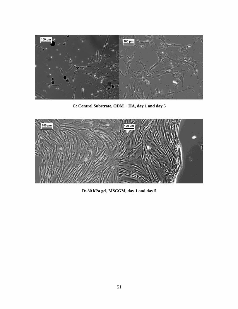
51
C: Control Substrate, ODM + HA, day 1 and day 5
D: 30 kPa gel, MSCGM, day 1 and day 5
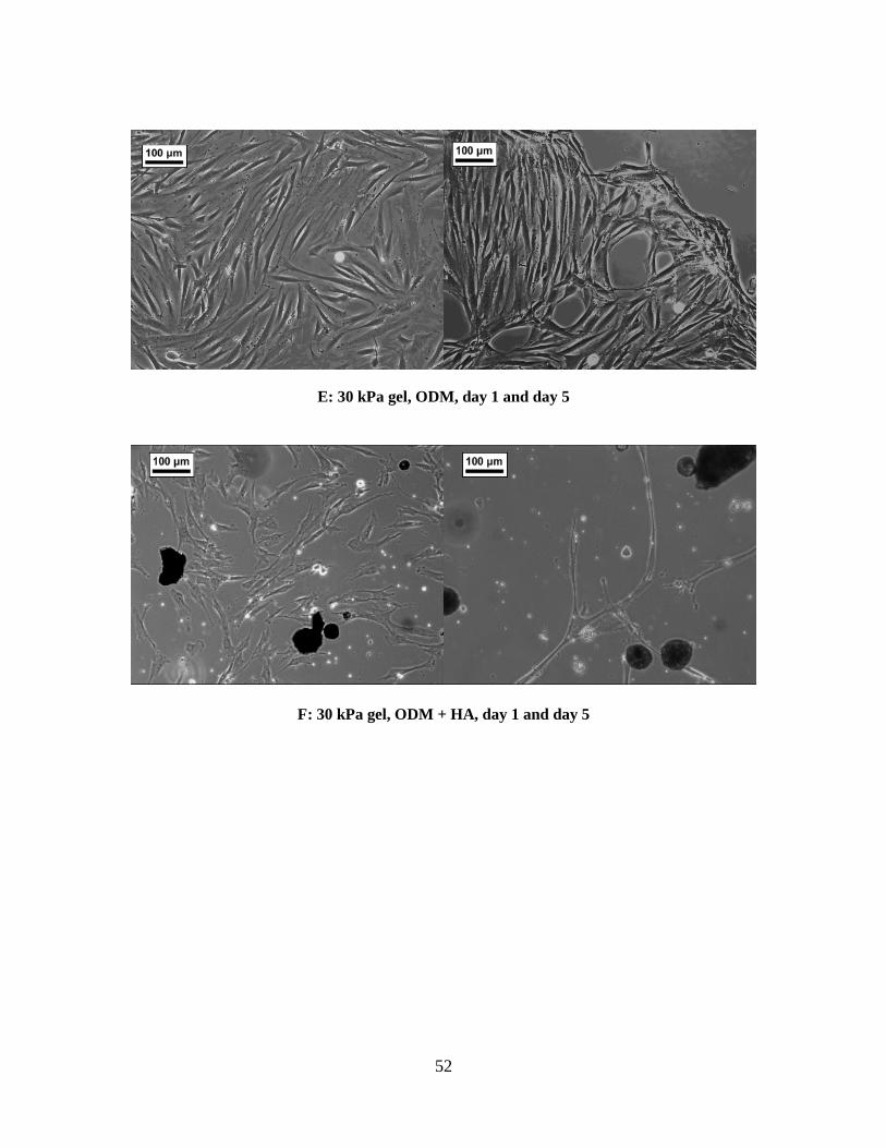
52
E: 30 kPa gel, ODM, day 1 and day 5
F: 30 kPa gel, ODM + HA, day 1 and day 5

53
G: 100 kPa gel, MSCGM, day 1 and day 5
H: 100 kPa gel, ODM, day 1 and day 5

54
I: 100 kPa gel, ODM + HA, day 1 and day 5
Figure 3.5 (above): Representative light microscope images of the cells under each condition
and at days 1 (left) and 5 (right). All scale bars are 100 µm.
3.3.2 Immunocytochemistry Imaging
The immunocytochemistry was done to determine the presence of osteopontin
(also called bone sialoprotein), a structural protein found in the extracellular matrix.
Notably, it is a prominent component of the mineralized extracellular matrices of bones
and teeth.(Hao et al., 1997; Shi et al., 2005; Sodek, Ganss, & McKee, 2000) It is
commonly involved in many biological processes, but it appears essential in the
remodeling and maintenance of bones and teeth.((Sodek et al., 2000)) In the fluorescence
images below, osteopontin is labeled purple.
The green filaments seen in the fluorescence images are actin filaments. They are
used to get a reference for the cellular outline. They do not show the cell membrane, but

55
rather the cytoskeleton which gives a fairly accurate representation of the cellular
membrane location. Note that all images were taken using a 40X oil immersion lens.

56
Figure 3.6: Confocal microscopy images of the cells in each condition, labeled on the top
and left side of the figure. Note the osteopontin is purple and the actin is green.
Control 30 kPa gel 100 kPa gel M
SC
GM
O
DM
O
DM
+ H
A

57
3.3.3 ALP and BCA Assays
Figure 3.7: ALP Specific Activity under each condition on the glass control substrate.
Figure 3.8: ALP Specific Activity under each condition on the 30 kPa gel.
0
0.0002
0.0004
0.0006
0.0008
0.001
0.0012
0.0014
0.0016
Day 0 Day 1 Day 3 Day 5
ALP
Sp
eci
fic
Act
ivit
y (u
mo
l S/m
g p
rote
in/m
in)
Time
Glass Control
MSCGM
ODM
ODM + HA
0
0.0002
0.0004
0.0006
0.0008
0.001
0.0012
0.0014
0.0016
Day 0 Day 1 Day 3 Day 5
ALP
Sp
eci
fic
Act
ivit
y (u
mo
l S/m
g p
rote
in/m
in)
Time
30 kPa Gel
MSCGM
ODM
ODM + HA

58
Figure 3.9: ALP Specific Activity under each condition on the 100 kPa gel.
It was seen that the specific activity of alkaline phosphatase was fairly constant
over the time period tested in all conditions. It was also evident that the values for the
standard deviations were low as the data was consistent within the each sample cell
lysate.
0
0.0002
0.0004
0.0006
0.0008
0.001
0.0012
0.0014
0.0016
Day 0 Day 1 Day 3 Day 5
ALP
Sp
eci
fic
Act
ivit
y (u
mo
l S/m
g p
rote
in/m
in)
Time
100 kPa Gel
MSCGM
ODM
ODM + HA
59
3.4 Discussion
3.4.1 Basic Imaging
In Figure 3.5, it was observed that there was a significant difference in the
behavior of cells on the glass control substrate as opposed to either polyacrylamide
substrate. On the glass coverslips, the cells spread out and grew normally. Neither the
osteogenic differentiation media nor the hydroxyapatite particles seemed to have a
difference on the morphology of the cells. However, the substrate stiffness had a major
effect. On both the 30 kPa and 100 kPa gels, the cells migrated to parts of the gel and
formed aggregates that had very defined edges. No morphological differences were seen
between the media types, but the HA particles did seem to have a negative effect on the
cells. It was noted that seemed to be fewer living cells on the gels with hydroxyapatite on
them. In all other conditions, the cells grew and multiplied well.
The clustering of cells on gels could be due to anchorage dependency. It is known
that cells need a substrate to attach to, and stiffer surfaces usually are preferred. The
surfaces the cells were exposed to might not have been stiff enough. In that case, they
migrated to form a rigid aggregate where they could anchor to each other to obtain their
firm attachment.(Engler et al., 2006)
Another possible explanation for the aggregation is a defect in the gel. The gels
were not always in pristine condition due to the handling of them with a scalpel or
forceps, so there could have been defects in the gel. Cells could have migrated towards
those defects to settle inside them to have more surface area to attach.

60
3.4.2 Immunocytochemistry Imaging
Similar aggregates were seen with confocal imaging (see Figure 3.6), but they
were not as common. It was noticed that osteopontin (purple in the images) was found in
every cell centered around the nucleus. This was expected because osteopontin is not
specific to bone-like tissue, it is just more prevalent in osseous tissues. One observation
of note was that there were osteopontin-filled nodules stemming from cells only on the
gels in osteogenic differentiation media. As it was noted before, it is thought that dentin
is formed in nodules.(Hao et al., 1997)The nodules present could an early sign of
osteogenic differentiation. In the images, hydroxyapatite appeared to have little to no
effect on the cells.
3.4.3 ALP and BCA Assays
The consistency of the specific activity of ALP seen in Figures 3.7-3.9 in almost
all conditions is curious. Morphological changes seen in both imaging modalities show
that the gel had some effect on the cells, while the ALP data suggest otherwise. One
observation is that for the control substrate and the stiffer (100 kPa) gel, the cells in ODM
has higher specific activities than the cells in MSCGM. This is expected, as the ODM is
supposed to induce osteogenic differentiation which should lead to an increase in activity
of alkaline phosphatase. This was not the case for the 30 kPa gel, so the lack of stiffness
may have hindered differentiation towards an osseous lineage. In previous experiments
testing substrate elasticity, more compliant substrates induced differentiation down a
neurogenic or myogenic lineage.(Engler et al., 2006)

61
In an extension of this study by another lab member, Laura Datko, an increase in
ALP specific activity was seen over longer time periods in cells in ODM. Therefore, the
short culture times may not have allowed time for enough differentiation to occur to have
an effect on the amount of alkaline phosphatase. A longer time study would be needed to
determine if this was the case.(Datko, 2011)
The effect of the hydroxyapatite particles could not be determined through these
assays. At some time points in some conditions, the HA particles had a positive effect on
ALP, and in other samples the effect was negative.
Finally, while the data was steady throughout all conditions and time points, it
was seen that the control glass substrate induced the highest amount of ALP activity. This
follows the idea that a stiffer substrate (glass is on the order of 1000X as stiff) promotes
osteogenic differentiation. However, in contrast to Engler, et al, a stiff hydrogel did not
seem to promote differentiation down an osseous lineage.
3.5 Conclusions
The results show promise in that changing the stiffness of the substrate did have
an effect on both the growth behavior and alkaline phosphatase activity of human dental
pulp stem cells. However, the effect cannot be concluded due to the limited culture time
used and lack of volume of data. It can be noted, though, that morphological changes
towards a defined lineage were not seen with either imaging modality other than the
presence of osteopontin-filled nodules. Also, no significant changes in ALP were noticed

62
for any of the media conditions. One noticeable change in the cells was the migration and
aggregation of cells on gels towards each other or possibly toward a defect in the gel.
In comparison to the study performed by Engler, et al on bone marrow stem cells,
the results were not similar. In this study, no definite morphological changes were seen in
the cells regardless of substrate, so it appears that dental pulp stem cells behave
differently than bone marrow-derive stem cells.
3.6 Future Work
The goal of these studies was to determine the effect of substrate stiffness on
differentiation of dental pulp stem cells. Valuable data was obtained, but more data needs
to be collected at more time points, especially later time points, for conclusions from the
data to be drawn.
The tests performed in these experiments were designed to show signs of
differentiation down an osteogenic lineage. However, the overall goal of the research is
to determine the odontogenic potential of dental pulp stem cells. Both lineages could
show signs of the indicators tested in these experiments, so it is unknown which lineage
the cells are actually differentiating towards. In the future, proteins specific to teeth, such
as dentin sialophosphoprotein (DSPP) and dentin matrix protein I (DMP-1), will need to
be stained for and also Western blotting should be done for these proteins.
To better compare with other literature sources, differentiation down other
lineages should be attempted alongside the experiments. For example, neurogenic and

63
myogenic differentiation media should be used to see if that differentiation down those
pathways is possible as well.
64
3.7 References
Datko, L. (2011). Effects of microenvironment on growth and differentiation of human
dental pulp stem cells. (Masters, Clemson University).
Duplomb, L., Dagouassat, M., Jourdon, P., & Heymann, D. (2007). Concise review:
Embryonic stem cells: A new tool to study osteoblast and osteoclast differentiation.
Stem Cells (Dayton, Ohio), 25(3), 544-552. doi:10.1634/stemcells.2006-0395
Engler, A. J., Sen, S., Sweeney, H. L., & Discher, D. E. (2006). Matrix elasticity directs
stem cell lineage specification. Cell, 126(4), 677-689. doi:10.1016/j.cell.2006.06.044
Gronthos, S., Mankani, M., Brahim, J., Robey, P. G., & Shi, S. (2000). Postnatal human
dental pulp stem cells (DPSCs) in vitro and in vivo. Proceedings of the National
Academy of Sciences of the United States of America, 97(25), 13625-13630.
doi:10.1073/pnas.240309797
Hao, J., Shi, S., Niu, Z., Xun, Z., Yue, L., & Xiao, M. (1997). Mineralized nodule
formation by human dental papilla cells in culture. European Journal of Oral
Sciences, 105(4), 318-324.

65
Huang, A. H., Chen, Y. K., Lin, L. M., Shieh, T. Y., & Chan, A. W. (2008). Isolation and
characterization of dental pulp stem cells from a supernumerary tooth. Journal of
Oral Pathology & Medicine : Official Publication of the International Association of
Oral Pathologists and the American Academy of Oral Pathology, 37(9), 571-574.
doi:10.1111/j.1600-0714.2008.00654.x
Kadar, K., Kiraly, M., Porcsalmy, B., Molnar, B., Racz, G. Z., Blazsek, J., et al. (2009).
Differentiation potential of stem cells from human dental origin - promise for tissue
engineering. Journal of Physiology and Pharmacology : An Official Journal of the
Polish Physiological Society, 60 Suppl 7, 167-175.
King, D. J., & Noss, R. R. (1989). Toxicity of polyacrylamide and acrylamide monomer.
Reviews on Environmental Health, 8(1-4), 3-16.
Laino, G., d'Aquino, R., Graziano, A., Lanza, V., Carinci, F., Naro, F., et al. (2005). A
new population of human adult dental pulp stem cells: A useful source of living
autologous fibrous bone tissue (LAB). Journal of Bone and Mineral Research : The
Official Journal of the American Society for Bone and Mineral Research, 20(8),
1394-1402. doi:10.1359/JBMR.050325
Liu, H., Gronthos, S., & Shi, S. (2006). Dental pulp stem cells. Methods in Enzymology,
419, 99-113. doi:10.1016/S0076-6879(06)19005-9
Lonza. (2012). Culture media database. Retrieved 3/17, 2012, from
http://bio.lonza.com/3018.html#ext-comp-1003:lz_culturemedia_list:change

66
Napier University. (2007). Enzyme assays. Retrieved 3/17, 2012, from
http://learning.covcollege.ac.uk/content/Jorum/CHB_Intro-to-basic-prac-skills_LM-
1.2/page29.htm
Shi, S., Bartold, P. M., Miura, M., Seo, B. M., Robey, P. G., & Gronthos, S. (2005). The
efficacy of mesenchymal stem cells to regenerate and repair dental structures.
Orthodontics & Craniofacial Research, 8(3), 191-199. doi:10.1111/j.1601-
6343.2005.00331.x
Sodek, J., Ganss, B., & McKee, M. D. (2000). Osteopontin. Critical Reviews in Oral
Biology and Medicine : An Official Publication of the American Association of Oral
Biologists, 11(3), 279-303.
Thermo Scientific. (2012). Polacrylamide gels. Retrieved 3/17, 2012, from
http://www.piercenet.com/browse.cfm?fldID=21518847-2D72-475F-A5B9-
B236EC5B641E
Tse, J. R., & Engler, A. J. (2010). Preparation of hydrogel substrates with tunable
mechanical properties. Current Protocols in Cell Biology / Editorial Board, Juan
S.Bonifacino ...[Et Al.], Chapter 10, Unit 10.16.
doi:10.1002/0471143030.cb1016s47
Wang, Y. L., & Pelham, R. J.,Jr. (1998). Preparation of a flexible, porous polyacrylamide
substrate for mechanical studies of cultured cells. Methods in Enzymology, 298, 489-
496.

67
Zhang, W., Walboomers, X. F., Shi, S., Fan, M., & Jansen, J. A. (2006). Multilineage
differentiation potential of stem cells derived from human dental pulp after
cryopreservation. Tissue Engineering, 12(10), 2813-2823.
doi:10.1089/ten.2006.12.2813
Zhang, W., Walboomers, X. F., Wolke, J. G., Bian, Z., Fan, M. W., & Jansen, J. A.
(2005). Differentiation ability of rat postnatal dental pulp cells in vitro. Tissue
Engineering, 11(3-4), 357-368. doi:10.1089/ten.2005.11.357
68
CHAPTER 4: The Effect of Static Compressive Stress on the Growth and
Differentiation of Dental Pulp Stem Cells
4.1 Introduction
In 1892, Julius Wolff published a work on bone transformation that is still
applicable today. In his work, Wolff explained his model of how a bone’s morphology
develops. He theorized that a bone changes its external shape and internal architecture in
response to the forces and stresses acting on it. Though this has been challenged by some,
it is widely accepted as “Wolff’s Law.”(Team Bone, 2008)
There are forces in the body that constantly push and pull on our bones, cartilage,
teeth, and the rest of the body. Much research has been done on the effects of these
stresses on cartilage because it is commonly damaged and cannot regenerate. Studies
have shown that cartilage is responsive to compressive stresses. In one study, both static
and dynamic stresses had an effect on the biosynthesis of proteoglycans in cartilage
explants. Proteoglycans are ECM components heavily expressed in cartilage
cells.(Nugent et al., 2006)
Also, research has been performed to understand the effects of compressive
stresses on mesenchymal stem cells for cartilage regenerative medicine. It has been
shown that in the absence of any chondrogenic growth factors, dynamic compressive
stresses applied to bone marrow stem cells induce synthesis of proteoglycans, a ECM
component heavily expressed in cartilage cells.(Kisiday, Frisbie, McIlwraith, &
Grodzinsky, 2009) Also, compressive stress applied to bone marrow stem cells induced

69
chondrogenic gene expression. Further, if the chondrogenesis had already been induced
with transforming growth factor β1 (TGF-β1), the effects were amplified. (Miyanishi et
al., 2006) Another study indicated that the same results were consistent for mesenchymal
stem cells embedded within hydrogels.(Terraciano et al., 2007)
Up to this point, dental pulp stem cells have not been studied for their response to
mechanical stresses and compression. However, it is known that odontoblasts do respond
to mechanical stimuli.(Magloire, Couble, Thivichon-Prince, Maurin, & Bleicher, 2009)
Odontoblasts are the dentin-producing cells that originate from DPSCs. Also, dental pulp
stem cells that are subjected to orthodontic tooth movement forces have been thought to
differentiate within hours to form either osteoblasts or osteoclasts depending on the force
present. Compression forces induce osteoclastic differentiation, and tension forces induce
osteoblastic differentiation.(Sutherland et al., 2004; Zainal Ariffin, Yamamoto, Zainol
Abidin, Megat Abdul Wahab, & Zainal Ariffin, 2011)
The aim of this study is to determine the effects of static compression on the
differentiation and growth of dental pulp stem cells. This will be achieved by first
designing and fabricating a suitable compression chamber in which to test the cells. Then,
cells will be cultured under different compressive conditions and analyzed for
differentiation markers.

70
4.2 Experimental Design
4.2.1 Design of a Static Compression Chamber
To determine the effect of compressive stresses on dental pulp stem cells, a
suitable compressive device needed to be obtained. No such device was available on the
market, so a device was designed using SolidWorks. Requirements of the device were
that it would fit onto a standard 6-well culture plate such that it could provide
compression to cells seeded into collagen gels in the wells. The design was taken to
Clemson Machining and Technical Services to discuss possible design modifications, and
the custom compression chamber device was fabricated using 316L Stainless Steel. The
device was analyzed and tested for its usability. Figures 4.1 and 4.2 show original design
ideas from SolidWorks. The theory was that the cylinders would protrude into the wells
of the culture plate and provide the compressive force.
Figure 4.8: Bottom view of the initial SolidWorks design for the compression chamber

71
Figure 4.9: Top view of the initial SolidWorks design for the compression chamber
Though the design of the chamber was altered some in processing and fabrication,
it ultimately was designed for symmetrical compression to all wells. To test, a pressure
sensitive film was obtained (Fujifilm Prescale) and the device was tested for even
pressure distribution. This was done by placing the film on a gel and setting the
compression device onto the film.
4.2.2 Cell Acquisition and Isolation
In this experiment, porcine dental pulp stem cells (pDPSCs) were used instead of
human cells as the source of the human cells was no longer available. It is assumed that
porcine dental pulp is similar to human dental pulp, and the cells found in the dental pulp
would have stem cell characteristics.

72
Porcine jawbones were obtained from Snow Creek Meat Processing facility in
Seneca, SC and transported to Godley-Snell Animal Research Facility at Clemson
University. Molars were extracted by peeling away the gingiva and excavating the
alveolar bone beneath the molar. Once the root of the tooth was visible and free of
attachment to the bone, the tooth was removed using an elevator and dental forceps. The
molars were placed in sterile Hank’s Balance Salt Solution (HBSS) with 2% anti/anti
solution (penicillin/ streptomycin/ amphotericin B). The higher concentration of the
anti/anti solution was used because the external surface of the tooth could have had a
high amount of bacteria on it.
The teeth were rinsed with 70% ethanol and HBSS before the pulp chamber was
exposed. This was accomplished by cracking the root of the tooth with pliers. The pulp
was extracted using sterile forceps and placed in a sterile petri dish where the pulp was
minced using a scalpel. The pieces were rinsed using sterile HBSS with 1% anti/anti
solution. The pulp pieces were “digested” by incubating them in a 3mg/mL solution of
collagenase II for 1 hour at 37°C. The solution was then passed through a cell strainer
with a pore diameter of 70µm and the remaining solution was centrifuged at 1200 rpm for
10 minutes. The cell pellet was re-suspended in fresh MSCGM and plated onto cell
culture flasks. Cells were adherent after 5-7 days. Note that the teeth and pulp were kept
separate from each other throughout the whole process to reduce contamination.

73
4.2.3 Culture and Compression Conditions
To allow the cells to be under compression in a more controlled way, the cells
needed to be embedded in a 3-D gel construct. Since the stiffness of the gel would not be
altered, polyacrylamide was not the gel material chosen. For clinical translational
purposes, gels were made out of rat tail collagen type I (BD Biosciences).
To contain the collagen gel to a consistent shape and height, hollow cylindrical
molds that were 12 mm in diameter were formed with a PDMS Sylgard 184 Silicone
Elastomer kit (Dow Corning). The molds were then placed onto glass coverslips and set
into the wells of a standard 6-well cell culture plate. The molds can be seen in Figure 4.3.
Figure 4.10: The PDMS molds in a cell culture plate (right), and one individual mold (left)
Collagen gels were synthesized within the PDMS molds by mixing a 2mg/mL
collagen solution, HEPES, and 10X MEM in an 8:1:1 ratio to get the desired final
volume. Then, 600 µL of the collagenous mixture was added to each mold and cells were
quickly added before gelling occurred. The collagen-cell mixture was placed in the 37°C

74
incubator for 30 minutes to allow gelling to occur. The compression chamber piece
replaced the original culture plate top, and the collagen gels were observed under
compression. To determine usability and form a protocol, 7F2 osteoblasts were used
because they are an abundant and well-characterized cell line. To perform the actual tests,
both the porcine dental pulp stem cells previously discussed and the osteoblasts were
used. The media was varied between MSCGM and ODM.
To test the compression effects, different compressive conditions needed to
defined and tested. First, the loading weight could be altered by adding weights onto the
top of the compression device. Also, the schedule of time compressed vs. time
uncompressed could be varied. Taking all factors into account, a testing plan was devised
and it is summarized in Table 4.1 below. Note that the control used for the experiment
will be cells grown in collagen gels allowed to freely swell.
6 hours on/6 hours
off 12 hours on/ 12 hours
off 24 hours on/ 24 hours
off
No weight added
MSCGM MSCGM MSCGM
ODM ODM ODM
Add 1 kg MSCGM MSCGM MSCGM
ODM ODM ODM
Add 2 kg MSCGM MSCGM MSCGM
ODM ODM ODM
Figure 11.4: Experimental Design Conditions.

75
4.2.4 Testing Analysis
Similarly to before, cells were tested with BCA and ALP assays to determine total
protein content and ALP specific activity. However, since the cells were embedded in a
collagen gel, the gels first needed to be digested using a collagenase solution incubated at
37°C for 20 minutes. The cells were then strained through a 70 µm cell strainer, and the
resulting cells were re-suspended in PBS and the protocol from Appendix C was
followed as mentioned in Chapter 3. The control in the experiment was to use gels that
were allowed to freely swell (not under compression). Also, ALP and BCA assays were
performed on samples from days 1, 3, and 5.

76
4.3 Results
4.3.1 Compression Chamber
The chamber was fabricated by Clemson Machining and Technical Services, and
the final dimensions and measurables of the device are listed in Table 4.1.
Table 4.1: Physical specifications of the custom compression device
Specification Measurement
Weight 636 grams
Length 5.5 inches
Width 3.75 inches
Cylinder Depth 0.57 inches
Cylinder Diameter 0.5 inches
The device was fabricated from medical grade 316L stainless steel. By calculating
the total surface area of all of the cylinders and finding the force exerted by the weight of
the device, a theoretical pressure of 9.2 kPa was calculated for the pressure exerted on the
collagen gels. Images of the compression device can be seen in Figures 4.5-4.7 below.

77
Figure 4.12: Views of the compression device from the underneath (top left), top of the compression
device (top right), the device fitting onto a 6-well culture plate (middle), and a diagram of the
components of the compression chamber (bottom).

78
In testing for usability, the compression device passed. It performed all necessary
functions as needed. In symmetry tests with the pressure-sensitive film, the device was
found to be mostly symmetric with the pressure being distributed almost completely
evenly over all surfaces. The pressure film can be seen in Figure 4.6. Note that the film
was not quite large enough to fit all the cylinders on it, but enough data was seen to
assume symmetry.
Figure 4.13: The colorimetric pressure sensitive film results.
4.3.2 ALP and BCA Assays
The results presented in this section are preliminary, and not enough data has been
obtained due to time constraints and the fact that only one device was available for use.
Therefore, only data from day 1 of the experiments was able to be recorded for all
conditions. The data for dental pulp stem cells and 7F2 osteoblasts under three
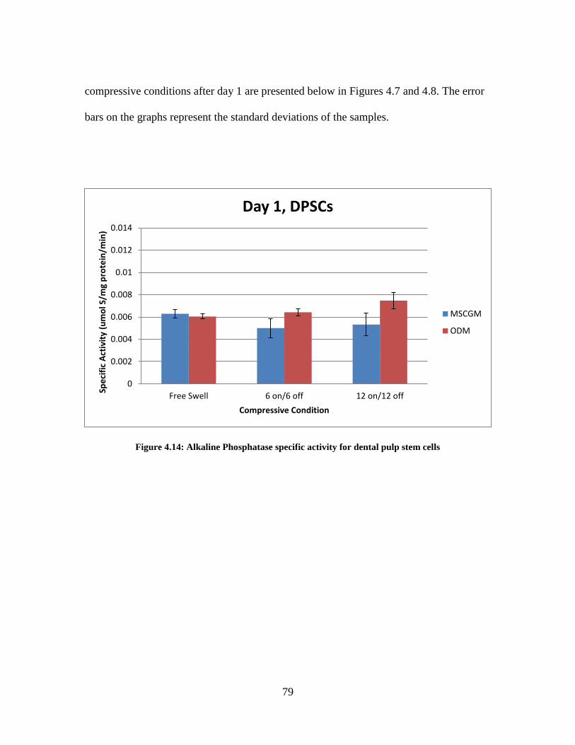
79
compressive conditions after day 1 are presented below in Figures 4.7 and 4.8. The error
bars on the graphs represent the standard deviations of the samples.
Figure 4.14: Alkaline Phosphatase specific activity for dental pulp stem cells
0
0.002
0.004
0.006
0.008
0.01
0.012
0.014
Free Swell 6 on/6 off 12 on/12 off
Spe
cifi
c A
ctiv
ity
(um
ol S
/mg
pro
tein
/min
)
Compressive Condition
Day 1, DPSCs
MSCGM
ODM

80
Figure 4.15: Alkaline Phosphatase specific activity for 7F2 Osteoblasts
To draw inferences and conclusions from this data, statistical analysis was
performed using Microsoft Excel. A T-Test was performed between the sets of three
measurements for each sample. Null (Ho) and alternative (Ha) hypothesis were as follows:
Ho: µ1 = µ2, Ha: µ1 ≠ µ2. A two-tailed T-Test was used with α = 0.05. Table 4.2 below
summarizes the results found from the T-Tests.
0
0.002
0.004
0.006
0.008
0.01
0.012
0.014
Free Swell 6 on/6 off 12 on/12 off
Spe
cifi
c A
ctiv
ity
(um
ol S
/mg
pro
tein
/min
)
Compressive Condition
Day 1, 7F2 Osteoblasts
MSCGM
ODM

81
Table 2.2: Comparison of means for each pair of conditions. Note that when a p-value is less than α,
the null hypothesis is rejected meaning that the samples are significantly different.
Constant
Conditions Population 1 Population 2
T-Test p-
value Decision
DPSC, ODM Free Swelling 6 on/6 off 0.158 Fail to Reject
Ho
DPSC, ODM Free Swelling 12 on/12 off 0.029 Reject Ho
DPSC, ODM 6 on/6 off 12 on/12 off 0.081 Fail to Reject
Ho
DPSC, MSCGM Free Swelling 6 on/6 off 0.073 Fail to Reject
Ho
DPSC, MSCGM Free Swelling 12 on/12 off 0.198 Fail to Reject
Ho
DPSC, MSCGM 6 on/6 off 12 on/12 off 0.702 Fail to Reject
Ho
7F2 OB ODM Free Swelling 6 on/6 off 0.049 Reject Ho
7F2 OB, ODM Free Swelling 12 on/12 off 0.022 Reject Ho
7F2 OB, ODM 6 on/6 off 12 on/12 off 0.122 Fail to Reject
Ho
7F2 OB,
MSCGM Free Swelling 6 on/6 off 0.232
Fail to Reject
Ho
7F2 OB,
MSCGM Free Swelling 12 on/12 off 0.913
Fail to Reject
Ho
7F2 OB,
MSCGM 6 on/6 off 12 on/12 off 0.009 Reject Ho
Free Swelling,
DPSC MSCGM ODM 0.401
Fail to Reject
Ho
Free Swelling,
7F2 OB MSCGM ODM 0.749
Fail to Reject
Ho
6 on/6 off, DPSC MSCGM ODM 0.051 Fail to Reject
Ho
6 on/6 off, 7F2
OB MSCGM ODM 0.111
Fail to Reject
Ho
12 on/12 off,
DPSC MSCGM ODM 0.040 Reject Ho
12 on/12 off, 7F2
OB MSCGM ODM 0.011 Reject Ho

82
It is seen in Table 4.2 that most of the conditions yielded similar results, and the
p-value for those samples was greater than α (α=0.05). There were some data
comparisons that showed p-values less than α, which indicates a statistically significant
difference in means. It can be noted that the osteoblasts in ODM have significant
differences in both compressive conditions compared to free swelling conditions. Also, it
is important to note that the only significant difference in the DPSCs due to compression
was seen in ODM when the cells were under 12 hours on/12 hours off of compressive
force. Also, the media conditions only seemed to have a significant effect under the 12
hours on/12 hours off condition for both types of cells.
4.4 Discussion and Conclusions
The compression device performed as necessary, and an acceptable protocol was
formed for testing cells. The PDMS and collagen gel components also were satisfactory
in performing their roles. The pressure exerted on the gels is applicable and is similar to
physiological pressures. In other studies, compressive pressures ranging from 1-100 kPa
were used(Nugent et al., 2006), so the baseline value of 9.2 kPa is acceptable. Also, the
assumption that the theoretical value is correct is supported by the symmetry of the
device. While it may not be exactly 9.2 kPa for all gels, the value is close. More weights
could still be added to reach higher pressures.
The ALP specific activity data shows very clearly that osteoblasts have more
alkaline phosphatase than DPSCs which was expected. Also, it can be seen that in
DPSCs, only one mean was shown to be significantly different from another due to

83
compression. This could indicate that the cells did not have a chance to differentiate yet.
This was very likely because the culture time was only 1 day. Also, statistical analysis
showed that the only difference due to different media types was seen under compressive
conditions, so it could be that the compression and media have an additive effect.
The osteoblasts on average produced more ALP under compression than free
swelling conditions. This could mean that the osteoblasts are activated by stress, similar
to odontoblasts being activated by dental procedures.(Magloire et al., 2009) The
differences in osteoblasts under compression were significant in samples in ODM. It can
be inferred that compression was able to have a greater effect on osteoblasts because they
were also differentiated into cells that respond to forces. The DPSCs may not naturally
respond as readily to forces, as it seems they need more time to differentiate before
compression has a significant effect.
Overall, the work presented in this study has shown that there is reason to
investigate the effect of compression on dental pulp stem cell growth and differentiation,
and this experiment has tested one method of testing static compression.
4.5 Future Work
In the future, testing at more time points should be performed to better understand
the differentiation of cells over time. Also, the pressure exerted on the cells in gels should
be varied to cover a larger range of physiological pressures and to compare to other
experiments. Also, Western blotting should be performed for bone proteins such as
osteopontin, but also dental-specific proteins like DSPP mentioned in a previous chapter.

84
Lastly, this concept needs to be applied to a dynamic compression study to better
simulate the pressures associated with chewing and other physiological forces.
85
4.6 References
Kisiday, J. D., Frisbie, D. D., McIlwraith, C. W., & Grodzinsky, A. J. (2009). Dynamic
compression stimulates proteoglycan synthesis by mesenchymal stem cells in the
absence of chondrogenic cytokines. Tissue Engineering.Part A, 15(10), 2817-2824.
doi:10.1089/ten.TEA.2008.0357
Magloire, H., Couble, M. L., Thivichon-Prince, B., Maurin, J. C., & Bleicher, F. (2009).
Odontoblast: A mechano-sensory cell. Journal of Experimental Zoology.Part B,
Molecular and Developmental Evolution, 312B(5), 416-424.
doi:10.1002/jez.b.21264
Miyanishi, K., Trindade, M. C., Lindsey, D. P., Beaupre, G. S., Carter, D. R., Goodman,
S. B., et al. (2006). Effects of hydrostatic pressure and transforming growth factor-
beta 3 on adult human mesenchymal stem cell chondrogenesis in vitro. Tissue
Engineering, 12(6), 1419-1428. doi:10.1089/ten.2006.12.1419
Nugent, G. E., Schmidt, T. A., Schumacher, B. L., Voegtline, M. S., Bae, W. C., Jadin,
K. D., et al. (2006). Static and dynamic compression regulate cartilage metabolism
of PRoteoGlycan 4 (PRG4). Biorheology, 43(3-4), 191-200.
Sutherland, M. K., Geoghegan, J. C., Yu, C., Turcott, E., Skonier, J. E., Winkler, D. G.,
et al. (2004). Sclerostin promotes the apoptosis of human osteoblastic cells: A novel
regulation of bone formation. Bone, 35(4), 828-835. doi:10.1016/j.bone.2004.05.023

86
Team Bone. (2008). Wolff's law. Retrieved 3/19, 2012, from
http://www.teambone.com/wolff.html
Terraciano, V., Hwang, N., Moroni, L., Park, H. B., Zhang, Z., Mizrahi, J., et al. (2007).
Differential response of adult and embryonic mesenchymal progenitor cells to
mechanical compression in hydrogels. Stem Cells (Dayton, Ohio), 25(11), 2730-
2738. doi:10.1634/stemcells.2007-0228
Zainal Ariffin, S. H., Yamamoto, Z., Zainol Abidin, I. Z., Megat Abdul Wahab, R., &
Zainal Ariffin, Z. (2011). Cellular and molecular changes in orthodontic tooth
movement. TheScientificWorldJournal, 11, 1788-1803. doi:10.1100/2011/761768
87
CHAPTER 5: Conclusions
Overall, tissue engineering of teeth is still in its preliminary stages, but the need
for a suitable replacement for dental materials is there. With all of the pathologies and no
material that can fully mimic the function of teeth, a new dental material is needed that
can better replace native dental tissue. Being able to re-grow healthy dental tissue within
the tooth would be the holy grail of dental tissue engineering. However, not enough is
known about the differentiation of stem cells into dental tissue. Also, what is known is
limited in its translational capacity. This research is focused on determining the micro-
environmental cues that stem cells need to induce differentiation down a bone or tooth-
like path.
Much has been done to characterize and investigate dental pulp stem cells, and the
results have been presented above. Among the major points are the observations that
growth patterns were affected by substrate stiffness and growth factors. However, the
culture times were not long enough to observe differentiation. It was also seen that
DPSCs do not behave in the same fashion that bone marrow stem cells do. To continue
this research, western blotting and staining for dental-specific proteins such as DSPP and
DMP-1 should be done to distinguish whether the differentiation was towards bone or
tooth-like tissue. Also, more repetition needs to be done for the results to have statistical
significance.
In the current work, it was seen that the setup used allows for testing of static
compression on cells seeded in gels. This has promise, but not enough data has been

88
collected for there to be conclusions drawn. More data will be collected for the cells
under different conditions and at more time points to test for statistical significance.

89
APPENDIX A: Polyacrylamide Gel Protocol
Source: Yu-li Wang Laboratory
Modified Acrylamide Substrate Protocol (by: Matthew Cupelli, Laura Datko, Dr.
Delphine Dean)
Materials:
22mm x 22mm square coverslips
Distilled water
NaOH pellets
3-aminopropyltrimethoxy silane
0.5% gluteraldehyde in PBS
1M HEPES (pH 8.5)
50mM HEPES (500uL of 1M HEPES per 10 mL of PBS, pH 8.5)
40% Acrylamide and 2% Bis
Ammonium Persulfate (10mg in 100μl of PBS – prepare immediately before use)
TEMED
Fibronectin (1 mg/mL in H2O)
Coverslip activation:
1. Put several NaOH pellets in a weigh boat and add distilled water. When they
dissolve, add coverslips for a few minutes, then remove and let dry on a kimwipe.
2. Coat one side of the coverslips (in a new weigh boat) with STERILE 3-
aminopropyltriethoxy silane
3. Incubate at room temperature for 5 minutes
4. Rinse with distilled water and shake for 5 minutes
5. Repeat #4 two more times **make sure to get all the APT off or the coverslips
will have a reddish tint when you add gluteraldehyde**
6. Coat the same side of the coverslips with 0.5% gluteraldehyde for 30 minutes at
RT
7. Repeat the rinsing process (steps 4 and 5)

90
Acrylamide preparation:
1. Make acrylamide solution in a 25 mL glass breaker according to the chart at the
end of protocol
2. Add 30μl ammonium persulfate and 20μl TEMED to the acrylamide solution; mix
gently
3. IMMEDIATELY pipet 20μl onto activated coverslip and quickly place another
coverslip on top
4. Leave remaining acrylamide in beaker. (Once this has polymerized, gel on
coverslip has also polymerized)
5. Flood the bottom of the dish with ~2mL of 50mM HEPES
6. Remove the top coverslip (with scalpel or forceps). Substrates can now be stored
in PBS for up to 2 weeks at 4°C
Final Acryl/Bis 40%Acrylamide 2%Bis 1M HEPES H20+Beads Young's Modulus
8%/0.1% 1000 ul 250 ul 50 ul 3700 ul 100 kN/m2
8/0.08 1000 200 50 3750 75
8/0.06 1000 150 50 3800 30
8/0.05 1000 125 50 3825 23
8/0.04 1000 100 50 3850 17
8/0.03 1000 75 50 3875 14
8/0.02 1000 50 50 3900 10
5/0.12 625 300 50 4025 33
5/0.10 625 250 50 4075 28
5/0.08 625 200 50 4125 24
5/0.06 625 150 50 4175 15
5/0.05 625 125 50 4200 ??
5/0.025 625 63 50 4262 7
3/0.10 375 250 50 4325 ??

91
Prepping substrate for cells:
1. Coat gels with 0.2 mg/mL sulfo-SANPAH in H2O for 10 minutes under long
wave UV
2. Rinse 2X with 50 mM HEPES (all sulfo-SANPAH waste into designated
container in lab)
3. Add 200μl fibronectin (125 ug/mL to get a 5 ug/cm2 coating) to substrate and
store overnight at 37°C
4. Rinse and store in PBS at 4°C for up to one week
5. Before plating, UV sterilize substrates for 15-30 minutes
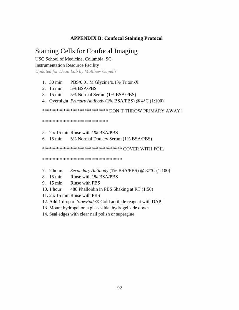
92
APPENDIX B: Confocal Staining Protocol
Staining Cells for Confocal Imaging USC School of Medicine, Columbia, SC
Instrumentation Resource Facility
Updated for Dean Lab by Matthew Cupelli
1. 30 min PBS/0.01 M Glycine/0.1% Triton-X
2. 15 min 5% BSA/PBS
3. 15 min 5% Normal Serum (1% BSA/PBS)
4. Overnight Primary Antibody (1% BSA/PBS) @ 4°C (1:100)
**************************** DON’T THROW PRIMARY AWAY!
****************************
5. 2 x 15 min Rinse with 1% BSA/PBS
6. 15 min 5% Normal Donkey Serum (1% BSA/PBS)
********************************** COVER WITH FOIL
**********************************
7. 2 hours Secondary Antibody (1% BSA/PBS) @ 37°C (1:100)
8. 15 min Rinse with 1% BSA/PBS
9. 15 min Rinse with PBS
10. 1 hour 488 Phalloidin in PBS Shaking at RT (1:50)
11. 2 x 15 min Rinse with PBS
12. Add 1 drop of SlowFade® Gold antifade reagent with DAPI
13. Mount hydrogel on a glass slide, hydrogel side down
14. Seal edges with clear nail polish or superglue

93
APPENDIX C: BCA and ALP Assay Protocol
Protocol for BCA and Alkaline Phosphatase Assays (from Bioengineering Department)
Collecting cell lysates:
1. Remove culture media and wash cells with PBS
2. Add 300 ul Mammalian-Protein Extraction Reagent ™(M-PER) solution to each
well
3. Incubate for 20 minutes at room temperature
4. During incubation period, label a microcentrifuge tube for each sample
5. Collect the cell lysates into the tubes (they can be frozen until ready for use)
BCA total protein assay:
1. Prepare working reagent (WR) as follows:
a. Mix in a 15 ml tube 5 ml of Reagent A and 100 ul of Reagent B
2. Using a stock solution of 1mg/ml of albumin, prepare standard curve as follows
(be very careful/accurate when pipetting these volumes as your results will
depend on the curve):
Standard # Stock Solution
(ul)
Water (ul) Final
Concentration
(ug/ml)
1 200 200 500
2 100 300 250
3 50 350 125
4 25 375 62.5
5 0 400 0
3. Perform the BCA assay in a 96 well-plate as follows:
a. Pipette 25 ul of each standard into 5 wells (in triplicate)
b. Pipette 25 ul of each sample (cell lysates) into separate wells (in
triplicate)
c. Add 200 ul WR to each well
d. Cover and incubate for 30 minutes at 37 C
e. Read the absorbance at 562 nm

94
f. Use Excel to create standard curve with standard #1-5 (Concentration
vs. Absorbance scatterplot + trendline) and use the trendline equation
to determine the total protein in your samples (ug/ml).
Principle of the method: The BCATM
Protein Assay is a detergent-compatible
formulation based on bicinchoninic acid (BCA) for the colorimetric detection and
quantitation of total protein. This method combines the well-known reduction of Cu+2
to
Cu+1
by protein in an alkaline medium (the biuret reaction) with the highly sensitive and
selective colorimetric detection of the cuprous cation (Cu+1
) using a unique reagent
containing bicinchoninic acid.1 The purple-colored reaction product of this assay is
formed by the chelation of two molecules of BCA with one cuprous ion. This water-
soluble complex exhibits a strong absorbance at 562 nm that is nearly linear with
increasing protein concentrations over a broad working range (20-20,000 ug/ml). The
BCATM
method is not a true end-point method; that is, the final color continues to
develop. However, following incubation, the rate of continued color development is
sufficiently slow to allow large numbers of samples to be assayed together.
(1)
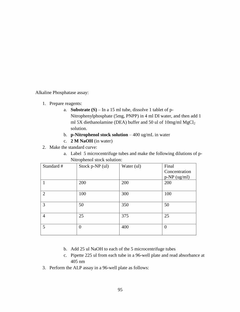
95
Alkaline Phosphatase assay:
1. Prepare reagents:
a. Substrate (S) – In a 15 ml tube, dissolve 1 tablet of p-
Nitrophenylphosphate (5mg, PNPP) in 4 ml DI water, and then add 1
ml 5X diethanolamine (DEA) buffer and 50 ul of 10mg/ml MgCl2
solution.
b. p-Nitrophenol stock solution – 400 ug/mL in water
c. 2 M NaOH (in water)
2. Make the standard curve:
a. Label 5 microcentrifuge tubes and make the following dilutions of p-
Nitrophenol stock solution:
Standard # Stock p-NP (ul) Water (ul) Final
Concentration
p-NP (ug/ml)
1 200 200 200
2 100 300 100
3 50 350 50
4 25 375 25
5 0 400 0
b. Add 25 ul NaOH to each of the 5 microcentrifuge tubes
c. Pipette 225 ul from each tube in a 96-well plate and read absorbance at
405 nm
3. Perform the ALP assay in a 96-well plate as follows:
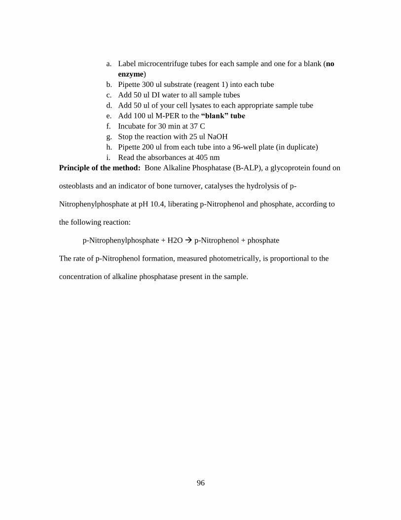
96
a. Label microcentrifuge tubes for each sample and one for a blank (no
enzyme)
b. Pipette 300 ul substrate (reagent 1) into each tube
c. Add 50 ul DI water to all sample tubes
d. Add 50 ul of your cell lysates to each appropriate sample tube
e. Add 100 ul M-PER to the “blank” tube
f. Incubate for 30 min at 37 C
g. Stop the reaction with 25 ul NaOH
h. Pipette 200 ul from each tube into a 96-well plate (in duplicate)
i. Read the absorbances at 405 nm
Principle of the method: Bone Alkaline Phosphatase (B-ALP), a glycoprotein found on
osteoblasts and an indicator of bone turnover, catalyses the hydrolysis of p-
Nitrophenylphosphate at pH 10.4, liberating p-Nitrophenol and phosphate, according to
the following reaction:
p-Nitrophenylphosphate + H2O p-Nitrophenol + phosphate
The rate of p-Nitrophenol formation, measured photometrically, is proportional to the
concentration of alkaline phosphatase present in the sample.