effects of high salinity, high temperature and ph on
TRANSCRIPT
DISEASES OF AQUATIC ORGANISMSDis Aquat Org
Vol. 101: 167–171, 2012doi: 10.3354/dao02511
Published November 8
INTRODUCTION
White spot syndrome virus (WSSV), the only member of the family Nimaviridae, is a rod-shapedenveloped virus with a double-stranded circularDNA genome encoding approximately 180 proteins(van Hulten et al. 2001, Yang et al. 2001, Chen et al.2002). The virion has a multilayered structure com-prising a nucleocapsid surrounded by a thick, lipid-containing envelope (Zhou et al. 2008). About 40structural proteins of the WSSV virion have been sofar identified by mass spectrometry analysis, includ-ing 8 nucleocapsid proteins: VP664, VP190, VP136,VP95, VP76, VP60, VP51 and VP15 (Tsai et al. 2004,Wu & Yang 2006, Xie et al. 2006, Li et al. 2007).Among them, VP664 is the major capsid protein reg-ularly distributed around the periphery of the nucle-ocapsid and it may contribute primarily to the assem-bly and morphogenesis of the virion (Leu et al. 2005).VP51 (also named VP466 or VP51C) is responsible forenvelopment of the nucleocapsid by direct interac-tion with envelope protein VP26 (Wan et al. 2008).
VP15 is a DNA-binding protein functionally similarto histone (Zhang et al. 2001, Witteveldt et al. 2005)and considered to be a core protein (Tsai et al. 2006).Reportedly, high salinity treatments could not disso-ciate WSSV capsid but could lead to the completeremoval of VP15 and genome from the viral nucleo-capsid (Tsai et al. 2006, Wu & Yang 2006). VP15 wasfound to contribute to the viral DNA packaging pro-cess by directly condensing viral DNA (Liu et al.2010, Sangsuriya et al. 2011).
The nucleocapsid is formed by assembling capsidproteins and viral genomic DNA or RNA. In additionto the protection of viral genomes, the capsid alsoparticipates in the delivering of viral genome duringthe infection process and encapsidation of viralgenome during the packaging of nascent progenyvirus (Bartenschlager et al. 1990, Hirsch et al. 1990).Although significant progress has been made toidentify the WSSV major capsid proteins, little workhas been done to analyze the properties of the viralcapsid itself. In the present study, we explored thestability of the WSSV capsid at high salinity, high
© Inter-Research 2012 · www.int-res.com*These authors contributed equally to this work**Corresponding author. Email: [email protected]
NOTE
Effects of high salinity, high temperature and pH oncapsid structure of white spot syndrome virus
Weiyu Chen1,2,*, Heng Zhang2,*, Li Gu2, Fang Li2, Feng Yang2,**
1College of Oceanography and Environmental Science of Xiamen University, Xiamen 361005, PR China2Key Laboratory of Marine Biogenetic Resources, Third Institute of Oceanography, Xiamen 361005, PR China
ABSTRACT: The structural stability of white spot syndrome virus (WSSV) capsids at high salinity,high temperature and various pH values was studied. To obtain the viral capsids, the nucleocap-sids were treated with high salinity. The results showed that high salinity treatment can cause thedissociation of VP15 and most of VP95 from the nucleocapsid, but there were no noticeable alter-ations in morphology and ultrastructure of the nucleocapsid and capsid. The capsids retained mor-phological integrity at temperatures <45°C but became aberrant at >60°C. In addition, the capsidswere relatively resistant to strong acid conditions and were tolerant to a broad pH range of 1 to 10.However, morphological change occurred at pH 10.5. The capsids broke up into small pieces atpH 11 and completely degraded in 0.1 and 1.0 M NaOH. These results indicated that the WSSVcapsid is acid-stable and alkali-labile.
KEY WORDS: WSSV · Capsid · Transmission electron microscopy · Structural stability
Resale or republication not permitted without written consent of the publisher
Dis Aquat Org 101: 167–171, 2012
temperature and various pH values, which will facil-itate our understanding of the assembly mechanismof the WSSV nucleocapsid or capsid and help todevelop appropriate control strategies.
MATERIALS AND METHODS
Purification of WSSV capsids
The WSSV virions and nucleocapsids were pre-pared and purified as previously described (Xie et al.2005, 2006). To obtain capsids, the nucleocapsids thathad been suspended in TNM buffer (20 mM Tris-HCl/pH 7.6, 150 mM NaCl, 2 mM MgCl2) weremixed with an equal volume of TNK high salinitybuffer (20 mM Tris-HCl/pH 7.6, 0.8 M NaCl, 0.8 MKCl). The solution became quite viscous due torelease of viral genomic DNA from the nucleocap-sids. Then, an equal volume of distilled water wasadded and mixed by inversion. The resulting fibrousprecipitate was washed twice with TNM buffer and
treated with DNase I (1 U µl−1) at room temperaturefor 2 h to remove the DNA. The capsids were sedi-mented by centrifugation at 2000 × g for 20 min at4°C and resuspended in TNM buffer for furtheranalysis. The viral genomic DNA in purified capsidswas extracted with phenol/chloroform and detectedby agarose gel electrophoresis.
Temperature and pH treatments
The capsids suspended in TNM buffer were incu-bated for 1 h at 37, 45, 60, 80 or 100°C. Then the cap-sid suspensions were immediately cooled on ice andexamined by transmission electron microscopy(TEM). Furthermore, to investigate the tolerance ofWSSV capsid to extreme pH, the capsids were incu-bated in 0.1 or 1 M HCl, as well as 0.1 or 1 M NaOHat room temperature for 1 h and then dialyzedovernight against TMS buffer at 4°C for TEM andsodium dodecyl sulphate polyacrylamide gel electro-phoresis (SDS-PAGE) analyses, respectively. In order
168
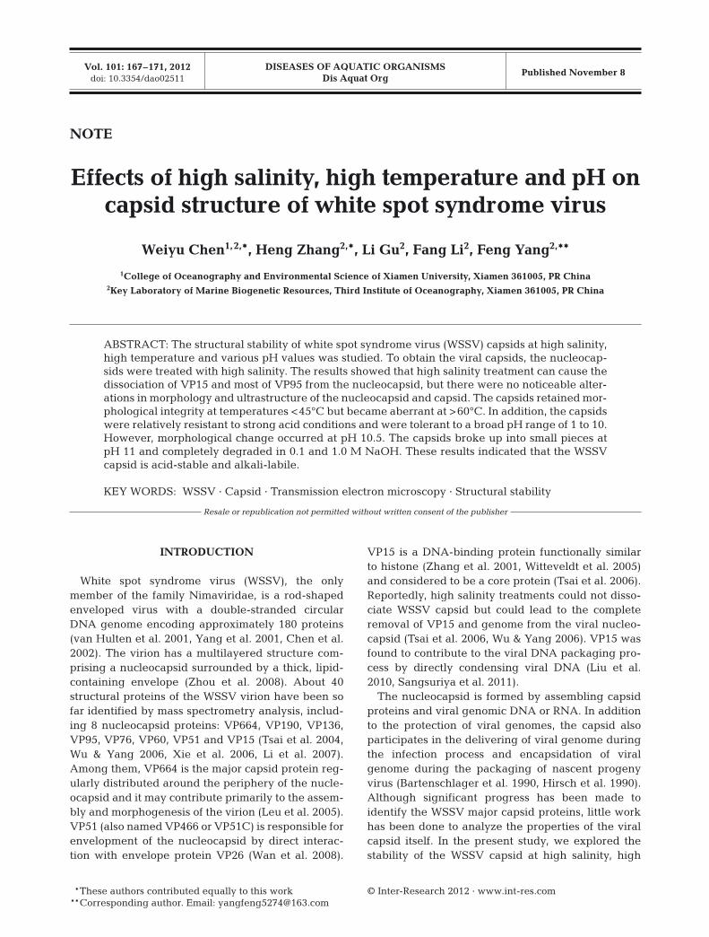
Fig. 1. Electron micrographs of (a) purified white spot syndrome virus (WSSV) nucleocapsids, (b) empty purified capsids, and heat-treated capsids at (c) 37, (d) 60, (e) 80 and (f) 100°C
Chen et al.: Structural stability of WSSV capsids
to study the stability of WSSV capsid under alkalineconditions, the capsids were incubated in 0.1 M car-bonate-bicarbonate buffer at pH 9, 9.5, 10, 10.5 or 11at room temperature for 1 h with slight shaking.Finally, the samples were examined by TEM.
Transmission electron microscopy (TEM)
Viral specimens were adsorbed onto 200 mesh carbon-coated nickel grids for 30 min at room tem-perature, and the excess liquid was carefully blottedwith Whatman filter paper. Then, the grids werewashed 3 times with distilled water and negativelystained with 2% phosphotungstic acid (PTA) for1 min. The excess stain was drained off with filterpaper and the specimens were examined with a TEM(JEM-1230, JEOL).
Sodium dodecyl sulfate-polyacrylamide gel electrophoresis (SDS-PAGE)
The nucleocapsids, capsids or treated sampleswere mixed with equal volumes of 2× Laemmlibuffer (Laemmli 1970) with 10% β-ME, heated at100°C for 10 min and separated by 12% polyacryl -amide gel. Protein bands were visualized usingCoomassie brilliant blue R-250 staining.
RESULTS
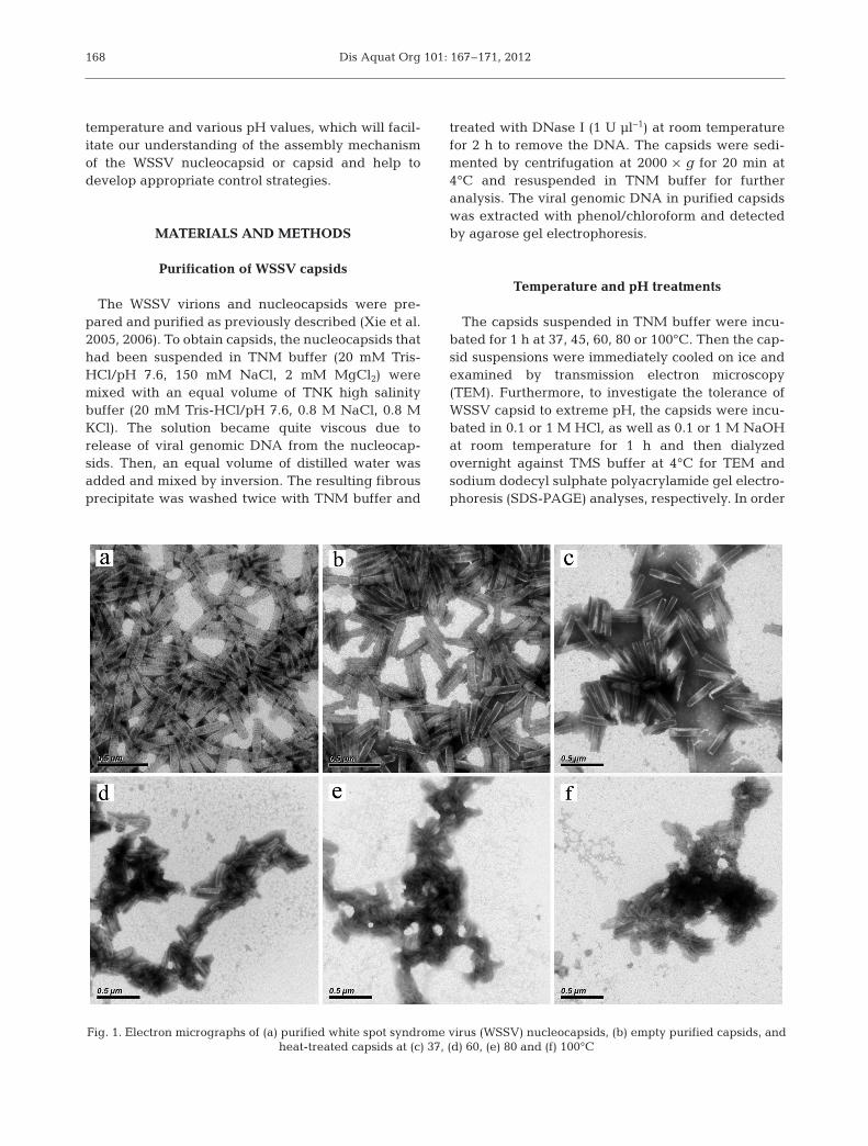
The WSSV capsids were prepared by high salinitytreatment. The viral genomic DNA was not detectedfrom purified capsids by agarose gel electrophoresis(data not shown). The TEM results revealed thatthere was no significant difference in the morpholog-ical appearance between the nucleocapsids (Fig. 1a)and capsids (Fig. 1b), and the profiles of the capsidsubunits (capsomers) were clearly visible on theirsurface, suggesting that ionic interactions betweencapsomers may make a relatively small contributionto the stability of the capsid. Whether perceptible dif-ferences in the protein composition between thenucleocapsids and capsids occurred was not clear. Inorder to visualize the protein components, the nucle-ocapsids and capsids were analyzed by SDS-PAGE.The results indicated that the high salinity treatmentcompletely removed the core protein VP15 from thenucleocapsids (Fig. 2, Lanes 1 and 2), indicating thatVP15 is not a structural component of the viral cap-sids. In addition, the VP95 protein was significantly
reduced in the capsids compared with the nucleo-capsids (Fig. 2, Lanes 1 and 2). The results of the highsalinity treatment suggested that the partial dissocia-tion of VP95 from the nucleocapsids may be relatedto the release of VP15 and viral DNA.
To estimate the effects of different temperature oncapsid structure, the capsids suspended in TNMbuffer were incubated for 1 h at 37, 45, 60, 80 or100°C. The TEM observation showed that no obviousconformation changes were observed in the capsidstreated at 37°C (Fig. 1c) or 45°C (data not shown).However, the surface structure of the capsid was nolonger visible after incubating at 60°C for 1 h(Fig. 1d). Heating up to 80 or 100°C, the capsids werefurther disrupted into amorphous masses (Fig. 1e,f).The results indicated that the structure of the capsidsis stable at a temperature lower than 45°C, but as thetemperature rises, the capsids form disorganizedaggregates due to thermal denaturation.
Moreover, to investigate the tolerance of WSSVcapsid to extreme pH, the capsids were incubated in0.1 or 1 M HCl, as well as 0.1 or 1 M NaOH. The TEMresults showed that the 0.1 M HCl treatment did not
169
Fig. 2. Sodium dodecyl sulphate polyacrylamide gel electro-phoresis (SDS-PAGE) analysis of purified white spot syn-drome virus (WSSV) nucleocapsid (Lane 1), empty capsids(Lane 2), 1 M HCl-treated capsids (Lane 3), 0.1 M-treatedcapsids (Lane 4), 1 M NaOH-treated capsids (Lane 5) and0.1 M NaOH-treated capsids (Lane 6) for structural proteins,including VP95 (asterisks) and core protein VP15 (arrow).
Lane M: low molecular mass protein marker
Dis Aquat Org 101: 167–171, 2012
cause significant change in capsid morphology, andcapsomeres remained clearly discernible (Fig. 3a,b).However, the capsid was disrupted into amorphousstructure that no longer retained the capsomericdetail, but its outline was still visible after treatmentwith 1 M HCl (Fig. 3c). The acid-treated protein com-ponents were further analyzed by SDS-PAGE. Asshown in Fig. 2, there was no obvious change in theprotein bands between HCl-treated and untreatedcapsids (Fig. 2, Lanes 2, 3 and 4). On the contrary,after 0.1 or 1 M NaOH treatment, no capsids or smallpieces were visible by TEM (data not shown), andalso no distinguishable protein bands were seen bySDS-PAGE (Fig. 2, Lanes 5 and 6), suggesting thatviral capsid proteins are likely to undergo degrada-tion under strong alkaline conditions.
To further examine the stability of WSSV capsidunder alkaline conditions, the capsids were incu-bated in 0.1 M carbonate-bicarbonate buffer at pH 9,9.5, 10, 10.5 or 11. The TEM results showed that viralcapsids appeared to have no significant morphologi-cal transformation after treatment at pH 9, 9.5 or 10
(Fig. 3d). However, at pH 10.5, the capsids appearedto undergo breakage and their structure becameloose (Fig. 3e). Surprisingly, the capsids were com-pletely disrupted into small patches at pH 11 (Fig. 3f).Although, alkaline conditions of pH 10.5 or greaterled to significant disruption of the capsid structure,we did not observe any free capsomeres.
DISCUSSION
In order to gain insight into the structure, organiza-tion and assembly of the WSSV nucleocapsid andcapsid, we examined their stability to high salinity,high temperature and extreme pH. After the highsalinity treatment, removal of the core protein VP15and viral genomic DNA appeared to have no effecton the morphology and ultrastructure of nucleocap-sid or capsid (Fig. 1a,b), suggesting that both VP15and DNA are not required for the maintenance of thecapsid structure. The data from SDS-PAGE showedthat, concomitant with the release of VP15, most of
170
Fig. 3. Electron microscopic analysis of (a) white spot syndrome virus (WSSV) capsids, (b) 0.1 M HCl-treated capsids, (c) 1 M HCl-treated capsids, and capsids incubated at (d) pH 10, (e) pH 10.5 and (f) pH 11

Chen et al.: Structural stability of WSSV capsids
the VP95 was also released from nucleocapsids(Fig. 2, Lanes 1 and 2). VP95 is a protein present inboth the viral envelope and nucleocapsid fractions(Xie et al. 2006), but its functional properties have notyet been examined. Based on the above experimen-tal results, we speculated that VP95 might participatein the release of virus nucleoprotein core consistingof VP15 and genomic DNA.
The pH dependence on the stability of the capsidswas assessed by incubating them at various pH val-ues. The capsids were found to be acid resistant butcomplete disruption took place under strong alkalineconditions (0.1 or 1 M NaOH). In addition, the cap-sids’ structure became broken and loose at pH 10.5(Fig. 3e) and disrupted into small patches at pH 11(Fig. 3f). Normally, for large DNA viruses, uncoating(loss of viral capsid) is required prior to viral genomedelivery into the nucleus. The above experimentaldata provided a suggestion that the uncoating ofWSSV may occur in a relatively alkaline compart-ment within the host cell. The mechanism of entryand uncoating of WSSV is unknown at present, andfurther studies are needed to verify our hypothesis.
In conclusion, in the present study, we found that(1) the high salinity treatment can cause the dissocia-tion of VP15 and most of VP95 from the nucleocapsid,but with no noticeable alterations in morphology andultrastructure of the nucleocapsid and capsid, whichsuggests that these peptides are not part of its struc-ture; (2) at temperatures lower than 45°C the capsidsretained their morphological integrity but becameaberrant at temperatures higher than 60°C; and(3) capsids are acid resistant but not alkaline resist-ant, as shown by stability at a broad pH range (1 to10) but degradation at pH 10.5.
Acknowledgements. This investigation was supported byNational Basic Research Program of China (973 Program)(No.2012CB114401), Natural Science Foundation of China(No. 31072243) and China Agriculture Research System-47.
LITERATURE CITED
Bartenschlager R, Junker-Niepmann M, Schaller H (1990)The P gene product of hepatitis B virus is required as astructural component for genomic RNA encapsidation.J Virol 64: 5324−5332
Chen LL, Leu JH, Huang CJ, Chou CM, Chen SM, WangCH, Lo CF, Kou GH (2002) Identification of a nucleocap-sid protein (VP35) gene of shrimp white spot syndromevirus and characterization of the motif important for targeting VP35 to the nuclei of transfected insect cells.Virology 293: 44−53
Hirsch RC, Lavine JE, Chang LJ, Varmus HE, Ganem D(1990) Polymerase gene products of hepatitis B virusesare required for genomic RNA packaging as well as forreverse transcription. Nature 344: 552−555
Laemmli UK (1970) Cleavage of structural proteins duringthe assembly of the head of bacteriophage T4. Nature227: 680−685
Leu JH, Tsai JM, Wang HC, Wang AHJ, Wang CH, KouGH, Lo CF (2005) The unique stacked rings in thenucleocapsid of the white spot syndrome virus virionare formed by the major structural protein VP664, thelargest viral structural protein ever found. J Virol 79: 140−149
Li Z, Lin Q, Chen J, Wu JL, Lim TK, Loh SS, Tang X, Hew CL(2007) Shotgun identification of the structural proteomeof shrimp white spot syndrome virus and iTRAQ differ-entiation of envelope and nucleocapsid subproteomes.Mol Cell Proteomics 6: 1609−1620
Liu Y, Wu J, Chen H, Hew CL, Yan J (2010) DNA conden-sates organized by the capsid protein VP15 in white spotsyndrome virus. Virology 408: 197−203
Sangsuriya P, Senapin S, Huang WP, Lo CF, Flegel TW(2011) Co-interactive DNA-binding between a novel,immunophilin-like shrimp protein and VP15 nucleocap-sid protein of white spot syndrome virus. PLoS ONE 6: e25420
Tsai JM, Wang HC, Leu JH, Hsiao HH, Wang AHJ, Kou GH,Lo CF (2004) Genomic and proteomic analysis of thirty-nine structural proteins of shrimp white spot syndromevirus. J Virol 78: 11360−11370
Tsai JM, Wang HC, Leu JH, Wang AH, Zhuang Y, WalkerPJ, Kou GH, Lo CF (2006) Identification of the nucleocap-sid, tegument, and envelope proteins of the shrimp whitespot syndrome virus virion. J Virol 80: 3021−3029
van Hulten MCW, Witteveldt J, Peters S, Kloosterboer N andothers (2001) The white spot syndrome virus DNAgenome sequence. Virology 286: 7−22
Wan Q, Xu L, Yang F (2008) VP26 of white spot syndromevirus functions as a linker protein between the envelopeand nucleocapsid of virions by binding with VP51. J Virol82: 12598−12601
Witteveldt J, Vermeesch AMG, Langenhof M, de Lang A,Vlak JM, van Hulten MCW (2005) Nucleocapsid proteinVP15 is the basic DNA binding protein of white spot syn-drome virus of shrimp. Arch Virol 150: 1121−1133
Wu C, Yang F (2006) Localization studies of two white spotsyndrome virus structural proteins VP51 and VP76. VirolJ 3: 76
Xie X, Li H, Xu L, Yang F (2005) A simple and efficientmethod for purification of intact white spot syndromevirus (WSSV) viral particles. Virus Res 108: 63−67
Xie X, Xu L, Yang F (2006) Proteomic analysis of the majorenvelope and nucleocapsid proteins of white spot syn-drome virus. J Virol 80: 10615−10623
Yang F, He J, Lin X, Li Q, Pan D, Zhang X, Xu X (2001) Com-plete genome sequence of the shrimp white spot bacilli-form virus. J Virol 75: 11811−11820
Zhang X, Xu X, Hew CL (2001) The structure and functionof a gene encoding a basic peptide from prawn whitespot syndrome virus. Virus Res 79: 137−144
Zhou Q, Li H, Qi YP, Yang F (2008) Lipid of white-spot syn-drome virus originating from host-cell nuclei. J Gen Virol89: 2909−2914
171
Editorial responsibility: Grant Stentiford,Weymouth, UK
Submitted: May 21, 2012; Accepted: July 27, 2012Proofs received from author(s): October 10, 2012