effects of smoking marijuana on brain perfusion and cognition
TRANSCRIPT

N
EUROPSYCHOPHARMACOLOGY
2002
–
VOL
.
26
,
NO
.
6
© 2002 American College of NeuropsychopharmacologyPublished by Elsevier Science Inc. 0893-133X/02/$–see front matter655 Avenue of the Americas, New York, NY 10010 PII S0893-133X(01)00425-0
Effects of Smoking Marijuana on Brain Perfusion and Cognition
Daniel S. O’Leary, Ph.D., Robert I. Block, Ph.D., Julie A. Koeppel, B.S., Michael Flaum, M.D.,Susan K. Schultz, M.D., Nancy C. Andreasen, M.D., Ph.D., Laura Boles Ponto, Ph.D.,
G. Leonard Watkins, Ph.D., Richard R. Hurtig, Ph.D., and Richard D. Hichwa, Ph.D.
The effects of smoking marijuana on regional cerebral blood flow (rCBF) and cognitive performance were assessed in 12 recreational users in a double-blinded, placebo-controlled study. PET with [
15
Oxygen]-labeled water ([
15
O]H
2
O) was used to measure rCBF before and after smoking of marijuana and placebo cigarettes, as subjects repeatedly performed an auditory attention task. Smoking marijuana resulted in intoxication, as assessed by a behavioral rating scale, but did not significantly alter mean behavioral performance on the attention task. Heart rate and blood pressure increased dramatically following smoking of marijuana but not placebo cigarettes. However, mean global CBF did not change significantly. Increased rCBF was observed in orbital and mesial frontal lobes, insula, temporal poles, anterior cingulate, as well as in the
cerebellum. The increases in rCBF in anterior brain regions were predominantly in “paralimbic” regions and may be related to marijuana’s mood-related effects. Reduced rCBF was observed in temporal lobe auditory regions, in visual cortex, and in brain regions that may be part of an attentional network (parietal lobe, frontal lobe and thalamus). These rCBF decreases may be the neural basis of perceptual and cognitive alterations that occur with acute marijuana intoxication. There was no significant rCBF change in the nucleus accumbens or other reward-related brain regions, nor in basal ganglia or hippocampus, which have a high density of cannabinoid receptors.
[Neuropsychopharmacology 26:802–816, 2002]
© 2002 Elsevier Science Inc. All rights reserved.Published by Elsevier Science Inc.
KEY
WORDS
:
Cerebral blood flow; Marijuana; Positron emission tomography; THC; Brain; Imaging; Cognition; Attention
Marijuana (
Cannabis sativa
) has been the most widelyused illicit drug in the United States for a number of de-
cades. Following steady declines throughout the late1970s and the 1980s, the prevalence of marijuana useamong youth skyrocketed in the early and middle1990s, and has remained high (SAMHSA 1998, 1999;Johnston et al. 1999). Marijuana is typically smoked, re-sulting in subjective effects that may include euphoria,depersonalization, altered time sense, lethargy, drowsi-ness, confusion, and anxiety (Solowij 1998). Smokingmarijuana may also result in impairment on sensory,motor, and cognitive tasks (Block et al. 1992; Heishmanet al. 1990).
Over 60 cannabinoids have been identified in theplant
Cannabis sativa
, but delta-9-tetrahydrocannabinol(THC) appears to be the major psychoactive ingredientof marijuana (Harvey 1999). THC has a long biologicalhalf-life (4–12 days) because of extensive accumulationof the drug in fatty tissue (Huestis 1999). Two types of
From the Mental Health Clinical Research Center (DSO’L, JAK,MF, SKS, NCA), Department of Anesthesia (RLB), Department ofPsychiatry (MF, SKS, NCA), Department of Radiology (LBP, GLW,RDH), and Department of Speech Pathology and Audiology (RRH),University of Iowa College of Medicine, Iowa City, IA 52242.
Address correspondence to: Daniel S. O’Leary, Ph.D., 2911JPP,200 Hawkins Drive, Iowa City, Iowa 52242-1057, Tel.: (319) 356-3897, Fax: (319) 356-2587, E-mail: [email protected]
Received May 14, 2001; revised October 8, 2001; accepted Novem-ber 10, 2001.
Online publication: 11/30/01 at www.acnp.org/citations/Npp113001216.

N
EUROPSYCHOPHARMACOLOGY
2002
–
VOL
.
26
,
NO
.
6
Acute Marijuana Effects
803
cannabinoid receptors have been identified in mam-mals, CB
1
and CB
2
(Pertwee 1997). There have been sug-gestions that one function of the cannabinoid system isto modulate dopaminergic (DA) activity (Biegon andKerman 1995; Loeber and Yurgelun-Todd 1999). Can-nabinoid receptors have a distribution in the brain thatis similar to that of DA (high concentrations in the basalganglia, hippocampus), but there is also a high densityof cannabinoid receptors in the cerebellum (Herkenham1992). High density bands of cannabinoid receptorshave also been found in the prefrontal cortex in humanpost-mortem tissue, particularly around the cingulateand superior frontal gyri (Biegon and Kerman 1995).
The few imaging studies that have assessed the ef-fects of marijuana and THC on brain blood flow andmetabolism in humans have produced conflicting re-sults. An early single photon emission computerized to-mography (SPECT) study by Mathew and colleaguesusing the
133
Xenon inhalation technique compared ex-perienced (a minimum of 10 marijuana cigarettes aweek for 3 years), and inexperienced marijuana usersbefore and after smoking marijuana or placebo(Mathew et al. 1989). In both groups the effects of smok-ing placebo were similar to the effects of smoking mari-juana. However, the inexperienced smokers showed adecrease in global cerebral blood flow (gCBF) that wassignificantly greater for marijuana than placebo,whereas the experienced smokers showed increasedCBF that was the same following marijuana and pla-cebo. In a later
133
Xenon SPECT study by Mathew andcolleagues (Mathew et al. 1992) assessed regional cere-bral blood flow (rCBF) following smoking marijuanacigarettes of two different potencies and placebo in 20experienced male smokers. Smoking marijuana in-creased global CBF significantly more than placebo,and the greatest increases in rCBF were in the frontallobes and in the right hemisphere. Mathew et al. (1997)have more recently reported rCBF measurements ob-tained in 32 normal volunteers using PET with[
15
O]H
2
O before and after intravenous (I.V.) infusion ofeither of two doses of THC or a placebo, given underdouble blind conditions. THC resulted in increasedrCBF bilaterally in the frontal lobes, the insula, anteriorcingulate and in subcortical regions, with the largest ef-fects in the right hemisphere.
Volkow et al. (1991a,b) assessed regional glucose me-tabolism (rGluM) with PET and [
18
F]FDG followingTHC injection in eight male volunteers who were occa-sional users of marijuana (a mean of one marijuana cig-arette every two months). Volkow et al. found a greatdeal of individual variability in the changes in cerebralglucose metabolism induced by THC. Three of eightsubjects in their study showed an increase, threeshowed a decrease, and two had no change in globalmetabolism in the cerebrum. However, all subjectsshowed an increase in normalized metabolism in the
cerebellum (12% increase over baseline), and the cere-bellar increase had a significant correlation with subjec-tive ratings of intoxication. These findings are consis-tent with the high density of cannabinoid receptors inthe cerebellum (Herkenham et al. 1990). Volkow et al.(1996) described the cerebral metabolic changes in-duced by THC in eight chronic users of marijuana andeight non-users. At baseline, the chronic marijuana us-ers had significantly less relative metabolic activity inthe cerebellum than did normal control subjects. Cere-bellar activity was increased in both non-users andchronic users of marijuana, but only the chronic groupshowed significant increases in orbitofrontal cortex,prefrontal cortex and basal ganglia.
The brain imaging studies reviewed above all uti-lized a “resting” baseline condition in which subjectsperform no explicit task when the effects of marijuanaor THC on the brain are assessed. Studies of the restingstate represent an important first step in understandingthe behavioral and physiological effects of marijuana.However, assessing the effects of marijuana only in aresting state confounds the direct effects of marijuanaon cerebral function with changes in subjective experi-ence and cognitive state that result from intoxication.Recent PET studies carried out in our laboratory mea-sured rCBF using [
15
O]H
2
O prior to and followingsmoking of a marijuana cigarette, and controlled thesubjects’ mental activities by engaging them in a audi-tory attention task (O’Leary et al. 2000). This initialstudy of five subjects found that smoking marijuana in-creased rCBF in a number of “paralimbic” brain regions(e.g., orbital frontal lobes, insula, temporal poles) and inanterior cingulate and cerebellum. Large reductions inrCBF were observed in temporal lobe regions that aresensitive to auditory attention effects.
The present study involved a second, independentsample of 12 volunteers who were occasional recre-ational smokers of marijuana. Unlike the study re-ported in O’Leary et al. (2000), the present study in-cluded the use of a placebo cigarette (marijuana withthe THC removed), as well as an active marijuana ciga-rette, and utilized a double blind design. We recruited12 new subjects who were assessed with PET beforeand after smoking either marijuana or placebo ciga-rettes in a single 8-injection study. Arterial lines wereplaced in all subjects to permit calculation of quantita-tive cerebral blood flow.
METHODS
The subjects were 12 healthy volunteers (6 males, 6 fe-males, mean age 30.5, sd
�
8.6 years) who were occa-sional recreational users of marijuana. They reportedtheir current use of marijuana as no more frequently

804
D. S. O’Leary et al. N
EUROPSYCHOPHARMACOLOGY
2002
–
VOL
.
26
,
NO
.
6
than 10 times a month (mean
�
2.7 times a month overthe past year), and had an average duration of usage ofsix years. All of the subjects were right handed. Subjectswere asked to refrain from smoking marijuana forseven days prior to the PET study. A urine screeningtest (TRIAGE
R
Drugs of Abuse Panel Kit) was adminis-tered to all subjects on the day of their PET scan. Allsubjects had a negative value for THC and six otherdrugs of abuse, indicating that they had not smokedmarijuana for at least four days prior to the study. Allsubjects provided written consent in compliance withthe guidelines of the University of Iowa InstitutionalReview Board and Radiation Protection Committee.
As detailed in Table 1, there were nine PET condi-tions. An initial “scout” condition was used to familiar-ize subjects with procedures and to measure arrival timeof the bolus of [
15
O]H
2
O in the brain. This was followedby eight PET image acquisition conditions. The scoutand first PET imaging condition utilized an auditorychoice reaction time (RT) task, which matched the di-chotic tests in number of trials, durations of auditorystimulation, and motor responses. It utilized easily dis-criminable, binaurally presented stimuli, consisting of 21pure tones (1319 Hz, 500 ms duration), randomly mixedwith 104 bursts of white noise (500 ms duration). Sub-jects responded to each tone by pressing a button withtheir right thumb as soon as they detected a target tone.
For the dichotic conditions, subjects were instructedto attend only to their left ear. On each trial, pairs ofnonsense words were delivered simultaneously to theleft and right ears via foam insert earphones at 80 dBSPL. The nonsense words were digitized naturalspeech, about 500 ms long. The initial and final conso-nants were stops (i.e., /p/, /b/, /d/, /t/, /g/, and /k/),which were combined with one of five vowels. Eachtrial consisted of 125 pairs of stimuli presented in ran-dom order with an interstimulus interval of 800 ms, in-
cluding 21 trials with the target presented to the at-tended ear and 10 with the target presented to theunattended ear. Subjects responded to the target in theattended ear by pressing a button with their right thumbas fast as possible. Responses were scored as hits if theyoccurred within 200–2000 ms after a target presented tothe attended ear. Responses and reaction times were re-corded by a personal computer. During the PET session,testing for both the dichotic tests and the control test be-gan 70 s before the estimated time of bolus arrival in thebrain and lasted for an additional 100 s.
After the first dichotic condition a marijuana or pla-cebo cigarette was smoked (half of the subjects smokedmarijuana first and half placebo). Both marijuana andplacebo cigarettes were obtained from the National In-stitute of Drug Abuse. The marijuana cigarettes con-tained a moderate amount of THC (
�
20 mg), and theplacebo contained marijuana with the THC removed.Neither the subject nor the staff in the PET imagingsuite at that time knew which cigarette contained activeTHC or was the placebo. The subject smoked the ciga-rettes, which were held in a hemostat, while in a re-clined position on the PET couch. A paced smokingprocedure similar to that described in Block et al. (1992)was used with subjects inhaling for 5 s, holding thesmoke in their lungs for 5 s, and then exhaling. The sub-jects rested for 25 s and were then again told to inhale,hold and exhale. This continued until the cigarette was toosmall to be smoked. Subjects exhaled into a dome- shapeddevice suspended about 12 inches above their head andthe smoke was vented from the PET camera room.
Following smoking, subjects were repositioned inthe PET camera by lining up marks drawn on their skinand laser guide lights. Subject’s heads were constrainedonly lightly by tape but images from each PET condi-tion were co-registered with each individual’s MRI im-age before analysis as described below. If subjects couldnot be adequately realigned another transmission scanwas performed for attenuation correction of the PETimages from the remaining conditions (see below). Af-ter smoking the initial cigarette, the subject performedthree dichotic conditions with PET imaging at approxi-mate 15 min intervals. A second cigarette was thensmoked, the subject repositioned, and three more di-chotic conditions with PET imaging were conducted.
A “highness” rating scale (0 “not at all”, 10 “highestever”) was administered verbally after each PET imageacquisition and immediately after each smoking condi-tion. The Beck Anxiety inventory was also administeredat a preliminary session, and after the first, fourth, andseventh PET image acquisitions. Heart rate and bloodpressure were recorded after each PET image acquisi-tion and immediately after each smoking session. Twoblood samples were obtained from the arterial line aftereach PET image acquisition and immediately followingeach smoking session. One of the blood samples was
Table 1.
PET Activation Tasks and Schedule for Smoking THC and Placebo Cigarettes
Condition
1
Activation Task
Scout (no image acquisition) Reaction Time TaskCondition 1 Reaction Time TaskCondition 2 Dichotic Task
Subject smokes THC (n
�
6) or placebo cigarette (n
�
6)Condition 3 Dichotic TaskCondition 4 Dichotic TaskCondition 5 Dichotic Task
Subject smokes THC or placebo cigarette Dichotic Task
Condition 6 Dichotic TaskCondition 7 Dichotic TaskCondition 8 Dichotic Task
1
Order of smoking marijuana and placebo cigarettes was counter-bal-anced across subjects.

N
EUROPSYCHOPHARMACOLOGY
2002
–
VOL
.
26
,
NO
.
6
Acute Marijuana Effects
805
placed on ice and immediately delivered to the Univer-sity of Iowa Hospitals and Clinics Specimen ControlLaboratory for analysis of total carbon dioxide (TCO
2
),partial pressure of CO
2
and carboxyhemoglobin. Theother sample was immediately centrifuged. From thissample plasma was pipetted, placed in a freezer at –20C, and later sent as a batch to NIDA’s Radioimmunoas-say Laboratory in Research Triangle Park, North Caro-lina for analysis of THC levels.
PET Data Acquisition
Each subject had an arterial catheter placed in the radialartery of one arm for blood sampling and a venouscatheter in the antecubital vein of the other arm for in-jection of the [
15
O]H
2
O. They were then positioned inthe PET camera. Laser guide lights were used to alignthe subject so that the most caudal slice was alignedwith the auditory meatus and the outer canthus of theeyes. The subjects’ heads were lightly taped and markswere drawn on their skin for use in realignment prior toeach image acquisition. A rotating pin source of[
68
GE]germanium was used to acquire a transmissionscan for attenuation correction of the emission images.The scout condition used a 15 mCi dose and the re-maining conditions utilized 50 mCi of [
15
O]H
2
O. PETimage acquisition conditions were repeated at approxi-mately 15 min intervals except for the conditions fol-lowing smoking. Smoking the THC and placebo ciga-rettes took approximately 15–20 min and PET imagingcommenced approximately 10–15 min following smok-ing. Subjects remained in the PET Center following thestudy until their heart rate and blood pressure returnedto baseline, they reported no symptoms of anxiety, andreported that they were no longer intoxicated (a high-ness rating of 3 or less). They were then placed in a caband driven to their home and were asked to remain athome for the remainder of the day.
Regional cerebral blood flow (rCBF) was measuredusing the bolus [
15
O]H
2
O method with a GE4096PLUSScanner (Herscovitch et al. 1983; Hichwa et al. 1995).For each emission scan, fifteen slices (6.5 mm center-to-center) were acquired with a 10 cm axial field ofview. Dynamic imaging and arterial blood samplingwere acquired over a 100-s interval following venousinjection. The dynamic imaging data were summedfrom the 40 s immediately following bolus transit, de-termined by time-activity curves from a region of inter-est over a cerebral artery. The summed images and ar-terial blood samples were then used to calculate tissueperfusion in mL/min/100 g tissue using the autoradio-graphic method (for details see Hurtig et al. 1994; Wol-lenweber et al. 1997). Quantitative flow images wereprocessed by the Image Processing Laboratory (IPL) ofthe Iowa Mental Health Clinical Research Center.
MR Imaging
The MR images were acquired for each subject in a 1.5Tesla General Electric Signa scanner using an SPGR se-quence, flip angle of 40 degrees, TE of 5 ms, TR of 24ms, and with 2 NEX. The contiguous 1.5 mm thick coro-nal slices were processed by the IPL using locally-developed BRAINS software (Andreasen et al. 1993).
PET and MRI Processing
Each subject’s MR images were processed utilizing acombination of automated methods and hand editing, re-sulting in a brain that was aligned in a standardized coor-dinate space. (Talairach and Tournoux 1988; Cizadlo et al.1994). The outlines of the PET images were automaticallyidentified with an edge detection algorithm and the PETimages for each condition for each subject were co-regis-tered with their MR images using a variance minimiza-tion program (Woods et al. 1992). An 18 mm Hanning fil-ter was applied to the PET images for each condition toeliminate residual anatomical variability.
Following spatial normalization and filtering, within-subject subtractions were computed to compare a num-ber of different conditions. The subtractions were fol-lowed by across-subject averaging of the subtractionimages and computation of voxel by voxel
t
-tests ofthe rCBF changes. Significant regions of activation werecalculated on the
t
-map images, using a technique thatcorrects for the large number of voxel by voxel
t
-testsperformed, the lack of independence between voxels,and the resolution of the processed PET images (Wors-ley et al. 1992).
The pre-smoking auditory RT condition was subtractedfrom the pre-smoking dichotic condition and from the con-ditions following smoking of THC and placebo. These sub-tractions highlight the cognitive activation resulting fromthe dichotic listening task pre- and post-smoking and per-mit a qualitative assessment of attention-related changes inactivation due to smoking marijuana and placebo. A sec-ond set of analyses subtracted the pre-smoking dichoticcondition from the dichotic conditions following smokingof marijuana and placebo. These analyses allowed assess-ment of the effects of smoking marijuana and placebo dur-ing the performance of the same cognitive task. Finally,subtraction of the post-smoking placebo condition fromthe post-smoking marijuana condition allowed assessmentof the effects of THC on rCBF controlling for the specific ef-fects of smoking and the cognitive task.
RESULTS
Behavioral and Physiological Data
Prior to smoking, the mean subjective rating of themagnitude of intoxication or “high” (10
�
“highest

806
D. S. O’Leary et al. N
EUROPSYCHOPHARMACOLOGY
2002
–
VOL
.
26
,
NO
.
6
ever”) was 0; the mean ratings (and standard devia-tions) for the three conditions following smoking of themarijuana cigarette were: 7.4 (1.7), 7.3 (1.5), 7.0 (1.8).The ratings following placebo were: 4.7 (3.6), 4.4 (3.6),3.5 (3.7). Since half of the subjects smoked the mari-juana cigarettes first, the highness ratings followingplacebo for these subjects were influenced by residualmarijuana effects. This is indicated by the fact that thesix subjects who smoked placebo first had mean ratingsof 1.8, 1.6, 0.7 for the three PET conditions followingsmoking of the placebo cigarette.
The subjects performed extremely well on the base-line auditory RT task (mean
�
666.8 ms, sd
�
109.4)and on the dichotic tasks, with over 98% correct detec-tions of left ear targets in every dichotic condition.Mean reaction time for the dichotic tasks was 887.9 ms(sd
�
148.1) for the pre-smoking dichotic condition. Forthe three conditions following smoking marijuana theRTs were: 845.4 (155.6), 914.0 (163.4), and 923.2 (195.4)ms. The RTs for the three conditions following smokingof placebo were: 911.3 (226.4), 975.3 (216.7), and 986.0(228.8) ms. Paired
t
-tests indicated that none of the RTsfor the post-smoking conditions differed significantlyfrom the pre-smoking RT.
As can be seen in Table 2, mean heart rate (HR) in-creased dramatically following smoking of marijuanabut not placebo cigarettes. Compared with the pre-smoking dichotic condition (66.2 beats/min) HR in-creased significantly in the PET condition followingsmoking marijuana (106.8 beats/min) t
�
�
6.1,
p
�
.001, as well as in the next two conditions (93.5 and 82.6beats/min respectively, with t
�
�
5.3,
p
�
.001, and t
�
4.5,
p
�
.001). HR changes in three conditions followingsmoking placebo (75.4 (12.0), 72.6 (121.3), 70.2 (11.1))were non-significant in comparison to the pre-smokingdichotic condition.
Blood pressure also increased after smoking mari-juana, but the changes were more variable across indi-
viduals and less dramatic. In the PET condition follow-ing smoking of marijuana diastolic pressure wassignificantly higher than in the pre-smoking condition(pre-smoking mean
�
69.2 mm Hg (sd
�
7.1), post-smoking mean
�
79.2 mm Hg (sd
�
14.5), t
�
�
2.7,
p
�
.02). Diastolic pressure remained significantly higher inthe second post-marijuana condition (mean
�
76.9 mmHg (sd
�
9.4), t
�
�
3.3,
p
�
.007) than in the pre-smok-ing condition, but was no longer significantly higher inthe third post-marijuana condition (70.5 mm Hg (sd
�
8.3) ns). After smoking placebo, diastolic pressure wasnot significantly different from the pre-smoking condi-tion at any time point. Systolic pressure was also notsignificantly higher after smoking marijuana or placebofor any condition.
The mean (and standard deviation) values for THCin plasma are given in Table 2. These were obtainedfrom blood samples obtained immediately after thecompletion of PET imaging for each of the post-smok-ing conditions. A value of 122.1 ngr/mL (sd
�
104.3)was measured in a sample that was obtained immedi-ately following smoking of the marijuana cigarette. Thisdropped quickly to the tabled value of 37.1 ng/mL ob-served in the sample obtained after the first PET acqui-sition following smoking, which occurred approxi-mately 15 min later. Thus, THC levels drop off rapidlyin the first few minutes following smoking. We wereunable to observe the acute effects of these high THClevels on rCBF because of the time required to reposi-tion the subject in the scanner following smoking.
Table 3 lists the correlations (Pearson) of THC levelswith behavioral task performance (reaction time), thehighness rating, heartrate, and blood pressure. Behav-ioral performance did not correlate significantly withTHC levels, perhaps because of the large variability inthe plasma THC measure (see standard deviations ofTHC in Table 2). Subjective ratings of “highness” alsodid not correlate significantly with THC levels, except
Table 2.
Mean (and standard deviation) Heart Rate, Blood Pressure, Whole Brain PET Counts, and Whole Brain Blood Flow (WBBF) for each PET Condition
Condition Heart Rate(beats/min)
Blood Pressure(systolic/diastolic) Pet Counts WBBF
THC Levels(ng/mL)
1
Scout (RT Task) 67.3 (8.4) 122/68 (14/9) . . .
2
. . . . . . Pre-Smoke RT Task 64.5 (8.7) 124/70 (10/7) 935 (199) 50.1 (5.7) . . . Pre-Smoke Dichotic task 66.2 (7.0) 124/69 (10/7) 940 (211) 51.9 (6.5) . . . Post-Smoke THC1
3
106.8 (25.3) 131/75 (20/10) 707 (184) 49.6 (5.5) 37.1 (27.1)Post-Smoke THC2 93.5 (20.8) 130/77 (18/9) 783 (231) 50.9 (6.5) 14.8 (10.5)Post-Smoke THC3 82.6 (15.5) 130/72 (13/8) 807 (218) 51.5 (9.7) 11.4 (7.3)Post-Smoke Placebo1 75.4 (12.0) 129/72 (13/8) 856 (217) 49.9 (7.6) 3.9 (4.5)Post-Smoke Placebo2 72.6 (12.3) 127/71 (13/8) 921 (200) 51.7 (7.8) 3.0 (3.9)Post-Smoke Placebo3 70.2 (11.1) 126/71 (14.9) 964 (185) 51.5 (9.7) 2.6 (3.6)
1
Levels of THC in plasma in nanograms per milliliter.
2
The scout condition used only 15 millicurries of [
15
O]water to obtain bolus arrival time. No PET image was acquired.
3
The same attend-left dichotic task, with differing randomizations of stimuli, was used pre-smoking condition, and all of the post-smoking con-ditions.

N
EUROPSYCHOPHARMACOLOGY
2002
–
VOL
.
26
,
NO
.
6
Acute Marijuana Effects
807
for the rating that immediately followed smoking theplacebo cigarette. Except for the assessment immedi-ately following smoking marijuana, heart rate showed aconsistently significant relationship with THC level, butblood pressure did not.
PET Data
Whole brain PET counts (a measure of the concentra-tion of the radiotracer in the brain) dropped followingsmoking of marijuana in comparison to the pre-smok-ing condition (from 940 to 707, t � 4.9, p � .0001, see Ta-ble 2), and remained significantly lower for the nexttwo conditions (t � 3.6, p � .004 and t � 3.4, p � . 006).PET counts were also lower in the first condition fol-lowing smoking placebo (mean � 856, t � 2.6, p � .03),but this appeared to be a carryover effect from smokingmarijuana. The six subjects who smoked marijuana firsthad a mean PET count of 810 for the condition follow-ing smoking placebo, whereas the six subjects whosmoked placebo first had a mean PET count of 902 fol-lowing placebo. All 12 subjects showed a pattern of in-creased HR and decreased PET counts when comparingthe conditions prior to and following smoking mari-juana. In contrast, mean whole brain blood flow(WBBF) did not change significantly following smokingof either marijuana or placebo cigarettes. In the condi-tion immediately prior to smoking marijuana, meanWBBF was 51.9, and was 49.6 ml/min/100 g followingsmoking. Six of the subjects had increased WBBF andhalf had decreased WBBF following smoking mari-juana.
A t-threshold of 3.61 (uncorrected p � .0005) wasused for all of the following t-map analyses, as well as avolume threshold of 100 contiguous voxels at or abovethis t-threshold. Table 4 lists the results of the PET t-map analysis in which the pre-smoking RT baselinecondition was subtracted from the dichotic listeningconditions immediately prior and immediately follow-ing smoking of placebo and marijuana. Prior to smok-ing, the subtraction of the RT baseline from the dichoticcondition replicated our previous findings (O’Leary et
al. 1996a, 1997; Block et al. 2000), with dichotic listeningresulting in large increases in rCBF in left (L) and right(R) superior temporal gyri (STG), and with no other re-gions showing significantly increased rCBF. Prior tosmoking, there were only a few small regions withlower rCBF in the dichotic condition than in the RTbaseline, which is also in line with our previous work.Smoking a placebo cigarette did not alter the pattern ofactivation in L and R STG for the dichotic minus base-line conditions. There were, however, additional smallactivations in L anterior temporal lobe, and R frontallobe. Regions with lower rCBF in the dichotic than RTcondition following placebo were found in L posteriorcingulate, L superior parietal lobe, and L hippocampus.As discussed below, some of the rCBF differences be-tween the pre-smoking condition and the condition fol-lowing placebo appear to have been residual effects ofmarijuana since half of the subjects smoked marijuanaprior to smoking placebo.
As seen in Table 4, smoking marijuana resulted indramatic changes in rCBF when the pre-smoking base-line condition was subtracted from the dichotic condi-tion immediately following smoking. In contrast to thepre-smoking and placebo analyses there were no signif-icant activations in L or R STG. Extensive bilateral re-gions of ventral frontal and temporal lobes and insulahad increased rCBF following smoking marijuana, asdid the anterior cingulate and cerebellum. A number ofregions also showed lower rCBF following smokingmarijuana. These included several regions of L frontallobe, L parietal lobe, L insula, and an extensive regionof precuneus.
Table 5 and Figure 1 display the analyses directlysubtracting the pre-smoking dichotic condition fromthe dichotic conditions immediately following smokingof a placebo and marijuana cigarette. These analyses al-low comparison of the effects of smoking placebo ver-sus marijuana on rCBF when subjects are performingthe same cognitive task. Smoking a placebo cigarette re-sulted in relatively little change in rCBF from the pre-smoking condition (small increases in R and L frontallobes and a small decrease in L parietal lobe). As in the
Table 3. Correlations Between Levels of Plasma THC and Reaction Time, Heart Rate and Blood Pressure
ConditionReaction Time
(msec)Highness
Rating Heart rate Systolic BP Diastolic BP
Immediately Post Smoking . . . �.04 .32 .17 .05Post-Smoking THC1 �.42 .02 .66* .31 .34Post-Smoking THC2 �.25 .25 .66* .30 .48Post-Smoking THC3 .10 .17 .69* .32 .47Post-Smoking Placebo1 .39 .70** .60* .54 .51Post-Smoking Placebo2 .26 .33 .72** .25 .36Post-Smoking Placebo3 .33 .34 .77** .31 .52
* p � .05** p � .01

808 D. S. O’Leary et al. NEUROPSYCHOPHARMACOLOGY 2002–VOL. 26, NO. 6
RT baseline analysis, smoking marijuana resulted in in-creased rCBF in a number of regions in ventral and me-sial frontal lobe, insula, temporal poles, and cerebellum.Smoking marijuana also resulted in decreased rCBF bi-
laterally in a number of regions in left and right frontallobes, in L STG, and in R occipital lobe.
Table 6 presents the results of a direct comparison ofthe two post-smoking conditions. When the condition
Table 4. PET t-Map Analyses of rCBF Differences Obtained by Subtracting the Pre-Smoking Reaction Time Baseline Condition from the Pre- and Post-Smoking Placebo and Marijuana Dichotic Conditions
Brain Regions with Significantly Higher Regional Cerebral Blood Flow (rCBF) in Dichotic than Baseline Condition
Region X Y Z1 t-value Volume2
Pre-Smoking DichoticCondition minus Baseline
L. Sup. Temp. Gyrus 3 �52 �1 5 10.0 16.7R. Sup. Temp. Gyrus 55 �16 3 9.0 11.6
Post-Smoking Placebo Condition minus Baseline
L. Sup. Temp. Gyrus �55 �16 5 6.5 10.0R. Sup. Temp. Gyrus 56 �7 3 5.7 6.3L. Temporal Pole �49 12 �18 4.5 0.7R. Straight gyrus 13 28 �17 4.5 0.7R. Orbital FL 31 10 �15 4.3 0.4R. Dorsolateral FL 50 26 �11 4.3 0.3
Post-Smoking Marijuana Condition minus Baseline
R. Ventral Forebrain4 43 8 �16 7.7 41.5L. Ventral Forebrain5 �47 5 �15 7.2 39.6Anterior Cingulate 4 35 0 4.9 1.7Anterior Cingulate 1 31 16 4.4 0.6L. Cerebellum �30 �66 �32 5.6 15.2R. Cerebellum 30 �66 �37 5.8 8.7R. Cerebellum 19 �77 �20 4.8 1.1
Brain Regions with Significantly Lower rCBF in Dichotic than Baseline Condition
Pre-Smoking Dichotic Conditionminus Baseline Condition
L. Straight Gyrus �6 27 �21 �6.0 0.9L. Mesial Frontal Lobe �7 53 0 �4.8 0.8R. Orbital Frontal Lobe 30 38 �14 �4.3 0.4
Post-Smoking Placebo Condition minus Baseline
Posterior Cingulate �5 �50 25 �5.5 5.4L. Parietal �32 �61 51 �6.7 2.4L. Hippocampus �18 �25 �12 �4.0 0.2
Post-Smoking Marijuana Condition minus Baseline
L. Frontal Lobe(BA8) �24 20 44 �5.1 1.6L. Motor Strip (BA4) �39 �19 34 �4.8 1.8L. Insula �32 �15 12 �4.1 0.2L. Parietal �4 �42 47 �6.5 14.0Precuneus 0 �58 21 �6.7 19.2R. precuneus 11 �63 35 �4.3 0.5
1Coordinates of maximum t-value from Talairach & Tornoux Atlas X � mm to right (�) or left (-) of inter-hemispheric fissure, Y � mm anterior (�) or posterior to anterior commissure, Z � mm superior (�) or infe-rior (-) to a plane passing through the anterior and posterior commissures.
2Volume in cubic centimeters of region in which contiguous voxels exceed a t-threshold of 3.61 (p � .0005uncorrected). Only regions with more than 100 contiguous voxels are reported.
3Superior Temporal Gyrus (Brodmann’s areas 22/41/42)4Extensive region of activation that includes R temporal pole, R middle temporal gyrus, R insula, and ante-
rior cigulate.5Extensive activation including L temporal lobe, L insula, and L sup. temp. gyrus.

NEUROPSYCHOPHARMACOLOGY 2002–VOL. 26, NO. 6 Acute Marijuana Effects 809
following smoking placebo was subtracted from thecondition following smoking marijuana there wereagain regions of increased rCBF in L and R temporalpoles, L ventral frontal lobe, R insula, R putamen, andin the cerebellum. Regions with lower rCBF followingmarijuana were observed in a number of bilateral fron-tal regions, in L STG and in R occipital lobe.
Because half of the subjects smoked marijuana priorto smoking placebo, the post-placebo condition mayhave been influenced by residual effects of marijuana,which was smoked approximately 50–60 min prior tosmoking placebo. This would attenuate the effects ofthe marijuana versus placebo analysis because the post-placebo condition would also have marijuana effects forhalf of the subjects, which would be common to the twoconditions and be subtracted out. To assess possiblecarryover effects separate analyses were performed forthe two sets of six subjects who smoked placebo andmarijuana in different orders.
For the analysis in which the pre-smoking baselinecondition was subtracted from the condition that imme-diately followed smoking placebo, the placebo-first group
had positive activations only in L and R STG. Therefore,the activations outside of STG following smoking pla-cebo noted in Table 4 appear to be due to effects ofsmoking marijuana. Similarly, for the analysis in whichthe pre-smoking dichotic condition was subtracted fromthe condition immediately following smoking placebo,the placebo-first group had no significant regions of in-creased rCBF following smoking placebo. These analy-ses indicate that smoking placebo did not increase rCBFin any brain region. There was also no evidence thatsmoking placebo decreased rCBF in any brain region.
DISCUSSION
Smoking marijuana significantly increased HR andblood pressure, and resulted in extensive changes inrCBF in comparison to pre-smoking conditions, and tothe conditions following smoking placebo. The rCBFchanges we observed reflect the direct changes causedby smoking marijuana upon brain metabolism andblood flow, as well as less direct effects resulting from
Table 5. PET t-Map Analyses of rCBF Differences Obtained by Subtracting the Pre-Smoking Dichotic Condition from the Pre- and Post-Smoking Placebo and Marijuana Dichotic Conditions
Brain Regions with Significantly Higher rCBF in Post-Smoking Conditions
Region X Y Z t-value Volume
Higher rCBF After Smoking PlaceboR. Orbital Frontal Lobe 16 43 �13 3.9 0.3L. Frontal lobe �26 53 5 4.3 0.3
Higher rCBF After Smoking MarijuanaL. Ventral Forebrain �30 16 �17 7.3 29.3R. Insula/Temporal Pole 40 12 �23 7.8 23.0
Anterior Cingulate 0 28 �17 5.0 1.0R. Cerebellum 5 �63 �37 6.2 14.7L. Cerebellum �35 �76 �23 4.6 1.1
Brain Regions with Significantly Lower rCBF in Post-Smoking Conditions
Lower rCBF After Smoking PlaceboL. Sup Parietal �31 �63 50 �4.0 1.3
Lower rCBF After Smoking MarijuanaR. Sup. Temp. Gyrus 50 �20 8 �5.2 2.8L. Sup. Temp. Gyrus �47 �23 10 �5.8 2.7L. Motor Strip �37 �7 39 �4.1 0.4R. Motor Strip 40 �20 39 �4.2 0.4R. Caudate 15 �21 16 �4.4 0.4L. Sup Parietal �49 �36 50 �5.4 1.3Precuneus �13 �57 30 �5.1 1.2Mesial Parietal 10 �62 37 �4.1 0.3Mesial Occipital 0 �71 5 �5.6 3.7Occipital/Parietal �9 �68 20 �4.7 1.0R. Occipital 27 �76 16 �4.4 0.5

810 D. S. O’Leary et al. NEUROPSYCHOPHARMACOLOGY 2002–VOL. 26, NO. 6
its intoxicating and mood-enhancing effects. Smokingmarijuana increased rCBF in ventral forebrain regionsthat have extensive interconnections with the limbicsystem. As discussed below, these rCBF increases in“paralimbic” brain regions (Mesulam and Mufson1982) had both similarities and differences to those re-ported in previous studies (Volkow et al. 1991a,b, 1996:Mathew et al. 1989, 1992, 1997), and may underlie thechanges in affect that are frequently caused by smokingmarijuana. Large regions of the cerebellum had rCBFincreases, which have also been reported in previousstudies, and may be associated with the intoxicationcaused by marijuana (Volkow et al. 1996).
Smoking marijuana also resulted in decreased rCBFin a number of brain regions, and altered the pattern ofrCBF during the performance of an auditory attentiontask. Temporal lobe auditory regions that have consis-tently shown robust activation during the dichotic taskin our laboratory (O’Leary et al. 1996, 1997; Block 2000),and in others’ (Hugdahl et al.1999, 2000), did not showrCBF increases that were significantly different from abaseline condition. Additionally, marijuana decreasedrCBF in comparison to the baseline condition in brainregions that have been found in a number of studies tobe involved in attentional modulation of sensory pro-cessing (Chelazzi and Corbetta 2000).
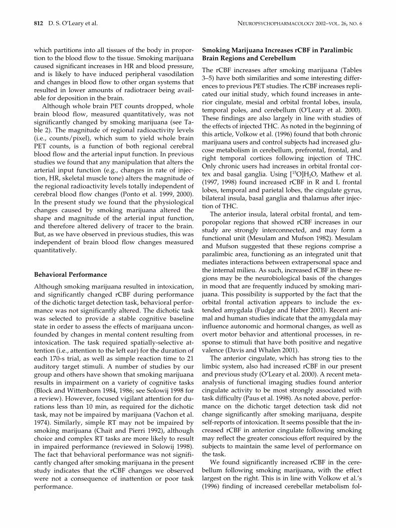
Figure 1. PET t-map image for the pre-smoking minus post-smoking marijuana analysis (in color) overlaid on an averageMR image for the 12 subjects in the study. Subjects were performing the same dichotic task prior to and following smoking.The three columns on the right are axial, sagittal, and coronal views of the unthresholded t-map, with t values ranging from�6.0 (purple) to �6.0 (red) as illustrated on the pallet on the right. The three views in the column on the left represent thesame t-map overlaid on the average MRI, but with the t values thresholded at a value of 3.61 (uncorrected p � .0005). Radio-logical convention is followed and the crosshairs are at the same location in all three views. The axial view shows a region inthe anterior cingulate that has significantly higher t values (i.e., greater rCBF) after smoking marijuana, and regions in left andright superior temporal gyrus and occipital lobe that have lower t values (decreased rCBF) after smoking. The sagittal viewagain illustrates the increase in anterior cingulate rCBF and the decrease in rCBF in occipital lobe after smoking marijuana,and additionally illustrates a region in the inferior cerebellum that has higher rCBF following smoking. The coronal viewsillustrate bilateral regions of the temporal poles, insula and orbital frontal lobe that have significantly higher rCBF after smok-ing marijuana. Note that the regions of increased rCBF in the coronal view are lateral and ventral to the basal ganglia.

NEUROPSYCHOPHARMACOLOGY 2002–VOL. 26, NO. 6 Acute Marijuana Effects 811
Whereas rCBF increases were localized to ventralforebrain and cerebellum, decreases in rCBF were local-ized to brain regions that mediate sensory processingand attention. These findings suggest that it may bepossible to isolate the mood-enhancing effects of mari-juana (rCBF increases in ventral forebrain) from mari-juana’s effects on perception, attention and behavior (de-creased rCBF in sensory regions and attention-relatedbrain systems).
Smoking Marijuana Increases Heartrate and Blood Pressure and Lowers Whole Brain PET Counts
Smoking marijuana increased HR by a mean of 40.6beats/min in the PET condition immediately followingsmoking, with all 12 subjects showing this effect (in-creases ranged from 8 to 82 beats/min). The HR in-crease dropped slowly, remaining significantly ele-vated in the second (27.3 beats/min higher) and third(24.2 beats/min higher) PET conditions followingsmoking (about 35 and 50 min respectively after smok-ing). Diastolic blood pressure also increased after smok-ing marijuana, but the changes were smaller and morevariable across individuals. Smoking marijuana did notresult in a significant change in systolic blood pressure.
A recent review of the cardiovascular effects of mari-juana noted that a dose-related tachycardia is a consis-tent finding in human studies, with increases in HR of30–60% over control rates (Trouve and Nahas 1999).Trouve and Nahous note that marijuana consistentlyproduces peripheral vasodilation in humans, but that
the findings concerning blood pressure are less consis-tent. The most frequent finding is an increase in bloodpressure (which is not seen in all subjects) when thesubject is supine, but significant hypotension when thesubject is upright.
In contrast to the increases found in humans, animalstudies have consistently found evidence of decreasedHR and blood pressure after injection of cannabinoids(for a review see Kunos et al. 2000a,b; Hillard 2000). Itseems unlikely that the difference is due to the effects ofsmoking rather than injecting THC, since we found nosignificant cardiovascular effects in the present studywhen subjects smoked marijuana cigarettes with mostof the THC removed. Animal studies have also foundthat injection of cannabinoids results in peripheral va-sodilation. Endogenous lipidlike substances have beenidentified recently, (e.g., anandamide) that bind to can-nabinoid receptors. It has been found that both plant-derived cannabinoids and the endogenous ligandsproduce decreased HR and hypotension in animals byactivating CB1 receptors in the periphery (Kunos et al.2000a,b). Anandamide has also been shown to cause va-sodilation in mesenteric vascular beds that is indepen-dent of CB1 or CB2 receptors (Kunos et al. 2000b).
The results of the present study are consistent withprevious human studies in finding that smoking mari-juana increases HR dramatically, and causes less consis-tent increases in blood pressure. We also observed asignificant drop in whole brain PET counts in thepresent study, which may be related to the cardiovascu-lar effects of marijuana. [15O]water is a diffusible tracer
Table 6. PET t-Map Analyses of rCBF Differences Obtained by Subtracting the Post-Smoking Placebo Condition from the Post-Smoking Marijuana Conditions
Brain Regions with Significantly Higher rCBF After Smoking Marijuana than Placebo
Region X Y Z t-value Volume2
L. Straight Gyrus �8 22 �8 6.4 9.3R. Insula/Putamen 28 8 �10 5.8 2.7R. Temporal Pole 44 8 �26 6.4 9.1L. Temporal Pole �46 4 �30 5.8 7.3R. Cerebellum 34 �61 �34 5.1 6.9R. Cerebellum 15 �79 �15 4.3 0.9L. Cerebellum �50 �62 �32 4.3 0.3
Brain Regions with Significantly Lower rCBF After Smoking Marijuana than Placebo
L. Sup Temp Gyrus �44 �23 12 �5.0 1.3L. Frontal Lobe (BA8/9) �38 12 27 �4.3 0.3L. Frontal Lobe (BA9) �30 50 5 �5.3 0.8L. Frontal Lobe (BA6) �31 4 45 �3.8 0.2R. Frontal Lobe (BA6) 25 11 44 �4.2 0.7R. Frontal Lobe (BA4/6) 35 �16 50 �4.7 1.2R. Frontal Lobe (BA4) 43 �22 39 �4.1 0.2R. Occipital (BA17) 1 �75 7 �4.2 0.3
812 D. S. O’Leary et al. NEUROPSYCHOPHARMACOLOGY 2002–VOL. 26, NO. 6
which partitions into all tissues of the body in propor-tion to the blood flow to the tissue. Smoking marijuanacaused significant increases in HR and blood pressure,and is likely to have induced peripheral vasodilationand changes in blood flow to other organ systems thatresulted in lower amounts of radiotracer being avail-able for deposition in the brain.
Although whole brain PET counts dropped, wholebrain blood flow, measured quantitatively, was notsignificantly changed by smoking marijuana (see Ta-ble 2). The magnitude of regional radioactivity levels(i.e., counts/pixel), which sum to yield whole brainPET counts, is a function of both regional cerebralblood flow and the arterial input function. In previousstudies we found that any manipulation that alters thearterial input function (e.g., changes in rate of injec-tion, HR, skeletal muscle tone) alters the magnitude ofthe regional radioactivity levels totally independent ofcerebral blood flow changes (Ponto et al. 1999, 2000).In the present study we found that the physiologicalchanges caused by smoking marijuana altered theshape and magnitude of the arterial input function,and therefore altered delivery of tracer to the brain.But, as we have observed in previous studies, this wasindependent of brain blood flow changes measuredquantitatively.
Behavioral Performance
Although smoking marijuana resulted in intoxication,and significantly changed rCBF during performanceof the dichotic target detection task, behavioral perfor-mance was not significantly altered. The dichotic taskwas selected to provide a stable cognitive baselinestate in order to assess the effects of marijuana uncon-founded by changes in mental content resulting fromintoxication. The task required spatially-selective at-tention (i.e., attention to the left ear) for the duration ofeach 170-s trial, as well as simple reaction time to 21auditory target stimuli. A number of studies by ourgroup and others have shown that smoking marijuanaresults in impairment on a variety of cognitive tasks(Block and Wittenborn 1984, 1986; see Solowij 1998 fora review). However, focused vigilant attention for du-rations less than 10 min, as required for the dichotictask, may not be impaired by marijuana (Vachon et al.1974). Similarly, simple RT may not be impaired bysmoking marijuana (Chait and Pierri 1992), althoughchoice and complex RT tasks are more likely to resultin impaired performance (reviewed in Solowij 1998).The fact that behavioral performance was not signifi-cantly changed after smoking marijuana in the presentstudy indicates that the rCBF changes we observedwere not a consequence of inattention or poor taskperformance.
Smoking Marijuana Increases rCBF in Paralimbic Brain Regions and Cerebellum
The rCBF increases after smoking marijuana (Tables3–5) have both similarities and some interesting differ-ences to previous PET studies. The rCBF increases repli-cated our initial study, which found increases in ante-rior cingulate, mesial and orbital frontal lobes, insula,temporal poles, and cerebellum (O’Leary et al. 2000).These findings are also largely in line with studies ofthe effects of injected THC. As noted in the beginning ofthis article, Volkow et al. (1996) found that both chronicmarijuana users and control subjects had increased glu-cose metabolism in cerebellum, prefrontal, frontal, andright temporal cortices following injection of THC.Only chronic users had increases in orbital frontal cor-tex and basal ganglia. Using [15O]H2O, Mathew et al.(1997, 1998) found increased rCBF in R and L frontallobes, temporal and parietal lobes, the cingulate gyrus,bilateral insula, basal ganglia and thalamus after injec-tion of THC.
The anterior insula, lateral orbital frontal, and tem-poropolar regions that showed rCBF increases in ourstudy are strongly interconnected, and may form afunctional unit (Mesulam and Mufson 1982). Mesulamand Mufson suggested that these regions comprise aparalimbic area, functioning as an integrated unit thatmediates interactions between extrapersonal space andthe internal milieu. As such, increased rCBF in these re-gions may be the neurobiological basis of the changesin mood that are frequently induced by smoking mari-juana. This possibility is supported by the fact that theorbital frontal activation appears to include the ex-tended amygdala (Fudge and Haber 2001). Recent ani-mal and human studies indicate that the amygdala mayinfluence autonomic and hormonal changes, as well asovert motor behavior and attentional processes, in re-sponse to stimuli that have both positive and negativevalence (Davis and Whalen 2001).
The anterior cingulate, which has strong ties to thelimbic system, also had increased rCBF in our presentand previous study (O’Leary et al. 2000). A recent meta-analysis of functional imaging studies found anteriorcingulate activity to be most strongly associated withtask difficulty (Paus et al. 1998). As noted above, perfor-mance on the dichotic target detection task did notchange significantly after smoking marijuana, despiteself-reports of intoxication. It seems possible that the in-creased rCBF in anterior cingulate following smokingmay reflect the greater conscious effort required by thesubjects to maintain the same level of performance onthe task.
We found significantly increased rCBF in the cere-bellum following smoking marijuana, with the effectlargest on the right. This is in line with Volkow et al.’s(1996) finding of increased cerebellar metabolism fol-

NEUROPSYCHOPHARMACOLOGY 2002–VOL. 26, NO. 6 Acute Marijuana Effects 813
lowing THC injection. Volkow et al. found that thecerebellar increase had a significant correlation withsubjective ratings of intoxication. In contrast, Mathewet al. (1997) found that ratings of intoxication corre-lated significantly with frontal lobe rCBF in 32 volun-teers. In an expansion of his sample to 46 volunteers,Mathew et al. (1998) found significantly increasedrCBF in the cerebellum, but reported that not all sub-jects showed this effect. Subjects who had decreasedcerebellar rCBF also had a disturbance of time sense.We plan to perform correlational analyses of ratings ofintoxication with rCBF, using an approach that allowsassessment of the correlation of a “seed voxel” or anexternal vector such as intoxication ratings with rCBFin every voxel of the PET image (Friston et al. 1993).Because this technique has relatively low power withsmall numbers of subjects, however, we plan to per-form this analysis after the completion of a study thatis currently underway, which will double the numberof available subjects.
Smoking Marijuana Does Not Increase rCBF in Basal Ganglia, Nucleus Accumbens, and Hippocampus
The major discrepancy between our findings and thosefrom other laboratories involve the basal ganglia. PETstudies using injected THC have found significant in-creases in basal ganglia rCBF, whereas our data showsrCBF increases that are ventral and lateral to the basalganglia. It is also possible that injected THC has differ-ent effects on brain metabolism and blood flow thandoes smoked marijuana. However, another explanationfor the divergent findings is that the studies by Volkowet al. (1996) and Mathew et al. (1992) utilized ROIsbased upon predefined templates that averaged activityover relatively large volumes of tissue. This could haveresulted in activations near to, but outside of, basal gan-glia being averaged into basal ganglia ROIs.
The present study utilized individually co-registeredPET and high-resolution MR images that permittedmore precise anatomical localization than in previousstudies. AIR software (Woods et al. 1992) was used toregister each of the eight PET conditions for each indi-vidual to their MR image. Landmarks on the MRI werethen used to place all images into a standardized stereo-taxic atlas space (Talairach and Tournoux 1988), whichallowed averaging of both PET and MR image sets. Thelenticular nuclei can be visualized on the average MRI,and, as seen in Figure 1, significant PET activations areventral and lateral to these structures. Although the nu-cleus accumbens is a small structure that is difficult tovisualize with MRI, its general location can be found onthe average MRI. Significant areas of activation were lat-eral to this, suggesting that marijuana did not increaserCBF significantly in “reward-related” brain regions.
The hippocampi as well as the basal ganglia containa high density of cannabinoid receptors, and also failedto show significant rCBF changes following smoking ofmarijuana. Inspection of the PET t-map images in themesial temporal lobe, aided by the co-registered MRimage, showed no region close to the hippocamal for-mation with significantly increased or decreased rCBF.This lack of rCBF change in brain regions with highdensities of cannabinoid receptors indicates that thelarge blood flow changes observed in the present studyresult from increased synaptic activity downstreamfrom the receptor binding sites. Our failure to find rCBFchanges in brain regions rich in cannabinoid receptors(except for cerebellum) indicates that the immediatemetabolic effects of THC binding to cannabinoid recep-tors are relatively brief.
It remains controversial whether marijuana is anatypical or anomalous addictive drug, which interactswith brain reward systems differently than drugssuch as methamphetamine that directly activate brainDA systems (e.g., Gardner and Vorel 1998). Our find-ing of no rCBF changes in the nucleus accumbens andbasal ganglia may be taken as support for the positionthat smoking marijuana does not directly activate re-ward-relevant DA neurons in the nucleus accumbensand/or basal ganglia. However, because the subjectshad to be repositioned in the PET camera after smok-ing, our rCBF measures occurred approximately 15min after smoking marijuana, which may have beentoo late to observe the direct effects of THC binding toreceptors. It is also possible that our technique, whichassessed rCBF changes using subtraction analysis andstatistical mapping, may not have been sensitive tosubtle changes in metabolism in reward-related brainregions.
Smoking Marijuana Decreases rCBF in Sensory Cortices and in Attention-Related Brain Regions
Smoking marijuana did not significantly change perfor-mance on the dichotic listening task but did result indecreased rCBF in auditory processing regions of thetemporal lobes that had been activated by the dichotictask prior to smoking. As can be seen in Table 4, sub-jects performing the dichotic listening task had lowerrCBF in Heschl’s gyrus in the temporal lobe after smok-ing marijuana than they did prior to smoking. It is im-portant to note that smoking marijuana did not result indecreased rCBF in Heschl’s gyrus in comparison to thepre-smoking baseline condition (see Table 3). Rather,the effect of marijuana was to decrease the magnitudeand extent of the activation resulting from the dichotictask. The regions showing decreased rCBF containsboth primary and secondary auditory cortex (Liegeois-Chauvel, Musolino and Chauvel 1991). Thus the effectof smoking marijuana was to eliminate the task-related

814 D. S. O’Leary et al. NEUROPSYCHOPHARMACOLOGY 2002–VOL. 26, NO. 6
activation of auditory processing regions that is nor-mally caused by the dichotic task.
In previous studies of dichotic listening we havefound that activation in STG reflects attentional pro-cessing in normal volunteers, and we have found ab-normal activation patterns in STG in subject popula-tions that may have attentional impairment (seeO’Leary, in press for a review). In normal volunteers,attention to the right ear increased the spatial extentand magnitude of rCBF to a greater degree in left STG,which was contralateral to the direction of attention,than in right STG (O’Leary et al. 1996a). Attending tothe left ear reversed this asymmetry, with greater rCBFincreases in right than left STG. This finding has beenreplicated in normal volunteers in two other studiesperformed by our group (Hurtig et al. unpublishedmanuscript; Block et al. 2000). In the present study wefound that both left and right STG showed significantactivation in the pre-smoking baseline minus pre-smok-ing dichotic analysis (see Table 2). In contrast to ourprevious studies using an attend-left condition, left STGhad a larger t-max (t � 10.0 vs. 9.0) and greater volume(16.7 vs. 11.6 cc) of activation than the right STG. Theactivation observed in both left and right STG due tothe pre-smoking dichotic listening task in the presentstudy was larger than in previous studies, and differ-ences in the characteristics of this subject sample mayexplain the differences in rCBF asymmetry. We werenot able to observe changes in rCBF asymmetry due tothe direction of attention because attend right condi-tions were not included in the present study. This com-parison is included in a companion study currently un-derway in our laboratory.
A group of individuals with schizophrenia assessedin a previous study had similar rCBF patterns in STG toa volunteer group when attending to the right ear, butfailed to show the normal change in rCBF asymmetrywhen attending to the left ear (O’Leary et al. 1996b).That is, the patient group maintained a left greater thanright asymmetry regardless of where attention was di-rected. We recently found that chronic users of mari-juana (minimum use of more than 7 times weekly formore than 2 years) also had an abnormal pattern ofasymmetry when attending to the left ear (Block et al.2000). The subjects were tested after 26 h of monitoredabstinence using the same baseline dichotic stimuliused in the present study, but with both attend left andattend right conditions. The chronic marijuana users re-sembled schizophrenics in showing greater left thanright STG activation when attention was directed to ei-ther the left or right ears. The abnormal pattern of acti-vation observed in both populations may reflect an in-ability to voluntarily activate the right hemisphereduring the more difficult attend-left condition and tothereby reverse the normal left hemisphere advantagefor linguistic stimuli.
As can be seen in Table 4, smoking marijuana de-creased rCBF bilaterally in the precentral sulcas/gyrus(i.e. motor strip), and in left parietal lobe in the vicinityof the intraparietal sulcas. Chelazzi and Corbetta (2000)recently reviewed PET imaging studies in which sub-jects covertly directed attention to visual stimuli in pe-ripheral locations. Regions that were found to showconsistent attention-related activation included the pre-central sulcus/gyrus, and areas of parietal cortex (thepostcentral and intraparietal sulcus) that showed de-creased blood flow in the present study. Bushara et al.(1999) assessed the frontal and parietal regions thatwere activated during auditory and visual spatial local-ization tasks. They found regions in the frontal and pa-rietal lobes that were uniquely activated during audi-tory and visual tasks as well as “supra-modal” regionsthat responded to both modalities. A previous study byour group (O’Leary et al. 1997) contrasted attention toauditory and to visual stimuli. A subtraction of a rest-ing baseline condition from averaged attend left and at-tend right dichotic conditions revealed activations inright parietal lobe (BA 7), left frontal lobe (BA 4), rightinsula, and right cerebellum, but the network activatedby auditory attention was much less extensive than thatactivated by visual attention. Thus, smoking marijuanadecreased rCBF in frontal and parietal regions that havebeen found to play a role in the attentional enhance-ment of sensory processing, which may explain thelower rCBF observed in auditory cortices in the dichoticconditions following smoking.
ACKNOWLEDGMENTS
This research was supported in part by the National Insti-tute of Drug Abuse Grant DA10551 and by MHCRC43271.
REFERENCES
Andreasen NC, Cizadlo T, Harris G, Swayze V, O’Leary DS,Cohen G, Ehrhardt J, Yuh WTC (1993): Voxel process-ing techniques for the antemortem study of neuroanat-omy and neuropathology using magnetic resonanceimaging. J Neuropsychiatry Clin Neurosci 5:121–130
Biegon A, Kerman I (1995): Quantitiative autoradiographyof cannabinoid receptors in the human brain post mor-tem. In Biegon A, Volkow (eds), Sites of Drug Action inthe Human Brain. Boca Raton, FL, CRC Press, pp 65–74
Block RI, Farinpour R, Braverman K (1992): Effects of mari-juana smoking on cognition and their relationship tosmoking technique. Pharmacology Biochemistry &Behavior 43:907–917
Block RI, Wittenborn JR (1984): Marijuana effects on seman-tic memory: verification of common and uncommoncategory members. Psychol Rep 55:503–512
Block RI, O’Leary DS, Augustinack JC, Boles Ponto LL, Gho-neim MM, Hurtig RR, Hall JA, Nathan PE (2000): Effects

NEUROPSYCHOPHARMACOLOGY 2002–VOL. 26, NO. 6 Acute Marijuana Effects 815
of frequent marijuana use on attention-related regionalcerebral blood flow. Society for Society for Neuro-science Abstracts 26(Part 2):2080
Block RI, Wittenborn JR (1986): Marijuana effects on speedof memory retrieval on a letter matching task. Int JAddict 21:281–285
Bushara KO, Weeks RA, Ishii K, Catalan MJ, Tian B, Raus-checker JP, Hallett M (1999): Modality-specific frontaland parietal areas for auditory and visual spatial local-ization in humans. Nat Neurosci 2:759–766
Chait LD, Pierri J (1992): Effects of smoked marijuana onhuman performance: a critical review. In Murphy L,Bartke A (eds), Marijuana/Cannabinoids: Neurobiol-ogy and Neurophysiology. Boca Raton, CRC Press, pp387–423
Chelazzi L, Corbetta M (2000): Cortical mechanisms of visu-ospatial attention in the primate brain. In M Gazziniga(ed), The New Cognitive Neurosciences. 2nd ed. Cam-bridge, MA, MIT Press, pp 648–663
Cizadlo T, Andreasen NC, Zeien G, Rajarethinam R, HarrisG, O’Leary DS, Swayze VW, Arndt S, Hichwa R,Ehrhardt J, Yuh WTC (1994): Image registration issuesin the analysis of multiple-injection 150H20 PET studies:BRAINFIT. Proceedings from SPIE–The InternationalSociety for Optical Engineering 2168:234–245
Davis M, Whalen PJ (2001): The amygdala: Vigilance andemotion. Mol Psychiatry 6:13–34
Friston KJ, Frith CD, Frackowiak RSJ (1993). Time-depen-dent changes in effective connectivity measured withPET. Human Brain Mapping 1:69–79
Fudge HL, Haber JN (2001): Bed nucleus of the stria termi-nalis and extended amygdala inputs to dopamine sub-populations in primates. Neuroscience 104:807–827
Gardner EL, Vorel SR (1998): Cannabinoid transmission andreward-related events. Neurobiol Dis 5:502–533
Harvey DJ (1999): Absorption, distribution and biotransfor-mation of the cannabinoids. In Nahas GG, Sutin KM,Harvey DJ, Agurell S (eds), Marijuana and Medicine.Totawa NJ, Humana Press, pp 91–103
Heishman SJ, Huertis MA, Henningfield JE, Cone EJ (1990):Acute and residual effects of marijuana: profiles ofplasma THC levels, physiological, subjective, and per-formance measures. Pharmacology Biochemistry andBehavior 37:561–565
Herkenham M, Lynn AB, Little MD, Johnson MR, MelvinLS, De Costa BR, Rice KC (1990): Cannabinoid receptorlocalization in brain. Proceedings of the National Acad-emy of Sciences 87:1932–1936
Herkenham M (1992): Cannabinoid receptor localization inbrain: relationship to motor and reward systems. AnnNY Acad Sci 654:19–32
Herscovitch, P, Markham J, Raichle ME (1983): Brain bloodflow measured with intravenous H215O. I. Theory anderror analysis. Journal of Nuclear Medicine 24:782–789
Hichwa RD, Ponto LLB, Watkins GL (1995): Clinical bloodflow measurements with [15O]water and positron emis-sion tomography (PET). In Emran AM (ed), SymposiumProceedings of the International Symposium on“Chemists’ Views on Imaging Centers.” New York, Ple-num, pp 401–417
Hillard CJ (2000): Endocannabinoids and vascular function.J Pharmacol Exp Ther 294:27–32
Huestis M (1999): Pharmacokinetics of THC in inhaled andoral preparations. In Nahas GG, Sutin KM, Harvey DJ,Agurell S (eds), Marijuana and Medicine, Totawa, NJ,Humana Press pp105–116.
Hugdahl K, Bronnick K, Kyllingsbaek S, Law I, Gade A,Paulsen OB (1999): Brain activation during dichotic pre-sentations of consonant-vowel and musical instrumentstimuli: A 15O-PET study. Neuropsychologia 37:431–440
Hugdahl K, Law I, Kyllingsbaek S, Bronnick K, Gade A,Paulson OB (2000): Effects of attention on dichotic lis-tening: An 15O-PET study. Hum Brain Mapp 10:87–97
Hurtig RR, Hichwa RD, O’Leary DS, Boles Ponto LL, Naray-ana S, Watkins GL, Andreasen NC (1994): Effects of tim-ing and duration of cognitive activation in [15O]waterPET studies. J Cereb Blood Flow Metab 14:423–430
Johnston LD, O’Malley PM, Bachman JG (1999): Drug trendsin 1999 are mixed. Monitoring the Future Study. Uni-versity of Michigan News and Information Services
Kunos G, Jarai Z, Batkai S, Goparaju SK, Ishac EJ, Liu J,Wang L, Wagner JA (2000a): Endocannabinoids as car-diovascular modulators. Chem Phys Lipids 108:159–168
Kunos G, Jarai Z, Varga K, Liu J, Wang L, Wagner JA (2000b):Cardiovascular effects of endocannabinoids–the plotthickens. Prostaglandins Other Lipid Mediat 61:71–84
Liegeois-Chauvel C, Musolino A, Chauvel P (1991): Local-ization of the primary auditory area in man. Brain114:139–153
Loeber RT, Yurgelun-Todd DA (1999): Human neuroimag-ing of acute and chronic marijuana use: implications forfrontocerebellar dysfunction. Human Psychopharma-cology ClinExp 14:291–304
Mathew RJ, Wilson WH, Tant SR (1989): Acute changes incerebral blood flow associated with marijuana smoking.Acta Psychiatr Scand 79:118–128
Mathew RJ, Wilson WH, Humphreys DF, Lowe JV, WietheKE (1992): Regional cerebral blood flow after marijuanasmoking. J Cereb Blood Flow Metab 12:750–758
Mathew RJ, Wilson WH, Coleman RE, Turkington TG,DeGrado TR (1997): Marijuana intoxication and brainactivation in marijuana smokers. Life Sci 60:2075–2089
Mathew RJ, Wilson WH, Turkington TG, Coleman RE(1998): Cerebellar activity and disturbed time senseafter THC. Brain Res 797(2):183–189
Mesulam, M-M, Mufson, E.J. (1982): Insula of the old worldmonkey. III: Efferent cortical output and comments onfunction. J Comp Neurolog 212: 38–52
O’Leary DS. Effects of attention on hemispheric asymmetry.In Hugdahl K, Davidson R (eds), Brain Asymmetry,Cambridge, MA, MIT Press (in press)
O’Leary DS, Andreasen NC, Hurtig RR, Hichwa RD, Wat-kins GL, Boles Ponto LL, Rogers M, Kirchner PT(1996a): A regional cerebral blood flow study of lan-guage and auditory attention. Brain Lang 53:20–39
O’Leary DS, Andreasen NC, Hurtig RR, Kesler ML, RogersM, Arndt S, Cizadlo T, Watkins GL, Ponto LLB, KirchnerPT, Hichwa RD (1996b): Auditory attentinal deficits inpatients with schizophrenia: a positron emission tomog-raphy (PET) sutdy. Arc Gen Psychiatry 53:633–641

816 D. S. O’Leary et al. NEUROPSYCHOPHARMACOLOGY 2002–VOL. 26, NO. 6
O’Leary DS, Andreasen NC, Hurtig RR, Torres IJ, FlashmanLA, Kesler ML, Ponto LLB, Watkins GL, Hichwa RD(1997): Auditory and visual attention assessed withPET. Hum Brain Mapp 5:422–436
O’Leary DS, Block RI, Flaum M, Schultz SK, Boles Ponto LL,Watkins GL, Hurtig RR, Andreasen NC, Hichwa RD(2000): Acute marijuana effects on rCBF and cognition:A PET study. Neuroreport 11:1–7
Paus T, Koski L, Caramanos Z, Westbury C (1998): Regionaldifferences in the effects of task difficulty and motoroutput on blood flow response in the human anteriorcingulate cortex: a review of 107 PET activation studies.Neuroreport 9:R37–47
Pertwee RG (1997): Pharmacology of cannabinoid CB1 andCB2 receptors. Pharmacol Ther 74:129–180
Ponto LLB, Popowski LA, Gisolfi CV, Johnson AK, MadsenMT, Watkins GL, Hichwa RD, Bushnell D (1999): Influ-ence of hydration state and cognitive performance onthe CNS deposition of freely-diffusible substances.AAPS PharmSci (supplement) 1:S-123
Ponto LLB, Narayana S, Grabowski TJ (2000): Pharmacoki-netic examination of the interaction between skeletalmuscle blood flow and CNS deposition of freely-diffus-ible substances. AAPSPharmSci [serial on the internet].2(4): electronic. SAMHSA, National Household Surveyon Drug Abuse (1998, 1999)
SAMHSA (1998, 1999): National Household Survey on DrugAbuse, Rockville, MD, Author
Solowij N (1998): Cannabis and Cognitive Functioning,Cambridge, Cambridge University Press
Talairach J, Tournoux P (1988): Co-planar Stereotaxic Atlasof the Human Brain: 3-D Proportional System: AnApproach to Cerebral Imaging. New York, Thieme
Trouve R, Nahas G (1999): Caardiovascular effects of mari-juana and cannabinoids. In Nahas GG, Sutin KM, Har-vey DJ, Agurel S (eds), Marijuana and Medicine.Totowa, NJ, Humana Press, pp 291–304
Vachon L, Sulkowski A, Rich E (1974): Marijuana effects onlearning, attention, and time estimation. Psychophar-macologicia 39:1–11
Volkow ND, Gillespie H, Mullani M, Tancredi L, Hollister L,Ivanovic M, Grant C (1991a): Use of positron emissiontomography to investigate the action of marijuana inthe human brain. Adv Biosci 80:3–11
Volkow ND, Gillespie H, Mullani N, Tancredi L, Grant C,Ivanovic M, Hollister L (1991b): Cerebellar metabolicactivation by delta-9-tetrahydrocannabinol in humanbrain: A study with positron emission tomography and18F–2-fluorof-2-deoxyglucose. Psychiatry Research.Neuroimaging 40:69–78
Volkow ND, Gillespie H, Mullani N, Tancredi L, Grant C,Valentine A, Hollister L (1996): Brain glucose metabo-lism in chronic marijuana users at baseline and duringmarijuana intoxication. Psychiatry Research: Neuroim-aging 67:29–38
Wollenweber SD, Hichwa RD, Ponto LLB (1997): A simpleon-line arterial time-activity curve detector for [O-15]water studies (1997). IEEE Trans Nuclear Science 44:1613–1617
Woods RP, Cherry SR, Mazziotta JC (1992): Rapid auto-mated algorithm for aligning and reslicing PET images.J Comput Assist Tomogr 16:620–633
Worsley K, Evans A, Marret S, Neelin P (1992): A threedimensional statistical analysis for CBF activation stud-ies in the human brain. J Cereb Blood Flow Metab12:900–918