efficient degradation of the organic uv filter
TRANSCRIPT

HAL Id: hal-03217024https://hal.archives-ouvertes.fr/hal-03217024
Submitted on 4 May 2021
HAL is a multi-disciplinary open accessarchive for the deposit and dissemination of sci-entific research documents, whether they are pub-lished or not. The documents may come fromteaching and research institutions in France orabroad, or from public or private research centers.
L’archive ouverte pluridisciplinaire HAL, estdestinée au dépôt et à la diffusion de documentsscientifiques de niveau recherche, publiés ou non,émanant des établissements d’enseignement et derecherche français ou étrangers, des laboratoirespublics ou privés.
Efficient degradation of the organic UV filterbenzophenone-3 by Sphingomonas wittichii strain
BP14P isolated from WWTP sludgeSonja Fagervold, Clémence Rohée, Alice Rodrigues, Didier Stien, Philippe
Lebaron
To cite this version:Sonja Fagervold, Clémence Rohée, Alice Rodrigues, Didier Stien, Philippe Lebaron. Efficientdegradation of the organic UV filter benzophenone-3 by Sphingomonas wittichii strain BP14P iso-lated from WWTP sludge. Science of the Total Environment, Elsevier, 2021, 758, pp.143674.�10.1016/j.scitotenv.2020.143674�. �hal-03217024�
1
Efficient degradation of the organic UV filter benzophenone-3 by Sphingomonas 1
wittichii strain BP14P isolated from WWTP sludge 2
3
S. K. Fagervold1*, C. Rohée2, A. M. S. Rodrigues1, D. Stien1 and P. Lebaron1 4
5
1 Sorbonne Université, CNRS, Laboratoire de Biodiversité et Biotechnologies 6
Microbiennes, LBBM, Observatoire Océanologique, 66650 Banyuls-sur-mer, France 7
2 Pierre Fabre Dermo-Cosmétique, Centre de Recherche & Développement Pierre 8
Fabre, 31000 Toulouse 9
10
* Corresponding author: Sonja K. Fagervold. [email protected]. 11
Address: Observatoire Océanologique de Banyuls, Laboratoire de Biodiversité et 12
Biotechnologies Microbiennes, Avenue Pierre Fabre, 66650 Banyuls/mer, France 13
14
15
Abstract 16
Benzophenone-3 (BP3) is a widely used organic UV filter present in many 17
environmental compartments. One way BP3 is released into the environment is 18
through effluents from wastewater treatment plants (WWTPs). These plants are 19
possible sources for degradation activity as WWTP sludge can potentially degrade 20
BP3. Our goal was to identify the BP3 degrading microorganism(s) in WWTP sludge 21
and to investigate whether the degradation was co-metabolic. Initial WWTP sludge 22
microcosms spiked with BP3 showed 100% degradation after 20 days. Multiple 23

2
transfers of these microcosms, while maintaining a strong selective pressure for 24
BP3 degradation capabilities, resulted in the dominance of one bacterial strain. This 25
strain, Sphingomonas wittichii BP14P was subsequently isolated and shown to 26
degrade BP3 in a growth dependent manner. Strain BP14P utilized BP3 as the sole 27
energy and carbon source and completely degraded BP3 after 7 days in minimal 28
media. We tested the capability of BP14P to degrade nine other UV filters, but the 29
degradation ability seemed to be restricted to BP3. However, whether this 30
specificity is due to the lack of degradation genes, cellular transport or low 31
bioavailability of the other UV filters remained unclear. The efficient degradation of 32
BP3 by a group of bacteria well known for their potential for xenobiotic degradation 33
is an important step forward for a complete risk assessment of the long-term 34
environmental impact of BP3. 35
36
Keywords: Sunscreens, BP3 degradation, growth-dependent, Sphingomonas 37
wittichii 38
39
1. Introduction 40
Benzophenone-3 (BP3) is an organic UV filter commonly used in sunscreen 41
formulations and other cosmetic products. Due to its widespread use, BP3 is now 42
found in many environmental compartments such as freshwater and seawater, 43
benthic sediments and aquatic biota (see Mao et al. (2018) for a recent review). 44
Further, BP3 has been found in bird eggs (Molins-Delgado et al., 2017) and even in 45
human breast tissue (Barr et al., 2018) suggesting that BP3 can biomagnify. Also, it 46

3
has been demonstrated that BP3 can be absorbed through the skin but the effects of 47
elevated systemic BP3 concentrations in humans are still unclear (Suh et al., 2020). 48
BP3 has been shown to be toxic in several species used for toxicological testing 49
including fish, corals and algae (see recent review and references therein (Lozano et 50
al., 2020)). Thus, the toxicity of BP3 combined with the possibility of 51
bioaccumulation indicates that BP3 might pose an ecological risk (Díaz-Cruz et al., 52
2019). 53
54
Wastewater treatment plants (WWTPs) are a major source of BP3 to the 55
environment, in addition to recreational water activities and industrial wastewater 56
discharges (Mao et al., 2018). Indeed, BP3 has been found in influents and effluents 57
of WWTPs (Ramos et al., 2016; Mao et al., 2018) in relatively high concentrations. 58
Often, the concentrations of BP3 are lower in the effluents of WWTPs than the 59
influents. This difference can be attributed to sorption to activated sludge and/or 60
biodegradation. 61
62
Biodegradation of BP3 has been shown to occur in different systems. Liu and 63
collegues observed degradation of BP3 in activated sludge microcosms (Liu et al., 64
2012) and in microcosms using aquifer materials as an innoculum (Liu et al., 2013). 65
The data from this latter study was used as input in a model to better understand 66
the fate of UV filters, including BP3, in aquifers (Rodríguez-Escales and Sanchez-Vila, 67
2020). Among others, this model took into account sorption processses, both to 68
sediments and biomass, but also assumed that the UV-filters were degraded in a co-69

4
metabolic manner with the oxidation of sediment organic matter as the main 70
process. 71
72
The microorganisms responsible for the degradation in the studies described above 73
were not identified (Liu et al., 2012; Liu et al., 2013). So far the only bacterial strain 74
shown to be capable of BP3 degradation is Methyliphilus sp. strain FP-6, which 75
degraded BP3 co-metabolically when grown with several other carbon substrates 76
(Jin et al., 2019). The white root fungus Trametes versicolor can also degrade BP3 77
(Badia-Fabregat et al., 2012; Gago-Ferrero et al., 2012) but it is not known if this 78
degradation was a co-metabolic process since dried and sterilized sludge was used 79
as solid support in these experiments. 80
81
The objective of our study was to investigate the potential degradation of BP3 in 82
microcosms with WWTP sludge, to determine whether the BP3 degradation is 83
indeed co-metabolic and whether any bacterial species exist that can completely 84
mineralize BP3. 85
86
87
2. Materials and methods. 88
89
2.1 Chemicals 90
The UV filters used for biodegradation studies, 2-hydroxy-4-methoxybenzophenone 91
(BP3) and methylene bis-benzotriazolyl tetramethylbutylphenol (MBBT) were 92
5
purchased from Sigma-Aldrich (Lyon, France), while 2-ethylhexyl salicylate (ES), 93
homosalate (HS), butyl methoxydibenzoylmethane (BM), octocrylene (OC), 94
diethylamino hydroxybenzoyl hexyl benzoate (DHHB), diethylhexyl butamido 95
triazone (DBT), and ethylhexyl triazone (ET) were kindly provided to us by Pierre 96
Fabre Pharmaceuticals. Analytical standards for the above-mentioned UV filters 97
were purchased from Sigma-Aldrich (Saint-Quentin-Fallavier, France). Analytical-98
grade dichloromethane (DCM), 1-methyl-2-pyrrolidinone (NMP), methanol, formic 99
acid (98 %) and molecular grade formaldehyde were obtained from Sigma-Aldrich. 100
Pure water was obtained from an Elga Purelab Flex System (Veolia LabWater STI, 101
Antony, France). Glassware was cleaned with DCM and calcinated at 450 °C for 2 102
hours to remove all traces of organic matter. 103
104
2.2 Sampling of sludge, initial culture and enrichment culture setup 105
The sludge was sampled from the Argelès-sur-mer (France) wastewater treatment 106
plant (WWTP) on August 24th, 2018, from the bottom of a clarifying tank. The 107
sludge was collected in glass mason jars and stored at 4 °C until utilized for 108
degradation experiments. The sludge contained 2765 (+/- 310) mg/L total 109
suspended solids (TSS) and the initial BP3 concentration was below the detection 110
limit for our analysis (4.6 ng/g, see Fagervold et al. (2019)). For the initial 111
degradation experiments, 100 mL sludge was mixed with 500 mL freshwater 112
mineral media (OECD301) in a 1-L glass bottle (live). BP3 was added in methanol (2 113
mg/mL) to a final concentration of 10 µg/ml. A sterile control (SC) was prepared by 114
autoclaving twice (48 hours apart) a 1-L bottle containing sludge and media at 120 115

6
°C for 20 minutes, followed by the addition of sodium azide (1 %) and then BP3. The 116
two bottles (one live and one SC) were incubated at 25 °C in the dark and were 117
systematically shaken and opened inside a fume hood twice per week to allow 118
aeration. 119
120
An enrichment culture was initiated with inoculum from the initial flask that 121
showed degradation. The final theoretical BP3 concentration was about 10 µg/mL 122
for Enrichment 1 (the first transfer), but this concentration was increased to a 123
theoretical concentration of 100 µg/mL for Enrichment 2 and subsequent 124
enrichments. To obtain a concentration of 100 µg/mL, 2 mL of a solution of BP3 in 125
acetone (2.5 mg/mL) was added to 100-mL Erlenmeyer flasks containing 2 g of inert 126
sand. The acetone was left to evaporate overnight. Mineral media (50 mL, as above) 127
was added and the flasks were autoclaved. Each month, the cultures were 128
transferred (1% vol/vol) into new flasks with sand and BP3 (see Figure 1 for 129
diagram of the experimental setup). The cultures were incubated at 25 °C in the 130
dark on a platform shaker at 100 rpm. 131
132
Fig. 1. Schematic of the experimental design. 133

7
Table 1 134 Selected chemical properties and structure of the UV filters used in this study 135 (ordered by increasing molecular weight). Note the abbreviations that are used 136 throughout. 137 138
139 140
Compound name (abbreviation) [CAS number]
MW Biodegradation ratea
Freshwater solubility at
25 °C (mg/L)b
LogPc Chemical structure
Benzophenone-3 (BP3) [131-57-7]
228.2 100 % 130 3.514
2-Ethylhexyl salicylate (ES)
[118-60-5] 250.3 15 %
28 % with R2B or BP3
137 5.335
Homosalate (HS) [118-56-9] 262.3 18 % with R2B
or BP3
< LOD (LOD = 0.85)
5.431
Butyl
methoxydibenzoylmethane (BM) [70356-09-1]
310.4 n.d.
19 5.391
Octocrylene (OC)
[6197-30-4] 361.5 n.d. 9.1 7.083
Diethylamino
hydroxybenzoyl hexyl benzoate (DHHB)
[302776-68-7]
397.5 n.d.
Not measured
7.078
bis-Ethylhexyloxyphenol methoxyphenyl triazine (BEMT) [187393-00-6]
627.8 n.d. 0.3 10.627
Methylene bis-benzotriazolyl
tetramethylbutylphenol (MBBT) [103597-45-1]
658.9 n.d.
< LOD (LOD = 0.04)
15.451
Diethylhexyl butamido
triazone (DBT) [154702-15-5]
766.0 n.d.
0.2 12.004
Ethylhexyl triazone (ET)
[88122-99-0]
823.1 n.d.
Not measured
14.589
a Degradation rate at day 14, when no degradation is seen in SC, and when p < 0.05 compared to SC with a Student t-test. n.d. stands for no degradation. b Experimental data. See protocol in Fagervold et al. (2019). LOD: Limit of detection. c According to Reaxys database (https://www.reaxys.com/, accessed on June 9, 2020)

8
2.3 Biodegradation assays 141
Biodegradation assays were conducted using 15-mL glass tubes with Teflon lined 142
caps (Pyrex, Analytic lab, France). The tubes were set up as for the enrichment 143
cultures, but with 0.2 g sand, 3 mL media, and care was taken so that for each batch 144
of degradation assays, the exact same amount of BP3 (around 100 µg/mL) was 145
added to the tubes. Three tubes were sacrificed for each time-point. The tubes were 146
incubated at 25 °C after inoculation. For the enrichment cultures, the inoculum for 147
the assay was 150 µL of supernatant after rigorous mixing of the enrichment 148
culture. Inoculum preparation for the biodegradation assay of strain BP14P was 149
done as follows: The strain was grown in 100 % R2B broth (Thermo Fisher 150
Scientific, Illkirch-Graffenstaden, France) for about 40 h until the culture had 151
reached an optical density at 600nm of approximately 1.0. The cell suspension was 152
centrifuged at 4200 g for 10 min and subsequently washed 2 times with minimal 153
media before a final resuspension in minimal media (4% v/v). The cultures were 154
incubated at 25 °C in the dark on a rotary shaker at 100 rpm. 155
156
2.4 BP3 extraction 157
Two different protocols were used for the extraction of BP3, one for the initial 158
biodegradation experiments and another for the biodegradation assays for 159
enrichment cultures and isolated strains. 160
161
For the initial cultures containing WWTP sludge, 5 mL slurry was taken in duplicate 162
from each flask for each timepoint, both live and SC. The slurry was centrifuged (950 163

9
x g for 20 min) to separate the aqueous and solid phases. The aqueous and solid 164
phases were then extracted and analyzed separately. The supernatant (aqueous 165
phase) was transferred to a new tube and subjected to liquid/liquid extraction with 166
acidified (0.1 % formic acid) DCM. This was achieved by adding 5 mL acidified DCM 167
to the aqueous sample, mixing by vortexing and shaking overnight at 25 °C in the 168
dark. The DCM fraction was decanted off and dehydrated with Na2SO4. The DCM was 169
then evaporated using the HT-4X system (Genevac, Biopharma Technologies, 170
Diemoz, France) and the residue was dissolved in 1 mL of NMP/water (8:2, v/v) 171
before analysis. 172
173
The solid phase was extracted as described by Fagervold and coworkers (Fagervold 174
et al., 2019) with modifications. Briefly, the pellet was frozen at –80 °C, lyophilized 175
and extracted twice with 2 ml acidified (0.1% formic acid) DCM/methanol mix (1:1, 176
vol/vol) and sonicated for 10 min. The supernatants were combined, evaporated 177
and re-dissolved in NMP/water (8:2, v/v) before HPLC analysis. 178
179
For the biodegradation assays, the whole tube containing sand and liquid media was 180
sacrificed for extraction at each timepoint. DCM (3 mL) containing an exact known 181
amount of MBBT (internal standard) was added to each tube. The tubes were mixed 182
by vortexing twice for 10 seconds, 1 h apart and shaken overnight at 100 rpm at 25 183
°C in the dark. After a brief sonication, 1 mL of the DCM phase was transferred to a 184
HPLC vial and diluted 20X in DCM before HPLC analysis. 185
186

10
2.5 HPLC analysis 187
The UV filters were analyzed using an Ultimate 3000TM HPLC system, equipped with 188
a DAD detector (Thermo Fisher Scientific) and a Phenomenex Kinetex Biphenyl 2.6 189
µm, 150 × 4.6 mm column as previously described (Fagervold et al., 2019) with 190
some minor modifications. The injection volume was 5 µl and the samples were 191
diluted in 100% DCM. We expanded the analysis described in Fagervold et al. 192
(2019) to include all targeted UV-filters. The calibration curves and retention times 193
of all the different UV filters (Table 1) were determined as described by Fagervold 194
and coworkers (Fagervold et al., 2019). The recovery rate was above 85 % for the 195
extraction of BP3 in the initial sludge microcosms. The recovery rate for the 196
degradation assays were variable for the different UV filters, ranging from 63 % for 197
ES to 93 % for OC. However, the extraction efficiency was repeatable for each UV 198
filter over time, which is the most important factor for the detection of degradation 199
activity. 200
201
For determination of possible BP3 degradation products, we also performed a 202
UHPLC-MS/MS analyses as described by Stien and coworkers (Stien et al., 2019). 203
The data were acquired using the Trace FinderTM 3.1 software and were processed 204
with FreeStyleTM and Compound DiscovererTM (all from Thermo Fisher Scientific). 205
206
2.6 Microbial community analysis of enrichment cultures 207
DNA was extracted using a Maxwell® 16 LEV Blood DNA Kit (Promega, 208
Charbonnières-les-Bains, France) with the Maxwell® 16 MDx Instrument (AS3000; 209
11
Promega) following the manufacturer’s instructions and with an initial lysing step. 210
Briefly, 2 mL of supernatant was transferred to a microcentrifuge tube and 211
centrifuged at 14 500 x g for 10 minutes. The pellet was resuspended in 300 µL 212
MilliQ water and 300 mL of the lysing buffer from the Maxwell® 16 LEV Blood DNA 213
Kit was added to “lysing matrix B beads” (MP Biochemicals, Illkirch, France) and 214
homogenized for 20 s at 6 m/s on a FastPrep-24™ 5G Instrument (MP biochemicals). 215
After extraction, the DNA was stored at –80 °C until further analysis. 216
The composition and diversity of the bacterial communities in enrichment cultures 217
were determined by Automated Ribosomal Intergenic Spacer Analysis (ARISA) and 218
by Illumina sequencing (RTL genomics, Texas, USA). The ARISA was performed as 219
described by Fisher et al. (Fisher MM and Triplett EW, 1999) with minor 220
modifications. Briefly, for the PCR we utilized the intergenic spacer primers 1406F 221
and 23SRY (Fisher MM and Triplett EW, 1999) and the KAPA2G Fast HotStart 222
ReadyMix (Roche, Analytic Lab, St Mathieu de Treviers, France) with the following 223
PCR cycling parameters: 94 °C for 3 minutes, 30 cycles of 94 °C for 15 s, 55 °C for 15 224
s and 72 °C for 30 s, then a final elongation of 72 °C for 1 min. The products were 225
purified, denatured and injected into a 16 capillary Applied Biosystems Sequencer 226
3130XL (Thermo Fisher Scientific) together with the internal standard 227
MapMarker® X-Rhodamine Labeled 50-1000 bp (Bioventures Inc., Tennessee, USA) 228
for the determination of peak lengths. For the analysis, individual fragment lengths 229
(within a 0.1 bp “error range”) were considered as separate Operational Taxonomic 230
Units (OTUs) and the relative abundance of each OTU was calculated by dividing the 231
area of the peak by the total area of the chromatogram after 200 bp. Furthermore, 232

12
we only took into account peaks contributing to more than 1 % of the relative peak 233
area. Concerning the Illumina sequencing analysis, the DNA was sent to RTL 234
genomics where paired end, 2×300 bp MiSeq Sequencing was performed using 235
standard conditions of the sequencing provider. We opted for dual indexing and 236
used the bacterial primers 341F (CCTACGGGNGGCWGCAG) and 806R 237
(GACTACHVGGGTATCTAATCC) (Herlemann et al., 2011). The sequences were 238
processed using Qiime2 (Bolyen et al., 2019). Briefly, the sequences were quality 239
filtered, stitched, denoised and clustered using dada2. Representative sequences 240
were classified using the Naive Bayes classifier and the Silva 132 database as a 241
reference. Our two samples (Enr 11 and Enr 12) yielded about 41000 and 47000 242
sequences, of which 39000 and 44000, respectively, belonged to a single OTU. There 243
were a total of 37 OTUs in the two samples, but most were present at very low 244
relative abundances. 245
246
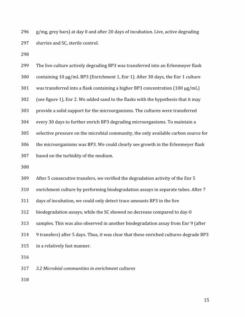
2.7 Strain isolation and sequencing 247
The supernatant of an actively degrading enrichment culture transferred 7 times 248
(Enrichment 7, or Enr 7) was diluted 100 fold and spread on R2B agar and 249
incubated for 3 days at 25 °C in the dark. Several colony morphologies were 250
observed and each morphotype was re-streaked on R2B agar to ensure purity. 251
These strains were tested for BP3 degradation activity and one positive strain, 252
BP14P, was further characterized. DNA was extracted as described above and 253
sequenced on the same sequencer used for ARISA. Here, we used universal bacterial 254
primers 27F and 1492R in the first PCR and then the internal primers 907R, 804F 255

13
and S8 for the sequencing reactions realized by the dideoxy reaction Sanger 256
sequencing using the BigDye™ Terminator v3.1 Cycle Sequencing Kit (Thermo 257
Fischer) and the manufacturer's protocol. 258
259
2.8 Flow Cytometry 260
Strain BP14P cells were counted throughout the degradation assay. The glass tubes 261
were vortexed and 1 mL of cell suspension was transferred to a microcentrifuge 262
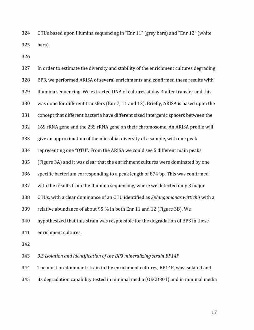
tube before adding glutaraldehyde (1% v/v final concentration). The tubes were 263
incubated at 4 °C in the dark for 15 minutes before flash freezing with liquid N2. The 264
samples were kept at –80 °C until analysis. Samples were defrosted at room 265
temperature and incubated with the nucleic acid stain SYBRGreen I (Molecular 266
Probes) for 15 min at room temperature in the dark before enumeration (Marie et 267
al., 1997) using a FACSCanto flow cytometer (BD-Biosciences) equipped with optics 268
fiber emitted light (407, 488 and 633 nm). Fluorescent 1.002 μm beads 269
(Polysciences Inc., Europe) were added to each sample as an internal standard to 270
normalize cell properties and to compare cell populations. Accurate analyzed 271
volumes and subsequent estimations of cell concentrations were calculated using 272
Becton-Dickinson TrucountTM beads. Total bacterial cells (TBA) were enumerated 273
according to the variations of light scatter properties (relative cell size) and green 274
fluorescence related to nucleic acid content. 275
276
277
3. Results and discussion 278

14
279
3.1 Degradation of BP3 in initial sludge microcosms and enrichment cultures 280
To detect BP3 degradation, the concentration of BP3 in the amended flask 281
containing active sludge was compared with the amended flask containing sterilized 282
sludge. The theoretical added total BP3 concentration was 10 µg/mL slurry and the 283
slurry contained approximately 2 mg/mL dry matter. We analyzed the aqueous and 284
the solid phase separately and it was clear that some BP3 partitioned onto the solid 285
phase in the slurry (Figure 2). This fraction was bioavailable since we could not 286
detect any BP3 in either the solid or the aqueous phase after 20 days of incubation. 287
The concentration in the SC remained approximately the same after 20 days, 288
showing that this is not an artifact due to abiotic sorption processes. Thus, we 289
observed a complete degradation of BP3 in live cultures compared to the SC after 20 290
days. 291
292
293
Fig. 2. BP3 concentrations in initial microcosms with WWTP sludge. BP3 294
concentrations in the aqueous phase (μg/mL, white bars) and in the solid phase (μ 295
15
g/mg, grey bars) at day 0 and after 20 days of incubation. Live, active degrading 296
slurries and SC, sterile control. 297
298
The live culture actively degrading BP3 was transferred into an Erlenmeyer flask 299
containing 10 µg/mL BP3 (Enrichment 1, Enr 1). After 30 days, the Enr 1 culture 300
was transferred into a flask containing a higher BP3 concentration (100 µg/mL) 301
(see figure 1), Enr 2. We added sand to the flasks with the hypothesis that it may 302
provide a solid support for the microorganisms. The cultures were transferred 303
every 30 days to further enrich BP3 degrading microorganisms. To maintain a 304
selective pressure on the microbial community, the only available carbon source for 305
the microorganisms was BP3. We could clearly see growth in the Erlenmeyer flask 306
based on the turbidity of the medium. 307
308
After 5 consecutive transfers, we verified the degradation activity of the Enr 5 309
enrichment culture by performing biodegradation assays in separate tubes. After 7 310
days of incubation, we could only detect trace amounts BP3 in the live 311
biodegradation assays, while the SC showed no decrease compared to day-0 312
samples. This was also observed in another biodegradation assay from Enr 9 (after 313
9 transfers) after 5 days. Thus, it was clear that these enriched cultures degrade BP3 314
in a relatively fast manner. 315
316
3.2 Microbial communities in enrichment cultures 317
318

16
319
Fig. 3. Analysis of the microbial community diversity in BP3 degrading enrichment 320
cultures. (A) ARISA analysis of enrichment cultures “Enr 7” (black bars), “Enr 11” 321
(grey bars) and “Enr 12” (white bars) where each peak is one “OTU” and the relative 322
abundance is based upon area of each peak. (B) Relative abundance of different 323
17
OTUs based upon Illumina sequencing in “Enr 11” (grey bars) and “Enr 12” (white 324
bars). 325
326
In order to estimate the diversity and stability of the enrichment cultures degrading 327
BP3, we performed ARISA of several enrichments and confirmed these results with 328
Illumina sequencing. We extracted DNA of cultures at day-4 after transfer and this 329
was done for different transfers (Enr 7, 11 and 12). Briefly, ARISA is based upon the 330
concept that different bacteria have different sized intergenic spacers between the 331
16S rRNA gene and the 23S rRNA gene on their chromosome. An ARISA profile will 332
give an approximation of the microbial diversity of a sample, with one peak 333
representing one “OTU”. From the ARISA we could see 5 different main peaks 334
(Figure 3A) and it was clear that the enrichment cultures were dominated by one 335
specific bacterium corresponding to a peak length of 874 bp. This was confirmed 336
with the results from the Illumina sequencing, where we detected only 3 major 337
OTUs, with a clear dominance of an OTU identified as Sphingomonas wittichii with a 338
relative abundance of about 95 % in both Enr 11 and 12 (Figure 3B). We 339
hypothesized that this strain was responsible for the degradation of BP3 in these 340
enrichment cultures. 341
342
3.3 Isolation and identification of the BP3 mineralizing strain BP14P 343
The most predominant strain in the enrichment cultures, BP14P, was isolated and 344
its degradation capability tested in minimal media (OECD301) and in minimal media 345

18
amended with 10 % R2B media. This was done to investigate whether strain BP14P 346
would degrade BP3 when other carbon sources were present. 347
348
349
350
Fig. 4. Degradation of BP3 by Sphingomonas wittichiii sp. strain BP14P coupled to 351
growth. BP3 concentrations in sterile controls (black), BP3 concentrations in live 352
cultures with minimal media (open circles), BP3 concentrations in live cultures with 353
minimal media and 10 % R2B media (black circles), cells per ml in cultures with 354
minimal media (open triangles) and cells per ml in cultures with minimal media and 355
10% R2B media (black triangles). 356
357
358

19
Strain BP14P degraded BP3 relatively fast as we could detect an almost 50% 359
degradation after one day in cultures amended with R2B media, and 90% 360
degradation after 4 days in both cultures with R2B but also in cultures with minimal 361
media (Figure 4). This degradation was clearly linked to growth since the 362
concentration of cells increased with time in cultures where the only carbon source 363
was BP3. We hypothesize that this strain completely mineralized BP3 and used BP3 364
as both carbon and energy source. To the best of our knowledge, this is the first 365
report showing a growth dependent degradation of BP3 in a pure strain. 366
367
A BLAST comparison of the 16S rRNA gene sequence of strain BP14P (1150 bp) 368
showed that it was 100 % identical to Sphingomonas wittichii DC-6, isolated from 369
activated sludge (KC410868) and only one base pair different (1149/1150) to the 370
widely studied strain S. wittichii RW1 (NR_074268.1). DC-6 can mineralize the 371
herbicides alachlor, acetochlor, and butachlor (Chen et al., 2013) through N-372
dealkylation activity by a Rieske non-heme iron oxygenase system (Chen et al., 373
2014). S. wittichii RW1 was first isolated from an urban river sample (Wittich et al., 374
1992) and is able to degrade a plethora of aromatic compounds, like chlorinated 375
dibenzo-p-dioxins (DDs) and dibenzofurans (DFs) and to grow on DD and DF as sole 376
carbon and energy sources (Wittich et al., 1992). The first step in the degradation of 377
DD and DF is carried out by a dioxygenase and interestingly many of the genes 378
responsible for degradation of aromatic compounds are present on megaplasmids in 379
both strains Sphingomonas wittichii RW1 and DC-6 (Armengaud et al., 1998; Cheng 380
20
et al., 2019; Miller et al., 2010). Indeed, this feature seems to be common in 381
Sphingomonas that degrade xenobiotics (Basta et al., 2004). 382
383
3.4 Putative degradation pathway of BP3 384
During the HPLC-UV analysis, we could clearly detect an UV absorbing peak at an 385
earlier retention time than BP3 in some samples. To identify this peak and other 386
putative degradation products or intermediates, we analyzed several samples using 387
LC-MS/MS and molecular networks (MN) constructed by means of the Global 388
Natural Products Social Molecular Networking (GNPS) platform (Wang et al., 2016) 389
(https://gnps.ucsd.edu/). GNPS is a MS/MS data community sharing and curation 390
platform. Its MN tool organizes all MS/MS spectra recorded in complex mixtures of 391
compounds by families of analogous compounds, each grouped in one MN. This 392
enables the user to highlight possible degradation products in samples without any 393
presupposition of the chemical structure. In the present MN analysis, the number of 394
similar fragments was set to 2, and the minimum cosine for linking two parent ions 395
was set to 0.7. With these parameters, only one compound clustered with BP3, with 396
a parent mass at m/z 215.070 ([M+H]+). This compound was identified as BP1, a 397
BP3 demethylation product (Figure 5). BP1 was detected in high concentrations in 398
day 1 samples of the degradation assay with S. wittichii BP14P when R2B was 399
added. At this timepoint, BP3 was already about 50% degraded. This peak was 400
detected only at trace levels in the following timepoints, meaning that BP1 was 401
rapidly degraded. This initial demethylation reaction has been previously reported 402
to be a probable degradation step in activated sludge microcosms (Liu et al., 2012) 403

21
and as one probable pathway for BP3 degradation by Methylophilus sp. strain FP-6 404
(Jin et al., 2019). 405
406

22
407
Fig. 5. Putative degradation pathway of BP3. 408
409

23
We investigated other possible degradation pathways by submitting the BP3 410
structure to an online pathway prediction tool (Gao et al., 2010) and then searching 411
for particular exact masses of the predicted products (m/z of corresponding [M+H]+ 412
ions) in all the LC-MS profiles of our samples. Based upon the prediction, another 413
possible first step is a hydroxylation of the benzene ring (Compound 2 in Figure 5). 414
An ion corresponding to this compound was detected in some of our samples, as 415
was compound 4. However, we did not detect compound 3 in any of our samples. 416
Based upon these results we hypothesize that the main degradation pathway 417
involves an initial demethylation of BP3 to BP1, followed by a hydroxylation to give 418
4. However, an initial hydroxylation of BP3 yielding compound 2 and a following 419
demethylation to result in compound 4 might be a minor pathway. 420
421
Jin and colleagues identified other compounds in Methylophilus sp. strain FP-6 422
cultures degrading BP3 (Jin et al., 2019). The presence of these compounds, not 423
depicted in Figure 5, suggested a degradation pathway involving hydroxylation of 424
BP3 and ring cleavage, but no demethylation. We did not find any evidence for this 425
pathway in S. wittichii BP14P. This might be due to the different nature (co-426
metabolism versus growth linked) of the degradation process of the two different 427
strains in question. 428
429
Compounds 5-9 are examples of possible products after cleavage based upon the 430
prediction system. However, these low molecular weight compounds could not be 431
detected by our LC system that analyzed molecules between 133 Da and 2000 Da. 432

24
Compound 10 was detected by Liu et al. (2012), but was not predicted by the 433
prediction tool. 434
435
3.5 UV filter degradation capabilities of strain BP14P are limited 436
We tested the capability of S. wittichii BP14P to degrade other UV filters, listed in 437
Table 1. Indeed, Miller et al. (2010) suggest that one of the reasons why S. wittichii 438
RW1 is so successful at degrading substrates with low water solubility might be 439
because of the abundance of TonB-dependent receptors, which can act as 440
transporters of aromatic compounds. Further, because the expression of some 441
dioxygenases (like dioxygenases dxnA1A2) have shown to be substrate dependent 442
(Armengaud et al., 1998), we also added BP3 to parallel assays to investigate 443
whether the putative expression of genes involved in degradation of BP3 can 444
stimulate the degradation of other aromatic compounds. Accordingly, we tested the 445
UV filters listed in Table 1 alone in biodegradation assays, as well as with extra 446
carbon (10% R2B) and with BP3. 447
448
We incubated S. wittichii sp. BP14P with the 9 different UV filters for 14 days. BP3 449
was over 95% degraded in all the assays where it was added. However, we detected 450
very little degradation of the other UV filters (Table 1). The only other UV filter that 451
was partially degraded with no other carbon sources present was ES (15 % after 14 452
days). S. wittichii BP14P degraded ES slightly more when BP3 and other carbon 453
sources were present, namely 28 % after 14 days. Finally, HS was 18 % degraded 454
after 14 days when BP3 and other carbon sources were present. 455

25
456
To summarize, S. wittichii BP14P did not degrade other UV filters to a great extent. 457
This specificity could be due to limited bioavailability of the other UV filters, a 458
limitation of putative transporters, or potentially, a limitation in the genes involved 459
in degradation. 460
461
3.6 What drives the specificity of BP3 degradation by strain BP14P? 462
Other researchers have observed that BP3 can be biodegraded. Liu et al. (2012) 463
found that BP3 was completely degraded after 42 days of incubation in both aerobic 464
and anaerobic microcosms also using inoculum from a WWTP. This suggests that 465
several groups of bacteria could be possible degraders, but this was not investigated 466
by Liu and collegues. Methylophilus sp. strain FP-6 was shown to co-metabolically 467
degrade BP3, degrading 15 % of BP3 after 8 days when no other carbon source was 468
provided but degrading BP3 to a higher extent when other carbon sources were 469
present (Jin et al., 2019). Based on the data provided, we believe that Methylophilus 470
sp. strain FP-6 degraded BP3 co-metabolically. Strikingly, strain BP14P was very 471
specifically enriched in the cultures through many transfers, suggesting there were 472
no other bacteria present in the sludge that could degrade BP3 in a growth 473
dependent manner or in an equally efficient manner as strain BP14P. 474
475
Even though the 16S rRNA gene of strain BP14P was 99.99% and 100% identical to 476
S. wittichii RW1 and DC-6, respectively, the functional similarity between these 477
strains remains to be investigated through a more polyphasic approach, including 478

26
genome sequencing. Interestingly, S. wittichii RW1 has a very specific dioxygenase 479
activity that enables it to degrade DD and DF (Armengaud et al., 1998) and thus we 480
hypothesize that S. wittichii BP14P degrades BP3 through the activity of at least one 481
specific mono- or dioxygenase responsible for the initial demethylation (see figure 482
4) or another limiting step in the degradation pathway. We base this hypothesis on 483
the fact that the mono- or dioxygenase activity needed for BP3 degradation did not 484
seem to be widely distributed as no other bacteria were enriched in these cultures. 485
However, whether it uses a homologue of the S. wittichii RW1 enzyme, remains to be 486
investigated. 487
488
S. wittichii strains seem to be especially suited to degrade xenobiotics. For example 489
S. wittichii RW1 has extreme redundancy in its use of aromatic compound 490
metabolism (Coronado et al., 2012). This group of microorganisms generally 491
contains megaplasmids containing degradation genes and horizontal gene transfer 492
seems to be very common. Importantly, it is well known that S. wittichii RW1 can 493
survive and retain its degradation capability in soil when amended for 494
bioremediation purposes (Megharaj et al., 1997). All this makes it particularly 495
fascinating to enlarge the degradation repertoire of this group to include UV filters 496
and BP3. 497
498
4. Conclusions and perspectives 499
The extensive use of BP3 has resulted in ubiquitous BP3 contamination in various 500
environments, including in WWTPs and in many aquatic ecosystems (Fagervold et 501

27
al, 2019). For a complete environmental risk assessment of BP3, it is important to 502
understand its fate and all the processes the molecule is subjected to. Degradation, 503
though either co-metabolism or mineralization is an important factor in this 504
assessment. We show here that BP3 can be completely degraded in a few days in 505
WWTPs by S. wittichii BP14P. Since BP3 can be utilized by a bacterium as an energy 506
source it then ensues that BP3 has the potential to be degraded in other 507
environmental compartments than WWTP sludge. As a consequence, the long-term 508
accumulation of BP3 in the environment could be less likely than previously 509
assumed. However, once released into marine ecosystems, it is as yet unknown 510
whether BP3 can be degraded. This question should be the subject of future studies, 511
as well as performing similar degradation testing on other sunscreen UV filters, with 512
a priority on the most toxic UV filters including octinoxate, octocrylene and 513
homosalate 514
515
Acknowledgements: 516
We would like to thank Cécile Villette for performing the sequencing of strain BP14P 517
and Maeva Duboeuf for help with chemical extractions. This work was carried out in 518
conjunction with the European Marine Biological Resource Centre (EMBRC-ERIC-519
Banyuls-sur-Mer Oceanographic Observatory, OOB). We are grateful to the 520
BIO2MAR platform for providing access to instrumentation and especially to Nyree 521
West for technical help concerning the ARISA procedure and for reading through the 522
manuscript. We would like to thank Christophe Salmeron of the BioPIC Imaging and 523
Cytometry platform (OOB) for performing the flow cytometry and the technical 524

28
support of EMBRC-France, whose French state funds are managed by the ANR 525
within the Investments of the Future program under reference ANR-10-INBS-02. We 526
would also like to thank the local council (Communauté de Communes Albères-Côte 527
Vermeille-Illibéris) who provided access to the WWTP site for sampling. This work 528
was financially supported by the Pierre Fabre Dermo-Cosmetic Laboratories in 529
France. 530
531
Figure captions 532
Figure 1. Schematic of the experimental design. 533
534
Figure 2. BP3 concentrations in initial microcosms with WWTP sludge. BP3 535
concentrations in the aqueous phase (μg/mL, white bars) and in the solid phase (μ 536
g/mg, grey bars) at day 0 and after 20 days of incubation. Live, active degrading 537
slurries and SC, sterile control. 538
539
Figure 3. Analysis of the microbial community diversity in BP3 degrading 540
enrichment cultures. (A) ARISA analysis of enrichment cultures Enr 7 (black bars), 541
Enr 11 (grey bars) and Enr 12 (white bars) where each peak is one OTU and the 542
relative abundance is based upon area of each peak. (B) Relative abundance of 543
different OTUs based upon Illumina sequencing in Enr 11 (grey bars) and Enr 12 544
(white bars). 545
546

29
Figure 4. Degradation of BP3 by Sphingomonas wittichiii sp. strain BP14P (left axis) 547
and growth followed by flow cytometry (right axis). BP3 concentrations in sterile 548
controls (SC, black square), BP3 concentrations in live cultures with minimal media 549
(open circles), BP3 concentrations in live cultures with minimal media 550
supplemented with 10 % R2B media (black circles), cell concentrations of cultures 551
with minimal media (open triangles) or minimal media supplemented with 10% 552
R2B media (black triangles). 553
554
555
Figure 5. Putative degradation pathway of BP3. Hypothesized degradation pathway 556
of BP3 based upon the online prediction tool “EAWAG-BBD Pathway Prediction 557
System”(http://eawag-bbd.ethz.ch/predict/) 558
559
560
561
562
563
References: 564
Armengaud, J., Happe, B., Timmis, K.N., 1998. Genetic analysis of dioxin dioxygenase 565
of Sphingomonas sp. Strain RW1: catabolic genes dispersed on the genome. J. 566
Bacteriol. 180, 3954–3966. 567
Badia-Fabregat, M., Rodríguez-Rodríguez, C.E., Gago-Ferrero, P., Olivares, A., Piña, B., 568
Díaz-Cruz, M.S., Vicent, T., Barceló, D., Caminal, G., 2012. Degradation of UV 569

30
filters in sewage sludge and 4-MBC in liquid medium by the ligninolytic 570
fungus Trametes versicolor. J. Environ. Manage. 104, 114–120. 571
https://doi.org/10.1016/j.jenvman.2012.03.039 572
Barr, L., Alamer, M., Darbre, P.D., 2018. Measurement of concentrations of four 573
chemical ultraviolet filters in human breast tissue at serial locations across 574
the breast. J. Appl. Toxicol. JAT 38, 1112–1120. 575
https://doi.org/10.1002/jat.3621 576
Basta, T., Keck, A., Klein, J., Stolz, A., 2004. Detection and Characterization of 577
Conjugative Degradative Plasmids in Xenobiotic-Degrading Sphingomonas 578
Strains. J. Bacteriol. 186, 3862. https://doi.org/10.1128/JB.186.12.3862-579
3872.2004 580
Bolyen, E., Rideout, J.R., Dillon, M.R., Bokulich, N.A., Abnet, C.C., Al-Ghalith, G.A., 581
Alexander, H., Alm, E.J., Arumugam, M., Asnicar, F., Bai, Y., Bisanz, J.E., 582
Bittinger, K., Brejnrod, A., Brislawn, C.J., Brown, C.T., Callahan, B.J., Caraballo-583
Rodríguez, A.M., Chase, J., Cope, E.K., Da Silva, R., Diener, C., Dorrestein, P.C., 584
Douglas, G.M., Durall, D.M., Duvallet, C., Edwardson, C.F., Ernst, M., Estaki, M., 585
Fouquier, J., Gauglitz, J.M., Gibbons, S.M., Gibson, D.L., Gonzalez, A., Gorlick, K., 586
Guo, J., Hillmann, B., Holmes, S., Holste, H., Huttenhower, C., Huttley, G.A., 587
Janssen, S., Jarmusch, A.K., Jiang, L., Kaehler, B.D., Kang, K.B., Keefe, C.R., Keim, 588
P., Kelley, S.T., Knights, D., Koester, I., Kosciolek, T., Kreps, J., Langille, M.G.I., 589
Lee, J., Ley, R., Liu, Y.-X., Loftfield, E., Lozupone, C., Maher, M., Marotz, C., 590
Martin, B.D., McDonald, D., McIver, L.J., Melnik, A.V., Metcalf, J.L., Morgan, S.C., 591
Morton, J.T., Naimey, A.T., Navas-Molina, J.A., Nothias, L.F., Orchanian, S.B., 592

31
Pearson, T., Peoples, S.L., Petras, D., Preuss, M.L., Pruesse, E., Rasmussen, L.B., 593
Rivers, A., Robeson, M.S., Rosenthal, P., Segata, N., Shaffer, M., Shiffer, A., 594
Sinha, R., Song, S.J., Spear, J.R., Swafford, A.D., Thompson, L.R., Torres, P.J., 595
Trinh, P., Tripathi, A., Turnbaugh, P.J., Ul-Hasan, S., van der Hooft, J.J.J., Vargas, 596
F., Vázquez-Baeza, Y., Vogtmann, E., von Hippel, M., Walters, W., Wan, Y., 597
Wang, M., Warren, J., Weber, K.C., Williamson, C.H.D., Willis, A.D., Xu, Z.Z., 598
Zaneveld, J.R., Zhang, Y., Zhu, Q., Knight, R., Caporaso, J.G., 2019. Reproducible, 599
interactive, scalable and extensible microbiome data science using QIIME 2. 600
Nat. Biotechnol. 37, 852–857. https://doi.org/10.1038/s41587-019-0209-9 601
Chen, Q., Wang, C.-H., Deng, S.-K., Wu, Y.-D., Li, Y., Yao, L., Jiang, J.-D., Yan, X., He, J., Li, 602
S.-P., 2014. Novel Three-Component Rieske Non-Heme Iron Oxygenase 603
System Catalyzing the N-Dealkylation of Chloroacetanilide Herbicides in 604
Sphingomonads DC-6 and DC-2. Appl. Environ. Microbiol. 80, 5078. 605
https://doi.org/10.1128/AEM.00659-14 606
Chen, Q., Yao, L., Wang, C., Deng, S., Chu, C., He, J., 2013. Isolation and 607
characterization of acetochlor-degrading strain Sphingomonas sp. DC-6 and 608
preliminary studies on its metabolic pathway. J. Agric. Sci. Technol. Beijing 609
15, 67–74. 610
Cheng, M., Yan, X., He, J., Qiu, J., Chen, Q., 2019. Comparative genome analysis reveals 611
the evolution of chloroacetanilide herbicide mineralization in Sphingomonas 612
wittichii DC-6. Arch. Microbiol. 201, 907–918. 613
https://doi.org/10.1007/s00203-019-01660-w 614

32
Coronado, E., Roggo, C., Johnson, D., van der Meer, J.R., 2012. Genome-Wide Analysis 615
of Salicylate and Dibenzofuran Metabolism in Sphingomonas Wittichii RW1. 616
Front. Microbiol. 3, 300. https://doi.org/10.3389/fmicb.2012.00300 617
Díaz-Cruz, M.S., Molins-Delgado, D., Serra-Roig, M.P., Kalogianni, E., Skoulikidis, 618
N.Th., Barceló, D., 2019. Personal care products reconnaissance in EVROTAS 619
river (Greece): Water-sediment partition and bioaccumulation in fish. Sci. 620
Total Environ. 651, 3079–3089. 621
https://doi.org/10.1016/j.scitotenv.2018.10.008 622
Fagervold, S.K., Rodrigues, A.S., Rohée, C., Roe, R., Bourrain, M., Stien, D., Lebaron, P., 623
2019. Occurrence and Environmental Distribution of 5 UV Filters During the 624
Summer Season in Different Water Bodies. Water. Air. Soil Pollut. 230, 172. 625
https://doi.org/10.1007/s11270-019-4217-7 626
Fisher, M.M., Triplett, E.W., 1999. Automated approach for ribosomal intergenic 627
spacer analysis of microbial diversity and its application to freshwater 628
bacterial communities. Appl Env. Microbiol 65, 4630–4636. 629
Gago-Ferrero, P., Badia-Fabregat, M., Olivares, A., Piña, B., Blánquez, P., Vicent, T., 630
Caminal, G., Díaz-Cruz, M.S., Barceló, D., 2012. Evaluation of fungal- and 631
photo-degradation as potential treatments for the removal of sunscreens BP3 632
and BP1. Sci. Total Environ. 427–428, 355–363. 633
https://doi.org/10.1016/j.scitotenv.2012.03.089 634
Gao, J., Ellis, L.B., Wackett, L.P., 2010. The University of Minnesota 635
Biocatalysis/Biodegradation Database: improving public access. Nucleic 636
Acids Res 38, D488. 637
33
Herlemann, D.P., Labrenz, M., Jürgens, K., Bertilsson, S., Waniek, J.J., Andersson, A.F., 638
2011. Transitions in bacterial communities along the 2000 km salinity 639
gradient of the Baltic Sea. ISME J. 5, 1571–1579. 640
https://doi.org/10.1038/ismej.2011.41 641
Jin, C., Geng, Z., Pang, X., Zhang, Y., Wang, G., Ji, J., Li, X., Guan, C., 2019. Isolation and 642
characterization of a novel benzophenone-3-degrading bacterium 643
Methylophilus sp. strain FP-6. Ecotoxicol. Environ. Saf. 186, 109780. 644
https://doi.org/10.1016/j.ecoenv.2019.109780 645
Liu, Y.-S., Ying, G.-G., Shareef, A., Kookana, R.S., 2013. Degradation of Six Selected 646
Ultraviolet Filters in Aquifer Materials Under Various Redox Conditions. 647
Groundw. Monit. Remediat. 33, 79–88. 648
https://doi.org/10.1111/gwmr.12027 649
Liu, Y.-S., Ying, G.-G., Shareef, A., Kookana, R.S., 2012. Biodegradation of the 650
ultraviolet filter benzophenone-3 under different redox conditions. Environ. 651
Toxicol. Chem. 31, 289–295. https://doi.org/10.1002/etc.749 652
Lozano C., Givens J., Stien D., Matallana-Surget S., Lebaron P., 2020. Bioaccumulation 653
and Toxicological Effects of UV-Filters on Marine Species, in: The Handbook 654
of Environmental Chemistry. Springer, Berlin, Heidelberg, pp. 1–46. 655
Mao, F., He, Y., Gin, K., 2018. Occurrence and fate of benzophenone-type UV filters in 656
aquatic environments: A review. Environ. Sci. Water Res. Technol. 5. 657
https://doi.org/10.1039/C8EW00539G 658

34
Marie, D., Partensky, F., Jacquet, S., Vaulot, D., 1997. Enumeration and Cell Cycle 659
Analysis of Natural Populations of Marine Picoplankton by Flow Cytometry 660
Using the Nucleic Acid Stain SYBR Green I. Appl. Environ. Microbiol. 63, 186. 661
Megharaj, M., Wittich, R.-M., Blasco, R., Pieper, D.H., Timmis, K.N., 1997. Superior 662
survival and degradation of dibenzo-p-dioxin and dibenzofuran in soil by 663
soil-adapted Sphingomonas sp. strain RW1. Appl. Microbiol. Biotechnol. 48, 664
109–114. https://doi.org/10.1007/s002530051024 665
Miller, T.R., Delcher, A.L., Salzberg, S.L., Saunders, E., Detter, J.C., Halden, R.U., 2010. 666
Genome sequence of the dioxin-mineralizing bacterium Sphingomonas 667
wittichii RW1. J. Bacteriol. 192, 6101–6102. 668
https://doi.org/10.1128/JB.01030-10 669
Molins-Delgado, D., Máñez, M., Andreu, A., Hiraldo, F., Eljarrat, E., Barceló, D., Díaz-670
Cruz, M.S., 2017. A Potential New Threat to Wild Life: Presence of UV Filters 671
in Bird Eggs from a Preserved Area. Environ. Sci. Technol. 51, 10983–10990. 672
https://doi.org/10.1021/acs.est.7b03300 673
Ramos, S., Homem, V., Alves, A., Santos, L., 2016. A review of organic UV-filters in 674
wastewater treatment plants. Environ. Int. 86, 24–44. 675
https://doi.org/10.1016/j.envint.2015.10.004 676
Rodríguez-Escales, P., Sanchez-Vila, X., 2020. Modeling the fate of UV filters in 677
subsurface: Co-metabolic degradation and the role of biomass in sorption 678
processes. Water Res. 168, 115192. 679
https://doi.org/10.1016/j.watres.2019.115192 680

35
Stien, D., Clergeaud, F., Rodrigues, A.M.S., Lebaron, K., Pillot, R., Romans, P., 681
Fagervold, S., Lebaron, P., 2019. Metabolomics Reveal That Octocrylene 682
Accumulates in Pocillopora damicornis Tissues as Fatty Acid Conjugates and 683
Triggers Coral Cell Mitochondrial Dysfunction. Anal. Chem. 91, 990–995. 684
https://doi.org/10.1021/acs.analchem.8b04187 685
Suh, S., Pham, C., Smith, J., Mesinkovska, N.A., 2020. The banned sunscreen 686
ingredients and their impact on human health: a systematic review. Int. J. 687
Dermatol. https://doi.org/10.1111/ijd.14824 688
Wang, M., Carver, J.J., Phelan, V.V., Sanchez, L.M., Garg, N., Peng, Y., Nguyen, D.D., 689
Watrous, J., Kapono, C.A., Luzzatto-Knaan, T., Porto, C., Bouslimani, A., Melnik, 690
A.V., Meehan, M.J., Liu, W.-T., Crüsemann, M., Boudreau, P.D., Esquenazi, E., 691
Sandoval-Calderón, M., Kersten, R.D., Pace, L.A., Quinn, R.A., Duncan, K.R., 692
Hsu, C.-C., Floros, D.J., Gavilan, R.G., Kleigrewe, K., Northen, T., Dutton, R.J., 693
Parrot, D., Carlson, E.E., Aigle, B., Michelsen, C.F., Jelsbak, L., Sohlenkamp, C., 694
Pevzner, P., Edlund, A., McLean, J., Piel, J., Murphy, B.T., Gerwick, L., Liaw, C.-695
C., Yang, Y.-L., Humpf, H.-U., Maansson, M., Keyzers, R.A., Sims, A.C., Johnson, 696
A.R., Sidebottom, A.M., Sedio, B.E., Klitgaard, A., Larson, C.B., Boya P, C.A., 697
Torres-Mendoza, D., Gonzalez, D.J., Silva, D.B., Marques, L.M., Demarque, D.P., 698
Pociute, E., O’Neill, E.C., Briand, E., Helfrich, E.J.N., Granatosky, E.A., Glukhov, 699
E., Ryffel, F., Houson, H., Mohimani, H., Kharbush, J.J., Zeng, Y., Vorholt, J.A., 700
Kurita, K.L., Charusanti, P., McPhail, K.L., Nielsen, K.F., Vuong, L., Elfeki, M., 701
Traxler, M.F., Engene, N., Koyama, N., Vining, O.B., Baric, R., Silva, R.R., 702
Mascuch, S.J., Tomasi, S., Jenkins, S., Macherla, V., Hoffman, T., Agarwal, V., 703

36
Williams, P.G., Dai, J., Neupane, R., Gurr, J., Rodríguez, A.M.C., Lamsa, A., Zhang, 704
C., Dorrestein, K., Duggan, B.M., Almaliti, J., Allard, P.-M., Phapale, P., Nothias, 705
L.-F., Alexandrov, T., Litaudon, M., Wolfender, J.-L., Kyle, J.E., Metz, T.O., 706
Peryea, T., Nguyen, D.-T., VanLeer, D., Shinn, P., Jadhav, A., Müller, R., Waters, 707
K.M., Shi, W., Liu, X., Zhang, L., Knight, R., Jensen, P.R., Palsson, B.Ø., Pogliano, 708
K., Linington, R.G., Gutiérrez, M., Lopes, N.P., Gerwick, W.H., Moore, B.S., 709
Dorrestein, P.C., Bandeira, N., 2016. Sharing and community curation of mass 710
spectrometry data with Global Natural Products Social Molecular 711
Networking. Nat. Biotechnol. 34, 828–837. 712
https://doi.org/10.1038/nbt.3597 713
Wittich, R.M., Wilkes, H., Sinnwell, V., Francke, W., Fortnagel, P., 1992. Metabolism of 714
dibenzo-p-dioxin by Sphingomonas sp. strain RW1. Appl. Environ. Microbiol. 715
58, 1005. 716
717
718
719