ekg board review 2012.ppt - mycmemedia.mycme.com/documents/39/ekg_9665.pdf · 9/26/2012 1 ekg...
TRANSCRIPT

9/26/2012
1
EKG REVIEW for UMDNJ-PA CME Review Course
Presented by Carol J. Sadley, M.Ed., PA-C
June 8, 2012
Normal Cardiac Conduction
www.healcentral.org/content/collections/ECG/ecg_ccs.gif
UMDNJ PANCE/PANRE Review Course
Conduction Pathway
Sino atrial (SA) node (in RA) fires
Signal travels across atria to AV node
Slight pause then signal travels down Slight pause, then signal travels down Bundle of His
His Bundle branches into Right & Left bundles (and further into fasicles)
Ending at the Purkinje fibers
Slight pause, then repolarization occursUMDNJ PANCE/PANRE Review Course

9/26/2012
2
EEKKGEK’sEKG Nomenclature
www.commons.wikimedia.org/wiki/File:EKG_complex.png
UMDNJ PANCE/PANRE Review Course
Normal 12-lead EKG
UMDNJ PANCE/PANRE Review Course
Heart Rate determination
Start with a QRS on a heavy black line
Use sequence to label rate at subsequent lineslines
UMDNJ PANCE/PANRE Review Course

9/26/2012
3
Six Second Strip (aka 30 big blocks)
UMDNJ PANCE/PANRE Review Course
Calculate the rate: 62 is correct (or just a little faster than 60)
UMDNJ PANCE/PANRE Review Course
Rate of Impulse Formation
S-A Node = 60-100 bpm
A V Node = 40 60 bpm A-V Node = 40-60 bpm
Ventricle = 20-40 bpm
UMDNJ PANCE/PANRE Review Course

9/26/2012
4
Normal Sinus Rhythm
www/healcentral.org/content/collections/ECG/ecg_nsr.gif
UMDNJ PANCE/PANRE Review Course
Sinus Bradycardia
www.healtcentral.org/content/collections/ECG/ecg_sb.gif
Sinus Bradycardia
NSR criteria, but heart rate is < 60
Can be normal especially in well-trained athletes or people taking B-blockersathletes or people taking B blockers
Most common rhythm disturbance seen in early stages of AMI
If symptomatic, treat with Atropine, consider pacing (TransCutaneous Pacing/TCP)
UMDNJ PANCE/PANRE Review Course

9/26/2012
5
Sick Sinus Syndrome
Associated w/ sinus arrest, s-a exit block, or persistent sinus bradycardia < 45 bpm
Seen in elderly, @ w/ atrial fibrillation, but often asymptomaticoften asymptomatic
Patchy fibrosis of SA node and conduction
May be caused by drug therapy, sarcoid, amyloidosis, cardiomyopathies
Treat with pacemaker if symptomatic
UMDNJ PANCE/PANRE Review Course
Sinus Tachycardia
www.healtcentral.org/content/collections/ECG/ecg_st.gif
Sinus Tachycardia
NSR criteria, but HR is >100<180
Normal under many conditions: exercise, fever hyperthyroid CHF COPD ETOHfever, hyperthyroid, CHF, COPD, ETOH
Treat cause first
If unstable/pt. symptomatic, immediate synchronized cardioversion
If stable, try vagal maneuvers,
B-blockers, +/- RFA used definitivelyUMDNJ PANCE/PANRE Review Course

9/26/2012
6
Sinus Arrhythmia
UMDNJ PANCE/PANRE Review Course
Sinus Arrhythmia
Normal physiological mechanism 2o vagal influence (young and/or old)
Barely detectable rate changes Barely detectable rate changes corresponding to the phases of respiration
Slight increase in rate during inspiration
Slight decrease in rate during expiration
UMDNJ PANCE/PANRE Review Course
Sinus Arrhythmia
www.healtcentral.org/content/collections/ECG/ecg_sa.gif
UMDNJ PANCE/PANRE Review Course

9/26/2012
7
Premature Atrial Contractions
Arrive earlier than expected next beat Different shape to p wave (because it
comes from a different and irritable focus in the atria)
Normal/narrow QRS complex Followed by a compensatory pause Usually benign, provide reassurance
UMDNJ PANCE/PANRE Review Course
Premature Atrial Contractions
www.healtcentral.org/content/collections/ECG/ecg_pac.gif
UMDNJ PANCE/PANRE Review Course
(P)SVT/Supraventricular Tachycardia
Rate 140-240 and regular, w/ narrow QRS Sudden/abrupt start/stop AKA “a-v nodal reentry tachycardia” (AVNRT)
Responds (slows or terminates) to vagal maneuvers, carotid massage (R then L: not both!)
Pharmacologic agents: (IV) adenosine -6 mg bolus or verapamil - 2.5 mg bolus
+/- Cardioversion (100 j) if symptomaticUMDNJ PANCE/PANRE Review Course
9/26/2012
8
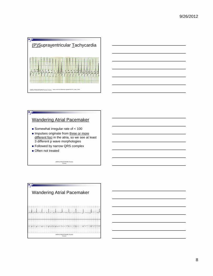
(P)Supraventricular Tachycardia
www.commons.wikimedia.org/wiki/File:SVT_Lead_II.JPGUMDNJ PANCE/PANRE Review Course
Wandering Atrial Pacemaker
Somewhat irregular rate of < 100
Impulses originate from three or more different foci in the atria so we see at leastdifferent foci in the atria, so we see at least 3 different p wave morphologies
Followed by narrow QRS complex
Often not treated
UMDNJ PANCE/PANRE Review Course
Wandering Atrial Pacemaker
UMDNJ PANCE/PANRE Review Course
9/26/2012
9
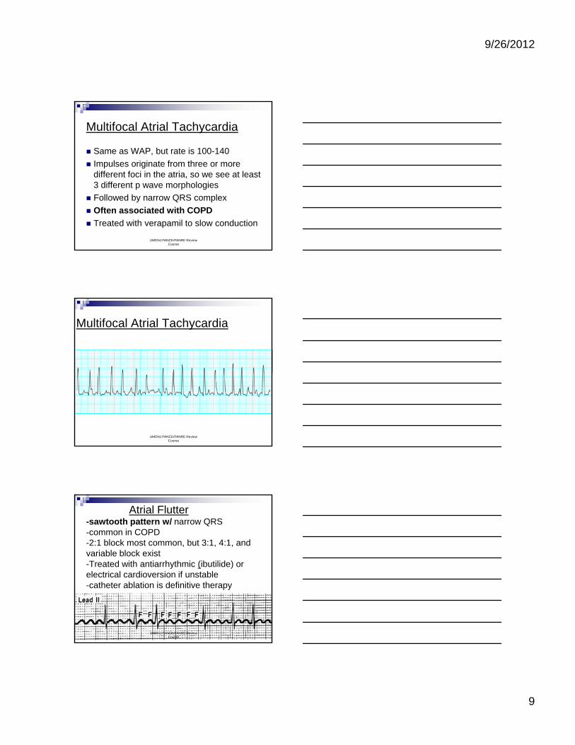
Multifocal Atrial Tachycardia
Same as WAP, but rate is 100-140
Impulses originate from three or more different foci in the atria so we see at leastdifferent foci in the atria, so we see at least 3 different p wave morphologies
Followed by narrow QRS complex
Often associated with COPD
Treated with verapamil to slow conduction
UMDNJ PANCE/PANRE Review Course
Multifocal Atrial Tachycardia
UMDNJ PANCE/PANRE Review Course
Atrial Flutter-sawtooth pattern w/ narrow QRS -common in COPD-2:1 block most common, but 3:1, 4:1, and variable block exist-Treated with antiarrhythmic (ibutilide) or y ( )electrical cardioversion if unstable-catheter ablation is definitive therapy
UMDNJ PANCE/PANRE Review Course

9/26/2012
10
Atrial Flutter with 2:1 block; ventricular rate is ~150 bpm
UMDNJ PANCE/PANRE Review Course
Atrial Flutter with variable block
www.healtcentral.org/content/collections/ECG/ecg_aflutter.gif
UMDNJ PANCE/PANRE Review Course
Atrial Fibrillation No common/distinct/visible p waves Irregularly irregular w/ narrow QRS New onset: check thyroid function Treated with b-blocker, Ca blocker, , ,
cardioversion or ?permanently with radio frequency ablation (RFA)
UMDNJ PANCE/PANRE Review Course

9/26/2012
11
Atrial Fibrillation
www.healtcentral.org/content/collections/ECG/ecg_afib.gif
Atrial Fibrillation
Common causes: HTN, CHD, myopathyr/o thyrotoxicosis and ETOH use/abuse
Danger due to potential for blood to Danger due to potential for blood to coagulate in RA sending clot to brain: CVA
First goal in AF = rate control
Second goal = Cardioversion or ablation
Third goal in chronic AF = anticoagulation
UMDNJ PANCE/PANRE Review Course
Chronic Atrial Fibrillation Tx
Anticoagulation most commonly treated with Coumadin (warfarin), Pradaxa (dabigatran) or Xarelto (rivaroxaban)( g ) ( )
Levels must be monitored regularly (PT, INR) for coumadin-not Pradaxa/Xarelto
On occasion, aspirin and/or Plavix (clopidogrel) is used for this purpose if ASA allergic
UMDNJ PANCE/PANRE Review Course

9/26/2012
12
Normal Cardiac Pacemakers
Atrial: SA Node – rate = 60-100
Junctional: AV Node rate = 40 60 Junctional: AV Node – rate = 40-60
Ventricular: ventricle – rate = 20-40
UMDNJ PANCE/PANRE Review Course
Junctional Rhythm
www.healtcentral.org/content/collections/ECG/ecg_junc.gif
Junctional Rhythm
Regular rhythm (so NOT a-fib)
Normal/narrow QRS (wide possible)
Absent, retrograde, or inverted p waves
Rate usually 40-60 bpm
Often seen with digitalis toxicity
UMDNJ PANCE/PANRE Review Course

9/26/2012
13
Premature Junctional Contraction (PJC) PJC is early, narrow QRS, followed by
compensatory pause, and with absent, inverted, or retrograde p wave
UMDNJ PANCE/PANRE Review Course
Accelerated Junctional Rhythm
Regular rhythm with narrow/normal QRS
Rate > 60 but < 100
P wave is absent inverted or retrograde P wave is absent, inverted, or retrograde
UMDNJ PANCE/PANRE Review Course
Junctional Tachycardia
Regular rhythm
Rate > 100
P waves are absent inverted retrograde P waves are absent, inverted, retrograde
UMDNJ PANCE/PANRE Review Course

9/26/2012
14
Normal Cardiac Pacemakers
Atrial: SA Node – rate = 60-100
Junctional: AV Node rate = 40 60 Junctional: AV Node – rate = 40-60
Ventricular: ventricle – rate = 20-40
UMDNJ PANCE/PANRE Review Course
Idioventricular Rhythm– treat with Pacemaker
www.healtcentral.org/content/collections/ECG/ecg_ivr.gif
UMDNJ PANCE/PANRE Review Course
Accelerated IVR (AIVR): common after MI: do not treat
www.healtcentral.org/content/collections/ECG/ecg_aivr.gifUMDNJ PANCE/PANRE Review Course

9/26/2012
15
PVCs (Premature Ventricular Contraction)
UMDNJ PANCE/PANRE Review Course
Premature Ventricular Contractions
Most common ventricular arrhythmia
Beat is early, wide, bizarre
Sticks out like a “sore thumb”
Followed by a compensatory pause
Rare/occasional PVCs may be normal, especially if they resolve with activity
Check electrolytes, thyroid, occult heart dz
If symptomatic, look for cause and treat with b-blockers first then consider ablation
UMDNJ PANCE/PANRE Review Course
Unifocal PVC
www.healtcentral.org/content/collections/ECG/ecg_pvc.gifUMDNJ PANCE/PANRE Review Course

9/26/2012
16
Multifocal PVCs
www.healtcentral.org/content/collections/ECG/ecg_pvc.gif
UMDNJ PANCE/PANRE Review Course
Ventricular Bigeminy
www.healtcentral.org/content/collections/ECG/ecg_bigem.gifUMDNJ PANCE/PANRE Review Course
Ventricular Couplet
www.healtcentral.org/content/collections/ECG/ecg_pairs.gifUMDNJ PANCE/PANRE Review Course

9/26/2012
17
“Salvo” or 3-beat Run of VT
www.healtcentral.org/content/collections/ECG/ecg_trip.gifUMDNJ PANCE/PANRE Review Course
“R-on-T” phenomenon Danger: may initiate Torsade/VT/VF
Find cause; Tx with lidocaine or amiodarone then cardiovert (100-360 J)
www.healtcentral.org/content/collections/ECG/ecg_ront.gif
UMDNJ PANCE/PANRE Review Course
Ventricular Tachycardia
A run of > 3 PVCs in a row
Rate regular and > 100 (160-240)
May be mono or polymorphic
C h K+ h Common cause = hypo- K+ or hypo-magnesemia
Non-sustained: <30 sec., spont. terminate
UMDNJ PANCE/PANRE Review Course

9/26/2012
18
Ventricular Tachycardia Monomorphic: more commonly associated
with a healed infarction
If symptomatic, tx with cardioversion; drugs = lidocaine or amiodarone; ODdrugs = lidocaine or amiodarone; OD pace if recurrent; definitive therapy = ICD
UMDNJ PANCE/PANRE Review Course
Torsades de Pointes
Polymorphic VT
UMDNJ PANCE/PANRE Review Course
Polymorphic VT
Torsades de pointes means “twisting of the points”
Often associated with Long QT intervals
M b it l b t ft lt f May be congenital, but often results from electrolyte imbalance (K, Mg, Ca)
Treat with B-blockers or temporary pacing (if pulse is present)
Do NOT treat with antiarrhythmics as they prolong the QT interval.
UMDNJ PANCE/PANRE Review Course

9/26/2012
19
Ventricular Fibrillation
www.healtcentral.org/content/collections/ECG/ecg_vf.gif
UMDNJ PANCE/PANRE Review Course
(Coarse) Ventricular Fibrillation Most common cause of SCD (sudden cardiac death)
Chaotic, irregular rhythm; no true QRS
No pulse on PE; treat with CPR/defibrillation/ACLS protocols
www.commons.wikimedia.org/wiki/File:EKG_VF.jpg
UMDNJ PANCE/PANRE Review Course
Asystole
No electrical activity/no defib; look for cause and treat specific abnormality
www.healtcentral.org/content/collections/ECG/ecg_asys.gif
UMDNJ PANCE/PANRE Review Course

9/26/2012
20
Ventricular Rhythms: compared
UMDNJ PANCE/PANRE Review Course
AV Conduction Blocks
An AV Conduction Block is any obstruction or delay of the normal conduction between the SA node and the Purkinje fibers.j
Most commonly occurs between AV node and His bundle
3 varieties of AV Block: 1st, 2nd, 3rd
degree
UMDNJ PANCE/PANRE Review Course
First Degree AV Block
All normal except PR interval >.2 sec (1 big block); often seen in athletes and with bradycardia; no treatment requiredy ; q
www.healcentral.org/content/collections/ECG/ecg_1_AV_block
UMDNJ PANCE/PANRE Review Course

9/26/2012
21
Second degree AV BlockWenckebach (aka Mobitz type I)
Gradually lengthening PR interval until a QRS complex is dropped.
Look for “grouped beating” in the EKG Look for grouped beating in the EKG
No treatment required; +/- EP studies
www.healcentral.org/content/collections/ECG/ecg_2A_V_block.gif
UMDNJ PANCE/PANRE Review Course
Second Degree AV Block – Mobitz II
PR interval remains constant
Intermittent dropping of QRS
Usually @ with organic heart diseasey @ g
Pacemaker usually indicated
UMDNJ PANCE/PANRE Review Course
Third Degree (aka) Complete HeartBlock
www.healtcentral.org/content/collections/ECG/ecg_chb.gif

9/26/2012
22
Third Degree/Complete Heart Block
No relationship between p waves and QRS (p waves “marching through”)
More p waves than QRS complexes
Ventricular response often slow (IVR)
Treat with permanent pacemaker
UMDNJ PANCE/PANRE Review Course
Third Degree A-V Block with Pace-maker Insertion Treatment
www.healcentral.org/content/collections/ECG/ecg_3_AV_block
UMDNJ PANCE/PANRE Review Course
Pacemakers A power source connected to electrodes
Most popular type = dual chamber multiple programmable units
www.commons.wikimedia.org/wiki/File:ECG/pacemaker_dualchamber.jpg

9/26/2012
23
Single Chamber Pacemaker
www.healtcentral.org/content/collections/ECG/ecg_pacer.gifUMDNJ PANCE/PANRE Review Course
Pacemaker Wire Placement
www.healtcentral.org/content/collections/ECG/ecg_lead_wire.gif
UMDNJ PANCE/PANRE Review Course
Dual Chamber Pacing (aka AV sequential pacing)
www.healtcentral.org/content/collections/ECG/ecg_av_pace.gif
UMDNJ PANCE/PANRE Review Course

9/26/2012
24
Pre-excitation Syndromes
Accessory AV conduction pathways
Result in “short circuits” or shortcuts from SA node to AV nodeSA node to AV node
Usually diagnosed on 12-lead EKG
2 types: Wolff-Parkinson-White Syndrome
and Lown-Ganong-Levine Syndrome
UMDNJ PANCE/PANRE Review Course
WPW and LGL: shortcuts in conduction
www.healtcentral.org/content/collections/ECG/ecg517.gif
UMDNJ PANCE/PANRE Review Course
Wolff-Parkinson-White Syndrome
Accessory pathway via “bundle of Kent” Appears as short PR interval (<.08) Presence of delta wavePresence of delta wave
Predisposes to tachyarrhythmias (A-fib—VF)
Treated with amiodarone or sotalol; if unstable cardiovert, then RFA definitively
UMDNJ PANCE/PANRE Review Course
9/26/2012
25
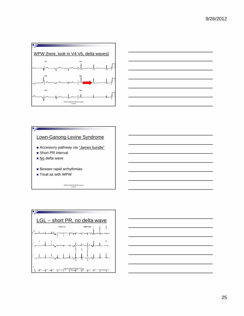
WPW (here, look in V4-V6, delta waves)
UMDNJ PANCE/PANRE Review Course
Lown-Ganong-Levine Syndrome
Accessory pathway via “James bundle”
Short PR interval
No delta wave No delta wave
Beware rapid arrhythmias
Treat as with WPW
UMDNJ PANCE/PANRE Review Course
LGL – short PR, no delta wave
UMDNJ PANCE/PANRE Review Course

9/26/2012
26
Mean QRS Vector and Axis
Down and to pt.’s left
www.commons.wikimedia.org/wiki/File:ECG_Eomtjpvem_vect0.svgUMDNJ PANCE/PANRE Review
Course
EKG Axis Determination(Who’s triangle?) Dr. Einthoven !
www.commons.wikimedia.org/wiki/File:ECG_Einthoven_vect.2.svg
UMDNJ PANCE/PANRE Review Course
Normal 12-lead EKG
UMDNJ PANCE/PANRE Review Course

9/26/2012
27
12 Lead EKG Arrangement
I AVR
II AVL
V1 V4
V2 V5II AVL
III AVF
Rhythm strip (1-3 leads)
V2 V5
V3 V6
UMDNJ PANCE/PANRE Review Course
Axis determination short cut
Look at the QRS deflection
Using leads I and AVF: (up =+; down = -)Up in I and Up in AVF = Normal AxisUp in I and Up in AVF = Normal Axis
Up in I and Down in AVF = Left Axis Deviation
Down in I and Up in AVF = Right Axis Dev.
Down in I and Down in AVF = Extreme RAD
UMDNJ PANCE/PANRE Review Course
Left Axis Deviation (LAD)

9/26/2012
28
Right Axis Deviation (RAD)
UMDNJ PANCE/PANRE Review Course
Bundle Branch Block (BBB)
www.commons.wikimedia.org/wiki/File:Reizeleitungssystem_RSB.png
UMDNJ PANCE/PANRE Review Course
LBBB Diagnosis Criteria
Wide (>.12 sec) QRS RSR’ in V5 or V6 (aka “dog ears”) Associated with ST depression in I, AVL, V5,
d V6and V6 Often associated with CAD New LBBB (@ w/ CP) = “STEMI equivalent”
BEWARE: Difficult to diagnose MI via EKG in presence of LBBB.
UMDNJ PANCE/PANRE Review Course

9/26/2012
29
Left Bundle Branch Block
UMDNJ PANCE/PANRE Review Course
Criteria to Diagnose RBBB
Wide QRS (> .12 sec) RSR’ in V1 or V2 (aka “rabbit ears”) May have associated ST depression in V1,May have associated ST depression in V1,
V2, +/- V3
Can be found in a “normal” EKG (i.e., usually not associated with CAD)
UMDNJ PANCE/PANRE Review Course
RBBB: RSR’ in V1/V2
UMDNJ PANCE/PANRE Review Course

9/26/2012
30
Right BBB with A-fib
UMDNJ PANCE/PANRE Review Course
Hemiblocks
Blocks occurring in either the Anterior or Posterior Divisions (fascicles) of the Left Bundle Branch (just distal to the AV node)(j )
Often result from diminished blood supply, i.e. MI or ischemia
QRS may be normal or wide
Major effect on EKG is axis deviation
UMDNJ PANCE/PANRE Review Course
Hypertrophy and Enlargement
Hypertrophy = ventricles; enlargement = atria
Specific EKG leads show evidence of Specific EKG leads show evidence of these findings
ALL EKG findings of hypertrophy and enlargement must be verified with ECHOCARDIOGRAM ! ! !
UMDNJ PANCE/PANRE Review Course

9/26/2012
31
Examples of LVH & RVH
www.commons.wikimedia.org/wiki/File:Heart_Left_ventricular_hypertrophy_sa.jpg
www.commons.wikimedia.org/wiki/File:Heart/kabulki_syndrome.jpgUMDNJ PANCE/PANRE Review Course
Left Atrial Enlargement
Look for biphasic p wave in V1 with large terminal portion
LAE:
May be associated with wide p waves elsewhere
May result from mitral stenosis, HTN
UMDNJ PANCE/PANRE Review Course
Left Atrial Enlargement
UMDNJ PANCE/PANRE Review Course

9/26/2012
32
Right Atrial Enlargement
Look for tall (>2.5 mm)peaked p waves
RAE:
in II, III, AVF
Results from tricuspid stenosis, pulmonary HTN, severe lung dz
UMDNJ PANCE/PANRE Review Course
Right Atrial Enlargement: look for tall p waves (>2.5mm) in II, III, AVF
UMDNJ PANCE/PANRE Review Course
Left Ventricular Hypertrophy
S in V 1/2 + R in V 5/6 > 35 mm
R in I + S in III > 25 mm
R in AVL > 13 mm
Often caused by HTN and valvular dz
Often associated with LAD & depressed ST seg (aka “ventricular strain” pattern)
Criteria not totally applicable < 35 y/o or thin
UMDNJ PANCE/PANRE Review Course
9/26/2012
33
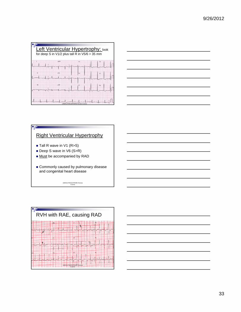
Left Ventricular Hypertrophy: look for deep S in V1/2 plus tall R in V5/6 > 35 mm
UMDNJ PANCE/PANRE Review Course
Right Ventricular Hypertrophy
Tall R wave in V1 (R>S)
Deep S wave in V6 (S>R)
Must be accompanied by RAD Must be accompanied by RAD
Commonly caused by pulmonary disease and congenital heart disease
UMDNJ PANCE/PANRE Review Course
RVH with RAE, causing RAD
Make changes
UMDNJ PANCE/PANRE Review Course
9/26/2012
34
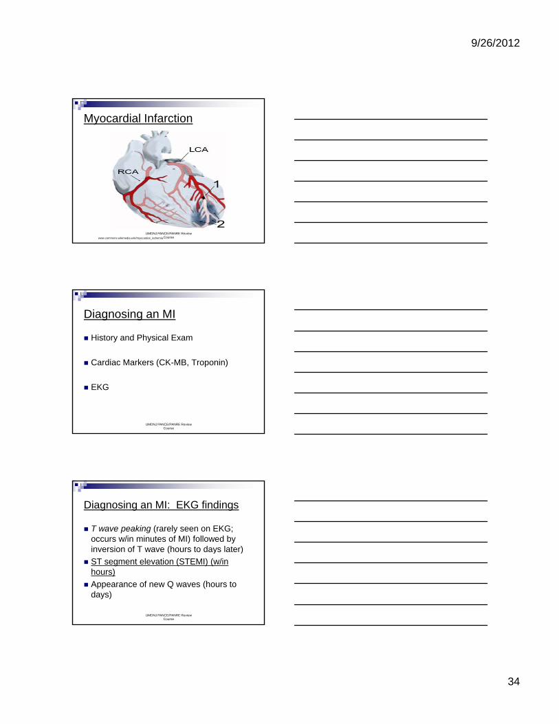
Myocardial Infarction
www.commons.wikimedia.wiki/myocardial_ischemia
UMDNJ PANCE/PANRE Review Course
Diagnosing an MI
History and Physical Exam
Cardiac Markers (CK MB Troponin) Cardiac Markers (CK-MB, Troponin)
EKG
UMDNJ PANCE/PANRE Review Course
Diagnosing an MI: EKG findings
T wave peaking (rarely seen on EKG; occurs w/in minutes of MI) followed by inversion of T wave (hours to days later)( y )
ST segment elevation (STEMI) (w/in hours)
Appearance of new Q waves (hours to days)
UMDNJ PANCE/PANRE Review Course

9/26/2012
35
Evolving EKG changes of MI
UMDNJ PANCE/PANRE Review Course
Evolution of Acute MI ST-changes
www.commons.wikimedia.org/wiki/File:12_Lead_EKG_ST_Elevation_tracing_only..jpg
UMDNJ PANCE/PANRE Review Course
Localizing an MI
Anterior Wall MI: Supplied by Left Anterior Descending Artery
Seen as EKG changes in I, V1-V4 Often a very deadly MIO te a e y dead y Often associated with “poor R-wave
progression”
V1 & V2 are mainly considered “septal” leads, so many MIs are “antero-septal”.
UMDNJ PANCE/PANRE Review Course

9/26/2012
36
Leads I, V1-V4 = Anterior MI
www.commons.wikimedia.org/wiki/File: AMI_scheme.png
UMDNJ PANCE/PANRE Review Course
Acute Antero-septal wall STEMI
UMDNJ PANCE/PANRE Review Course
Inferior Wall MI
Inferior or diaphragmatic portion of the heart is supplied mostly by the Right Coronary Arteryy y
Can be associated with Right ventricular infarction
EKG changes are seen in II, III, AVF
UMDNJ PANCE/PANRE Review Course

9/26/2012
37
II, III, AVF( +/- V6 ST elevations) = Inferior MI
www.commons.wikimedia.org/wiki/File:AMI_scheme.png
UMDNJ PANCE/PANRE Review Course
Inferior Wall MI
UMDNJ PANCE/PANRE Review Course
Lateral Wall MI
Lateral portion of the heart is supplied by the Left Circumflex Artery.
EKG changes seen in I, AVL, V5 & V6
UMDNJ PANCE/PANRE Review Course
9/26/2012
38
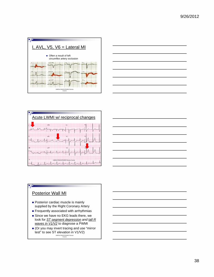
I, AVL, V5, V6 = Lateral MI
Often a result of left circumflex artery occlusion
UMDNJ PANCE/PANRE Review Course
Acute LWMI w/ reciprocal changes
UMDNJ PANCE/PANRE Review Course
Posterior Wall MI
Posterior cardiac muscle is mainly supplied by the Right Coronary Artery
Frequently associated with arrhythmias Frequently associated with arrhythmias
Since we have no EKG leads there, we look for ST segment depression and tall R waves in V1/V2 to diagnose a PWMI
(Or you may invert tracing and use “mirror test” to see ST elevation in V1/V2)
UMDNJ PANCE/PANRE Review Course

9/26/2012
39
Look in V1 for “mirror image”: Tall R wave in V1, +/- ST dep
UMDNJ PANCE/PANRE Review Course
Reciprocal Changes
Sometimes, the dramatic EKG changes in the infarct area produce opposing changes in distant leads (or those opposite the ( ppinfarct location.
In an Acute MI with ST elevations, ST depression may appear in a distant lead.
These ST depressions are called “reciprocal changes”
UMDNJ PANCE/PANRE Review Course
Non-Q Wave or Non-STEMI MIs
Not all MIs produce Q waves or ST elevations
On EKG a non-Q Wave or non-STEMI On EKG, a non Q Wave or non STEMI only shows: T wave inversion and ST depression
Must use History and Cardiac Markers to make diagnosis
UMDNJ PANCE/PANRE Review Course
9/26/2012
40
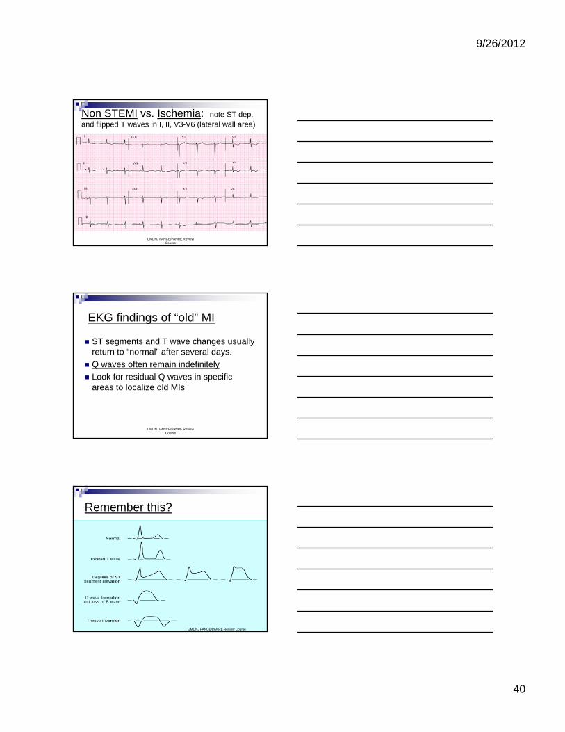
Non STEMI vs. Ischemia: note ST dep. and flipped T waves in I, II, V3-V6 (lateral wall area)
UMDNJ PANCE/PANRE Review Course
EKG findings of “old” MI
ST segments and T wave changes usually return to “normal” after several days.
Q waves often remain indefinitely Q waves often remain indefinitely
Look for residual Q waves in specific areas to localize old MIs
UMDNJ PANCE/PANRE Review Course
Remember this?
UMDNJ PANCE/PANRE Review Course
9/26/2012
41
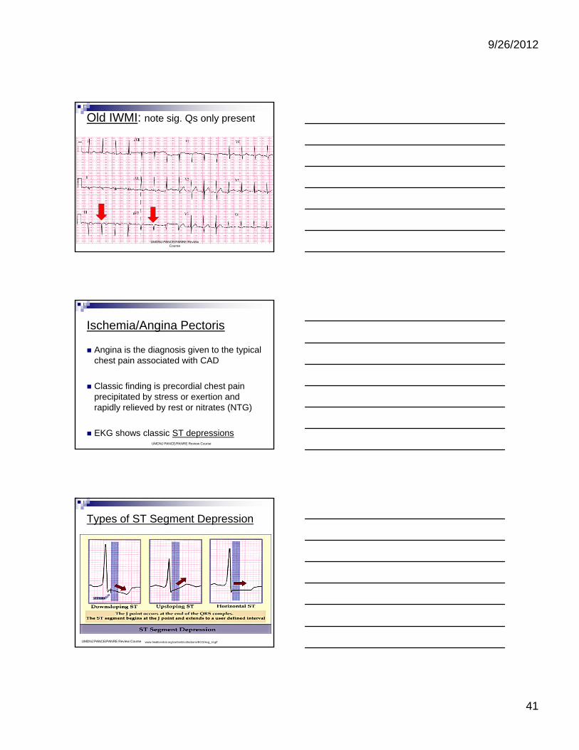
Old IWMI: note sig. Qs only present
UMDNJ PANCE/PANRE Review Course
Ischemia/Angina Pectoris
Angina is the diagnosis given to the typical chest pain associated with CAD
Classic finding is precordial chest pain precipitated by stress or exertion and rapidly relieved by rest or nitrates (NTG)
EKG shows classic ST depressionsUMDNJ PANCE/PANRE Review Course
Types of ST Segment Depression
www.healtcentral.org/content/collections/ECG/ecg_st.gifUMDNJ PANCE/PANRE Review Course

9/26/2012
42
Ischemia: EKG w/ ST depression
ST depression > 1 mm = significant
< 1mm – “non-specific ST segment changes”
www.commons.wikimedia.org/wiki/File:StressECG_STDepression.jpgUMDNJ PANCE/PANRE Review Course
Antero(septal) wall Ischemia
UMDNJ PANCE/PANRE Review Course
Inferolateral wall ischemia (XST)
UMDNJ PANCE/PANRE Review Course

9/26/2012
43
Benign ST changes: Early repolarization (see diffuse ST elevations)
UMDNJ PANCE/PANRE Review Course
Prinzmetal’s (Variant) Angina
Angina-like chest pain often a result of coronary artery spasm
Associated with ST elevation on EKG Associated with ST elevation on EKG
Thought to be a reversible injury, with ST segments returning to baseline after treatment with nitroglycerin
UMDNJ PANCE/PANRE Review Course
Miscellaneous EKG Diagnoses
Certain effects may be recognized by their characteristic appearance on EKG
The EKG “alerts us” to the diagnosis—it The EKG alerts us to the diagnosis it does NOT make the diagnosis
It can act as another “clue” in making a diagnosis
UMDNJ PANCE/PANRE Review Course
9/26/2012
44
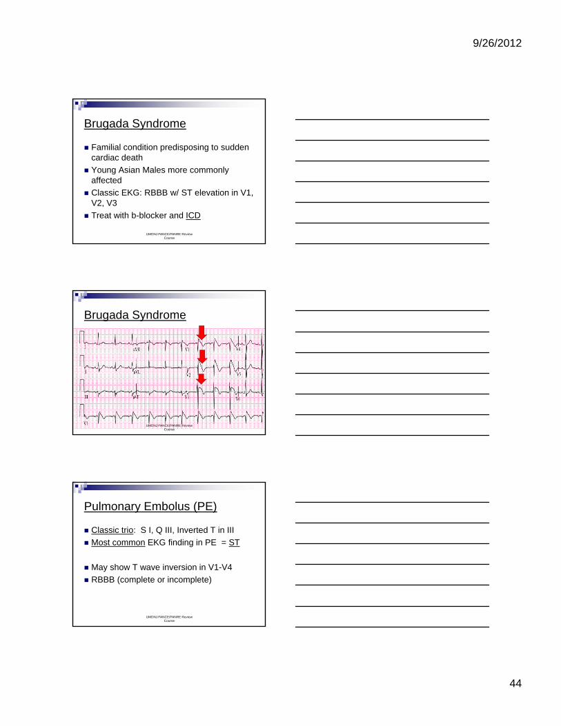
Brugada Syndrome
Familial condition predisposing to sudden cardiac death
Young Asian Males more commonly Young Asian Males more commonly affected
Classic EKG: RBBB w/ ST elevation in V1, V2, V3
Treat with b-blocker and ICD
UMDNJ PANCE/PANRE Review Course
Brugada Syndrome
UMDNJ PANCE/PANRE Review Course
Pulmonary Embolus (PE)
Classic trio: S I, Q III, Inverted T in III
Most common EKG finding in PE = ST
May show T wave inversion in V1-V4
RBBB (complete or incomplete)
UMDNJ PANCE/PANRE Review Course

9/26/2012
45
Pulmonary Embolus (PE)
UMDNJ PANCE/PANRE Review Course
Hyperkalemia (high potassium)
The potassium ion is critical in the cardiac conduction cycle at the cellular level
The range of potassium is very narrow The range of potassium is very narrow
Classic finding is tall, peaked T waves
As the levels increase, the p wave flattens while the QRS widens – beware!
UMDNJ PANCE/PANRE Review Course
Hyperkalemia progression
UMDNJ PANCE/PANRE Review Course

9/26/2012
46
Hyperkalemia: (early stage); note tall, peaked T waves in V1-V4
UMDNJ PANCE/PANRE Review Course
Note wide QRS, absent p throughout EKG in advanced hyperkalemia
UMDNJ PANCE/PANRE Review Course
Hypokalemia: Classic findings:flattened T wave and new U wave
UMDNJ PANCE/PANRE Review Course

9/26/2012
47
Hypokalemia: u wave in V2, V3
UMDNJ PANCE/PANRE Review Course
Hypocalcemia Levels of calcium drop below normal
QT lengthens—DANGER!
UMDNJ PANCE/PANRE Review Course
Hypocalemia with long QT seen in all leads here
UMDNJ PANCE/PANRE Review Course

9/26/2012
48
Long QT Syndrome
May be an inherited syndrome (Romano-Ward syndrome)
May be due to a variety of drugs May be due to a variety of drugs (quinidine, sotalol, abx such as the quinolones, and antidepressants)
May lead to “R-on-T” or Torsades
Treat with b-blocker, Mg, pacer, ICD
UMDNJ PANCE/PANRE Review Course
Hypothermia
J/Osborne wave (usually with bradycardia)
UMDNJ PANCE/PANRE Review Course
Digitalis: effect and toxicity
Digitalis effect causes “scooped” ST seg and is normal
Digitalis excess and toxicity can cause AV Digitalis excess and toxicity can cause AV blocks and ventricular rhythms
UMDNJ PANCE/PANRE Review Course

9/26/2012
49
Pericarditis: inflammation of the pericardial sac – acute or chronic
www.commons.wikimedia.org/wiki/File: ChronicInfectivePericarditis.jpg
UMDNJ PANCE/PANRE Review Course
Acute pericarditis: diffuse ST elevations
Chronic pericarditis: diffuse ST depressions
UMDNJ PANCE/PANRE Review Course
Acute Pericarditis w/ diffuse ST elevation
UMDNJ PANCE/PANRE Review Course

9/26/2012
50
Electrical Alternans
Associated with a large pericardial effusion
Electrical axis of the heart varies with each beat due to the heart “floating in fluid-filledbeat due to the heart floating in fluid filled sac”
Results in varying amplitude (alternating large and small) of EKG beats (and pulse on PE = pulses alternans)
UMDNJ PANCE/PANRE Review Course
Electrical Alternans: seen in various leads here
UMDNJ PANCE/PANRE Review Course
Hypertrophic Cardiomyopathy
Previously known as IHSS, may occur as obstructive (HOCM), dilated, or restrictive
Causes SCD in some instances (young)
EKG may show Q waves in many leads, LVH, LAD, and some deeply inverted T waves.
Treated (if diagnosed) by placement of ICD
UMDNJ PANCE/PANRE Review Course

9/26/2012
51
Hypertrophic Cardiomyopathy
www.commons.wikimedia.org/wiki/File:Heart/left/ventricular/hypertrophy/sa.jpg
UMDNJ PANCE/PANRE Review Course
Implantable Cardioverter Defibrillator (ICD)
www.commons.wikimedia.org/wiki/File:AICD.jpg
UMDNJ PANCE/PANRE Review Course
References:
Scheidt, S., Basic Electrocardiography, Vol. 36, Summit, NJ; CIBA-GEIGY Corp., 1996
Thaler, Malcolm S., The Only EKG Book You’ll E N d Si th Editi LWW 2010Ever Need, Sixth Edition; LWW, 2010
McPhee, S., Papadakis, M., Rabow, M. Lange 2012 Current Medical Diagnosis & Treatment; 51st Ed., McGraw Hill, 2012
www.healcentral.org
www.commons.wikimedia.orgUMDNJ PANCE/PANRE Review
Course
9/26/2012
52
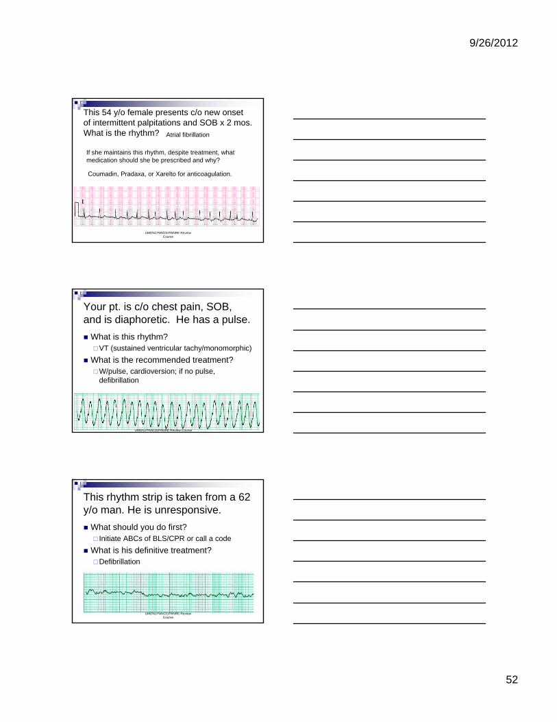
This 54 y/o female presents c/o new onset of intermittent palpitations and SOB x 2 mos.What is the rhythm? Atrial fibrillation
If she maintains this rhythm, despite treatment, what medication should she be prescribed and why?
UMDNJ PANCE/PANRE Review Course
Coumadin, Pradaxa, or Xarelto for anticoagulation.
Your pt. is c/o chest pain, SOB, and is diaphoretic. He has a pulse.
What is this rhythm?VT (sustained ventricular tachy/monomorphic)
What is the recommended treatment? What is the recommended treatment?W/pulse, cardioversion; if no pulse,
defibrillation
UMDNJ PANCE/PANRE Review Course
This rhythm strip is taken from a 62 y/o man. He is unresponsive.
What should you do first? Initiate ABCs of BLS/CPR or call a code
What is his definitive treatment? What is his definitive treatment?Defibrillation
UMDNJ PANCE/PANRE Review Course

9/26/2012
53
This is a 44 y/o man who presents c/o mild CP and nausea x 2 hours.
UMDNJ PANCE/PANRE Review Course
44 y/o man with CP & nausea
--Red flags/findings?
ST elevations in II, III, AVF, V5, V6
--Other findings?
Reciprocal changes in I, AVL, V1
--Rate, rhythm, axis?
110, ST, normalUMDNJ PANCE/PANRE Review Course
This is a 55 y/o female with a h/o DM who presents c/o dyspnea x 2 hours.
UMDNJ PANCE/PANRE Review Course

9/26/2012
54
55 y/o female diabetic w/ dyspnea
Red flags?
ST elevations in I, AVL, (?)V5, V6
Tall R wave and ST depression in V1 Tall R wave and ST depression in V1
Findings/Interpretation?
RSR’ in V1/V2 = RBBB
Acute postero-lateral wall MI
Reciprocal changes in II, III, AVFUMDNJ PANCE/PANRE Review Course
This EKG is taken on a 25 y/o male who presents for a pre-employment physical.
UMDNJ PANCE/PANRE Review Course
What is the most likely diagnosis here? -- Why?
Brugada Syndrome (note classic ST shape in V1-V3)
UMDNJ PANCE/PANRE Review Course

9/26/2012
55
Remember this T-shirt?
I LOVE CARDIOLOGY
Thanks for your attention and GOOD LUCK!UMDNJ PANCE/PANRE Review Course