ekzein meaning “to boil out”; word ek means “out”, while zema
TRANSCRIPT

CAS1 Research PAPER
Disease Review
March 2021
Atopic Dermatitis: Treatment Practices And Prevention- The Ayurvedic Way
Usha Nagavarapu
Abstract
Atopic dermatitis (AD), collectively known as eczema, is a chronic inflammatory skin disease
that is characterized by a dysfunctional skin barrier. AD, also called atopic eczema, is a
recurrent inflammatory skin condition that affects 15-20% of children and 1-3% of
adults worldwide. It is a chronic condition characterized by acute flare-ups of eczematous
pruritic lesions that causes the skin to become inflamed and irritated. Sadly, the prevalence
of this disorder is increasing, and it poses a significant burden on health-care resources and
patients' quality of life. It is evident that dermatologists have many treatment options and
guidelines for AD. Still, the treatment does not seem to address AD individuals’ underlying
issues, as they vary from patient to patient. Several patients and families with AD are seeking
a more holistic approach to help address and find a cure for the disorder. There is an
increased emphasis on natural therapies, and alternative medicines such as Traditional
Chinese Medicine (TCM), acupuncture, and Ayurveda, in conjunction with Western medical
treatments are being pursued. This review attempts to summarize some of the available
treatment options to understand the integrative holistic approach needed to treat an AD
individual.
Keywords: eczema, atopic dermatitis; Ayurveda
Introduction:
Atopic dermatitis (AD), collectively known as eczema, is a chronic inflammatory skin disease
that is characterized by a dysfunctional skin barrier.1 The word eczema comes from the Greek
word ekzein meaning “to boil out”; word ek means “out”, while zema means boiling.2 The
exact cause of eczema is not known. People with eczema do have the IgE antibodies
(immunoglobulin E) produced by the immune system as part of allergic reactions. Eczema

can be a tough and vexing condition accompanied by overwhelming psychological
challenges faced by the patients. AD is a chronic condition that causes the skin to become
inflamed and irritated, making it extremely itchy. It can come and go for years or throughout
life and can overlap with other types of eczema. The disorder is often observed to be
associated with redness, swelling, “weeping” clear fluid, crusting and scaling.
AD is a common condition and anyone can get the disease, but it is commonly seen to begin
in childhood. Living with atopic dermatitis can often be arduous, but treatment can help
control symptoms. In United States (US) and Europe, the prevalence of AD among children is
estimated to be approximately 20%, and among adults is estimated to range between 7% and
14% in adults. The condition is extremely widespread in the US, and it is estimated that over
31 million Americans suffer from some type of eczema. In the US, AD is the most common type
of eczema, affecting over 9.6 million children and about 16.5 million adults. An estimated $364
million to $3.8 billion is spent on treating and managing AD each year.3 It therefore accounts
for a significant economic global burden.4 among skin disease treatments.
In approximately 80% of affected individuals2, eczema can begin during childhood. However,
it can also begin during adolescence and even in adulthood; the disorder can range from mild
to severe.5 AD is typically considered the first step of the ‘atopic march’, associated with a
risk of developing asthma, or food allergy.6 The lipid barrier of skin is generally reduced in
people suffering from eczema, compared to people who do not have the disorder. Trans
epidermal water low (TEWL) from the skin is also common, leading to a dry and compromised
skin. Once the skin barrier is compromised, different irritants and materials can easily
penetrate the skin and make it vulnerable to infections. The immune system also overreacts
to environmental allergens and causes inflamed, irritated, or sore skin.7 “Itch that rashes” is
characteristic feature of eczema.8
An agreement seems to exist among the research community that the cause of AD is due to a
combination of both genetic and environmental factors. Genes responsible for encoding
protein S100, or filaggrin, proteases and their inhibitors have been studied extensively and
have been found to be associated with AD especially with epidermal barrier dysfunction.1
These genes are involved in altering the normal skin function in a myriad of ways. In addition,
these genetic predispositions are interpreted as leading to an increase in probability of
developing atopic dermatitis if one of the parents suffers from it.9 A recent study correlated

the defects in the filaggrin gene to eczema and asthma.10, 11 Advanced genetics methods such
as genome-wide association (GWA) and single nucleotide polymorphism (SNP) have been
employed to directly associate AD with genes of innate/adaptive immune systems, human
leukocyte antigens (HLA), cytokines, chemokines, drug-metabolizing genes and various
other genes.11 A number of environmental triggers and causes such as allergens, poor
hygiene, and diet have also been attributed to dysfunctional skin barrier.12
Treatment of this condition usually begins through a self-assessment followed by a diagnosis
by a physician or a dermatologist. Patients are often recommended to avoid allergens that
may trigger an inflammatory response and to further avoid any itching that may lead to a
compromised skin barrier. Topical steroids, antihistamines, and emollients are some of the
common medications prescribed to treat eczema.13
Outside of Western medicine, the use of alternative therapies such as Chinese herbal
preparations, homeopathy, yoga, Ayurveda, or massage therapy as a complementary therapy
(or an integrative method) to manage AD/eczema symptoms is growing. Ayurveda, a
traditional holistic form of medicine that began in India seeks to bring the mind, body, and
soul into balance by employing herbs, oils, diet, massage, and mind-body practices like yoga
and meditation to balance the life humors, or doshas. Ayurveda believes that good health is
the result of three humors or forces of the body being in balance, and diseases happen when
they are out of balance. The three doshas are called vata, pitta, and kapha.
Ayurveda describes skin diseases as twak rogas or kushtha and eczema as a type of kshudra
kushtha, or minor skin ailments.2,14,15 Ayurveda
describes eczema as a group of inflammatory
conditions which can be either acute or
chronic. Different causes for the disorder are
described and various configurations have
been reported. Acute eczema is usually seen as
a rapidly evolving, red, blistered, and swollen
rash while chronic eczema is a long term,
irritated, sensitive, thickened, lichenified often
accompanied by puritic skin.16 Based on the
presentation, Charaka and Sushruta correlate eczema with Vicarcika.17,18 According to
सकण ड् ूः पि्का श्यावा बहुस्रावा पवचर्चिका||२६||
The clinical features of Vicharchika
• Color – blackish brown
• Nature –excessive exudation, eruptions
• Associated symptoms – pruritus
https://www.carakasamhitaonline.com/index.ph
p/Kushtha_Chikitsa, 2.6.1111.Vicārcikā kuṣṭha

Charaka, Vicarcika is caused by an increase in kapha and reduced blood supply, leading to
blackish brown discoloration which is further accompanied by obstructive changes. These
later affect the local immunity and gives rise to a chance for dermatophytes to penetrate the
skin barrier. This results in eruptions, thereby causing excessive exudation. Secondary
infection and reduced blood supply leads to pruritus. This condition is often compared with
wet eczema.17 Vicarcika or eczema, is also described as ‘Rakta Pradoshaja Vikara’ meaning
that it has involvement of all the three doshas, with kapha playing a dominant role.19,20,21 In
addition, Sushrata Samhita describes Vicarcika as psoriasis, and eczema is described as small
pustules or pimples characterized by an itching, burning secretion and appearing on the
surface of the body; these are also termed Fakma (eczema).18 Eczema flares happen
frequently in these patients; therefore, finding a treatment plan that manages these flares is
important for the wellbeing of the patient.20 Charaka has mentioned that in all the chronic
diseases, regenerative rasayana drugs can be prescribed to help build rasa and dhatus i.e.,
body cells and tissues.22
Western Medicine Perspective
AD is a multifactorial disease with genetics playing an important role in the presentation of
the disorder, and filaggrin’s role in skin barrier impairment in the development of AD is well
documented. In addition, environmental factors and microbial involvement are also
recognized to play a role in the development of the disease. Western medicine focuses on
mitigating the adverse factors that are known to cause the condition, with the help of
pharmaceutical treatments. People with eczema tend to have an over-reactive immune
system. When triggered by a substance inside or outside the body, the immune
system responds by initiating inflammation. It is this inflammatory response that causes the
itchy, painful, rash-like symptoms common to several types of eczema. Unfortunately, AD
usually presents itself in early childhood; within children, its prevalence is 20% and continues
to rise globally.10 It is a complex disease, affecting all strata of life alike.9, 23
Types of Eczema
There are seven primary forms of eczema (Table 1), each with different triggers, symptoms,
and treatments.

Table 1: Types of Eczema
Type Symptoms Treatment
1 Atopic
dermatitis
This is the most severe and long-lasting
form of eczema. It's characterized by
inflamed skin that may crack and release
a clear fluid when scratched (an effect
known as "weeping").24 The patches are
often infected with Staphylococcus
aureus (staph)
Avoiding triggers, stress, and
maintaining a good routine and
lifestyle. Topical corticosteroids, non-
steroidal topicals and biologics are
often used. Often antimicrobial
treatments are needed. The allergens
need to be addressed
2 Contact
dermatitis
This is an allergic or the irritant kind of
skin flare up; it is usually localized. The
irritant contact dermatitis accounts for
about 80%.25 This kind does not run in
families and is not linked to other allergic
conditions such as hay fever or asthma.
Steroids
3 Neurodermat
itis
Is a common type of eczema that affects
about 12% of the population with intense
itching and scratching along with
symptoms of neurodermatitis, In some
cases itching is related to pleasure. 26
Chronic scratching causes itchy patches
of skin to become dry, leathery and
thickened known as lichenification, and
neurodermatitis is also known as lichen
simplex chronicus.
Corticosteroids, calcineurin inhibitors
and salicylic acid ointments, zinc oxide
paste, medicated patches that contain
lidocaine, oral medications such as
antihistamines, colloidal oatmeal, and
lifestyle changes, including yoga, and
relaxation technique.
4 Dyshidrotic
eczema
This form is more common in people who
suffer forms of eczema. It likely has a
genetic component and presents as small
and itchy blisters on the palms of hands,
soles of feet, and edges of the fingers and
toes. The cause of dyshidrotic eczema is
not known, though a fungal component is
Staying away from triggers, preventing
stress, having a regular skincare
routine. Use ceramides based topicals
to help repair the skin barrier, topical
corticosteroids, cool compress, and
anti-fungal medication. Prescribe light
therapy, topical calcineurin inhibitors
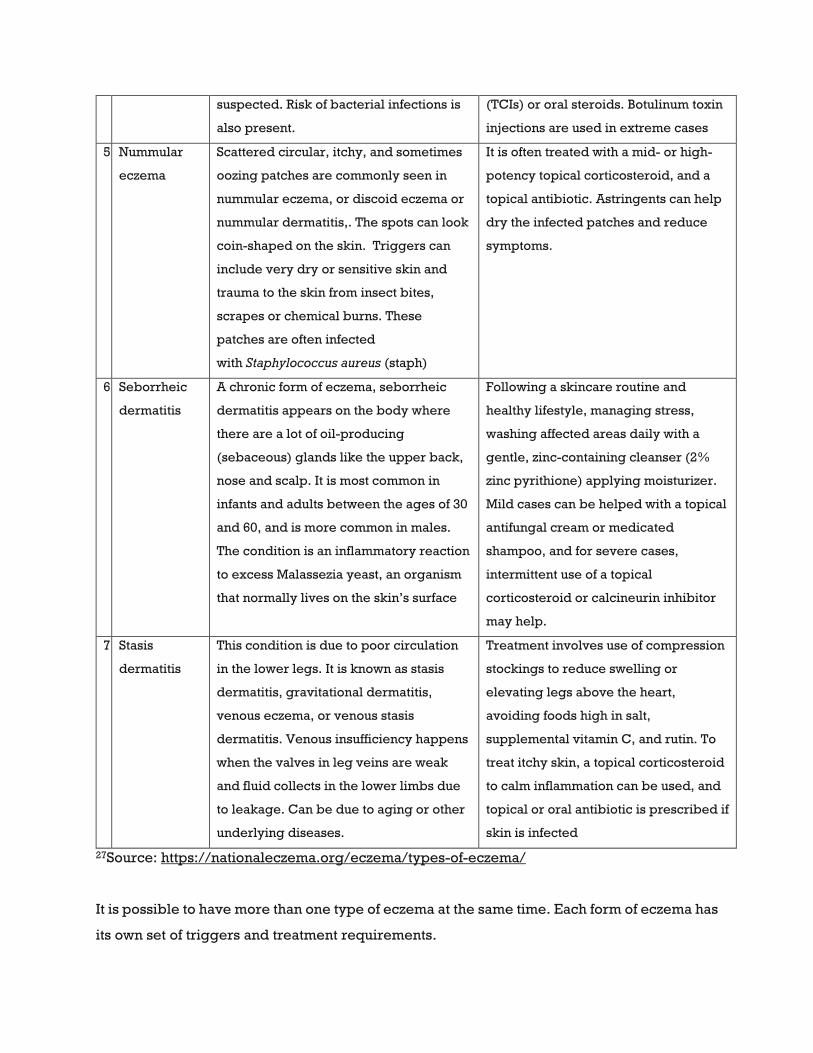
suspected. Risk of bacterial infections is
also present.
(TCIs) or oral steroids. Botulinum toxin
injections are used in extreme cases
5 Nummular
eczema
Scattered circular, itchy, and sometimes
oozing patches are commonly seen in
nummular eczema, or discoid eczema or
nummular dermatitis,. The spots can look
coin-shaped on the skin. Triggers can
include very dry or sensitive skin and
trauma to the skin from insect bites,
scrapes or chemical burns. These
patches are often infected
with Staphylococcus aureus (staph)
It is often treated with a mid- or high-
potency topical corticosteroid, and a
topical antibiotic. Astringents can help
dry the infected patches and reduce
symptoms.
6 Seborrheic
dermatitis
A chronic form of eczema, seborrheic
dermatitis appears on the body where
there are a lot of oil-producing
(sebaceous) glands like the upper back,
nose and scalp. It is most common in
infants and adults between the ages of 30
and 60, and is more common in males.
The condition is an inflammatory reaction
to excess Malassezia yeast, an organism
that normally lives on the skin’s surface
Following a skincare routine and
healthy lifestyle, managing stress,
washing affected areas daily with a
gentle, zinc-containing cleanser (2%
zinc pyrithione) applying moisturizer.
Mild cases can be helped with a topical
antifungal cream or medicated
shampoo, and for severe cases,
intermittent use of a topical
corticosteroid or calcineurin inhibitor
may help.
7 Stasis
dermatitis
This condition is due to poor circulation
in the lower legs. It is known as stasis
dermatitis, gravitational dermatitis,
venous eczema, or venous stasis
dermatitis. Venous insufficiency happens
when the valves in leg veins are weak
and fluid collects in the lower limbs due
to leakage. Can be due to aging or other
underlying diseases.
Treatment involves use of compression
stockings to reduce swelling or
elevating legs above the heart,
avoiding foods high in salt,
supplemental vitamin C, and rutin. To
treat itchy skin, a topical corticosteroid
to calm inflammation can be used, and
topical or oral antibiotic is prescribed if
skin is infected
27Source: https://nationaleczema.org/eczema/types-of-eczema/
It is possible to have more than one type of eczema at the same time. Each form of eczema has
its own set of triggers and treatment requirements.

Pathophysiology- how much do we know?
Eczema or AD is a condition that causes a person to develop patches of dry, itchy skin on their
body. It often develops as a result of inflammation in the body, so eating foods that do not
result in inflammation may help reduce symptoms. It is a chronic ailment characterized by
distinct flares and pruritus, altering between persistent flares with itching and complete
remission. These can be very distinct and vary in intensity, frequency, and duration among
individuals affected by AD. Over-the-counter creams and medications can help to
reduce inflammation. Research to understand the causative factors of eczema, and to
elucidate the genetics of AD has been an ongoing effort. The disease is highly complex and
with involvement of multiple factors, the pathophysiology is still evolving, and a lot remains
unknown. AD usually appears early in life. A constant interaction of a dysfunctional epidermal
barrier and the environmental irritants dictate the development of the disease. The
predisposed genetic factors also play a role in shaping the disorder. A primary immune
dysfunction is suspected which results in IgE sensitization and allergic inflammation. A
secondary epithelial barrier disturbance has been proposed and some are of the belief that a
primary defect in the epithelial barrier leads to secondary immunologic dysregulation and
later inflammation. Recent studies have also indicated that allergy in AD individuals could be
due to the disease itself. A need exists to look for alternative ways of preventing the onset of
AD.28
Mechanisms
In healthy individuals, balance between important subsets of T cells (eg, Th1, Th2, Th17, Th22
and a biphasic inflammation is seen. A Th2-biased immune response (IL-4, IL-13, including
thymic stromal lymphopoietin (TSLP) and eosinophils is predominant in the initial and acute
phase of AD, while a Th1/Th0 is prevalent in chronic AD skin lesions (IFN-γ, IL-12, IL-5 and
GM-CSF).27 It is proposed that a primary immune dysfunction may lead to an imbalance in the
T cell subsets potentially increasing Th2 cells. The Th2 cells are known to influence a rise in
type 2 cytokines such as interleukin IL–4, IL-5, and IL-13 which further trigger an increase in
IgE from plasma cells. In individuals with chronic AD though, the Th1 cells have been shown
to dominate and Th17 cells have been found to be elevated in patients with AD.29 Eosinophils
and mast cells have also been implicated in the pathogenesis of AD.30 It has been implicated

that basophils and group 2 innate lymphoid cells (ILC2s) are regulated by a family of
epithelial cell‒derived cytokines which are released from damaged keratinocytes, including
TSLP, IL-25, and IL-33. There are studies which indicate that a classical adaptive Th2 cell and
innate type 2 immune cells both play a critical role in the etiology of AD through interactions
with epidermal-derived cytokines. The innate immune system represents the first line of
defense against infections. In AD, a decrease in the antimicrobial peptides has been seen,
and in AD individuals, Staphylococcus aureus colonization is often detected.
Th2 cells are known to be significant sources of the itch-inducing cytokine or pruritogen IL-
31.31 A 2017 study identified that neuronal signaling of the type 2 cytokines IL-4 and IL-13
regulate AD-associated itch.32 Dupilumab, a dual IL-4 and IL-13 monoclonal antibody
(mAb) was developed and was approved in 2017 and has been advertised as a highly
effective treatment for AD. The US Food and Drug Administration (FDA) approved dupilumab
for the treatment of moderate-to-severe atopic dermatitis in adults which is not managed with
topical medications. It was the first biologic agent approved to treat AD. In October of 2018,
Dupilumab received approval by the FDA as an add-on maintenance therapy in patients with
moderate-to-severe asthma aged 12 years or older with an eosinophilic phenotype or with
oral corticosteroid-dependent asthma. Dupilumab is an antagonist, works by binding to IL-
4Rα, and further modulates signaling of both the IL-4 and IL-13 pathways. Dupilumab is given
as a subcutaneous injection and works by targeting the Th2 pathway to inhibit the
inflammatory responses that drives the symptoms of atopic dermatitis.33
AD individuals develop symptoms of AD as a result of skin barrier defects following entry of
antigens or allergens, which further stimulates the inflammatory cytokines pathway. There is
a possibility that antigens enter the system via the gut or the lungs as well. Xerosis and
ichthyosis are known to be associated signs in many AD patients, and studies have shown that
37 to 50% of people with ichthyosis vulgaris have atopic disease and up to 37% of people with
AD show clinical signs of ichthyosis vulgaris. Recent studies have shown a strong link between
mutations in the gene encoding filaggrin (FLG), a key epidermal barrier protein,
and ichthyosis vulgaris.34 Critical components involved in establishing the skin barrier
protection are in the outer layers of the epidermis, especially the proteins that form the tight
junction (TJ) and components of the innate immune system. FLG is a key protein in the
epidermis and for the formation of the skin barrier. FLG mutations have been associated with

early-onset AD.9, 28, 35 Interleukins IL- 4 and IL-13 are found detected in AD lesions and have
been shown to decrease expression of FLG in keratinocytes.1 The family of IL-1 has relevant
pro-inflammatory features, such as IL-1α, suggesting that the onset of inflammation may occur
due to changes in the skin barrier. These patients with AD with a deficiency in FLG expression
have decreased stratum corneum hydration, increased TEWL, and higher pH. Besides,
inflammation, and reduced protein expression in keratinocytes could be related to FLG
deficiency.36 Over 100 genes have been identified to be associated with AD,11,37, 38 including
genes like SPINK5/LEKT1 and DNA methylation differences have also been linked with
AD.39,40
AD exhibits a complex pathogenesis, compromised skin barrier, and an imbalance of the
immune system. The protective components of the skin barrier are present in the outer layers
of the epidermis including proteins such as FLG, the proteins that form the tight junction (TJ),
and components of the innate immune system. Changes in skin barrier control the
pathogenesis of AD, linking the structural changes with the innate and adaptive immune
system. Potent, cost-effective and safer therapies are essential to prevent, manage and treat
AD effectively.36
Treatment
Recommended first-line action to take to prevent AD flares involves reducing exposure to
potential allergens such as dust mites or pollen. Other measures which are equally important
for an AD individual is reducing dryness and developing good hygiene/skincare routine to
reduce itchiness or infection. Some of the common medications that are used by Western
Medicine for AD includes topical steroids, antihistamines, and antibiotics, calcineurin
inhibitors, phosphodiesterase-4 (PDE4) inhibitors, moisturizers and barrier repair agents,
and wet wraps. Topical steroids are among the most popular of these medications and have
been heavily used for the past 40 years in the treatment of AD.41 Most of them work by
constricting blood vessels, thus lessening the inflammation of the skin and the redness
associated with AD. Hydrocortisone was used first and since, many corticosteroid compounds
have now been licensed for treatment of AD. The development of the topical immuno-
modulators, tacrolimus and pimecrolimus are expensive and are not effective. Unfortunately,

this is not a safe solution, as it is well known that the long-term use of topical steroids can result
in many side-effects, such as folliculitis, skin atrophy, striae, erythema, and infection.
Antihistamines are a common medication prescribed to prevent allergic reactions and intense
itching for AD individuals. Itching is a common symptom in an inflammatory disease such as
AD and can damage the skin barrier, causing additional dryness and potentially infection.42
Itching is often caused by histamine, a molecule held accountable for triggering an
inflammatory response. Therefore, antihistamines are commonly prescribed to reduce
itching and associated inflammation, thereby preventing any secondary skin barrier damage.
Antibiotics are used to treat the symptoms associated with AD. They are usually prescribed
to prevent and eradicate skin colonization by Staphylococcus aureus (S. aureus) bacteria,
which is commonly seen in AD. This bacterium has been reported to increase inflammation
by releasing virulence factors such as superantigens and cytotoxins.43 Oral antibiotics are
used to battle the S. aureus infection.8 Another potential downside of antibiotics is that they
can result in side effects such as hypersensitivity and allergic reactions.13 Some oral systemic
therapies that are accessible to treat AD are cyclosporin, methotrexate, azathioprine, and
mycophenolate mofetil.33
Recently, mAb based treatment options have been developed to target molecules involved
in the inflammatory pathway. These targeted approaches allow for greater efficacy and limit
adverse side effects. Dupilumab, an anti-IL-4Rα mAb, is an FDA approved antibody treatment
for AD patients. Studies for other Th2 pathway targeted mAbs, including tralokinumab and
lebrikizumab are underway. Anti-IL-31 mAbs, to control pruritus, have shown benefit in the
treatment of moderate-to-severe AD. Some mAbs like omalizumab, rituximab, ustekinumab,
and secukinumab have had conflicting results in AD treatment; in a small study, fezakinumab,
an IL-22 monoclonal antibody, showed improvement in the treatment of severe AD by altering
epidermal hyperplasia. TNF-alpha inhibitors such as infliximab and etanercept have
demonstrated mixed results in small clinical trials with some reports of AD exacerbation in
patients taking infliximab. The topical JAK-kinase inhibitor tofacitinib improved AD in a phase
II trial while the oral form, baricitinib, is still under study. Several targeted anti-pruritic agents
such as tradipitant, aprepitant, serlopitant, asimdoline, and rosiglitazone are also currently
being studied.33,44

Integrative Medicine and Future
AD, a chronic inflammatory skin disease, is an extremely challenging and complicated
disorder, and heterogeneous with many underlying endotypes. It poses a substantial burden
on health-care resources and on an individual’s quality of life, which translates into stress,
lack of sleep, itchiness, dryness, and financial loss. A common characteristic of AD includes
dark patches, and dry, scaly, itchy skin, often seen as rashes on the knees, elbows, folds of
the joints, backs of the hands, or on the scalp. AD is known to present itself in early childhood
and initiates ‘atopic march’ which represents a typical sequence of atopic diseases in
childhood before the development of other allergic disorders later in life. It has been seen
that dermatologists have many treatment options and guidelines for AD. Still, the treatment
does not seem to address AD individuals underlying issues as they vary from patient to
patient.
Several patients and families with AD are seeking a more holistic approach to help address
and find a cure for the disorder. As mentioned previously, there is an increased emphasis on
natural therapies, and alternative medicines such as Traditional Chinese Medicine (TCM),
acupuncture, and Ayurveda, in conjunction with Western medical treatments, are being
pursued. Alternatively, researchers have affirmed that meditation is a practical method to
manage eczema flares. Participants in the National Eczema Association (NEA)-funded study
found that consistent meditation helped improve concentration and gave them a sense of
control over their itch.45
Ayurveda-Kushtha (Skin Diseases)
In Ayurveda, the disease is described by the name “Vicarcika”17,18. Virechana Karma, or
purgation therapy2, followed by systemic medications are considered as the best line of
management for skin disorders.46,47 Keeping eczema flares under control can effectively have
a positive impact on a patient’s quality of life.
Samprapti (Pathogenesis) and Roopa (Symptoms)
Vicarcika is Kshudra; Kushtha diseases have a tendency of exacerbations.14 The term eczema
is broadly applied to an array of persistent skin rashes characterized by redness, edema,
itching and dryness, with likely crusting, flaking, blistering, cracking, oozing, or bleeding.20
According to Ayurvedic classical texts, Vicarcika has following symptoms: Kandu (excessive
itching), Pidika (vesicle/boil/pustule), Shyavata (discoloration), Bahusrava (profuse oozing),
Lasikasrava Raji (marked lining/lichenification), Ruja (Pain), Rukshata (excessive dryness).
The main causative factor is Agnimandhya, or low fire.48 Sometimes, changes in skin
discoloration can be observed, especially when lesions are healing. Eczema is usually a dry
disorder with thick, scaly skin and hyperpigmentation.49 Visible criss-cross markings and
lichenification are also seen in these patients.50
The pathogenesis involved in the manifestation of Vicarcika, and Kustha roga in general, is
vitiation of Tridosha, predominantly of Kapha Dosha. The Sushruta Samhita defines Vicarcika’s
Roopa as excessive pain and itchng.18,51 Sushruta categorized Vicarcika (dry eczema) as Pitta
pradhan Kshudra kushtha. The Charak Samhita describes Vicarcika as having pimples which
are itchy, blackish, and with excessive discharge.14
Upasaya (Treatment)
Shodhana and Shamana therapy are the two types of treatment recommended for Kushtha
diseases, like eczema or Vicarcika.52 Shodhana Chikitsa, or purifying treatments, are used to
eliminate the vitiated doshas and are considered beneficial for skin diseases. In Ayurvedic
medicine, eczema is mainly treated with Panchakarma therapy. When a full Shodhana chikitsa
(i.e Panchakarma) is not possible due to weakened Ojas, Vicarcika is then treated by Shamana
chikitsa. Shamanoushadis or medicines are administered to help correct the dhatus.53 Oral
medicines for detoxification or purification are also used in the beginning of the treatment
followed by external topical application at the end.
Charaka has mentioned that in all the chronic diseases, Rasayana drugs should be
prescribed.22 Rasayanas or rejuvenating medicines are prescribed for nourishment to the
skin. Rasayana includes herbs like guduchi (Tinospora cardifolia) and bhringaraj (Eclipta alba).
Other herbs often used in Ayurveda to treat eczema symptom include cardamom, turmeric,
triphala, neem, and Indian sarsaparilla. It is known that stress can also give rise to eczema

symptoms, Ayurvedic herbs such as kava kava, winter cherry, and brahmi, that support the
nervous system can be helpful in controlling these symptoms. Yoga, massages, and
meditation can help to reduce stress. An Ayurvedic treatment plan for eczema includes
a diet plan, which should be rich in whole and unprocessed foods. Remedies like oatmeal
bath can be beneficial. Using coconut oil for dryness is recommended, and coconut has
antibacterial properties that may avert harmful, infection-causing bacteria from entering
cracked skin. Treatment herbs include hempseed oil, which can hydrate and strengthen the
skin while preventing bacterial infections, sunflower oil, which can reduce inflammation and
increase hydration, witch hazel, which can help control inflammations, and finally aloe vera
gel, which can be used to relieve inflamed skin and itching.
In a study, a patient was treated with Guduchyadikwath, Arogyavardhini Vati (internal
medication) and Triphala Churna, Vasa Churna and Karanja Tail (local application) for a month
and improvement in all symptoms was observed and skin became normal with no
discoloration.16 Similarly, in 2009, Mandip and Chandola20 performed a clinical study to
understand the role of Shirishadi decoction administered orally and simultaneously with
Snuhyadi lepa applied topically to manage AD/eczema. The study data showed that the
combination treatment resulted in a complete remission in 18.2% patients, a marked
improvement was observed in 42.4% patients and a moderate improvement was seen in
36.4% patients with a high recurrence rate of 80% in these patients. The same authors in
2010,20,47 decided to conduct another clinical study with Rasayana herbs such
as guduchi and bhringaraja, based on Charaka’s recommendation. In this study
guduchi and bhringaraja were given after Koshtha Shuddhi with Aragvadha (Cassia fistula)
Hima or cold infusion, and simultaneously with Shirishadi decoction orally and applying
Snuhyadi lepa externally. This combination provided complete remission to 22.6% of patients
with Vicarcika and controlled the recurrences of the disease in 89.5% of patients. The use of
Koshtha Shuddhi decreased the recurrence of the disease.
Guduchi is Kushthaghna Rasayana and has proved to have antiallergic effects. It is also an
antioxidant, an immunostimulant, and has hepatoprotective properties.54
Likewise, bhringaraja is also Kushthaghn Rasayana and Keshya (hair). It is an antioxidant, anti-
inflammatory, and has tonic actions. Based on the study data, it was concluded that the
addition of guduchi and bhringaraja Rasayana to Shirishadi decoction and Snuhyadi
lepa significantly controlled the recurrence of eczema. However, no significant cure rate
differences were observed between the Rasayana group and the no Rasayana group. The
results of a third study showed improved cure rate when Virechana Karma was performed
prior to the administration of guduchi-bhringaraja Rasayana and Shirishadi decoction orally
and Snuhyadi lepa externally. The cure rate increased to 81.3% for the patients
of Vicarcika and control in recurrences was observed.40,20 Other treatments using Rakta
mokshna, or leeches, have shown improvement of the symptoms of Vicarcika. According to
Sushruta, regular bloodletting can help develop resistance against all types of skin
diseases.51,55
Eczema affects the body and the mind. Ayurveda hopes to target this disease by improving
the immune system, managing eczema related symptoms, removing the toxins, and
strengthening the mind. Several alternative and Western treatment options are available to
effectively manage kushtha or skin diseases. A strategic treatment regimen combined with
lifestyle changes including stress management, yoga, massage therapy, good hygiene, and a
healthy diet can result in the prevention, control, and management of eczema flare ups (or
Vicarcika), and thus improve the patient's quality of life.
References
1 Pelc J, Czarnecka-Operacz M, Adamski Z. Structure and function of the epidermal barrier in patients
with atopic dermatitis - treatment options. Part one. Postepy Dermatol Alergol. 2018;35(1):1-5.
2 Hegde P, Hemanth DT, Emmi SV, Shilpa MP, Shindhe PS, Santosh YM. A case discussion on
eczema. Int J Ayurveda Res. 2010;1(4):268-270.
3 "The Socioeconomic Impact of Atopic Dermatitis in the United States" 24 Feb. 2008,
https://onlinelibrary.wiley.com/doi/abs/10.1111/j.1525-1470.2007.00572.x.
4 Simon Bylund, Laura B. von Kobyletzki, Marika Svalstedt and Åke Svensson. Prevalence and
Incidence of Atopic Dermatitis: A Systematic Review. Acta Derm Venereol 2020; 100: adv00160.
5 https://nationaleczema.org/eczema/
6 Martin MJ, Estravís M, García-Sánchez A, Dávila I, Isidoro-García M, Sanz C. Genetics and
Epigenetics of Atopic Dermatitis: An Updated Systematic Review. Genes (Basel). 2020 Apr
18;11(4):442.
7 Galli SJ, Tsai M, Piliponsky AM. The development of allergic inflammation. Nature.
2008;454(7203):445-454.
8 Nemeth V, Evans J. Eczema. [Updated 2020 Aug 11]. In: StatPearls [Internet]. Treasure Island (FL):
StatPearls Publishing; 2020 Jan-. Available from: https://www.ncbi.nlm.nih.gov/books/NBK538209/
9 McPherson T. Current Understanding in Pathogenesis of Atopic Dermatitis. Indian J Dermatol.
2016;61(6):649-655.
10 McLean WH. The allergy gene: how a mutation in a skin protein revealed a link between eczema
and asthma. F1000 Med Rep. 2011; 3:2. Published 2011 Jan 14.

11 Al-Shobaili HA, Ahmed AA, Alnomair N, Alobead ZA, Rasheed Z. Molecular Genetic of Atopic
dermatitis: An Update. Int J Health Sci (Qassim). 2016;10(1):96-120.
12Tsakok T, Woolf R, Smith CH, Weidinger S, Flohr C. Atopic dermatitis: the skin barrier and beyond.
Br J Dermatol. 2019 Mar;180(3):464-474.
13Atopic Dermatitis: An Overview - American Family Physician." 1 Jul. 2012,
https://www.aafp.org/afp/2012/0701/p35.html.
14 P. V. Sharma. Charaka Samhita. Chapter VII. Page 127-130.
https://archive.org/details/CharakaSamhitaTextWithEnglishTanslationP.V.Sharma/page/n157/mode
/2up. Chapter VII (21-26).
15 Charak Samhita Research, Training and Skill Development Centre (CSRTSDC). Chapter 7.
Management of Kushtha (Skin Diseases). Sthana (Section): Chikitsa Sthana- 2.6.1111.Vicārcikā kuṣṭha.
https://www.carakasamhitaonline.com/index.php?title=Kushtha_Chikitsa#11.Vic.C4.81rcik.C4.81_ku
.E1.B9.A3.E1.B9.ADha
16 Bharat Mungara and Shreeba Jadeja Anita Desai. Effect of Shamana chikitsa in Vicharchika (wsr.
Chronic Eczema): A case study. International Journal of Herbal Medicine 2017; 5(2): 38-40
17Charak Samhita Research, Training and Skill Development Centre (CSRTSDC). Chapter 7.
Management of Kushtha (Skin Diseases). Kushtha Chikitsa-Chikitsa Sthana- 4.1. 146.Vichārchikā
https://www.carakasamhitaonline.com/index.php?title=Kushtha_Chikitsa#6.Vich.C4.81rchik.C4.81
18 Kaviraj Kunja Lal Bhishagratna. An English translation of the Sushruta samhita, based on original
Sanskrit text. Nidana Sthanam; 1865. Chapter V, page 36-39.
https://archive.org/details/englishtranslati00susruoft/page/37/mode/1up.
19 Charak Samhita Research, Training and Skill Development Centre (CSRTSDC). Chapter 7.
Management of Kushtha (Skin Diseases). 2.7Dosha dominance in types of kushtha
https://www.carakasamhitaonline.com/index.php?title=Kushtha_Chikitsa#Dosha_dominance_in_typ
es_of_kushtha
20 Kaur M, Chandola HM. Role of rasayana in cure and prevention of recurrence of vicharchika
(eczema). Ayu. 2010;31(1):33-39. doi:10.4103/0974-8520.68207
21 Charak Samhita Research, Training and Skill Development Centre (CSRTSDC). Nidana Sthana
Chapter 5. Diagnosis and etiopathogenesis of Skin diseases
https://www.carakasamhitaonline.com/index.php?title=Kushtha_Nidana
22Charak Samhita Research, Training and Skill Development Centre (CSRTSDC). Chikitsa
Sthana Chapter 1. Rejuvenation therapy. 2.1.5Benefits of Rasayana
https://www.carakasamhitaonline.com/index.php?title=Rasayana_Adhyaya#Benefits_of_Rasayana
23 Severe Atopic Dermatitis Often Puts a Dent in Quality of Life - Medscape - Mar 04, 2021.
24 https://www.niams.nih.gov/health-topics/atopic-dermatitis
25 https://nationaleczema.org/eczema/types-of-eczema/contact-dermatitis/
26 Mochizuki H, Papoiu ADP, Nattkemper LA, Lin AC, Kraft RA, Coghill RC, Yosipovitch G. Scratching
Induces Overactivity in Motor-Related Regions and Reward System in Chronic Itch Patients. J Invest
Dermatol. 2015 Nov;135(11):2814-2823.
27 https://nationaleczema.org/eczema/types-of-eczema/
28 Nutten S: Atopic Dermatitis: Global Epidemiology and Risk Factors. Ann Nutr Metab 2015;66(suppl
1):8-16. 0
29 Koga C, Kabashima K, Shiraishi N, Kobayashi M, Tokura Y. Possible pathogenic role of Th17 cells
for atopic dermatitis. J Invest Dermatol. 2008 Nov;128(11):2625-2630.
30 https://emedicine.medscape.com/article/1049085-overview#a5
31 Cevikbas F, Wang X, Akiyama T, Kempkes C, Savinko T, Antal A, et al. A sensory neuron-
expressed IL-31 receptor mediates T helper cell-dependent itch: Involvement of TRPV1 and TRPA1. J
Allergy Clin Immunol. 2014 Feb. 133 (2):448-60.
32 Oetjen LK, Mack MR, Feng J, et al. Sensory Neurons Co-opt Classical Immune Signaling Pathways
to Mediate Chronic Itch. Cell. 2017 Sep 21. 171 (1):217-228.e13.
33 Del Rosso JQ. MONOCLONAL ANTIBODY THERAPIES for Atopic Dermatitis: Where Are We Now in
the Spectrum of Disease Management? J Clin Aesthet Dermatol. 2019;12(2):39-41.
34 Osawa R, Akiyama M, Shimizu H. Filaggrin gene defects and the risk of developing allergic
disorders. Allergol Int. 2011 Mar. 60(1):1-9.

35 Drislane C, Irvine AD. The role of filaggrin in atopic dermatitis and allergic disease. Ann Allergy
Asthma Immunol. 2020 Jan;124(1):36-43.
36 Zaniboni MC, Samorano LP, Orfali RL, Aoki V. Skin barrier in atopic dermatitis: beyond
filaggrin. An Bras Dermatol. 2016;91(4):472-478.
37 Paternoster L, Standl M, Waage J, Baurecht H, Hotze M, Strachan DP, et al. Multi‐ancestry genome‐wide association study of 21,000 cases and 95,000 controls identifies new risk loci for atopic
dermatitis. Nat Genet 2015; 47:1449–56.
38 Liang Y, Chang C, Lu Q. The genetics and epigenetics of atopic dermatitis‐filaggrin and other
polymorphisms. Clin Rev Allergy Immunol 2016; 51:315–28.
39 Barnes KC: An update on the genetics of atopic dermatitis: scratching the surface in 2009. J Allergy
Clin Immunol 2010; 125:16-29.
40 Boorgula, M.P., Taub, M.A., Rafaels, N. et al. Replicated methylation changes associated with
eczema herpeticum and allergic response. Clin Epigenet 11, 122 (2019).
41 Atherton DJ. Topical corticosteroids in atopic dermatitis. BMJ. 2003;327(7421):942-943.
42 "Histamine and antihistamines in atopic dermatitis - PubMed."
https://pubmed.ncbi.nlm.nih.gov/21618889/. Accessed 6 Jan. 2021.
43 "Staphylococcus aureus: an underestimated factor in the ...." 22 Feb. 2019,
https://www.ncbi.nlm.nih.gov/pmc/articles/PMC6409874/. Accessed 6 Jan. 2021.
44 Kalamaha K, Reis E, Newton S, Roche C, Julson J, Fernandes H, Rodrigues J. Atopic dermatitis: a
review of evolving targeted therapies. Expert Rev Clin Immunol. 2019 Mar;15(3):275-288. doi:
10.1080/1744666X.2019.1560267. Epub 2019 Jan 14. PMID: 30577713.
45 https://nationaleczema.org/meditation-ease-itch/
46 Gameti, *Rahul, Kori, V., Patel, K. S., & Rajagopalas, S. (2016). Ayurvedic Management Of Psoriasis
- A Case Study. International Journal of Ayurveda and Pharma Research, 4(11). Retrieved from
https://ijapr.in/index.php/ijapr/article/view/502
47 Kaur M, Chandola H. Role of Virechana Karma in cure and prevention of recurrence of Vicharchika
(Eczema). Ayu. 2012;33(4):505-510. doi:10.4103/0974-8520.110526
48 Vagbhatta, Ashtang Hridaya with Sarvangasundari commentary of Arunadatta & Ayurveda
Rasayana of Hemadri edited by Pt. Hari Sadashiva Shashtri, Chaukhambha Surbharati Prakashan,
Varanasi,reprint 2007; Nidana Sthana 12/1, Pg.513.
49 Kaviraj Kunja Lal Bhishagratna. An English translation of the Sushruta samhita, based on original
Sanskrit text. Nidana Sthanam; 1865. Chapter XIII, pages 88- 89.
https://archive.org/details/englishtranslati00susruoft/page/88/mode/2up?q=247.
50 Kasper: Harrison's Principles of Internal Medicine. 16th edition. Mc Graw Hill Medical Publishing
Divison: New Delhi; 2004. p. 289
51 Neelam, *Arya, Anita, S., V. K., G., & Rohit Kumar, K. (2016). AYURVEDIC MANAGEMENT OF
ECZEMA (VICHARCHIKA) - A REVIEW. International Journal of Ayurveda and Pharma Research, 4(4).
Retrieved from https://ijapr.in/index.php/ijapr/article/view/334
52 Kaviraj Kunja Lal Bhishagratna. An English translation of the Sushruta samhita, based on original
Sanskrit text. Chikitsa Sthanam; 1865. Chapter IX.
https://archive.org/details/englishtranslati00susruoft/page/346/mode/2up?q=247
53 Savalagimath Mahesh P, Rani Jyoti, Patil Santosh F. Ayurvedic management of vicharchika with
special reference to eczema: A case report. 2018, Volume: 11 (1): 92-96
54 Data Base on Medicinal Plants used in Ayurveda. 1st edition. III. New Delhi: CCRAS; 2001.
Anonymous; p. 257.
55 Sushruta, Sushruta Samhita with Nibandhasangraha of dalhanacharyaandNyayachandrikaPanchika
of Gayadasa.Yadava T. Shareera Sthana.7 Varanasi:ChaukhambhaSurabharatiPrakashana;
2004..383, 379p

Reference 1
Postepy Dermatol Alergol. 2018 Feb; 35(1): 1–5.
Published online 2018 Feb 20. doi: 10.5114/ada.2018.73159
PMCID: PMC5872242
PMID: 29599666
Structure and function of the epidermal barrier in patients with atopic dermatitis –
treatment options. Part one
Jagoda Pelc, Magdalena Czarnecka-Operacz, and Zygmunt Adamski
Abstract
Atopic dermatitis is a chronic, recurrent inflammatory skin disease, which is frequently
familial. The main cause of the disease seems to be a defect of the epidermal barrier
resulting from a genetic predisposition concerning the epidermis, functioning of the
immune system as well as environmental factors (which are not related to the immune
system). Genes responsible for encoding protein S100, filaggrin, proteases and their
inhibitors are the main genes related to the problem of epidermal barrier dysfunction.
There is a close connection between structural and immunological processes. Increased
expression of cytokine Th2 profile belongs to the latter category. The objective of the
present paper is to describe the influence of aforementioned factors on epidermis structure
and dysfunction which leads to clinical symptoms of atopic dermatitis.
Keywords: atopic dermatitis, epidermal barrier, filaggrin, cytokines
Reference 2
Int J Ayurveda Res. 2010 Oct-Dec; 1(4): 268–270.
doi: 10.4103/0974-7788.76792
A case discussion on eczema
Pallavi Hegde, D T Hemanth, S V Emmi, M P Shilpa, Pradeep S Shindhe, and Y M Santosh
Author information Article notes Copyright and License information Disclaimer
Abstract
Eczema is a form of dermatitis where inflammation of epidermis occurs. The exact cause of
eczema is not known. Although it is activated by the immune system and is related to
allergic reactions, it is not the same as other allergic reactions. In Ayurveda, the disease is
described by the name “Vicharchika.” Virechana is the best line of management for skin
disorders. Controlling eczema more effectively can make a radical improvement to the
patient's quality of life. A case report of 45-year-old male, who presented with complaints of
rashes over dorsum of both foot associated with intense itching and burning sensation,
oozing wound posterior to lateral malleolus and dorsum of left foot has been presented here.
Keywords: Eczema, vicharchika, virechana
Reference 3
The Socioeconomic Impact of Atopic Dermatitis in the United States: A Systematic Review
Anthony J. Mancini M.D.
Kellee Kaulback B.A., M.I.St.
Sarah L. Chamlin M.D.
https://doi.org/10.1111/j.1525-1470.2007.00572.x
Abstract: The aim of this study was to review studies examining the direct and indirect costs
of atopic dermatitis in the United States. A search was performed using OVID MEDLINE,
MEDLINE In‐Process and Other Non‐Indexed Citations, EMBASE, the International Agency
for Health Technology Assessment (INAHTA) database, and the Cochrane Library. All
abstracts were reviewed for the following criteria: original cost data, studies performed in
the United States, and English language. The search yielded 418 papers. Fifty‐nine papers
were reviewed in detail, and four studies were found that met the inclusion criteria. These
cost‐identification analyses estimated the cost of atopic dermatitis heterogeneously and
could not be compared directly. National cost estimates ranged widely, from $364 million to
$3.8 billion US dollars per year. The cost of atopic dermatitis is significant and will likely
increase in proportion to increasing disease prevalence. Measurement of the cost of atopic
dermatitis in the United States has been limited to direct cost‐identification analyses, with
few studies measuring the indirect cost of disease.
Reference 4
Prevalence and Incidence of Atopic Dermatitis: A Systematic Review
Simon Bylund1#, Laura B. von Kobyletzki2–4#, Marika Svalstedt5 and Åke Svensson3 1Department of Pediatrics, Örebro University Hospital, Örebro, 2University Healthcare Research
Center, Faculty of Medicine and Health, Örebro University, Örebro, 3Department of
Dermatology, Skåne University Hospital, Lund University, Malmö, 4Department of Occupational
and Environmental Dermatology, Skåne University Hospital, Malmö, and 5Hospital library,
Central Hospital Karlstad, Region of Värmland, Sweden #These authors contributed equally.
ABSTRACT
The primary objective of this study was to systematically review and analyse
epidemiological studies of the prevalence and incidence of atopic dermatitis (AD) during
childhood and adulthood, focusing on data from the 21st century. A systematic search of
PubMed, EMBASE and Google (manual search) was performed in June 2019, followed by
data abstraction and study quality assessment (Newcastle–Ottawa Scale). Cross-sectional
and longitudinal epidemiological studies of individuals with AD (doctor-diagnosed or
standardized definition) were included. Of 7,207 references reviewed, 378 moderate/good-
quality studies were included: 352 on prevalence of AD and 26 on incidence of AD. In the
21st century, the 1-year prevalence of doctor-diagnosed AD ranged from 1.2% in Asia to
17.1% in Europe in adults, and 0.96% to 22.6% in children in Asia. The 1-year incidence
ranged from 10.2 (95% confidence interval (95% CI) 9.9–10.6) in Italy to 95.6 (95% CI 93.4–
97.9) per 1,000 person-years in children in Scotland. There were few recent studies on
incidence of AD in the 21st century and no studies on adults only; most studies were
conducted in Europe and the USA. Epidemiological studies on childhood and adulthood AD
in different continents are still needed, especially on the incidence of AD during adulthood.
Reference 5
https://nationaleczema.org/eczema/
Reference 6
Review Genetics and Epigenetics of Atopic Dermatitis: An Updated Systematic Review Maria J
Martin 1,2,† , Miguel Estravís 1,2,3,† , Asunción García-Sánchez 1,2,3,* , Ignacio Dávila 1,2,4 ,
María Isidoro-García 1,2,5,6 and Catalina Sanz 1,2,
Genes 2020, 11, 442; doi:10.3390/genes11040442
Abstract: Background: Atopic dermatitis is a common inflammatory skin disorder that affects
up to 15–20% of the population and is characterized by recurrent eczematous lesions with
intense itching. As a heterogeneous disease, multiple factors have been suggested to
explain the nature of atopic dermatitis (AD), and its high prevalence makes it necessary to
periodically compile and update the new information available. In this systematic review,

the focus is set at the genetic and epigenetic studies carried out in the last years. Methods: A
systematic literature review was conducted in three scientific publication databases
(PubMed, Cochrane Library, and Scopus). The search was restricted to publications indexed
from July 2016 to December 2019, and keywords related to atopic dermatitis genetics and
epigenetics were used. Results: A total of 73 original papers met the inclusion criteria
established, including 9 epigenetic studies. A total of 62 genes and 5 intergenic regions
were described as associated with AD. Conclusion: Filaggrin (FLG) polymorphisms are
confirmed as key genetic determinants for AD development, but also epigenetic regulation
and other genes with functions mainly related to the immune system and extracellular
matrix, reinforcing the notion of skin homeostasis breakage in AD. Keywords: atopic
dermatitis; genetics; epigenetics; skin barrier; genetic association studies; DNA
methylation; omics
Reference 7
Nature. Author manuscript; available in PMC 2013 Feb 15.
Published in final edited form as:
Nature. 2008 Jul 24; 454(7203): 445–454.
doi: 10.1038/nature07204
The development of allergic inflammation
Stephen J. Galli,1,2 Mindy Tsai,1 and Adrian M. Piliponsky1
Abstract
Allergic disorders, such as anaphylaxis, hay fever, eczema and asthma, now afflict roughly
25% of people in the developed world. In allergic subjects, persistent or repetitive
exposure to allergens, which typically are intrinsically innocuous substances common in the
environment, results in chronic allergic inflammation. This in turn produces long-term
changes in the structure of the affected organs and substantial abnormalities in their
function. It is therefore important to understand the characteristics and consequences of
acute and chronic allergic inflammation, and in particular to explore how mast cells can
contribute to several features of this maladaptive pattern of immunological reactivity.
Reference 8
Eczema
Valerie Nemeth; Justin Evans.
Last Update: November 20, 2020.
Continuing Education Activity
Eczema, also known as atopic dermatitis, is a common chronic skin condition that can lead to
recurrent infections and poor quality of life if left untreated. This activity reviews the
evaluation and management of eczema and highlights the role of interprofessional teams in
improving outcomes for patients with this condition.
Objectives:
• Review the pathophysiology of eczema.
• Outline the adverse effects of poorly controlled eczema.
• Summarize the treatment options for eczema.
• Describe the importance of improving care coordination amongst the
interprofessional team to improve outcomes for patients with eczema.
Reference 9
Indian J Dermatol. 2016 Nov-Dec; 61(6): 649–655.
doi: 10.4103/0019-5154.193674
PMCID: PMC5122281

PMID: 27904184
Current Understanding in Pathogenesis of Atopic Dermatitis
Tess McPherson
Abstract
There have been advances in our understanding of the complex pathogenesis of atopic
eczema over the past few decades. This article examines the multiple factors which are
implicated in this process.
Keywords: Atopic dermatitis, atopic march, filaggrin
Reference 10
F1000 Med Rep. 2011; 3: 2.
Published online 2011 Jan 14. doi: 10.3410/M3-2
The allergy gene: how a mutation in a skin protein revealed a link between eczema and
asthma
W. H. Irwin McLean
Abstract
Ichthyosis vulgaris is a common genetic skin disorder characterized by dry, scaly skin.
About 1% of the European population have the full presentation of ichthyosis vulgaris; up to
10% have a milder, subclinical form. Atopic eczema is the most common, inflammatory skin
condition, affecting 20% of children. It is often accompanied by a number of other allergies,
including atopic asthma. Atopic eczema is a complex trait, where predisposing genes in
combination with environmental stimuli produce the disease. Recently, we reported the first
loss-of-function genetic mutations in the filaggrin gene as the cause of ichthyosis vulgaris.
We noted people with ichthyosis vulgaris also have atopic eczema (and vice versa) and that
the filaggrin gene sits in a known atopic eczema susceptibility locus. We went on to confirm
that filaggrin mutations, carried by up to 10% of the population, are the major genetic
predisposing factor for atopic eczema and the various allergies associated with atopic
eczema. Filaggrin is a highly abundant protein expressed in the uppermost part of the
epidermis that is critical to the formation and hydration of the stratum corneum—the
outermost dead cell layers responsible for the barrier function of the skin. Filaggrin
deficiency leads to a “leaky” skin barrier that allows higher than normal water loss
(explaining the dry, scaly skin), as well as allowing entry of allergens through the epidermis
where they trigger inflammatory and allergic immune responses (atopic eczema and
allergies). This work has placed the skin barrier at the center stage of eczema and allergy
research and has kick-started new therapy development programs aimed at repairing or
enhancing skin-barrier function as a means of treating or preventing these very common
diseases.
Reference 11
Molecular Genetic of Atopic dermatitis: An Update
Hani A. Al-Shobaili,(1) Ahmed A. Ahmed,(2) Naief Alnomair,(3) Zeiad Abdulaziz
Alobead,(4) and Zafar Rasheed(5
Abstract
Atopic dermatitis (AD) is a chronic multifactorial inflammatory skin disease. The
pathogenesis of AD remains unclear, but the disease results from dysfunctions of skin
barrier and immune response, where both genetic and environmental factors play a key
role. Recent studies demonstrate the substantial evidences that show a strong genetic
association with AD. As for example, AD patients have a positive family history and have a
concordance rate in twins. Moreover, several candidate genes have now been suspected
that play a central role in the genetic background of AD. In last decade advanced

procedures similar to genome-wide association (GWA) and single nucleotide polymorphism
(SNP) have been applied on different population and now it has been clarified that AD is
significantly associated with genes of innate/adaptive immune systems, human leukocyte
antigens (HLA), cytokines, chemokines, drug-metabolizing genes or various other genes. In
this review, we will highlight the recent advancements in the molecular genetics of AD,
especially on possible functional relevance of genetic variants discovered to date.
Keywords: Atopic dermatitis, molecular genetics, immune genes, cytokine, chemokine,
drug-metabolizing genes
Reference 12
Review Article
Atopic dermatitis: the skin barrier and beyond
T. Tsakok
R. Woolf
C.H. Smith
S. Weidinger
C. Flohr
First published: 03 July 2018
https://doi.org/10.1111/bjd.16934
Funding sources None.
Conflicts of interest S.W. has performed consultancy for Sanofi‐Genzyme, Astellas and
Novartis; has received independent research grants from Novartis, Biogen and Pfizer; and is
involved in performing clinical trials with many pharmaceutical companies manufacturing
drugs used for the treatment of conditions including psoriasis and atopic dermatitis. C.H.S.
has departmental funding from pharmaceutical companies involved in manufacturing
therapies for atopic dermatitis, including Roche, Regeneron and Novartis. C.F. has advised
Sanofi on their conventional systemic therapy registry EUROSTAD and Roche/Genentech on
the design of a biologics trial.
T.T. and R.W. contributed equally to this review.
Reference 13
Atopic Dermatitis: An Overview - American Family Physician." 1 Jul. 2012,
https://www.aafp.org/afp/2012/0701/p35.html.
REBECCA BERKE, MD; ARSHDEEP SINGH, MD; and MARK GURALNICK, MD, Kaiser
Permanente’s Woodland Hills Family Medicine Residency Program, Woodland Hills,
California
Am Fam Physician. 2012 Jul 1;86(1):35-42.
Patient information: See related handout on eczema, written by the authors of this article.
Atopic dermatitis, also known as atopic eczema, is a chronic pruritic skin condition affecting
approximately 17.8 million persons in the United States. It can lead to significant morbidity.
A simplified version of the U.K. Working Party’s Diagnostic Criteria can help make the
diagnosis. Asking about the presence and frequency of symptoms can allow physicians to
grade the severity of the disease and response to treatment. Management consists of
relieving symptoms and lengthening time between flare-ups. Regular, liberal use of
emollients is recommended. The primary pharmacologic treatment is topical
corticosteroids. Twice-daily or more frequent application has not been shown to be more
effective than once-daily application. A maintenance regimen of topical corticosteroids may
reduce relapse rates in patients who have recurrent moderate to severe atopic dermatitis.
Pimecrolimus and tacrolimus are calcineurin inhibitors that are recommended as second-
line treatment for persons with moderate to severe atopic dermatitis and who are at risk of

atrophy from topical corticosteroids. Although the U.S. Food and Drug Administration has
issued a boxed warning about a possible link between these medications and skin
malignancies and lymphoma, studies have not demonstrated a clear link. Topical and oral
antibiotics may be used to treat secondary bacterial infections, but are not effective in
preventing atopic dermatitis flare-ups. The effectiveness of alternative therapies, such as
Chinese herbal preparations, homeopathy, hypnotherapy/biofeedback, and massage
therapy, has not been established.
Reference 14
P. V. Sharma. Charaka Samhita. Chapter VII. Page 127-130.
https://archive.org/details/CharakaSamhitaTextWithEnglishTanslationP.V.Sharma/page/n1
57/mode/2up. Chapter VII. (21-26)
Reference 15
Charak Samhita Research, Training and Skill Development Centre (CSRTSDC). Chapter 7.
Management of Kushtha (Skin Diseases). Sthana (Section): Chikitsa Sthana-
2.6.1111.Vicārcikā kuṣṭha.
https://www.carakasamhitaonline.com/index.php?title=Kushtha_Chikitsa#11.Vic.C4.81rcik.
C4.81_ku.E1.B9.A3.E1.B9.ADha
Reference 16
International Journal of Herbal Medicine 2017; 5(2): 38-40
Effect of Shamana chikitsa in Vicharchika (wsr. Chronic Eczema): A case study
Bharat Mungara and Shreeba Jadeja Anita Desai
Abstract
Dermatitis, commonly known as eczema, is a common chronic, relapsing skin disease
characterized by pruritus, disrupted epidermal barrier function, and immunoglobulin E-
mediated sensitization to food and environmental allergens. Atopic dermatitis is a complex
disease that arises from interactions between genes and the environment. Eczema can be
co-related with Vicharchika. Vicharchika can be treated with Shodhana Chikitsa and
Shamana Chikitsa. Here, a female subject, aged 17 years, Student, living presently in
Ahmedabad, with the chief complains of kandu (itching) on affected sites. The other
associated symptoms were Burning, Fissure and scaling since 6 month. Patient first took
allopathy medication but didn’t get benefited then she wanted to take Ayurvedic
medication. After 1month of treatment with Guduchyadikwath, Arogyavardhini Vati (internal
medication) and Triphala Churna, Vasa Churna and Karanja Tail (local application) all
symptoms improved and skin became normal and no discoloration. Keywords: Eczema,
Dermatitis, Shamana Chikitsa, Vicharchika, Arogyavardhini Vati, Guduchyadi Kwatha
Reference 17
Charak Samhita Research, Training and Skill Development Centre (CSRTSDC). Chapter 7.
Management of Kushtha (Skin Diseases). Kushtha Chikitsa-Chikitsa Sthana- 4.1.146.Vichārchikā
https://www.carakasamhitaonline.com/index.php?title=Kushtha_Chikitsa#6.Vich.C4.81rchik.C
4.81
Reference 18
Kaviraj Kunja Lal Bhishagratna. An English translation of the Sushruta samhita, based on original
Sanskrit text. Nidana Sthanam; 1865. Chapter V, pages 36-39.
https://archive.org/details/englishtranslati00susruoft/page/37/mode/1up.
Reference 19
Charak Samhita Research, Training and Skill Development Centre (CSRTSDC). Chapter 7.
Management of Kushtha (Skin Diseases). 2.7Dosha dominance in types of kushtha
https://www.carakasamhitaonline.com/index.php?title=Kushtha_Chikitsa#Dosha_dominanc
e_in_types_of_kushtha
Reference 20
Role of Rasayana in Cure and Prevention of Recurrence of Vicharchika (Eczema)
Mandip Kaur* and H. M. Chandola**
2010;31(1):33-39. doi:10.4103/0974-8520.68207
Abstract
Generally, skin diseases run a chronic course and the recurrence is very common. Mandip
and Chandola (2009) reported that Shirishadi Decoction administered orally and
simultaneously Snuhyadi Lepa applied externally to the patients of Vicharchika (Eczema)
provided complete remission to 18.2% patients, marked improvement to 42.4% patients and
moderate improvement to 36.4% patients but the recurrence rate was very high i.e.
80%. Charaka, in the context of the treatment of Apasmara mentions that in all the chronic
diseases, Rasayana drugs should be prescribed. As eczema is a chronic disease and its
recurrences are very common, therefore, it was thought desirable to evaluate the role of
the Rasayana drugs in the cure and prevention of the recurrence of Vicharchika (Eczema). In
this study, total 38 patients of Vicharchika (Eczema) were registered, among which 31
patients completed the full course of treatment. These patients were first subjected
to Koshtha Shuddhi done with Aragvadha (Cassia fistula) Hima administered orally at bedtime
for initial eight days. Thereafter 30 ml of Shirishadi Decoction and 6 gm of Guduchi
(Tinospora cardifolia) and Bhringaraja (Eclipta alba) powder was given with Ghrita. Both the
drugs were given twice daily after meals orally. Simultaneously, Snuhyadi Lepa was applied
on the eczematous lesions. Results of the study showed that addition of Rasayana drugs
provided complete remission to 22.6% and checked the recurrence of the disease in the
89.5% patients of Vicharchika (Eczema).
Keywords: Vicharchika, Rasayana, Koshtha Shuddhi, Shirishadi Decoction, Snuhyadi
Lepa, Guduchi- Bhringaraja Rasayana, Eczema, Recurrence
Reference 21
Charak Samhita Research, Training and Skill Development Centre (CSRTSDC). Nidana
Sthana Chapter 5. Diagnosis and etiopathogenesis of Skin diseases
https://www.carakasamhitaonline.com/index.php?title=Kushtha_Nidana
Reference 22
Charak Samhita Research, Training and Skill Development Centre (CSRTSDC). Chikitsa
Sthana Chapter 1. Rejuvenation therapy. 2.1.5Benefits of Rasayana
https://www.carakasamhitaonline.com/index.php?title=Rasayana_Adhyaya#Benefits_of_Ra
sayana
Reference 23
Severe Atopic Dermatitis Often Puts a Dent in Quality of Life - Medscape - Mar 04, 2021.
Reference 24
https://www.niams.nih.gov/health-topics/atopic-dermatitis

Reference 25
https://nationaleczema.org/eczema/types-of-eczema/contact-dermatitis/
Reference 26
Scratching Induces Overactivity in Motor-Related Regions and Reward System in
Chronic Itch Patients
Hideki Mochizuki 1, Alexandru D P Papoiu 2, Leigh A Nattkemper 1, Andrew C Lin 1, Robert A
Kraft 3, Robert C Coghill 4, Gil Yosipovitch 5
Abstract
Scratching evokes a rewarding and pleasurable sensation, particularly in chronic itch
patients. To date, no study has investigated the cerebral activity during scratching in
chronic itch patients and whether it differs from that in healthy subjects. Using arterial spin
labeling functional magnetic resonance imaging, we analyzed and compared the cerebral
mechanism of self-scratching and its correlation with pleasurability in 10 patients with
chronic itch and in 10 healthy controls. Cowhage was applied to the right forearm to induce
itch. Scratching significantly attenuated the itch sensation (P<0.001) and evoked an
associated pleasurability. Scratching-induced pleasurability significantly activated the
reward system in the chronic itch and healthy groups, confirming that this reward system
has a crucial role in scratching-induced pleasurability. A higher activity during scratching in
chronic itch patients, versus healthy controls, was noted in brain regions related to motor
control and motivation to act, including the supplementary motor area, premotor cortex,
primary motor cortex, and midcingulate cortex, as well as the caudate nucleus involved in
the reward system. This overactivity may be associated with the addictive scratching and/or
neural hypersensitization.
Reference 27
Source: https://nationaleczema.org/eczema/types-of-eczema/
Reference 28
Atopic Dermatitis
Atopic Dermatitis: Global Epidemiology and Risk Factors
Nutten S
Ann Nutr Metab 2015;66(suppl 1):8-16
https://doi.org/10.1159/000370220
Abstract
Atopic dermatitis (AD) is a chronic inflammatory skin disease posing a significant burden on
health-care resources and patients' quality of life. It is a complex disease with a wide spectrum
of clinical presentations and combinations of symptoms. AD affects up to 20% of children and
up to 3% of adults; recent data show that its prevalence is still increasing, especially in low-
income countries. First manifestations of AD usually appear early in life and often precede
other allergic diseases such as asthma or allergic rhinitis. Individuals affected by AD usually
have genetically determined risk factors affecting the skin barrier function or the immune
system. However, genetic mutations alone might not be enough to cause clinical
manifestations of AD, and it is merely the interaction of a dysfunctional epidermal barrier in
genetically predisposed individuals with harmful effects of environmental agents which leads
to the development of the disease. AD has been described as an allergic skin disease, but
today, the contribution of allergic reactions to the initiation of AD is challenged, and it is
proposed that allergy is rather a consequence of AD in subjects with a concomitant underlying
atopic constitution. Treatment at best achieves symptom control rather than cure; there is thus
a strong need to identify alternatives for disease prevention.
Reference 29
J Invest Dermatol
2008 Nov;128(11):2625-2630.
Possible pathogenic role of Th17 cells for atopic dermatitis
Chizuko Koga 1, Kenji Kabashima 2, Noriko Shiraishi 3, Miwa Kobayashi 3, Yoshiki Tokura 3
Abstract
The critical role of IL-17 has recently been reported in a variety of conditions. Since IL-17
deeply participates in the pathogenesis of psoriasis and keratinocyte production of certain
cytokines, the involvement of T helper cell 17 (Th17) in atopic dermatitis (AD) is an issue to
be elucidated. To evaluate the participation of Th17 cells in AD, we successfully detected
circulating lymphocytes intracellularly positive for IL-17 by flow cytometry, and the IL-17+
cell population was found exclusively in CD3+CD4+ T cells. The percentage of Th17 cells was
increased in peripheral blood of AD patients and associated with severity of AD. There was a
significant correlation between the percentages of IL-17+ and IFN-gamma+ cells, although
percentage of Th17 cells was not closely related to Th1/Th2 balance. Immunohistochemically,
IL-17+ cells infiltrated in the papillary dermis of atopic eczema more markedly in the acute
than chronic lesions. Finally, IL-17 stimulated keratinocytes to produce GM-CSF, TNF-alpha,
IL-8, CXCL10, and VEGF. A marked synergistic effect between IL-17 and IL-22 was observed
on IL-8 production. The number of Th17 cells is increased in the peripheral blood and acute
lesional skin of AD. Th17 cells may exaggerate atopic eczema.
Reference 30
https://emedicine.medscape.com/article/1049085-overview#a5
Reference 31
J Allergy Clin Immunol
2014 Feb;133(2):448-60.
A sensory neuron-expressed IL-31 receptor mediates T helper cell-dependent itch:
Involvement of TRPV1 and TRPA1
Ferda Cevikbas 1, Xidao Wang 2, Tasuku Akiyama 3, Cordula Kempkes 4, Terhi
Savinko 5, Attila Antal 6, Gabriela Kukova 6, Timo Buhl 4, Akihiko Ikoma 4, Joerg
Buddenkotte 7, Vassili Soumelis 8, Micha Feld 6, Harri Alenius 5, Stacey R Dillon 9, Earl
Carstens 3, Bernhard Homey 10, Allan Basbaum 11, Martin Steinhoff 12
Abstract
Background: Although the cytokine IL-31 has been implicated in inflammatory and
lymphoma-associated itch, the cellular basis for its pruritic action is yet unclear.
Objective: We sought to determine whether immune cell-derived IL-31 directly stimulates
sensory neurons and to identify the molecular basis of IL-31-induced itch.
Methods: We used immunohistochemistry and quantitative real-time PCR to determine IL-
31 expression levels in mice and human subjects. Immunohistochemistry,
immunofluorescence, quantitative real-time PCR, in vivo pharmacology, Western blotting,
single-cell calcium imaging, and electrophysiology were used to examine the distribution,
functionality, and cellular basis of the neuronal IL-31 receptor α in mice and human subjects.
Results: Among all immune and resident skin cells examined, IL-31 was predominantly
produced by TH2 and, to a significantly lesser extent, mature dendritic cells. Cutaneous and
intrathecal injections of IL-31 evoked intense itch, and its concentrations increased
significantly in murine atopy-like dermatitis skin. Both human and mouse dorsal root ganglia
neurons express IL-31RA, largely in neurons that coexpress transient receptor potential
cation channel vanilloid subtype 1 (TRPV1). IL-31-induced itch was significantly reduced in
TRPV1-deficient and transient receptor channel potential cation channel ankyrin subtype 1
(TRPA1)-deficient mice but not in c-kit or proteinase-activated receptor 2 mice. In cultured
primary sensory neurons IL-31 triggered Ca(2+) release and extracellular signal-regulated
kinase 1/2 phosphorylation, inhibition of which blocked IL-31 signaling in vitro and reduced
IL-31-induced scratching in vivo.
Conclusion: IL-31RA is a functional receptor expressed by a small subpopulation of IL-
31RA(+)/TRPV1(+)/TRPA1(+) neurons and is a critical neuroimmune link between TH2 cells
and sensory nerves for the generation of T cell-mediated itch. Thus targeting neuronal IL-
31RA might be effective in the management of TH2-mediated itch, including atopic
dermatitis and cutaneous T-cell lymphoma.
Keywords: AD; AITC; Allyl isothiocyanate; Atopic dermatitis; Cytokine; DRG; Dorsal root
ganglia; ERK; Extracellular signal-regulated kinase; GRPR; Gastrin-releasing peptide
receptor; HBSS; Hanks balanced salt solution; High-power field; IB4; Isolectin B4; KO;
Knockout; MEK; Mas-related G protein–coupled receptor; Mitogen-activated protein kinase
enzyme; Mrgpr; NPR-A; Natriuretic peptide receptor A; OSMRβ; OVA; Oncostatin M
receptor β; Ovalbumin; PAR-2; Proteinase-activated receptor 2; Quantitative real-time PCR;
SC; SEB; Spinal cord; Staphylococcal enterotoxin B; TG; TRPA1; TRPV1; Transient receptor
channel potential cation channel ankyrin subtype 1; Transient receptor potential cation
channel vanilloid subtype 1; Trigeminal ganglion; atopic dermatitis; hpf; qPCR; sensory
nerve; skin; transient receptor potential channel.
Reference 32
Cell
2017 Sep 21;171(1):217-228.e13.
doi: 10.1016/j.cell.2017.08.006. Epub 2017 Sep 7.
Sensory Neurons Co-opt Classical Immune Signaling Pathways to Mediate Chronic
Itch
Landon K Oetjen 1, Madison R Mack 1, Jing Feng 2, Timothy M Whelan 1, Haixia
Niu 1, Changxiong J Guo 2, Sisi Chen 3, Anna M Trier 1, Amy Z Xu 1, Shivani V Tripathi 1, Jialie
Luo 2, Xiaofei Gao 2, Lihua Yang 4, Samantha L Hamilton 4, Peter L Wang 5, Jonathan R
Brestoff 5, M Laurin Council 6, Richard Brasington 7, András Schaffer 8, Frank
Brombacher 9, Chyi-Song Hsieh 10, Robert W Gereau 4th 11, Mark J Miller 4, Zhou-Feng
Chen 2, Hongzhen Hu 2, Steve Davidson 3, Qin Liu 2, Brian S Kim 12
Abstract
Mammals have evolved neurophysiologic reflexes, such as coughing and scratching, to
expel invading pathogens and noxious environmental stimuli. It is well established that
these responses are also associated with chronic inflammatory diseases, including asthma
and atopic dermatitis. However, the mechanisms by which inflammatory pathways promote
sensations such as itch remain poorly understood. Here, we show that type 2 cytokines
directly activate sensory neurons in both mice and humans. Further, we demonstrate that
chronic itch is dependent on neuronal IL-4Rα and JAK1 signaling. We also observe that
patients with recalcitrant chronic itch that failed other immunosuppressive therapies
markedly improve when treated with JAK inhibitors. Thus, signaling mechanisms previously
ascribed to the immune system may represent novel therapeutic targets within the nervous
system. Collectively, this study reveals an evolutionarily conserved paradigm in which the
sensory nervous system employs classical immune signaling pathways to influence
mammalian behavior.
Keywords: IL-13; IL-4; IL-4Rα; JAK1; atopic dermatitis; itch; pruriceptor; pruritus; type 2
cytokines.
Reference 33
J Clin Aesthet Dermatol. 2019 Feb; 12(2): 39–41.
MONOCLONAL ANTIBODY THERAPIES for Atopic Dermatitis: Where Are We Now in
the Spectrum of Disease Management?
James Q. Del Rosso, DO
Abstract
Atopic dermatitis (AD) is a chronic disorder that requires thorough patient education and a
therapeutic management strategy designed to control flares, decrease recurrences, and
reduce pruritus. In cases that cannot be controlled by proper skin care and barrier repair,
topical therapy, and avoidance of triggers, systemic therapy is often required to control
flares and maintain remission. It is important for clinicians to avoid becoming overly
dependent on the intermittent use of systemic corticosteroid therapy to control flares,
without incorporating other treatment options that might more optimally control AD over
time. This article provides an overview of systemic therapies, including conventional oral
therapy options and injectable biologic agents, that modulate the immune dysregulation in
AD. Major emphasis is placed on the monoclonal antibodies currently available (e.g.,
dupilumab) for the treatment of AD, as well as those in latter stages of development, with a
focus on agents targeting IL-4and/or IL-13.
Keywords: Atopic dermatitis, calcineurin inhibitors, phosphodiesterase-4 inhibitors,
immunosuppressants, interleukin-4, interleukin-13
Reference 34
Review
Allergol Int
2011 Mar;60(1):1-9.
doi: 10.2332/allergolint.10-RAI-0270. Epub 2011 Dec 25.
Filaggrin gene defects and the risk of developing allergic disorders
Rinko Osawa 1, Masashi Akiyama, Hiroshi Shimizu
Abstract
Filaggrin is a key protein that facilitates terminal differentiation of the epidermis and
formation of the skin barrier. Mutations in the gene encoding filaggrin (FLG) have been
identified as the cause of ichthyosis vulgaris (IV) and have been shown to be major
predisposing factors for atopic dermatitis (AD). Approximately 40 loss-of-function FLG
mutations have been identified in patients with ichthyosis vulgaris (IV) and/or atopic
dermatitis (AD) in Europe and Asia. Major differences exist in the spectra of FLG mutations
observed between different ancestral groups. Notably, prevalent FLG mutations are distinct
between European and Asian populations. Many cohort studies on FLG mutations in AD have
revealed that approximately 25-50% of AD patients harbour filaggrin mutations as a
predisposing factor. In addition, FLG mutations are significantly associated with AD-
associated asthma. The risk for developing allergic rhinitis is also significantly higher with a
FLG mutation, both with and without accompanying AD. Recent studies have hypothesized

that skin barrier defects caused by FLG mutations allows allergens to penetrate the
epidermis and to interact with antigen-presenting cells, leading to the development of
atopic disorders including asthma. The restoration of skin barrier function seems a feasible
and promising strategy for prophylactic treatment of AD patients with FLG mutations.
Reference 35
Ann Allergy Asthma Immunol
2020 Jan;124(1):36-43.
doi: 10.1016/j.anai.2019.10.008. Epub 2019 Oct 14.
The role of filaggrin in atopic dermatitis and allergic disease
Catherine Drislane 1, Alan D Irvine 2
Abstract
Objective: To provide an overview of filaggrin biology and the role of filaggrin variants in
atopic dermatitis (AD) and allergic disease.
Data sources: We performed a PubMed literature review consisting mainly of studies
relating to filaggrin in the last 5 years.
Study selections: We selected articles that were found in PubMed using the search terms
filaggrin, atopic dermatitis, skin barrier, and atopy.
Results: Filaggrin plays an important role in the development of AD and allergic disease.
Novel methods in measuring filaggrin expression and identifying filaggrin mutations aid in
stratifying this patient cohort. We review new insights into understanding the role of
filaggrin in AD and allergic disease.
Conclusion: Filaggrin remains a very important player in the pathogenesis of atopic
dermatitis and allergic disease. This review looks at recent studies that aid our
understanding of this crucial epidermal protein.
Copyright © 2019. Published by Elsevier Inc.
Reference 36
An Bras Dermatol. 2016 Jul-Aug; 91(4): 472–478.
doi: 10.1590/abd1806-4841.20164412
PMCID: PMC4999106
Skin barrier in atopic dermatitis: beyond filaggrin*
Mariana Colombini Zaniboni,1 Luciana Paula Samorano,1 Raquel Leão Orfali,1 and Valéria
Aoki
Abstract
Atopic dermatitis is a chronic inflammatory skin disease with a complex pathogenesis,
where changes in skin barrier and imbalance of the immune system are relevant factors.
The skin forms a mechanic and immune barrier, regulating water loss from the internal to
the external environment, and protecting the individual from external aggressions, such as
microorganisms, ultraviolet radiation and physical trauma. Main components of the skin
barrier are located in the outer layers of the epidermis (such as filaggrin), the proteins that
form the tight junction (TJ) and components of the innate immune system. Recent data
involving skin barrier reveal new information regarding its structure and its role in the
mechanic-immunological defense; atopic dermatitis (AD) is an example of a disease related
to dysfunctions associated with this complex.
Keywords: Antimicrobial cationic peptides, Claudins, Dermatitis, atopic, Immunity, innate

Reference 37
Paternoster L, Standl M, Waage J, Baurecht H, Hotze M, Strachan DP, et al. Multi‐ancestry
genome‐wide association study of 21,000 cases and 95,000 controls identifies new risk loci
for atopic dermatitis. Nat Genet 2015;47:1449–56.
Reference 38
Liang Y, Chang C, Lu Q. The genetics and epigenetics of atopic dermatitis‐filaggrin and
other polymorphisms. Clin Rev Allergy Immunol 2016;51:315–28.
Reference 39
Barnes KC: An update on the genetics of atopic dermatitis: scratching the surface in 2009. J
Allergy Clin Immunol 2010;125:16-29.
Abstract
A genetic basis for atopic dermatitis (AD) has long been recognized. Historic documents
allude to family history of disease as a risk factor. Before characterization of the human
genome, heritability studies combined with family-based linkage studies supported the
definition of AD as a complex trait in that interactions between genes and environmental
factors and the interplay between multiple genes contribute to disease manifestation. A
summary of more than 100 published reports on genetic association studies through mid-
2009 implicates 81 genes, in 46 of which at least 1 positive association with AD has been
demonstrated. Of these, the gene encoding filaggrin (FLG) has been most consistently
replicated. Most candidate gene studies to date have focused on adaptive and innate
immune response genes, but there is increasing interest in skin barrier dysfunction genes.
This review examines the methods that have been used to identify susceptibility genes for
AD and how the underlying pathology of this disease has been used to select candidate
genes. Current challenges and the potential effect of new technologies are discussed.
Reference 40
Boorgula, M.P., Taub, M.A., Rafaels, N. et al. Replicated methylation changes associated with
eczema herpeticum and allergic response. Clin Epigenet 11, 122 (2019).
https://doi.org/10.1186/s13148-019-0714-1
Abstract
Background
Although epigenetic mechanisms are important risk factors for allergic disease, few studies
have evaluated DNA methylation differences associated with atopic dermatitis (AD), and
none has focused on AD with eczema herpeticum (ADEH+). We will determine how
methylation varies in AD individuals with/without EH and associated traits. We modeled
differences in genome-wide DNA methylation in whole blood cells from 90 ADEH+, 83
ADEH−, and 84 non-atopic, healthy control subjects, replicating in 36 ADEH+, 53 ADEH−,
and 55 non-atopic healthy control subjects. We adjusted for cell-type composition in our
models and used genome-wide and candidate-gene approaches.
Results
We replicated one CpG which was significantly differentially methylated by severity, with
suggestive replication at four others showing differential methylation by phenotype or
severity. Not adjusting for eosinophil content, we identified 490 significantly differentially
methylated CpGs (ADEH+ vs healthy controls, genome-wide). Many of these associated
with severity measures, especially eosinophil count (431/490 sites).
Conclusions
We identified a CpG in IL4 associated with serum tIgE levels, supporting a role for Th2
immune mediating mechanisms in AD. Changes in eosinophil level, a measure of disease
severity, are associated with methylation changes, providing a potential mechanism for
phenotypic changes in immune response-related traits.
Reference 41
BMJ. 2003 Oct 25; 327(7421): 942–943.
Topical corticosteroids in atopic dermatitis
Recent research reassures that they are safe and effective in the medium term
David J Atherton
Reference 42
"Histamine and antihistamines in atopic dermatitis - PubMed."
https://pubmed.ncbi.nlm.nih.gov/21618889/. Accessed 6 Jan. 2021.
Reference 43
"Staphylococcus aureus: an underestimated factor in the ...." 22 Feb. 2019,
https://www.ncbi.nlm.nih.gov/pmc/articles/PMC6409874/. Accessed 6 Jan. 2021.
Reference 44
Review
Expert Rev Clin Immunol
2019 Mar;15(3):275-288.
doi: 10.1080/1744666X.2019.1560267. Epub 2019 Jan 14.
Atopic dermatitis: a review of evolving targeted therapies
Kadra Kalamaha 1, Erin Reis 1, Shauna Newton 1, Conor Roche 1, Janet Julson 1, Hermina
Fernandes 1 2, Jonathan Rodrigues 1 3 4
Abstract
Atopic dermatitis (AD) is a chronic inflammatory skin condition, affecting a significant
number of patients of all ages. As we learn more about the pathogenesis of AD, new
targeted treatment options are being developed to better tailor its management. Currently,
a variety of biologic agents are utilized to target specific components and regulators of the
inflammatory pathways in allergic and inflammatory conditions. These targeted therapies
allow for greater efficacy while limiting adverse effects. Areas covered: This review
examines the current literature in respect to several different monoclonal antibodies that are
being studied toward a personalized approach in the treatment of AD. Expert opinion:
Several trials examining the use of biologics for AD have demonstrated mixed success.
While some have shown promise for improvement of clinical symptoms, there are several
barriers to support consistent use including cost, adverse effects, small sample sizes,
conflicting evidence, and lack of demonstrated long-term safety and efficacy. The ultimate
goal for future research is to develop biomarkers for different AD phenotypes in order to
allow for targeted therapy of AD.
Keywords: Atopic dermatitis; biologics; cytokines; eczema; immunomodulators;
interleukin; monoclonal antibodies.
Reference 45
https://nationaleczema.org/meditation-ease-itch/
Reference 46
Gameti, *Rahul, Kori, V., Patel, K. S., & Rajagopalas, S. (2016). AYURVEDIC MANAGEMENT
OF PSORIASIS - A CASE STUDY. International Journal of Ayurveda and Pharma Research,
4(11). Retrieved from https://ijapr.in/index.php/ijapr/article/view/502

Abstract
In Ayurveda, all types of skin diseases are described under one umbrella term of Kushtha.
Acharyas have described that all Kushthas have Tridosha involvement but the type of
Kushtha depends on the predominance of particular Doshas. Eka Kushtha is compared with
psoriasis due to its maximum resemblance. Psoriasis is a long-lasting autoimmune disease
characterized by patches of abnormal skin. These skin patches are typically red, itchy, and
scaly. They may vary in severity from small and localized to extensive, large and spread in
complete body. It typically presents with red patches and white scales on the top. Areas of
the body most commonly affected are the back of the forearms, shins, around the navel, and
the scalp. Psoriasis is generally thought to be a genetic disease which is triggered by
environmental factors. Other factors such as local trauma, general illness and stress are also
involved. In modern medicine, the cure of this disease is out of question as the cause is
unknown. Ayurveda propounds a holistic treatment approach for psoriasis. As per
Ayurvedic view point Vata, Pitta and Kapha vitiation are the major contributing pathological
factors in the body. The line of treatment of skin diseases are Shodhana and Shamana
therapy. Virechana Karma (purgation therapy) followed by internal medications are
considered as the best line of management for skin disorders. A case report of 13 year old
male child presented with well demarcated raised red scaling silvery patches on trunk and
back region, limbs with itching and burning will be presented in the full paper.
Reference 47
Kaur M, Chandola H. Role of Virechana Karma in cure and prevention of recurrence of
Vicharchika (Eczema). Ayu. 2012;33(4):505-510. doi:10.4103/0974-8520.110526
Abstract
Mandip and Chandola reported that administration of Rasayana (Guduchi and Bhringaraja)
after Koshtha Shuddhi with Aragvadha Hima and simultaneous giving of Shirishadi decoction
orally and applying of Snuhyadi Lepa externally provided complete remission to 22.6%
patients of Vicharchika (Eczema) and checked the recurrences of the disease in the 89.5%
patients. As in this group, cure rate was not up to the expectation; therefore, it was thought
desirable to see whether performing of Virechana Karma instead of Koshtha Shuddhi prior to
the administration of the above drugs enhances the cure rate for the Vicharchika (Eczema)
patients. For the present study, 39 patients of Vicharchika (Eczema) were registered, of
which 32 patients completed the full course of the treatment. These patients were
given Virechana after preparing with the proper internal Snehana, Abhyanga, and Svedana as
per classical method. After the Samsarjana Krama, they were administered
the Shirishadi decoction and Guduchi-Bhringraja Rasayana powder orally with simultaneous
local application of Snuhyadi Lepa on the eczematous lesions. The results of this study
showed that when Virechana Karma was performed prior to the administration of Guduchi-
Bhringaraja Rasayana and Shirishadi decoction orally and SnuhyadiLepa externally, it not
only increased the cure rate to 81.3% in the patients of Vicharchika (Eczema) but also
checked the recurrences to great extent as only negligible number of the patients reported
the recurrence.
Keywords: Eczema, Koshtha Shuddhi, Rasayana, Vicharchika, Virechana Karma
Reference 48
Vagbhatta, Ashtang Hridaya with Sarvangasundari commentary of Arunadatta &
Ayurveda Rasayana of Hemadri edited by Pt. Hari Sadashiva Shashtri, Chaukhambha
Surbharati Prakashan, Varanasi,reprint 2007; Nidana Sthana 12/1, Pg.513.

Reference 49
Kaviraj Kunja Lal Bhishagratna. An English translation of the Sushruta samhita, based on
original Sanskrit text. Nidana Sthanam; 1865. Chapter XIII, pages 88- 89.
https://archive.org/details/englishtranslati00susruoft/page/88/mode/2up?q=247.
Reference 50
Kasper: Harrison's Principles of Internal Medicine. 16th edition. Mc Graw Hill Medical
Publishing Divison: New Delhi; 2004. p. 289
Reference 51
Neelam, *Arya, Anita, S., V. K., G., & Rohit Kumar, K. (2016). AYURVEDIC MANAGEMENT OF
ECZEMA (VICHARCHIKA) - A REVIEW. International Journal of Ayurveda and Pharma
Research, 4(4).
https://ijapr.in/index.php/ijapr/article/view/334
Abstract
In the Ayurvedic text all skin diseases were included under the Kushtarog. Which is
classified in two divisions i.e. Mahakushta and Kshudrakushta. Vicharchika is described
under Kshudrakushta. The clinical presentation of Vicharchika similar to Eczema in modern
dermatology. Eczema (also called atopic dermatitis) is characterized by dry itchy skin with
areas of poorly demarcated erythema and scale. In the acute phase eczema may be
vesicular and oozing, in the chronic phase it may become hyperpigmented and lichenified
(thickened). Excoriations (scratch marks) are frequently seen. The modern science has
greatly advanced, particularly in dermatology but there is no specific medicaments for sure
cure of eczema but symptomatic treatments like steroids are used, but they produce serious
side effects like nephrotoxicity, osteoporosis, skin cancer etc. Modern pharmacology whole
body. It brings a balance of body, mind and spirit. Ayurveda believes that All Dosha in
balance is essential for well-being. offers treatment for the symptom of eczema. However, it
does not provide treatment for the root. Therefore, recurrence is very common. Ayurveda
offers treatment for the root of eczema by cleansing vitiated Dosha and balancing
the Dosha and Dhatus.
Reference 52
Kaviraj Kunja Lal Bhishagratna. An English translation of the Sushruta samhita, based on
original Sanskrit text. Chikitsa Sthanam; 1865. Chapter IX.
https://archive.org/details/englishtranslati00susruoft/page/346/mode/2up?q=247
Reference 53
Savalagimath Mahesh P, Rani Jyoti, Patil Santosh F. Ayurvedic management
of vicharchika with special reference to eczema: A case report. 2018, Volume: 11 (1): 92-96
Reference 54
Data Base on Medicinal Plants used in Ayurveda. 1st edition. III. New Delhi: CCRAS; 2001.
Anonymous; p. 257.
Reference 55
Sushruta, SushrutaSamhita with Nibandhasangraha of
dalhanacharyaandNyayachandrikaPanchika of Gayadasa.Yadava T. Shareera Sthana.7
Varanasi:ChaukhambhaSurabharatiPrakashana; 2004..383, 379p