elisa for brucellosis detection based on … · µg/ml lysozyme; bugbuster (novagen, madison, wi)...
TRANSCRIPT
SoutheaSt aSian J trop Med public health
130 Vol 45 No. 1 January 2014
Correspondence: Thareerat Kalambaheti, De-partment of Microbiology and Immunology, Faculty of Tropical Medicine, Mahidol Uni-versity, 420/6 Ratchawithi Road, Ratchathewi, Bangkok 10400, Thailand.Tel: +66 (0) 2306 9100-09 ext 1592; Fax: +66 (0) 2643 5583E-mail: [email protected]
ELISA FOR BRUCELLOSIS DETECTION BASED ON THREE BRUCELLA RECOMBINANT PROTEINS
Pikun Thepsuriyanont1, Apiradee Intarapuk2, Panita Chanket1, Wittawat Tunyong1 and Thareerat Kalambaheti1
1Department of Microbiology and Immunology, Faculty of Tropical Medicine, Mahidol University; 2Faculty of Veterinary Medicine, Mahanakorn University of
Technology, Bangkok, Thailand
Abstract. Control of brucellosis among farm animals, wildlife and humans re-quire reliable diagnosis. Rose Bengal serological test (RBT) is based on lipopoly-saccharide antigen of Brucella, which may cross react with other gram-negative bacteria and produce false positive result. Immunoreactive proteins, such as outer-membrane protein BP26, ribosome recycling factor protein CP24 and Brucella lumazine synthase (BLS), previously reported to be recognized by infected sheep sera, were selected for production of recombinant proteins for use in an ELISA in order to investigate immune response among goats and cows, in comparison with commercial RBT. Cut-off value for ELISA was based on the immune response of in vitro fertilized goats and cows. Goats positive for Brucella culture or by RBT were ELISA positive for either IgG or IgM against at least one recombinant protein. For animals with negative RBT, animals with positive ELISA could be detected, and 61.6% possessed ELISA values as high as in infected animals. Thus, this ELISA procedure is proposed as an alternative to RBT for screening of brucellosis in farm animals.
Keywords: brucellosis, diagnosis, ELISA, recombinant protein
INTRODUCTION
Brucellosis is a major zoonotic disease of public health and animal welfare, and is of economic significance worldwide (Georgios and Nicolaos, 2005). The disease is caused by members of the genus Bru-cella (Cutler and Cutler, 2006; Rajashekara et al, 2004), and brucellosis presents a great
variety of clinical manifestations, which makes it difficult to diagnose based on clinical signs. Therefore, diagnosis must be confirmed directly by isolation of Bru-cella, mostly from blood culture or indi-rectly by detection of immune response against its antigens (Orduña et al, 2000).
At present, diagnosis of brucellosis is performed using serological techniques mainly based on the detection of anti-bodies against lipopolysaccharide (LPS) of smooth strain Brucella (Weynants et al, 1997). However, one of the major drawbacks in employing Brucella LPS is the substantial similarity of its O-polysaccharide to that of various other gram-negative bacteria, such as Escherichia
eliSa for bruceloSiS detection
Vol 45 No. 1 January 2014 131
coli O:116 and O:157, Salmonella group N (O:30) of Kaufmann-White, Pseudomonas multophilia, Vibrio cholerae and especially Yersinia enterocolitica O:9 (Cutler et al, 2005; Muñoz et al, 2005). Consequently, this results in serological cross reactivity and leads to low specificity.
Antigen preparation is from whole cell preparation or crude cell extract derived from Brucella and Rose Bengal test (RBT) is routinely used. These antigen preparations are hazardous, as they can cause disease directly through contaminated aerosols (Samartino et al, 1999). In eradication pro-grams, veterinarians cannot differentiate vaccinated from infected animals using LPS-based assays (Baldi et al, 2000). In addition, Brucella remains on the Center for Disease Control and Prevention (CDC) category B list of potential biological war-fare agents (Pappas et al, 2006).
Therefore, an important develop-ment goal in diagnosis of brucellosis is the identification of protein antigens that induce a robust antibody response during infection. Antigens shared by all Brucella species would be of extreme importance for diagnostic purposes. A few such antigens have been described in recent years. For example, Brucella lumazine synthase (BLS), an 18-kDa cytoplasmic protein present in all Brucella species, has been used in the diagnosis of human and canine brucellosis (Goldbaum et al, 1993, 1999). Another antigen found in all Brucella species is a 24-kDa protein known as CP24, a homologue of E. coli ribosome recycling factor (RRF) and other spe-cies, and is present in the cytosol of both smooth and rough Brucellae (Cassataro et al, 2002a,b). CP24 protein is recognized by sera from sheep experimentally infected with B. melitensis but not by sera from animals vaccinated with B. melitensis
attenuated strain Rev-1 (Cassataro et al, 2002a,b). In addition, recombinant CP24 protein heterologously expressed in E. coli is recognized by sera from Brucella-infected sheep (Vizcaíno et al, 1996). Another non-LPS group of antigens em-ployed for diagnostic purposes is that of the outer membrane proteins (OMPs) of Brucella spp. Of most interest is BP26 or Omp28 protein, which has been identi-fied simultaneously by three research groups as an immunodominant antigen in infected cattle, sheep, goat, and humans (Cloeckaert et al, 1996; Lindler et al, 1996; Rossetti et al, 1996). BP26 is named ac-cording to the nomenclature of Rossetti et al (1996) who was the first to publish the nucleotide sequence of bp26. BP2 is capable of detecting brucellosis in sheep caused by B. melitensis or B. ovis by means of competitive enzyme-linked immuno-sorbent assay (ELISA) using BP26-specific monoclonal antibodies (MAbs) (Lindler et al, 1996) or by an indirect ELISA using the protein partially purified from Brucella spp (Letesson et al, 1997), which provides good results in differentiating between B. melitensis-infected and B. melitensis Rev.1-vaccinated sheep.
This study describes the production of three recombinant proteins, namely, rBP26, rCP24 and rBLS. Immunogenicity of the recombinant proteins was studied by mouse immunization, and subsequent-ly, immunized mouse sera were subjected to reaction with native Brucella proteins in order to demonstrate that the epitopes of the three recombinant proteins were shared with those of Brucella endogenous proteins. These recombinant proteins were also evaluated for their immuno-logical reactivity to sera from normal and infected animals using western blot analysis and ELISA.

SoutheaSt aSian J trop Med public health
132 Vol 45 No. 1 January 2014
MATERIALS AND METHODS
Bacterial strains and reagentsBrucella strain used in the study was
a Thai B. melitensis isolate, identified and confirmed by multiplex PCR (Benjathum-marak and Kalambaheti, 2012). B. meliten-sis was isolated on Brucella agar (tryptic soy agar containing Brucella supplement (Oxoid, Hamshire, UK) and 6 antibiotics. Expression vectors, pRSET A, B and C, and E. coli BL21(DE3) containing pLysS and competent E. coli DH-5a strains em-ployed as host cells for the recombinant plasmid-constructions were from Invitro-gen (Paisley, UK). Sera samples
Goat and bovine sera were randomly collected from farms and subjected to RBT in order to determine the presence of antibodies to Brucella cell wall. Vaginal swabs were collected from both sero-positive and -negative animals, based on both Brucella culture results and RBT. Five groups of animal sera were included for ELISA tests: 7 goat sera with positive cultures, 8 goat sera positive with RBT, 60 goat sera with negative RBT, 6 cow sera with positive RBT, and 54 cow sera with negative RBT. All animal protocols were approved by the Faculty of Tropical Medicine, Mahidol University Animal Care and Use Committee.DNA cloning
Recombinant pRset expression plas-mids, each containing a target gene were constructed as follows. The primers for each target gene were designed so as to contain a specific restriction enzyme site was presented in each primer (Table 1). PCR (25 µl) contained 50-100 ng of Bru-cella genomic DNA, 1X PCR buffer, 200 µM each dNTP, 1 U Taq DNA polymerase, and 10 pmol of each primer pair. PCR
Tabl
e 1
Prim
ers
used
in c
onst
ruct
ion
of re
com
bina
nt B
ruce
lla p
rote
ins.
Prot
ein
M
W;
Prim
er (5
’_3’
) D
NA
C
orpo
rate
d
Size
of
(locu
s taq
of
kDa
st
rand
re
stric
tion
PC
R Br
ucell
a m
elite
nsis
enzy
me
size
pr
oduc
t (bp
)bv
1 st
r 16M
gen
ome)
Lum
azin
e sy
ntha
se”r
ibH
” 17
.22
TATG
AA
CTC
GA
GA
TGTC
CG
AA
CA
AG
AC
A
+ X
hoI
483
or B
LS; B
MEI
I058
9
TTA
GTC
GG
AA
TTC
CG
CG
GC
GA
TGC
GG
CTG
-
EcoR
I Bp
26; B
MEI
0536
26
.42
AC
GC
AG
CC
TCG
AG
GC
ATC
GC
CG
TCA
CC
GG
+
Xho
I 75
3
ATT
GA
CC
CA
TGG
GTT
ATA
GC
TGTT
TTC
G
- N
coI
Cp2
4;ur
idyl
ate
kina
se o
r 20
.72
TTTG
AC
TCG
AG
AG
AC
CTG
AA
AC
GC
CG
CA
+
Xho
I 84
0rib
osom
e re
cycl
ing
fact
or
C
TGC
ATG
AA
TTC
CC
CC
TCC
TTG
AC
AG
CA
-
EcoR
IBM
EI08
25
eliSa for bruceloSiS detection
Vol 45 No. 1 January 2014 133
amplicon and cognate plasmid vector were digested with the same restriction enzyme and subjected to ligation over-night. RibH2 and CP24 fragments were inserted into pRSET-B, while BP26 frag-ment was inserted into pRSET-C. Each recombinant plasmid was then transfected into competent E. coli DH5a and transfor-mants selected under ampicillin (100 µg/ml) and confirmed by PCR. Recombinant plasmid was then used to transform E. coli BL-21(DE3) containing pLysS under chloramphenicol (40 µg/ml) selection.
Heterologous expression and purification of recombinant proteins
Exponential-phase cultures of trans-formants containing recombinant plamids were treated with 1 mM IPTG for 3 hours. Induced cells, as well as un-induced cells, were lyzed by ready-mix lysis buf-fer containing 0.1% triton X-100 and 20 µg/ml lysozyme; Bugbuster (Novagen, Madison, WI) and centrifuged at 10,000g for 20 minutes at 4ºC. The pellets were subjected to sonication for 5 minutes with a pulse interval of 5 seconds by sonicator (Sonics, Newtown, CT) and then centrifuged at 10,000g for 20 minutes at 4ºC. The supernatants and bacterial cell lysates were analyzed by western blot-ting. In brief, proteins from SDS-PAGE were electrophoretically transferred onto PVDF membranes (Bio-rad, Hercules, CA), which then were incubated with 3% bovine serum albumin in TBS buffer (0.1M Tris buffer saline pH7.4) overnight at 4ºC, washed with TBS containing 0.05% Tween 20, incubated with anti-His alkaline phosphatase (AP)-conjugated antibod-ies (Southern Biotech, Birmingham, AL), washed and His-tagged bands visualized using nitro blue-tetrazolium chloride-5-bromo-4-chloro-3-indolyl phosphate substrate. Western blotting was also
conducted using goat sera and mouse anti-goat IgG AP-conjugated polyclonal antibodies (Southern Biotech, Birming-ham, AL).
After confirmation of solubility, recombinant proteins were purified by Ni-NTA agarose affinity column (QIA-GEN, Hilden, Germany). The His-tagged fusion proteins were eluted by a gradient of imidazole ranging from 0 to 0.5 M. The purified proteins were analyzed by SDS-PAGE and protein concentration was measured using Bio-rad Bradford protein assay. The purified proteins were dialyzed against 0.01 M phosphate-buffered saline (PBS) pH 7.2 and lyophilized. Mouse immunization
Five male ICR mice from National Laboratory Animal Center, Mahidol Uni-versity were injected intraperitoneally twice, at a two-week interval, with 10 µg of recombinant protein mixed at 1:1 ratio with Imject® Alum (Pierce Biotechnology, Rockford, IL). Five days after the last injection, mice were bled via the eye and serum antibody-titer against each recom-binant protein antigen was determined by ELISA. Blood was collected from mice with elevated antibody titers and sera were reacted by western blotting with Brucella whole cell lysate. RBT
A 25 µl aliquot of serum was pipetted onto a clean microscope slide and an equal volume of RBT antigen (BENGATEST; Synbiotics, San Diego, CA) was added. After mixing the agglutination reaction was recorded. Indirect ELISA detection of IgG and IgM
In brief, to each well of a micro-titer plate (Nunc, Roskilde, Sweden) was add-ed 100 µl aliquot of varying concentrations of recombinant proteins diluted in 0.1 M
SoutheaSt aSian J trop Med public health
134 Vol 45 No. 1 January 2014
carbonate buffer (pH 9.6) and the plates were incubated at 4ºC overnight. Each plate was then washed with 0.1M PBS pH7.2 containing 0.05% Tween-20 (PBS-Tween) and incubated with 200 µl aliquot/well of 1% bovine serum albumin (BSA) in PBS-Tween at 37oC for 1 hour. Wells were washed with PBS-Tween, 100 µl aliquot of serum samples diluted in PBS containing 0.2% gelatin and 0.2% BSA was added to each well and plates were incubated at 37ºC for 1 hour. Wells were washed with 200 µl aliquot of PBS-Tween and incu-bated with AP-conjugated anti-goat IgG or AP-conjugated anti-goat IgM (1:4,000 dilution), while AP-conjugated anti-cow IgG (1:4,000 dilution) was employed for cow sera. Plates were incubated for 1 hour, washed as described above and each well was treated in the dark for 1 hour with 100 µl aliquot of developing solution consist-ing of pNPP substrate (KPL, Gaithersberg, MD). The phosphatase reaction was ter-minated by addition of 50 µl aliquot of 5% EDTA solution and optical density (OD) at 405 nm was measured in an ELISA reader (Tecan Group, Männedorf, Switzerland). In order to ensure accuracy of ELISA as-say, control sera (one goat serum positive for Brucella culture and one negative for culture) were included in each plate. Each serum was tested in duplicate and data recorded as mean OD.
RESULTS
Cloning of Brucella genesPCR amplicons of each target gene
contained two different restriction en-zyme sites at each primer pair: XhoI and EcoRI for ribH2 and Cp24, and XhoI and NcoI for Bp26. Amplicons were digested with their respective enzymes, ligated to plasmid vectors similarly treated, pRsetB
for ribH2 and Cp24, and pRsetC for Bp26, and cloned in E. coli DH5a and subse-quently in E. coli BL-21(DE3) containing pLysS for heterologous expression. The recombinant plasmids contained 483, 630 and 545 bp inserts of ribH2, Bp26 and Cp24, respectively (data not shown).
Expression and purification of Brucella recombinant proteins
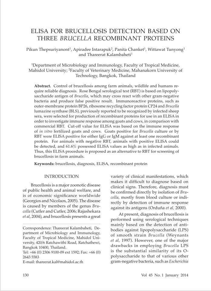
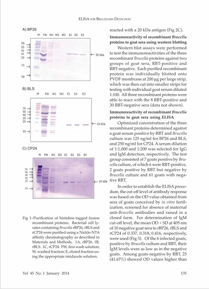
Following induction of protein ex-pression with 1 mM IPTG for 3 hours, bacterial cell lysates and supernatants from sonicated cell pellets were analyzed by western blotting using anti-His tag antibodies, revealing rBP26, rBLS and rCP24 fragments of 35, 23 and 24 kDa, respectively (data not shown). As the majority of the heterologously expressed recombinant proteins were in the soluble fraction (bacterial cell lysate), they were then purified using Ni-NTA affinity chro-matography. RBP26 was eluted with 50 mM imidazole (Fig 1A), and rBLS (Fig 1B) and rCP24 (Fig 1C) with 2 mM imidazole.
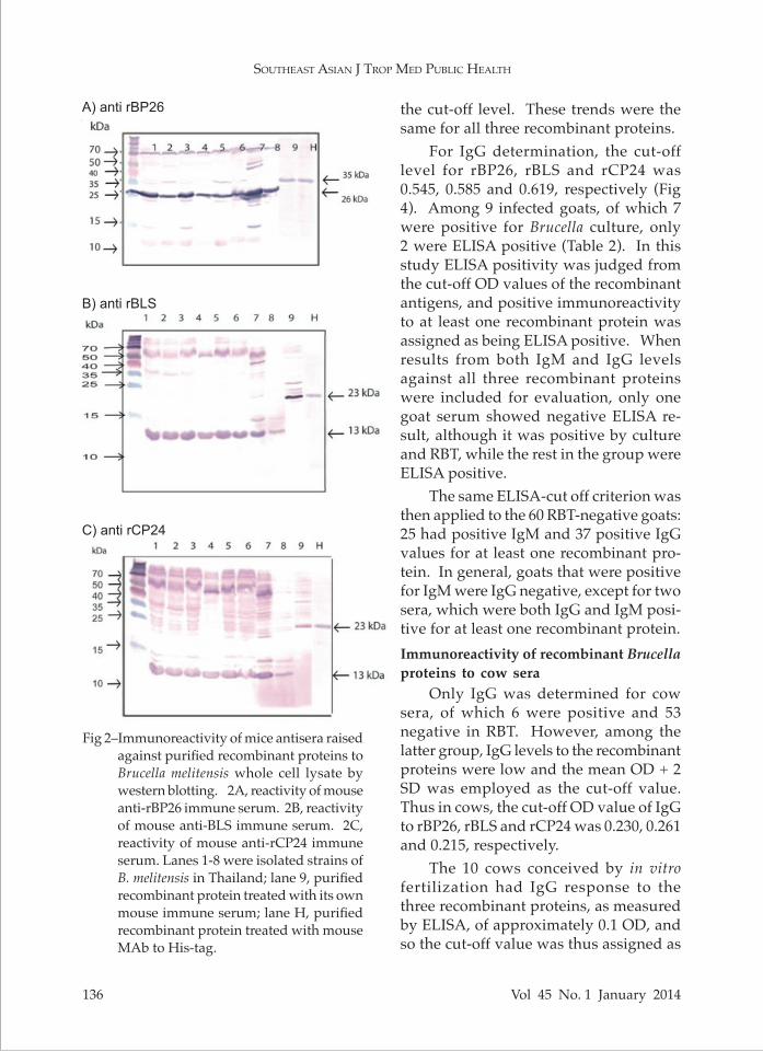
Reactivity of mice anti-recombinant pro-teins immune sera against Brucella whole cell lysate
The immunization regimen produced mice immune sera with reactivity to each recombinant protein of titers > 1:128,000 as determined by ELISA. Immune sera to the three purified recombinant proteins were then tested against Brucella whole cell lysate in order to evaluate their speci-ficities against the natural antigens, which should be 3-5 kDa smaller than their corresponding recombinant antigens. At 1:20,000 dilution, mouse anti-rBP26 (35 kDa) immune serum recognized a 26 kDa protein from Brucella whole cell ly-sate (Fig 2A), that raised against rBLS (23 kDa) recognized a 13 kDa antigen (Fig 2B) and anti-rCP24 (24 kDa) immune serum
eliSa for bruceloSiS detection
Vol 45 No. 1 January 2014 135
Fig 1–Purification of histidine-tagged fusion recombinant proteins. Bacterial cell ly-sates containing Brucella rBP26, rBLS and rCP24 were purified using a Nickle-NTA affinity chromatography as described in Materials and Methods. 1A, rBP26. 1B, rBLS. 1C, rCP24. FW, first wash solution; W, washed fraction; E, eluted fraction us-ing the appropriate imidazole solution.
reacted with a 20 kDa antigen (Fig 2C). Immunoreactivity of recombinant Brucella proteins to goat sera using western blotting
Western blot assays were performed to test the immunoreactivities of the three recombinant Brucella proteins against two groups of goat sera, RBT-positive and RBT-negative. Each purified recombinant protein was individually blotted onto PVDF membrane at 200 µg per large strip, which was then cut into smaller strips for testing with individual goat serum diluted 1:100. All three recombinant proteins were able to react with the 8 RBT-positive and 30 RBT-negative sera (data not shown). Immunoreactivity of recombinant Brucella proteins to goat sera using ELISA
Optimized concentration of the three recombinant proteins determined against a goat serum positive by RBT and Brucella culture was 125 ng/ml for BP26 and BLS, and 250 ng/ml for CP24. A serum dilution of 1:1,000 and 1:200 was selected for IgG and IgM detection, respectively. The test group consisted of 7 goats positive by Bru-cella culture, of which 6 were RBT-positive, 2 goats positive by RBT but negative by Brucella culture and 61 goats with nega-tive RBT.
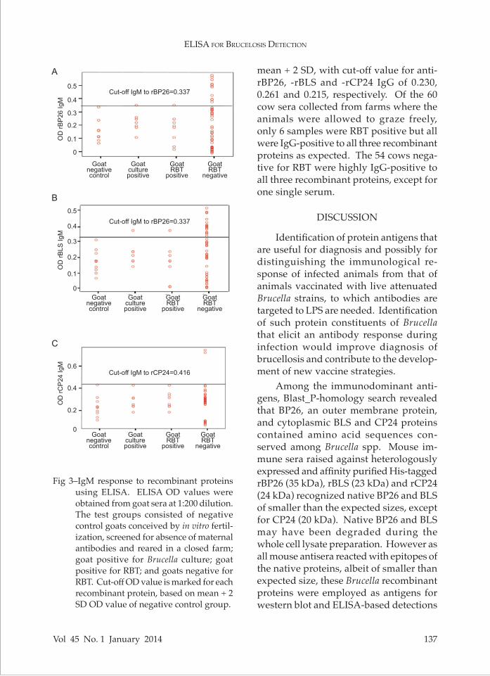
In order to establish the ELISA proce-dure, the cut-off level of antibody response was based on the OD value obtained from sera of goats conceived by in vitro fertil-ization, screened for absence of maternal anti-Brucella antibodies and raised in a closed farm. For determination of IgM cut-off level, the mean OD + SD at 405 nm of 10 negative goat sera to rBP26, rBLS and rCP24 of 0.337, 0.318, 0.416, respectively, were used (Fig 3). Of the 6 infected goats, positive by Brucella culture and RBT, their IgM levels were as low as in the negative goats. Among goats negative by RBT, 25 (41.67%) showed OD values higher than
A) BP26
B) BLS
C) CP24
SoutheaSt aSian J trop Med public health
136 Vol 45 No. 1 January 2014
the cut-off level. These trends were the same for all three recombinant proteins.
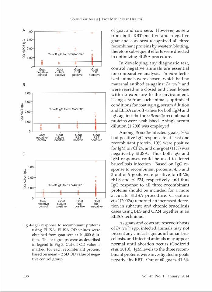
For IgG determination, the cut-off level for rBP26, rBLS and rCP24 was 0.545, 0.585 and 0.619, respectively (Fig 4). Among 9 infected goats, of which 7 were positive for Brucella culture, only 2 were ELISA positive (Table 2). In this study ELISA positivity was judged from the cut-off OD values of the recombinant antigens, and positive immunoreactivity to at least one recombinant protein was assigned as being ELISA positive. When results from both IgM and IgG levels against all three recombinant proteins were included for evaluation, only one goat serum showed negative ELISA re-sult, although it was positive by culture and RBT, while the rest in the group were ELISA positive.
The same ELISA-cut off criterion was then applied to the 60 RBT-negative goats: 25 had positive IgM and 37 positive IgG values for at least one recombinant pro-tein. In general, goats that were positive for IgM were IgG negative, except for two sera, which were both IgG and IgM posi-tive for at least one recombinant protein.Immunoreactivity of recombinant Brucella proteins to cow sera
Only IgG was determined for cow sera, of which 6 were positive and 53 negative in RBT. However, among the latter group, IgG levels to the recombinant proteins were low and the mean OD + 2 SD was employed as the cut-off value. Thus in cows, the cut-off OD value of IgG to rBP26, rBLS and rCP24 was 0.230, 0.261 and 0.215, respectively.
The 10 cows conceived by in vitro fertilization had IgG response to the three recombinant proteins, as measured by ELISA, of approximately 0.1 OD, and so the cut-off value was thus assigned as
Fig 2–Immunoreactivity of mice antisera raised against purified recombinant proteins to Brucella melitensis whole cell lysate by western blotting. 2A, reactivity of mouse anti-rBP26 immune serum. 2B, reactivity of mouse anti-BLS immune serum. 2C, reactivity of mouse anti-rCP24 immune serum. Lanes 1-8 were isolated strains of B. melitensis in Thailand; lane 9, purified recombinant protein treated with its own mouse immune serum; lane H, purified recombinant protein treated with mouse MAb to His-tag.
A) anti rBP26
B) anti rBLS
C) anti rCP24
eliSa for bruceloSiS detection
Vol 45 No. 1 January 2014 137
mean + 2 SD, with cut-off value for anti-rBP26, -rBLS and -rCP24 IgG of 0.230, 0.261 and 0.215, respectively. Of the 60 cow sera collected from farms where the animals were allowed to graze freely, only 6 samples were RBT positive but all were IgG-positive to all three recombinant proteins as expected. The 54 cows nega-tive for RBT were highly IgG-positive to all three recombinant proteins, except for one single serum.
DISCUSSION
Identification of protein antigens that are useful for diagnosis and possibly for distinguishing the immunological re-sponse of infected animals from that of animals vaccinated with live attenuated Brucella strains, to which antibodies are targeted to LPS are needed. Identification of such protein constituents of Brucella that elicit an antibody response during infection would improve diagnosis of brucellosis and contribute to the develop-ment of new vaccine strategies.
Among the immunodominant anti-gens, Blast_P-homology search revealed that BP26, an outer membrane protein, and cytoplasmic BLS and CP24 proteins contained amino acid sequences con-served among Brucella spp. Mouse im-mune sera raised against heterologously expressed and affinity purified His-tagged rBP26 (35 kDa), rBLS (23 kDa) and rCP24 (24 kDa) recognized native BP26 and BLS of smaller than the expected sizes, except for CP24 (20 kDa). Native BP26 and BLS may have been degraded during the whole cell lysate preparation. However as all mouse antisera reacted with epitopes of the native proteins, albeit of smaller than expected size, these Brucella recombinant proteins were employed as antigens for western blot and ELISA-based detections
Goatnegativecontrol
Goatculturepositive
GoatRBT
positive
GoatRBT
negative
Goatnegativecontrol
Goatculturepositive
GoatRBT
positive
GoatRBT
negative
Goatnegativecontrol
Goatculturepositive
GoatRBT
positive
GoatRBT
negative
Cut-off IgM to rBP26=0.337
Cut-off IgM to rBP26=0.337
Cut-off IgM to rCP24=0.416
0.5
0.4
0.3
0.2
0.1
0
0.5
0.4
0.3
0.2
0.1
0
0.6
0.4
0.2
0
OD
rB
LS
Ig
MO
D r
CP
24
Ig
MO
D r
BP
26
Ig
M
Fig 3–IgM response to recombinant proteins using ELISA. ELISA OD values were obtained from goat sera at 1:200 dilution. The test groups consisted of negative control goats conceived by in vitro fertil-ization, screened for absence of maternal antibodies and reared in a closed farm; goat positive for Brucella culture; goat positive for RBT; and goats negative for RBT. Cut-off OD value is marked for each recombinant protein, based on mean + 2 SD OD value of negative control group.
B
A
C
SoutheaSt aSian J trop Med public health
138 Vol 45 No. 1 January 2014
of goat and cow sera. However, as sera from both RBT-positive and -negative goat and cow sera recognized all three recombinant proteins by western blotting, therefore subsequent efforts were directed in optimizing ELISA procedure.
In developing any diagnostic test, control negative animals are essential for comparative analysis. In vitro fertil-ized animals were chosen, which had no maternal antibodies against Brucella and were reared in a closed and clean house with no exposure to the environment. Using sera from such animals, optimized conditions for coating Ag, serum dilution and ELISA cut-off values for both IgM and IgG against the three Brucella recombinant proteins were established. A single serum dilution (1:200) was employed.
Among Brucella-infected goats, 70% had positive IgG response to at least one recombinant protein, 10% were positive for IgM to rCP24, and one goat (11%) was negative by ELISA. Thus both IgG and IgM responses could be used to detect brucellosis infection. Based on IgG re-sponse to recombinant proteins, 4, 5 and 3 out of 9 goats were positive to rBP26; rBLS and rCP24, respectively and thus IgG response to all three recombinant proteins should be included for a more accurate ELISA procedure. Cassataro et al (2002a) reported an increased detec-tion in subacute and chronic brucellosis cases using BLS and CP24 together in an ELISA technique.
As goats and cows are reservoir hosts of Brucella spp, infected animals may not present any clinical signs as in human bru-cellosis, and infected animals may appear normal until abortion occurs (Godfroid et al, 2010). IgM levels to the three recom-binant proteins were investigated in goats negative by RBT. Out of 60 goats, 41.6%
Goatnegativecontrol
Goatculturepositive
GoatRBT
positive
GoatRBT
negative
Goatnegativecontrol
Goatculturepositive
GoatRBT
positive
GoatRBT
negative
Cut-off IgG to rBP26=0.545
Cut-off IgG to rBLS=0.585
Cut-off IgG to rCP24=0.619
Goatnegativecontrol
Goatculturepositive
GoatRBT
positive
GoatRBT
negative
4.00
3.00
2.00
1.00
0
OD
rB
P2
6 I
gG
4.00
3.00
2.00
1.00
0
OD
rB
LS
Ig
G
3.00
2.00
1.00
0
OD
rC
P2
4 I
gG
Fig 4–IgG response to recombinant proteins using ELISA. ELISA OD values were obtained from goat sera at 1:1,000 dilu-tion. The test groups were as described in legend to Fig 3. Cut-off OD value is marked for each recombinant protein, based on mean + 2 SD OD value of nega-tive control group.
B
A
C

eliSa for bruceloSiS detection
Vol 45 No. 1 January 2014 139
protein, except for only one cow, kept in a closed farm that had immunoreactivity similar to control negative animals. Thus our ELISA method was able to identify goats and cows in endemic area, which are reservoirs of Brucella, owing to their exposure to soil-borne Brucella. Abortion in these animals could occur whenever Brucella successfully evades the host im-mune response.
Chaudhuri et al (2010) has employed BP26/omp28 in ELISA to determine anti-body response among cattle, goats and dogs. Delpino et al (2003) could not ap-ply CP24 in ELISA test as the antibody response of the their Brucella-free animal was as high as in brucellosis animal.
RBT is the routine serological diag-nostic method for brucellosis. As the major antigen of this agglutination test is LPS, vaccinated animals would already have antibodies positive to RBT. Thus detection of antibody response to proteins
Sample No. BP26 BLS CP24 Cut-off IgM IgG IgM IgG 1:1,000 IgM IgG 1:1,000 ELISA-OD 1:200 1:1,000 1:200 1:1,000 1:200 1:1,000 validation 0.337 0.545 0.318 0.585 0.416 0.619
1CR 0.350 0.331 0.370 0.567 0.423 0.549 Positive2C 0.258 0.465 0.233 1.421 0.307 0.603 Positive3CR 0.244 0.268 0.235 0.361 0.311 0.366 Negative4CR 0.106 1.767 0.138 0.768 0.182 0.687 Positive5CR 0.217 2.007 0.209 0.425 0.240 0.350 Positive6CR 0.193 0.441 0.172 0.614 0.253 0.567 Positive7CR 0.181 3.790 0.175 0.496 0.233 0.422 Positive8R 0.006 0.935 0.034 0.960 0.348 0.982 Positive9R 0.010 0.508 0.016 0.677 0.332 0.648 Positive
Table 2Evaluation of ELISA-based procedure using recombinant proteins for detection of
Brucella-infected goat.
C, Culture positive; R, Rose Bengal positive.
were IgM positive, and 2 animals with the highest IgM levels were positive to cyto-plasmic proteins (rBLS and rCP24) and not the outer membrane protein (rBP26), suggesting that IgM was elicited during infection and cytoplasmic proteins are ap-propriate detecting-antigens. The number of positive cases for IgM response to rBLS was higher than rCP24, suggesting that IgM level might be detected by employing rBLS only, when those sera were positive to both rCP24 and rBLS. However there were a few cases of sera, that were positive to rCP24 only.
RBT could detect only 88% of posi-tives Brucella cases, and 70% of RBT-negative goat sera had immune response of either IgG or IgM to at least one of the three recombinant proteins, except for 2 sera (3%) that were positive for both IgM and IgG. Similarly, among cows raised in open farms, all had positive immunoreactivity to each recombinant
SoutheaSt aSian J trop Med public health
140 Vol 45 No. 1 January 2014
recognized by the immune response dur-ing Brucella infection is more preferable. The ELISA procedure reported in this study employed three Brucella recombi-nant proteins, BP26, BLS and CP26, as antigens to detect IGM and IgG in goat sera and IgG in cow sera. The critical negative control sera were obtained from animals conceived by in vitro fertilization, screened for absence of maternal anti-Brucella antibodies and raided in a closed farm system. Criterion for an infected goat is ELISA-based positive antibody to at least one of the three recombinant proteins. Thus positive IgM response indicates that the animal has recently acquired Brucella antigen, while high IgG response indicates chronic Brucella infec-tion. Our studies indicate that animals (goats and cows) in endemic areas can be identified as being reservoir hosts for Brucella if antibodies (IgM and/or IgG) to Brucella antigens are detected.
ACKNOWLEDGEMENTS
Support of a PhD student and labo-ratory supplies were from the Strategic Scholarships of the Frontier Research Network for the PhD Program, Office of the Higher Education Commission (CHE-PhD-THA), Ministry of Education, Thailand. We also thank the Faculty of Tropical Medicine, Mahidol University for providing facilities and budget for laboratory supplies.
REFERENCES
Baldi PC, Velikovsky CA, Braden BC, Giambar-tolomei GH, Fossati CA, Goldbaum FA, Structural, functional and immunological studies on a polymeric bacterial protein. Braz J Med Biol Res 2000; 33: 741-7.
Benjathummarak S, Kalambaheti T. Bruce-
ladder multiplex PCR for speciation of Brucella isolates in Thailand. Proceedings of the 1st ASEAN Plus Three Graduate Research Congress 2012: HS109-113.
Cassataro J, Delpino MV, Velikovsky CA, Bruno L, Fossati CA, Baldi PC. Diagnostic usefulness of antibodies against ribosome recycling factor from Brucella melitensis in human or canine brucellosis. Clin Diagn Lab Immunol 2002a; 9: 366-9.
Cassataro J, Velikovsky CA, Giambartolomei GH, et al. Immunogenicity of the Brucella melitensis recombinant ribosome recycling factor-homologous protein and its cDNA. Vaccine 2002b; 20: 1660-9.
Chaudhuri P, Prasad R, Kumar V, Basavaraj-appa AG. Recombinant OMP28 antigen-based indirect ELISA for serodiagnosis of bovine brucellosis. Molec Cell Probes 2010; 24: 142-5.
Cloeckaert A, Debbarh S, Vizcaino N, Saman E, Dubray G, Zygmunt M. Cloning nucleotide sequence, and expression of the Brucella melitensis bp26 gene coding for a protein immunogenic infected sheep. FEMS Microbiol Lett 1996; 140: 139-44.
Cutler SJ, Cutler RR. Brucellosis: the most common bacterial zoonosis. Biomed Scient 2006; 106: 336-41.
Cutler SJ, Whatmore AM, Commander NJ, Brucellosis-new aspects of an old disease. J Appl Microbiol 2005; 98: 1270-81.
Delpino MV, Cassataro J, Fossati CA, Baldi PC. Antibodies to the CP24 protein of Brucella melitensis lack diagnostic usefulness in ovine brucellosis. Vet Microbiol 2003; 93: 101-7.
Georgios P, Nicolaos A. Brucellosis. N Engl J Med 2005; 352: 2325-36.
Godfroid J, Nielsen K, Saengerman C. Diagno-sis of brucellosis in livestock and wildlife. Croat Med J 2010; 51: 296-305.
Goldbaum FA, Leoni J, Wallach JC, Fossati CA. Characterization of an 18-kilodalton Bru-cella cytoplasmic protein which appears to be a serological marker of active infection
eliSa for bruceloSiS detection
Vol 45 No. 1 January 2014 141
of both human and bovine brucellosis. J Clin Microbiol 1993; 31: 2141-5.
Goldbaum FA, Velikovsky CA, Baldi PC, Mörtl S, Bacher A, Fossati CA. The 18-kDa cy-toplasmic protein of Brucella species – an antigen useful for diagnosis– is a lumazine synthase. J Med Microbiol 1999; 48: 833-9.
Letesson J, Tibor A, van Eynde G, et al. Humoral immune responses of Brucella-infected cattle, sheep, and goats to eight purified recombinant Brucella proteins in an indi-rect enzyme-linked immunosorbent assay. Clin Diagn Lab Immunol 1997; 4: 556-64.
Lindler L, Hadfield L, Tall B, et al. Cloning of Brucella melitensis group3 antigen gene encoding Omp28, a protein recognized by the humoral immune response during human brucellosis. Infect Immun 1996; 64: 2490-9.
Muñoz PM, Marín CM, Monreal D, et al. Ef-ficacy of several serological tests and an-tigens for diagnosis of bovine brucellosis in the presence of false-positive serological results due to Yersinia enterocolitica O:9. Clin Diagn Lab Immunol 2005; 12: 141-51.
Orduña A, Almaraz A, Prado A, et al. Evalua-tion of an immunocapture-agglutination test (Brucellacapt) for serodiagnosis of human brucellosis. J Clin Microbiol 2000;
38: 4000-5.Pappas G, Panagopoulou P, Christou L, Akriti-
dis N. Biological weapons. Cell Mol Life Sci 2006; 63: 2229-36.
Rajashekara G, Glasner J, Glover D, et al. Com-parative whole genome hybridization reveals genomic islands in Brucella species. J Bacteriol 2004; 186: 5040-51.
Rossetti O, Arese A, Boschiroli M, Cravero S. Cloning of Brucella abortus gene and characterization of expressed 26-kilodal-ton periplasmic protein: potential use for diagnosis. J Clin Microbiol 1996; 34: 165-9.
Samartino L, Gall D, Gregoret R, Nielsen K. Validation of enzyme-linked immunosor-bent assays f or the diagnosis of bovine brucellosis. Vet Microbiol 1999; 70: 193-200.
Vizcaíno N, Cloeckaert A, Dubray G, Zygmunt MS. Cloning, nucleotide sequence, and expression of the gene coding for a ribo-some releasing factor-homologous protein of Brucella melitensis. Infect Immun 1996; 64: 4834-7.
Weynants V, Gilson D, Cloeckaert A, et al. Characterization of smooth lipopolysac-charides and O polysaccharides of Brucella species by competition binding assays with monoclonal antibodies. Infect Immun 1997; 65: 1939-43.