ems monitoring devices: tips and pitfallsgatheringofeagles.us/presentations2008/kathleen schrank et...
TRANSCRIPT

EMS Monitoring Devices: Tips and Pitfalls
Kathleen Schrank, MD, FACEP, FACP
City of Miami Fire Rescue
University of Miami Miller School of Medicine
Monitors and machines
Assist with patient assessment BUT:
– Do NOT replace eyes-on/hands-on care
– Are just one piece of clinical judgment
– ALL have pitfalls/malfunctions/limitations
– Are more complex than ever
TREAT THE PATIENT, NOT THE MACHINE

ETCO2 & CO2 Waveform Capnography
Used in ORs for decades, then ICUs, some EDs Primary goal: prevent hypoxia by early
identification of hypoventilation Indicates adequacy of ventilation and perfusion Verifies correct position of ETT or LMA Standard of care for patients under general
anesthesia
Rapidly expanding use in EMS Quick poll of EMS Medical Directors: Do you use waveform in ED or med practice? How many use ETCO2 device in EMS?
Which—colorimetric, # Bars, # + waveform?

Alternative tube confirmation devices
ETCO2 and Waveform for EMS
Confirm placement of ETT, Combi, King
Monitor tubes for dislodgement
– After defibs or movement
– Turnover of care at ED
Determine status of patient perfusion
– ROSC post arrest
– Adequacy of CPR
– Confirm if dead dead
Documentation of all the above

ETCO2 and Waveform in EMS
In non-arrest patients:
Determine status of patient ventilation
– Apnea
– Inadequate or excess ventilation
Asthma/COPD vs. CHF
Hyperglycemia vs. DKA

Definitions for training
“Tidal”—respiratory ebb and flow, like the tide
End-tidal CO2 is the # at end of expiration
ETCO2 vs. Serum PCO2
ETCO2 vs. CO2 Waveform Capnography
Capnometry vs. Capnography

Normal ETCO2 & CO2 Waveform
Capnography shows CO2 with ventilation
Nl ETCO2 is ~ 5%
(~ 35-37 mm Hg)
Gradient (PaCO2 to ETCO2): 5-6 mm Hg
ETCO2 estimates PaCO2 in pts with nl lungs
A = end of inhalation
B – D = exhalation
D = ETCO2
D – E = inhalation

Learning ETCO2/Waveform
TAKES TIME, PRACTICE, REFRESHER, QUESTIONS Tracings seem reversed at first
(because of our concept of our breathing pattern) Practice concept by breathing in time with strip Easy if strips look “classic”, but they often don‟t
(field vs. OR) Must consider more than 1 cause for abnormality:
– Ventilation vs. perfusion– Hypoperfusion v. hyperventilation v. tube above cords– Air trapping, dead space– Machine problem vs. Patient problem
Trouble-shooting “weird” waveform is perhaps the hardest EMS skill to master

Tachypneic, Hyperventilating
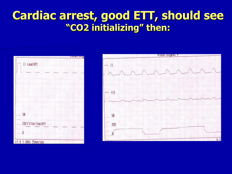
Cardiac arrest, good ETT, should see “CO2 initializing” then:

Arrest: ETT confirmed

„2002 Children’s Medical Center Corporation
ETT Confirmed, Low Perfusion
(slide from Dr. Baruch Krauss)

„2002 Children’s Medical Center Corporation
ETT good, VERY low perfusion(slide from Dr. Krauss)
Ability to switch to lower amplitude range would help

„2002 Children’s Medical Center Corporation
What we hope to see: ROSC!
(slide from Dr. Krauss)

But, where is this ETT?

Flat straight solid black line at 0
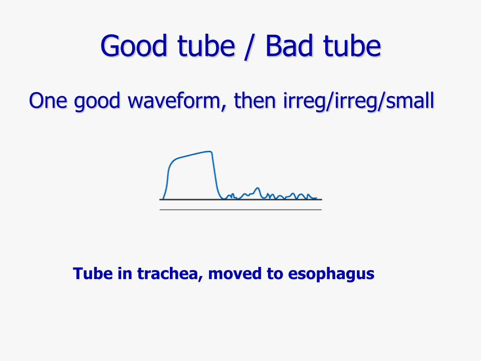
Good tube / Bad tube
One good waveform, then irreg/irreg/small
Tube in trachea, moved to esophagus

Playing with our LP 12
As the sensor is screwed in, you‟ll see:
Screw in more and then fully:

Playing with our LP 12
Microstream sensor screws in 3 rotations When nearly tight, waveform may go from:
- - - - to solid/flat/straight at zero (instead of pt tracing)
Tighten a little, get a waveform & low plateau Tighten more, higher plateau TIGHTEN UNTIL WINGS HORIZONTAL = full
waveform MIGHT LOOSEN during use—looks like tube moved (Tighten too far = broken sensor gold ring)
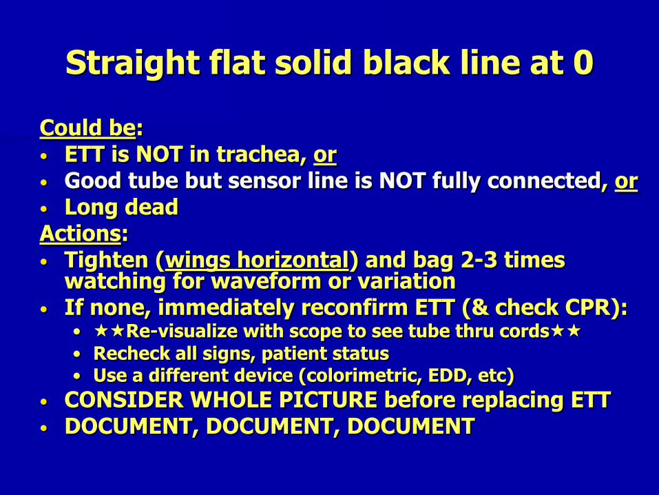
Straight flat solid black line at 0
Could be:• ETT is NOT in trachea, or• Good tube but sensor line is NOT fully connected, or• Long deadActions:• Tighten (wings horizontal) and bag 2-3 times
watching for waveform or variation• If none, immediately reconfirm ETT (& check CPR):
• Re-visualize with scope to see tube thru cords
• Recheck all signs, patient status• Use a different device (colorimetric, EDD, etc)
• CONSIDER WHOLE PICTURE before replacing ETT• DOCUMENT, DOCUMENT, DOCUMENT

More Trouble-shooting (LP12)
• Good waveform but low plateau:• Tube above the cords/hypopharynx, or• Sensor connection not quite tight, or• Perfusion poor
• Is it time to call the code (ET <10)?• Hard to read due to calibration scale 0-50• Be sure that sensor connection is good
• Did tube move after defib?• Tracing pauses after shock (or sensor now loose)
• Nasal sensor: variable readings• Solid straight flat black line at high #= ????

Chest Compressions + ETCO2
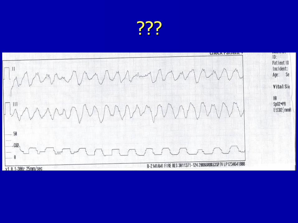
???
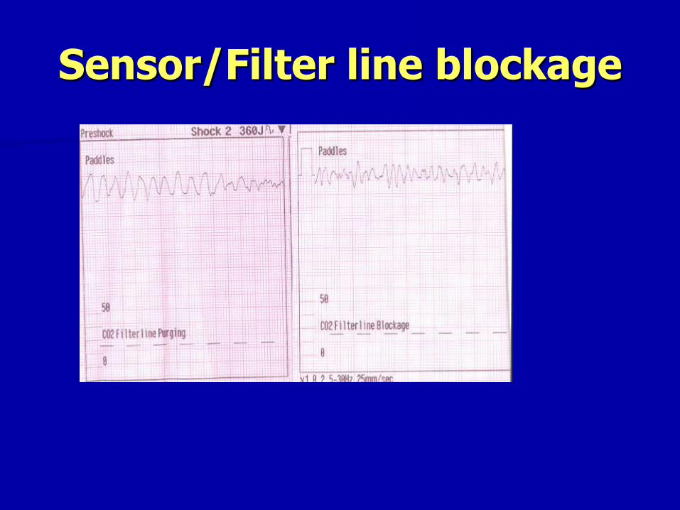
Sensor/Filter line blockage

Filter Line Blockage (LP 12)
„Purging…(30 seconds)…Filter Line Blocked‟
Will not re-start unless line disconnected 1st
BVM: Tight seal on mask may kink line
ETT:
Blood/fluids/vomitus = change filter line
Mechanical kink:
• Re-straighten line, then disconnect and reconnect
• If still bad, change to new filter line

ETCO2 Resources
www.capnography.com
Emscapnography.blogspot.com
Snohomishcountymedics.terapad.com
www.biotel.ws/protocolsHTML/Protocols 2004/capnographyinterpretation.asp
www.physio-control.com/learning/clinical-topics/capnography.aspx

More trouble-shooting
Pulse oximetry
CO monitoring
Pacer capture vs. electrical artifact
– Dispersion from gel in defib/pacing pads
– Must feel mechanical pulse to be sure, or
– Artifact “QRS” will change amplitude with mAs, heart QRS will not
12 lead computer interpretations
Automated BP readings

GENERAL PRINCIPLES #1
Quick differential: Problem with patient? machine? my brain?
Treat the patient, NOT the machine
Actions: Recheck patient‟s ABCs (hands-on) and stabilize
Recheck machine
PUT THE WHOLE PICTURE TOGETHER!
Psychology: Doubt: Often hard to trust self over machine
Denial: Often hard to set one‟s ego aside and use machine info (“That tube is in, damn it”)
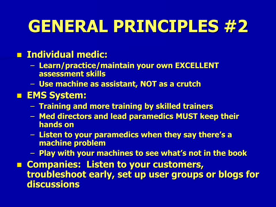
GENERAL PRINCIPLES #2
Individual medic:– Learn/practice/maintain your own EXCELLENT
assessment skills
– Use machine as assistant, NOT as a crutch
EMS System:– Training and more training by skilled trainers
– Med directors and lead paramedics MUST keep their hands on
– Listen to your paramedics when they say there‟s a machine problem
– Play with your machines to see what‟s not in the book
Companies: Listen to your customers, troubleshoot early, set up user groups or blogs for discussions