endometriosis foci differentiation by rapid lipid
TRANSCRIPT

1Scientific RepoRts | 7: 2546 | DOI:10.1038/s41598-017-02708-x
www.nature.com/scientificreports
Endometriosis foci differentiation by rapid lipid profiling using tissue spray ionization and high resolution mass spectrometryVitaliy V. Chagovets1, Zhihao Wang1,2, Alexey S. Kononikhin1,3, Natalia L. Starodubtseva1,3, Anna Borisova1, Dinara Salimova1, Igor A. Popov1,3, Andrey V. Kozachenko1, Konstantin Chingin2, Huanwen Chen 2, Vladimir E. Frankevich1, Leila V. Adamyan1 & Gennady T. Sukhikh1
Obtaining fast screening information on molecular composition of a tissue sample is of great importance for a disease biomarkers search and for online surgery control. In this study, high resolution mass spectrometry analysis of eutopic and ectopic endometrium tissues (90 samples) is done using direct tissue spray mass spectrometry in both positive and negative ion modes. The most abundant peaks in the both ion modes are those corresponding to lipids. Species of three lipid classes are observed, phosphatidylcholines (PC), sphingomyelins (SM) and phosphoethanolamines (PE). Direct tissue analysis gives mainly information on PC and SM lipids (29 species) in positive ion mode and PC, SM and PE lipids (50 species) in negative ion mode which gives complementary data for endometriosis foci differentiation. The biggest differences were found for phospholipids with polyunsaturated acyls and alkils. Although, tissue spray shows itself as appropriate tool for tissue investigation, caution should be paid to the interpretation of mass spectra because of their higher complexity with more possible adducts formation and multiple interferences must be taken into account. The present work extends the application of direct tissue analysis for the rapid differentiation between endometriotic tissues of different foci.
Endometriosis is an abundant gynecological pathology of poorly understood pathogenesis affecting 10% of women1. It is characterized by the extrauterine presence of endometrial glands and stroma. The disease affects mostly women in their reproductive age and can cause wide set of non-specific symptoms including infertility, dysmenorrhea, dyspareunia, and non-cyclic pelvic pain. The only reliable way to diagnose the pathology is sur-gical laparoscopy for the moment. Many efforts have been applied to find biomarkers of endometriosis and to develop less invasive methods to reveal the presence of early stages2. Such investigations were mainly devoted to body fluids screening3–7. Lack of the information about molecular composition of eutopic (inside uterine) and ectopic (extrauterine) endometrium is observed. There are hypotheses about alteration in composition of ectopic and eutopic endometrial tissues and in surrounding tissue which allows eutopic endometrium survival. Therefore, tissue investigation can shed some light on mechanisms of the disease and be used for validation of biomarkers found in fluids in low-invasive way. Data on eutopic and ectopic endometrium composition is also important for the development of the online surgery control to determine volume of an operation. This necessi-tates development of method for fast screening of big amount of tissue samples. Wide variety of approaches for tissue composition analysis are present nowadays8: matrix-assisted laser desorption/ionization imaging, desorp-tion electrospray ionization, etc. One of the ambient methods for a sample analysis with minimal pretreatment is suggested by Cooks’ group and named “leaf-spray”9. This method was further extrapolated for fast tissue analysis
1V.I. Kulakov Research Center for Obstetrics, Gynecology and Perinatology, Ministry of Healthcare of the Russian Federation, 4 Oparina str., 117997, Moscow, Russia. 2Jiangxi Key Laboratory for Mass Spectrometry and Instrumentation, East China University of Technology, 418 Guanglan road, 330013, Nanchang, China. 3Moscow Institute of Physics and Technology, 141700, Dolgoprudnyi, Moscow Region, Russia. Correspondence and requests for materials should be addressed to H.C. (email: [email protected]) or V.E.F. (email: [email protected])
Received: 20 January 2017
Accepted: 18 April 2017
Published: xx xx xxxx
OPEN

www.nature.com/scientificreports/
2Scientific RepoRts | 7: 2546 | DOI:10.1038/s41598-017-02708-x
and turned into different variations of tissue spray10–15. The method has been applied to the analysis of brain tis-sues and various cancer tumors10, 13.
The present work extends the application of direct tissue analysis for the rapid differentiation between endo-metriotic tissues of different foci. Features of different tissues are identified by conventional lipidomic approach including lipid extraction and following analysis by hydrophilic interaction liquid chromatography with electro-spray ionization mass spectrometry (HILIC-LC/ESI-MS)16, 17.
ResultsThe design of MS analysis of tissue samples and the ion source scheme are presented in Fig. 1. The following set-tings are varied until obtaining stable TIC in both positive and negative ion modes: distance from tissue tip to the mass spectrometer inlet is varied in the range 5–50 mm; applied potential in the range 2–5 kV, extracting solvent flow rate in the range 5–50 µL/min. Figure S1 shows extracted positive ion chromatograms of some ions obtained with optimized settings. Relative standard deviation of a peak intensity is within 5% for the same tissue piece and about 5–10% for neighboring pieces. Then the scheme of an analysis for every tissue sample is as follows. A piece of tissue of approximate size 2 × 1 × 1 mm is cut from a frozen sample, thawed and fixed on a needle. After that, an MS analysis starts. The suggested construction gives better tissue fixation and control.
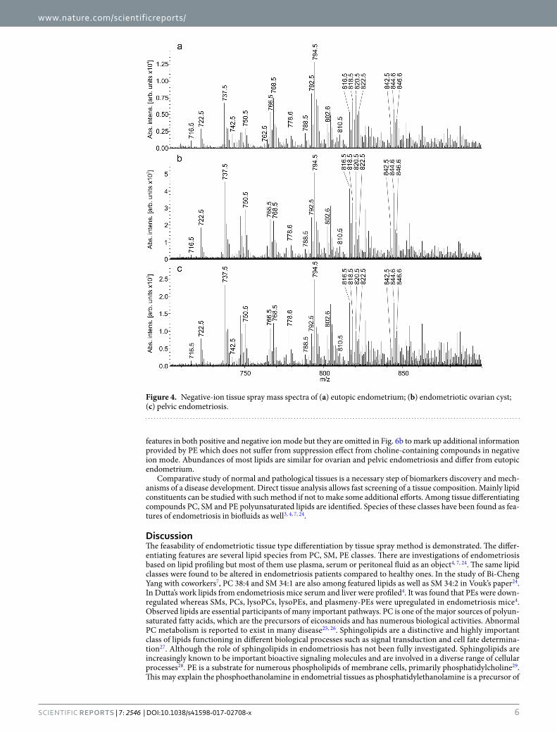
Positive ion mode. Representative mass spectra of three kinds of endometriotic tissue from one patient collected in positive ion mode are shown in Fig. 2. A total of 1159 ions in sample tissues in positive ion mode are detected over threshold of 200 counts in different tissue types. The most abundant m/z correspond to lipid species.
Figure 1. Flowchart of the experiment and schema of the direct tissue spray ion source.
Figure 2. Positive ion tissue spray mass spectra of (a) eutopic endometrium; (b) endometriotic ovarian cyst; (c) pelvic endometriosis.

www.nature.com/scientificreports/
3Scientific RepoRts | 7: 2546 | DOI:10.1038/s41598-017-02708-x
The identification is done in the following way illustrated for m/z 782.5685. An accurate mass of a compound which abundance exceeds a threshold in 200 counts is found from the mass spectrum. Tentative assignment of the compound is done based on LIPID MAPS data18 within 10 ppm mass accuracy. For the considered m/z it can be either protonated species of PC 36:4, PE 39:4 or sodiated species of PC 34:1, PE 37:1. MS/MS information about the fragmentation pattern of the considered m/z is used for better assignment (Figure S2a). Peaks with m/z 147 and 184 are observed in the tandem mass spectra. These peaks are characteristic for fragmentation of sodiated and protonated phosphatidylcholine, correspondingly, and originate from its polar head group19, 20. This fact leads to a conclusion that m/z 782 precursor ion corresponds to interfered protonated PC 36:4 and sodiated PC 34:1. Such peak overlapping is known problem upon lipids study in positive ion mode19, 21, 22. Identified lipid species are listed in Table 1 and some of them are marked with opened circles in Fig. 2. Additional lipids validation is done by HILIC-LC/MS analysis of tissue’s lipid extract using methods described earlier16, 17. Figure S3 represents resulting TIC from HILIC-LC/MS analysis of the lipid extract. Annotated chromatographic peaks correspond to lipid species observed in tissue spray experiments. Retention time of the observed lipids (Table S2) correlates with literature data16, 17 and confirms identification provided in Tables 1 and 2.
Series of peaks denoted with closed circles are worth of consideration (Fig. 2). Their profile is similar to that of the opened circle peaks. The difference between these two groups is 16 Da which can evidence about oxidation of lipids during direct tissue analysis. Such effect has been observed in another ambient method, DESI23. This conclusion is supported by the presence of m/z 163 peak in tandem mass spectra of m/z 799 (Figure S2b). It is also 16 Da bigger then the characteristic fragment of polar head group of sodiated PC. Another explanation of this series can be potassium cation attachment to the initial lipid molecules.
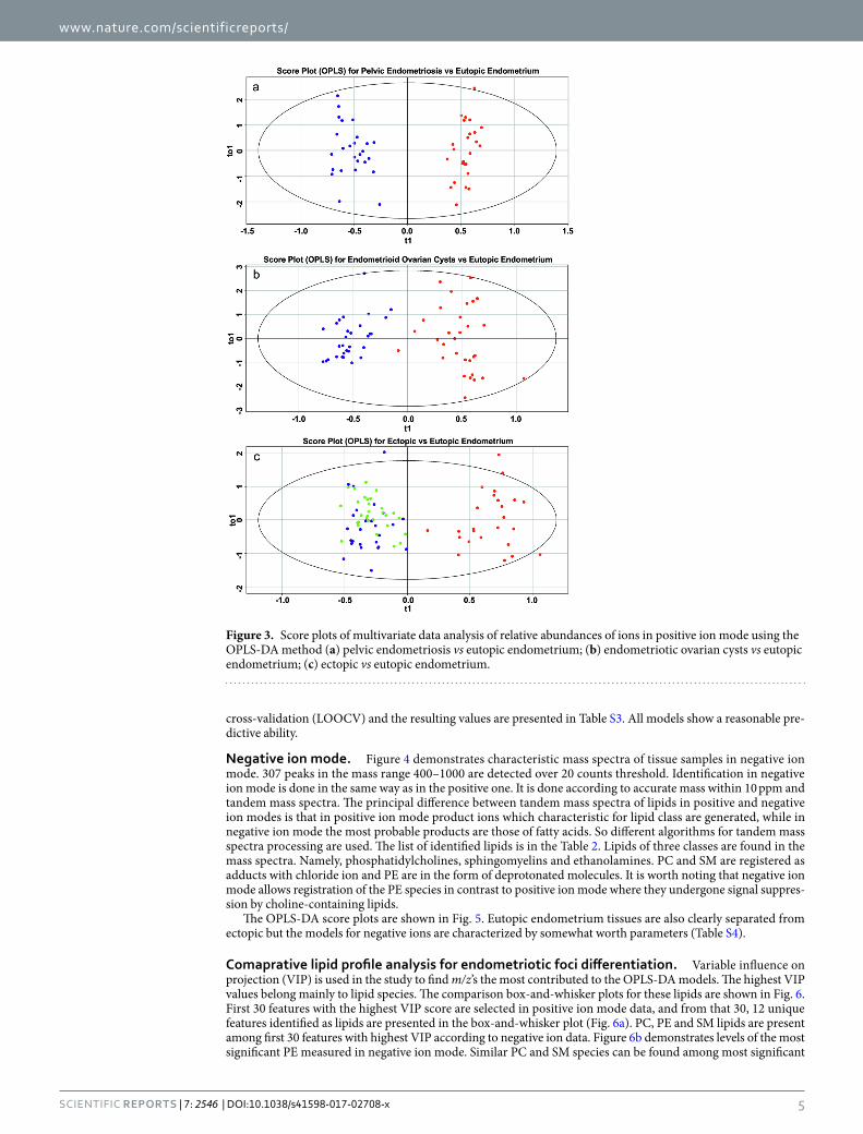
The supervised OPLS-DA model is used to separate different tissue types and find the differentially produced metabolites. As shown in Fig. 3, the OPLS-DA model can separate eutopic endometrium from ectopic one, while two types of endometriotic foci are not clustered demonstrating higher similarity compared to eutopic endome-trium (Fig. 3c). R2 values representing explained variance of the data are extracted from the models and listed in Table S3. Predictive capability of the models is estimated by Q2. This parameter is obtained with Leave-one-out
Lipid
[M + Na]+ [M + O + Na]+
Theoretical Experimen- tal Mass accuracy [ppm] Theoretical Experimen- tal Mass accuracy [ppm]
Phosphatidylcholines
PC 30:0 728.5200 728.5237 5 744.5160 n.d.
PC O-32:0 742.5721 742.5665 8 758.5635 n.d.
PC 32:1 754.5357 754.5393 5 770.5308 770.5278 4
PC 32:0 756.5513 756.5537 3 772.5476 772.5480 1
PC O-34:1 768.5877 768.5824 7 784.5805 784.5846 5
PC 34:2 780.5513 780.5544 4 796.5467 796.5436 4
PC 34:1 782.5670 782.5700 4 798.5624 798.5656 4
PC O-36:4 790.5721 790.5651 9 806.5647 806.5630 2
PC 36:4 804.5513 804.5543 4 820.5462 820.5444 2
PC 36:3 806.5670 806.5606 8 822.5456 822.5549 11
PC 36:2 808.5826 808.5856 4 824.5771 824.5736 4
PC 36:1 810.5983 810.6011 3 826.5911 826.5878 4
PC O-38:5 816.5877 816.5893 2 832.5814 832.5909 11
PC 38:7 826.5357 826.5368 1 842.5280 842.5401 14
PC 38:5 830.5670 830.5700 4 846.5605 846.5595 1
PC 38:4 832.5826 832.5859 4 848.5777 848.5749 3
PC 38:3 834.5983 834.5958 3 850.5932 850.5853 9
PC 40:7 854.5670 854.5654 2 870.5591 870.5661 8
PC 40:6 856.5826 856.5858 4 872.5739 872.5719 2
Sphingomyelins
SM 32:1 697.5254 697.5318 9 713.5203 713.5264 9
SM 33:1 711.5411 711.5477 9 727.5360 727.539 4
SM 34:2 723.5411 723.5477 9 739.5360 739.5421 8
SM 34:1 725.5567 725.5628 8 741.5516 741.5539 3
SM 36:1 753.5880 753.5934 7 769.5829 769.5848 2
SM 38:1 781.6193 781.6154 5 797.6142 797.6107 4
SM 40:1 809.6506 809.6539 4 825.6455 825.6507 6
SM 42:3 833.6506 833.6535 3 849.6455 849.6479 3
SM 42:2 835.6663 835.6737 9 851.6612 851.6645 4
SM 42:1 837.6819 837.6860 5 853.6768 853.6757 1
Table 1. Theoretical and experimental m/z values of lipid species identified in positive ion mode in tissue samples with measured mass accuracy.
www.nature.com/scientificreports/
4Scientific RepoRts | 7: 2546 | DOI:10.1038/s41598-017-02708-x
Lipid Theoretical Experimental Mass accuracy [ppm]
Phosphatidyletanoamines [M − H]−
PE O-34:2 700.5287 700.5357 10
PE 34:1 716.5236 716.5302 9
PE O-36:5 722.5130 722.5198 9
PE O-36:3 726.5443 726.5465 3
PE O-36:2 728.5599 728.5622 3
PE 36:4 738.5079 738.5062 2
PE 36:2 742.5392 742.5417 3
PE 36:1 744.5549 744.5569 3
PE O-38:7 746.5130 746.5145 2
PE O-38:6 748.5286 748.5323 5
PE O-38:5 750.5443 750.5491 6
PE 38:7 760.4922 760.4952 4
PE 38:6 762.5079 762.5052 4
PE 38:5 764.5235 764.5308 9
PE 38:4 766.5392 766.5364 4
PE O-40:6 776.5599 776.5565 4
PE O-40:5 778.5756 778.5728 4
PE 40:8 786.5079 786.5156 10
PE 40:7 788.5235 788.5256 3
PE 40:5 792.5548 792.5510 5
PE 40:4 794.5705 794.5743 5
PE 42:10 810.5079 810.5096 2
PE 42:9 812.5235 812.5269 4
Phosphatidylcholines [M + Cl]−
PC 30:0 740.50018 740.5013 2
PC O-32:0 754.55228 754.5545 3
PC 32:1 766.51588 766.5214 7
PC 32:0 768.53148 768.5325 1
PC O-34:1 780.56788 780.5639 5
PC 34:2 792.53148 792.5296 2
PC 34:1 794.54718 794.545 3
PC O-36:4 802.55228 802.5551 4
PC 36:4 816.53148 816.5299 2
PC 36:3 818.54718 818.5456 2
PC 36:2 820.56278 820.5605 3
PC 36:1 822.57848 822.5703 10
PC O-38:5 828.56788 828.5644 4
PC 38:7 838.51588 838.5127 4
PC 38:5 842.54718 842.5397 9
PC 38:4 844.56278 844.5558 8
PC 38:3 846.57848 846.5731 6
PC 40:7 866.54718 866.5383 10
PC 40:6 868.56278 868.5582 5
Sphingomyelins [M + Cl]−
SM 32:1 709.50558 709.5011 6
SM 33:1 723.52128 723.5198 2
SM 34:2 735.52128 735.5283 10
SM 34:1 737.53688 737.5351 2
SM 36:1 765.56818 765.5732 7
SM 42:3 845.63078 845.6332 3
SM 42:2 847.64648 847.6545 9
SM 42:1 849.66208 849.6568 6
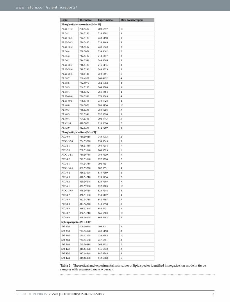
Table 2. Theoretical and experimental m/z values of lipid species identified in negative ion mode in tissue samples with measured mass accuracy.
www.nature.com/scientificreports/
5Scientific RepoRts | 7: 2546 | DOI:10.1038/s41598-017-02708-x
cross-validation (LOOCV) and the resulting values are presented in Table S3. All models show a reasonable pre-dictive ability.
Negative ion mode. Figure 4 demonstrates characteristic mass spectra of tissue samples in negative ion mode. 307 peaks in the mass range 400–1000 are detected over 20 counts threshold. Identification in negative ion mode is done in the same way as in the positive one. It is done according to accurate mass within 10 ppm and tandem mass spectra. The principal difference between tandem mass spectra of lipids in positive and negative ion modes is that in positive ion mode product ions which characteristic for lipid class are generated, while in negative ion mode the most probable products are those of fatty acids. So different algorithms for tandem mass spectra processing are used. The list of identified lipids is in the Table 2. Lipids of three classes are found in the mass spectra. Namely, phosphatidylcholines, sphingomyelins and ethanolamines. PC and SM are registered as adducts with chloride ion and PE are in the form of deprotonated molecules. It is worth noting that negative ion mode allows registration of the PE species in contrast to positive ion mode where they undergone signal suppres-sion by choline-containing lipids.
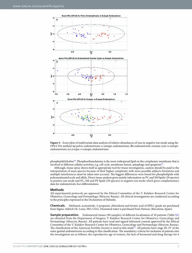
The OPLS-DA score plots are shown in Fig. 5. Eutopic endometrium tissues are also clearly separated from ectopic but the models for negative ions are characterized by somewhat worth parameters (Table S4).
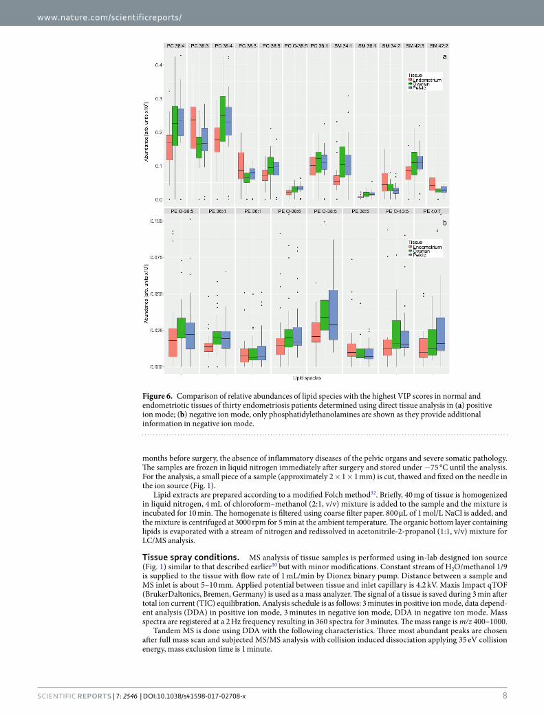
Comaprative lipid profile analysis for endometriotic foci differentiation. Variable influence on projection (VIP) is used in the study to find m/z’s the most contributed to the OPLS-DA models. The highest VIP values belong mainly to lipid species. The comparison box-and-whisker plots for these lipids are shown in Fig. 6. First 30 features with the highest VIP score are selected in positive ion mode data, and from that 30, 12 unique features identified as lipids are presented in the box-and-whisker plot (Fig. 6a). PC, PE and SM lipids are present among first 30 features with highest VIP according to negative ion data. Figure 6b demonstrates levels of the most significant PE measured in negative ion mode. Similar PC and SM species can be found among most significant
Figure 3. Score plots of multivariate data analysis of relative abundances of ions in positive ion mode using the OPLS-DA method (a) pelvic endometriosis vs eutopic endometrium; (b) endometriotic ovarian cysts vs eutopic endometrium; (c) ectopic vs eutopic endometrium.
www.nature.com/scientificreports/
6Scientific RepoRts | 7: 2546 | DOI:10.1038/s41598-017-02708-x
features in both positive and negative ion mode but they are omitted in Fig. 6b to mark up additional information provided by PE which does not suffer from suppression effect from choline-containing compounds in negative ion mode. Abundances of most lipids are similar for ovarian and pelvic endometriosis and differ from eutopic endometrium.
Comparative study of normal and pathological tissues is a necessary step of biomarkers discovery and mech-anisms of a disease development. Direct tissue analysis allows fast screening of a tissue composition. Mainly lipid constituents can be studied with such method if not to make some additional efforts. Among tissue differentiating compounds PC, SM and PE polyunsaturated lipids are identified. Species of these classes have been found as fea-tures of endometriosis in biofluids as well3, 4, 7, 24.
DiscussionThe feasability of endometriotic tissue type differentiation by tissue spray method is demonstrated. The differ-entiating features are several lipid species from PC, SM, PE classes. There are investigations of endometriosis based on lipid profiling but most of them use plasma, serum or peritoneal fluid as an object4, 7, 24. The same lipid classes were found to be altered in endometriosis patients compared to healthy ones. In the study of Bi-Cheng Yang with coworkers7, PC 38:4 and SM 34:1 are also among featured lipids as well as SM 34:2 in Vouk’s paper24. In Dutta’s work lipids from endometriosis mice serum and liver were profiled4. It was found that PEs were down-regulated whereas SMs, PCs, lysoPCs, lysoPEs, and plasmeny-PEs were upregulated in endometriosis mice4. Observed lipids are essential participants of many important pathways. PC is one of the major sources of polyun-saturated fatty acids, which are the precursors of eicosanoids and has numerous biological activities. Abnormal PC metabolism is reported to exist in many disease25, 26. Sphingolipids are a distinctive and highly important class of lipids functioning in different biological processes such as signal transduction and cell fate determina-tion27. Although the role of sphingolipids in endometriosis has not been fully investigated. Sphingolipids are increasingly known to be important bioactive signaling molecules and are involved in a diverse range of cellular processes28. PE is a substrate for numerous phospholipids of membrane cells, primarily phosphatidylcholine29. This may explain the phosphoethanolamine in endometrial tissues as phosphatidylethanolamine is a precursor of
Figure 4. Negative-ion tissue spray mass spectra of (a) eutopic endometrium; (b) endometriotic ovarian cyst; (c) pelvic endometriosis.
www.nature.com/scientificreports/
7Scientific RepoRts | 7: 2546 | DOI:10.1038/s41598-017-02708-x
phosphatidylcholine30. Phosphoethanolamine is the most widespread lipid on the cytoplasmic membrane that is involved in different cellular activities, e.g. cell cycle, membrane fusion, autophagy and apoptosis29.
Although, tissue spray shows itself as appropriate tool for tissue investigation, caution should be paid to the interpretation of mass spectra because of their higher complexity with more possible adducts formation and multiple interferences must be taken into account. The biggest differences were found for phospholipids with polyunsaturated acyls and alkils. Direct tissue analysis gives mainly information on PC and SM lipids (29 species) in positive ion mode and PC, SM and PE lipids (50 species) in negative ion mode which gives complementary data for endometriotic foci differentiation.
MethodsAll experimental protocols are approved by the Ethical Committee of the V. Kulakov Research Center for Obstetrics, Gynecology and Perinatology (Moscow, Russia). All clinical investigations are conducted according to the principles expressed in the Declaration of Helsinki.
Chemicals. Methanol, acetonitrile, 2-propanol, chloroform and formic acid of HPLC grade are purchased from Sigma-Aldrich (St. Louis, MO, USA). Deionized water is purchased from Panreac (Barcelona, Spain).
Sample preparation. Endometrial tissues (90 samples) of different localization of 30 patients (Table S1) are obtained from the Department of Surgery, V. Kulakov Research Center for Obstetrics, Gynecology and Perinatology (Moscow, Russia). All patients have read and signed Informed consent approved by the Ethical Committee of the V. Kulakov Research Center for Obstetrics, Gynecology and Perinatology (Moscow, Russia). The classification of the American Fertility Society is used in this study31. All patients have stage III–IV of the outer genital endometriosis according to this classification. The mandatory criteria for inclusion of patients into the investigation are as follows: the reproductive age of women, the lack of hormonal and drug therapy for 6
Figure 5. Score plots of multivariate data analysis of relative abundances of ions in negative ion mode using the OPLS-DA method (a) pelvic endometriosis vs eutopic endometrium; (b) endometriotic ovarian cysts vs eutopic endometrium; (c) ectopic vs eutopic endometrium.
www.nature.com/scientificreports/
8Scientific RepoRts | 7: 2546 | DOI:10.1038/s41598-017-02708-x
months before surgery, the absence of inflammatory diseases of the pelvic organs and severe somatic pathology. The samples are frozen in liquid nitrogen immediately after surgery and stored under −75 °C until the analysis. For the analysis, a small piece of a sample (approximately 2 × 1 × 1 mm) is cut, thawed and fixed on the needle in the ion source (Fig. 1).
Lipid extracts are prepared according to a modified Folch method32. Briefly, 40 mg of tissue is homogenized in liquid nitrogen, 4 mL of chloroform–methanol (2:1, v/v) mixture is added to the sample and the mixture is incubated for 10 min. The homogenate is filtered using coarse filter paper. 800 µL of 1 mol/L NaCl is added, and the mixture is centrifuged at 3000 rpm for 5 min at the ambient temperature. The organic bottom layer containing lipids is evaporated with a stream of nitrogen and redissolved in acetonitrile-2-propanol (1:1, v/v) mixture for LC/MS analysis.
Tissue spray conditions. MS analysis of tissue samples is performed using in-lab designed ion source (Fig. 1) similar to that described earlier10 but with minor modifications. Constant stream of H2O/methanol 1/9 is supplied to the tissue with flow rate of 1 mL/min by Dionex binary pump. Distance between a sample and MS inlet is about 5–10 mm. Applied potential between tissue and inlet capillary is 4.2 kV. Maxis Impact qTOF (BrukerDaltonics, Bremen, Germany) is used as a mass analyzer. The signal of a tissue is saved during 3 min after total ion current (TIC) equilibration. Analysis schedule is as follows: 3 minutes in positive ion mode, data depend-ent analysis (DDA) in positive ion mode, 3 minutes in negative ion mode, DDA in negative ion mode. Mass spectra are registered at a 2 Hz frequency resulting in 360 spectra for 3 minutes. The mass range is m/z 400–1000.
Tandem MS is done using DDA with the following characteristics. Three most abundant peaks are chosen after full mass scan and subjected MS/MS analysis with collision induced dissociation applying 35 eV collision energy, mass exclusion time is 1 minute.
Figure 6. Comparison of relative abundances of lipid species with the highest VIP scores in normal and endometriotic tissues of thirty endometriosis patients determined using direct tissue analysis in (a) positive ion mode; (b) negative ion mode, only phosphatidylethanolamines are shown as they provide additional information in negative ion mode.

www.nature.com/scientificreports/
9Scientific RepoRts | 7: 2546 | DOI:10.1038/s41598-017-02708-x
HILIC-LC conditions. Extract of endometriotic tissue is analyzed using a Spherisorb Si column (150 × 2.1 mm, 5 µm; Waters, Milford, MA, USA), a flow rate of 50 µL/min, an injection volume of 3 µL, column temperature of 40 °C and a mobile phase gradient: 0–0.5 min−6% B, 60.5 min−23% B, 61–64 min−6% B where solvent B is 5 mM aqueous ammonium acetate. Solvent A is acetonitrile16, 17. All LC/MS experiments are per-formed on the Dionex UltiMate 3000 liquid chromatograph (ThermoScientific, Germering, Germany) coupled to the Maxis Impact qTOF analyzer with ESI (Bruker Daltonics, Bremen, Germany).
ESI-MS conditions. Maxis Impact qTOF is used in the HILIC-LC/MS method with ESI (Bruker Daltonics, Bremen, Germany). Mass spectra are obtained in positive ion and negative ion modes in the mass range m/z 400–1000 with the following setting of tuning parameters: capillary voltage 4.1 kV in positive ion mode (3.0 kV in negative ion mode), pressure of the nebulizing gas 0.7 bar, drying gas flow rate 6 L/min, and temperature of the drying gas 200 °C.
Data analysis. Obtained mass spectra from each sample is averaged over 3 min and saved in m/z – Intensity tables using DataAnalysis software (BrukerDaltonica, Bremen, Germany). Thus obtained data is processed with scaling on TIC and peak alignment. Multivariate data analyses is performed using orthogonal projections onto latent structures discriminant analysis (OPLS-DA) method33 implemented in ropls package34. Multivariate mod-els are described using R2 and Q2 parameters, where R2 describes fraction of data that the model can explain using the latent variables, and Q2 describes part of data predicted by the model according to the cross validation.
Tentative lipid identification is conducted with in-lab created R code. The code searches a record in LIPID MAPS database18 within 10 ppm from the experimental m/z. More precise identification is done based on the MS/MS data for the peak under consideration, if it undergone MS/MS analysis.
References 1. Giudice, L. C. & Kao, L. C. Endometriosis. Lancet 364, 1789–1799, doi:10.1016/S0140-6736(04)17403-5 (2004). 2. Evers, J. L., Dunselman, G. A. & Van der Linden, P. J. Markers for endometriosis. Bailliere’s clinical obstetrics and gynaecology 7,
715–739 (1993). 3. Cordeiro, F. B. et al. Lipidomics analysis of follicular fluid by ESI-MS reveals potential biomarkers for ovarian endometriosis. J Assist
Reprod Genet, doi:10.1007/s10815-015-0592-1 (2015). 4. Dutta, M. et al. Metabolomics Reveals Altered Lipid Metabolism in a Mouse Model of Endometriosis. Journal of proteome research,
doi:10.1021/acs.jproteome.6b00197 (2016). 5. Gupta, S. et al. Serum and peritoneal abnormalities in endometriosis: potential use as diagnostic markers. Minerva ginecologica 58,
527–551 (2006). 6. Lee, Y. H. et al. Limited value of pro-inflammatory oxylipins and cytokines as circulating biomarkers in endometriosis - a targeted
‘omics study. Scientific reports 6, 26117, doi:10.1038/srep26117 (2016). 7. Yang, B.-C. et al. Serum metabolic profiling study of endometriosis by using wooden-tip electrospray ionization mass spectrometry.
Analytical Methods 7, 6125 (2015). 8. Laskin, J. & Lanekoff, I. Ambient Mass Spectrometry Imaging Using Direct Liquid Extraction Techniques. Analytical chemistry 88,
52–73, doi:10.1021/acs.analchem.5b04188 (2016). 9. Malaj, N., Ouyang, Z., Sindona, G. & Cooks, R. G. Analysis of pesticide residues by leaf spray mass spectrometry. Analytical Methods
4, 1913–1919, doi:10.1039/C2AY25222H (2012). 10. Kononikhin, A. et al. A novel direct spray-from-tissue ionization method for mass spectrometric analysis of human brain tumors.
Analytical and bioanalytical chemistry 407, 7797–7805, doi:10.1007/s00216-015-8947-0 (2015). 11. Liu, J., Cooks, R. G. & Ouyang, Z. Biological Tissue Diagnostics Using Needle Biopsy and Spray Ionization Mass Spectrometry.
Analytical chemistry 83, 9221–9225, doi:10.1021/ac202626f (2011). 12. Hu, B., Lai, Y. H., So, P. K., Chen, H. & Yao, Z. P. Direct ionization of biological tissue for mass spectrometric analysis. The Analyst
137, 3613–3619, doi:10.1039/c2an16223g (2012). 13. Wei, Y. et al. Tissue spray ionization mass spectrometry for rapid recognition of human lung squamous cell carcinoma. Scientific
reports 5, 10077, doi:10.1038/srep10077 (2015). 14. Zhang, H. et al. Direct characterization of bulk samples by internal extractive electrospray ionization mass spectrometry. Scientific
reports 3, 2495, doi:10.1038/srep02495 (2013). 15. Zhang, H. et al. Direct Assessment of Phytochemicals Inherent in Plant Tissues Using Extractive Electrospray Ionization Mass
Spectrometry. Journal of agricultural and food chemistry 61, 10691–10698, doi:10.1021/jf4032469 (2013). 16. Cifkova, E., Holcapek, M. & Lisa, M. Nontargeted lipidomic characterization of porcine organs using hydrophilic interaction liquid
chromatography and off-line two-dimensional liquid chromatography-electrospray ionization mass spectrometry. Lipids 48, 915–928, doi:10.1007/s11745-013-3820-4 (2013).
17. Lisa, M., Cifkova, E. & Holcapek, M. Lipidomic profiling of biological tissues using off-line two-dimensional high-performance liquid chromatography-mass spectrometry. Journal of chromatography. A 1218, 5146–5156, doi:10.1016/j.chroma.2011.05.081 (2011).
18. Sud, M. et al. LMSD: LIPID MAPS structure database. Nucleic Acids Research 35, D527–D532, doi:10.1093/nar/gkl838 (2007). 19. Al-Saad, K. A., Siems, W. F., Hill, H. H., Zabrouskov, V. & Knowles, N. R. Structural analysis of phosphatidylcholines by post-source
decay matrix-assisted laser desorption/ionization time-of-flight mass spectrometry. Journal of the American Society for Mass Spectrometry 14, 373–382, doi:10.1016/S1044-0305(03)00068-0 (2003).
20. Hsu, F. F. & Turk, J. Electrospray ionization with low-energy collisionally activated dissociation tandem mass spectrometry of glycerophospholipids: mechanisms of fragmentation and structural characterization. Journal of chromatography. B, Analytical technologies in the biomedical and life sciences 877, 2673–2695, doi:10.1016/j.jchromb.2009.02.033 (2009).
21. Schiller, J. et al. CsCl as an auxiliary reagent for the analysis of phosphatidylcholine mixtures by matrix-assisted laser desorption and ionization time-of-flight mass spectrometry (MALDI-TOF MS). Chemistry and physics of lipids 113, 123–131 (2001).
22. Chagovets, V. et al. Peculiarities of data interpretation upon direct tissue analysis by Fourier transform ion cyclotron resonance mass spectrometry. European journal of mass spectrometry 22, 123–126 (2016).
23. Pasilis, S. P., Kertesz, V. & Van Berkel, G. J. Unexpected analyte oxidation during desorption electrospray ionization-mass spectrometry. Analytical chemistry 80, 1208–1214, doi:10.1021/ac701791w (2008).
24. Vouk, K. et al. Discovery of phosphatidylcholines and sphingomyelins as biomarkers for ovarian endometriosis. Human reproduction 27, 2955–2965, doi:10.1093/humrep/des152 (2012).
25. Franca, P. et al. Abnormal Choline Phospholipid Metabolism in Breast and Ovary Cancer:Molecular Bases for Noninvasive Imaging Approaches. Current Medical Imaging Reviews 3, 123–137, doi:10.2174/157340507780619160 (2007).
26. Podo, F. Tumour phospholipid metabolism. NMR in biomedicine 12, 413–439 (1999).

www.nature.com/scientificreports/
1 0Scientific RepoRts | 7: 2546 | DOI:10.1038/s41598-017-02708-x
27. Lee, J., Yeganeh, B., Ermini, L. & Post, M. Sphingolipids as cell fate regulators in lung development and disease. Apoptosis 20, 740–757, doi:10.1007/s10495-015-1112-6 (2015).
28. Gault, C. R., Obeid, L. M. & Hannun, Y. A. An overview of sphingolipid metabolism: from synthesis to breakdown. Advances in experimental medicine and biology 688, 1–23 (2010).
29. Pavlovic, Z. & Bakovic, M. Regulation of Phosphatidylethanolamine Homeostasis—The Critical Role of CTP:Phosphoethanolamine Cytidylyltransferase (Pcyt2). International journal of molecular sciences 14, 2529–2550, doi:10.3390/ijms14022529 (2013).
30. Farine, L., Niemann, M., Schneider, A. & Bütikofer, P. Phosphatidylethanolamine and phosphatidylcholine biosynthesis by the Kennedy pathway occurs at different sites in Trypanosoma brucei. Scientific reports 5, 16787, doi:10.1038/srep16787 (2015).
31. Classification of Endometriosis. Fertility and sterility 32, 633–634, doi:10.1016/S0015-0282(16)44409-2. 32. Folch, J., Lees, M. & Sloane Stanley, G. H. A simple method for the isolation and purification of total lipides from animal tissues. The
Journal of biological chemistry 226, 497–509 (1957). 33. Trygg, J. & Wold, S. Orthogonal projections to latent structures (O-PLS). Journal of Chemometrics 16, 119–128, doi:10.1002/cem.695
(2002). 34. Thevenot, E. A., Roux, A., Xu, Y., Ezan, E. & Junot, C. Analysis of the Human Adult Urinary Metabolome Variations with Age, Body
Mass Index, and Gender by Implementing a Comprehensive Workflow for Univariate and OPLS Statistical Analyses. Journal of proteome research 14, 3322–3335, doi:10.1021/acs.jproteome.5b00354 (2015).
AcknowledgementsThis work was supported by Ministry of Education and Science of the RussianFederation grant (agreement No. 14.613.21.0059, RFMEFI61316X0059) and International Science & Technology Cooperation Program of China (No. 2015DFA40290), Science and Technology Planning Project at the Ministry of Science and Technology of Jiangxi Province, China (No. 20152ACB21013, 20161BBH80055).
Author ContributionsG.T.S., L.V.A., V.E.F., H.C. and A.V.K. conceived and designed the research; V.V.C., Z.W., A.S.K. and N.L.S. analyzed the experimental data and wrote the manuscript. I.A.P. and K.C. contributed significantly to the discussion of results and manuscript refinement. A.B. and D.S. performed tissue sample preparation and clinical consultation. Z.W., V.V.C. performed the experiments and prepared all figures. All authors reviewed the manuscript.
Additional InformationSupplementary information accompanies this paper at doi:10.1038/s41598-017-02708-xCompeting Interests: The authors declare that they have no competing interests.Publisher's note: Springer Nature remains neutral with regard to jurisdictional claims in published maps and institutional affiliations.
Open Access This article is licensed under a Creative Commons Attribution 4.0 International License, which permits use, sharing, adaptation, distribution and reproduction in any medium or
format, as long as you give appropriate credit to the original author(s) and the source, provide a link to the Cre-ative Commons license, and indicate if changes were made. The images or other third party material in this article are included in the article’s Creative Commons license, unless indicated otherwise in a credit line to the material. If material is not included in the article’s Creative Commons license and your intended use is not per-mitted by statutory regulation or exceeds the permitted use, you will need to obtain permission directly from the copyright holder. To view a copy of this license, visit http://creativecommons.org/licenses/by/4.0/. © The Author(s) 2017