endothelial progenitor cells (epcs). 2 epcs in cv diseasesepcs therapeuticspathophysiology...
TRANSCRIPT
Endothelial Progenitor Cells (EPCs)

2
EPCs in CV diseases
EPCsEPCs
TherapeuticsTherapeuticsPathophysiologyPathophysiology
AtherosclerosisAtherosclerosis
Heart diseaseHeart disease
Peripheral vascular diseasePeripheral vascular disease
CV risk factorsCV risk factors
Endothelial dysfunctionEndothelial dysfunction
CollateralsCollaterals
RestenosisRestenosis
CV diseaseCV disease
Courtesy of Arshed A. Quyyumi, MD.
3
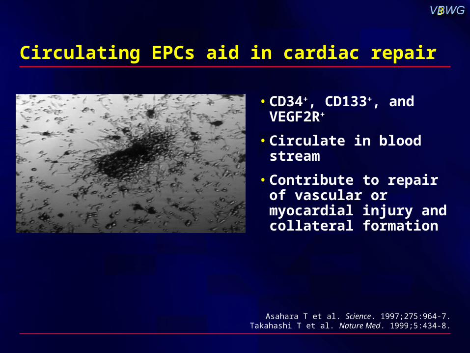
Circulating EPCs aid in cardiac repair
• CD34+, CD133+, and VEGF2R+
• Circulate in blood stream
• Contribute to repair of vascular or myocardial injury and collateral formation
Asahara T et al. Science. 1997;275:964-7.Takahashi T et al. Nature Med. 1999;5:434-8.

4
EPC physiology
• Originate in bone marrow
• Circulate in blood stream
• Number and function (proliferation, migration, homing) modulated by age, CV risk factors, and disease
• Release stimulated by organ and vascular injury
• Participate in vascular repair (collateralization) and re-endothelialization, partly by paracrine effects
• Circulating numbers by exercise and drugs (statins and ACE inhibitors)
• Independent predictors of endothelial dysfunction and long-term prognosis in patients with CAD
Hill JM et al. N Engl J Med. 2003;348:593-600.
5
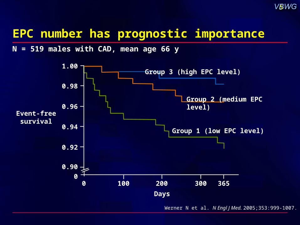
Werner N et al. N Engl J Med. 2005;353:999-1007.
N = 519 males with CAD, mean age 66 y
EPC number has prognostic importance
1.00
0.98
0.96
0.94
0.92
0.90
0100 200 300 365
Group 3 (high EPC level)
Group 2 (medium EPC level)
Group 1 (low EPC level)
0
Days
Event-freesurvival
6
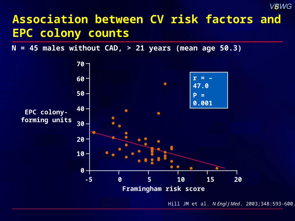
Association between CV risk factors and EPC colony counts
Framingham risk score
Hill JM et al. N Engl J Med. 2003;348:593-600.
N = 45 males without CAD, > 21 years (mean age 50.3)
r = –47.0
P = 0.001
-5 0 5 10 15 20
70
EPC colony-forming units
60
50
40
30
20
10
0

7
Mobilization of EPCs after myocardial infarction
Shintani S et al. Circulation. 2001;103:2776-9.
1 3 7 14 28
N = 16 patients with AMI, 8 controls
P < 0.001 P < 0.001
MNCCD34+ (/106WBCs)
Time after onset
Day
300
P < 0.001 P < 0.05
200
100
0
8
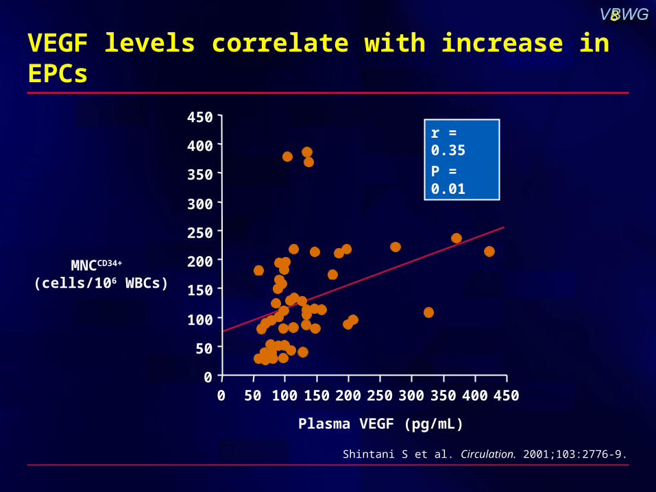
VEGF levels correlate with increase in EPCs
Shintani S et al. Circulation. 2001;103:2776-9.
MNCCD34+ (cells/106 WBCs)
Plasma VEGF (pg/mL)
r = 0.35
P = 0.01
0
50
100
150
200
250
300
350
400
450
0 50 100 150 200 250 300 350 400 450
9
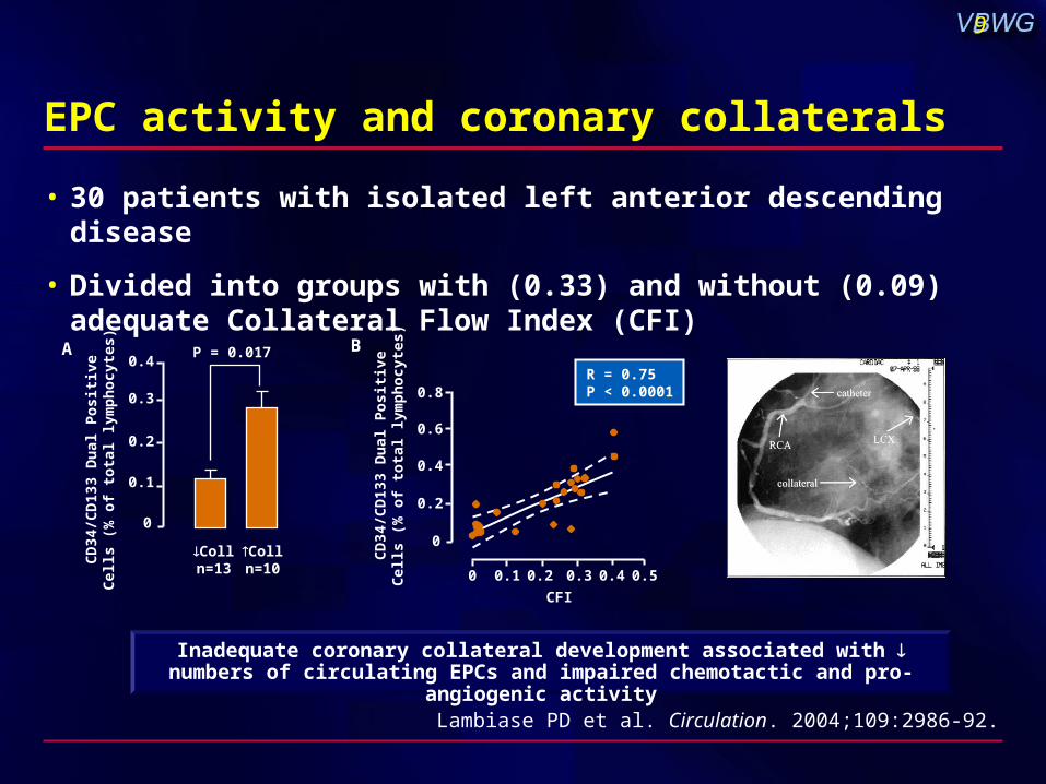
EPC activity and coronary collaterals
• 30 patients with isolated left anterior descending disease
• Divided into groups with (0.33) and without (0.09) adequate Collateral Flow Index (CFI)
Lambiase PD et al. Circulation. 2004;109:2986-92.
CD
34/C
D13
3 D
ual
Po
siti
veC
ells
(%
of
tota
l lym
ph
ocy
tes)
B
R = 0.75P < 0.0001
CFI
0.8
0.6
0.4
0.2
0
0 0.1 0.2 0.3 0.4 0.5
CD
34/C
D13
3 D
ual
Po
siti
veC
ells
(%
of
tota
l lym
ph
ocy
tes)
A0.4
0.3
0.2
0.1
0
Colln=13
Colln=10
P = 0.017
Inadequate coronary collateral development associated with numbers of circulating EPCs and impaired chemotactic and pro-angiogenic activity
10
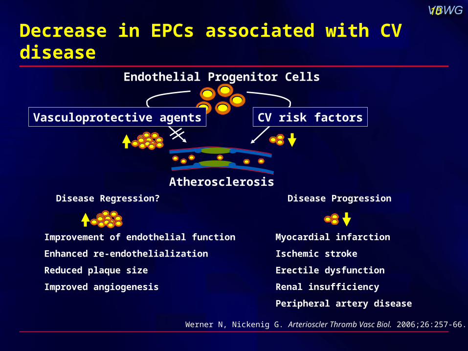
Decrease in EPCs associated with CV disease
Werner N, Nickenig G. Arterioscler Thromb Vasc Biol. 2006;26:257-66.
Endothelial Progenitor Cells
Atherosclerosis
Improvement of endothelial function
Enhanced re-endothelialization
Reduced plaque size
Improved angiogenesis
Myocardial infarction
Ischemic stroke
Erectile dysfunction
Renal insufficiency
Peripheral artery disease
Disease Regression? Disease Progression
Vasculoprotective agents CV risk factors