engineering gene circuits: foundations and applications
TRANSCRIPT

20Engineering Gene
Circuits: Foundationsand Applications
Dennis TuJiwon LeeTaylan OzdereTae Jun LeeLingchong YouDuke University, Durham
20.1 Introduction................................................................. 20-363
20.2 Designing Gene Circuits ............................................. 20-364Modeling-Guided Rational Design .
Refining a Circuit through Evolutionary Techniques
20.3 Programming Robust Circuit Behavior..................... 20-366Cellular Noise . Basic Regulatory
Motifs to Resist Noise . Cell–Cell Communication
20.4 Applications ................................................................. 20-370Gene Therapy . Drug Development .
Biotechnology Applications
20.5 Outlook ........................................................................ 20-372
Abstract
Synthetic biology has emerged as a useful approach to decoding fundamental laws underlying biological
control. Recent efforts have produced many exciting systems and generated substantial insights. These
progresses highlight the potential of synthetic biology to impact diverse areas including biology,
computation, engineering, and medicine.
20.1 Introduction
Biological systems often function reliably in diverse environments despite internal or external perturba-
tions. This behavior is often characterized as ‘‘robustness.’’ Based on extensive studies over the last
several decades, much of this robustness can be attributed to the control of gene expression through
complex cellular networks [1–4]. These networks are known to consist of various regulatory modules,
including feedback [5] and feed-forward [6] regulation and cell–cell communication [7]. With these
basic regulatory modules and motifs, researchers are now constructing artificial networks that mimic
nature to gain fundamental biological insight and understanding [8]. In addition, other artificial net-
works that are engineered with novel functions will serve as building blocks for future practical appli-
cations. These efforts form the foundation of the recent emergence of synthetic biology [3,9,10]. These
artificial networks are interchangeably called ‘‘synthetic gene circuits’’ or ‘‘engineered gene circuits.’’
Recent accomplishments in synthetic biology include engineered switches [11–14], oscillators [15,16],
logic gates [17–19], metabolic control [20], reengineered translational machinery [21], population control
[22] and pattern formation [23] using natural or synthetic [24] cell–cell communication, reengineered
viral genome [25], and hierarchically complex circuits built upon smaller, well-characterized
Tuan Vo-Dinh / Nanotechnology in Biology and Medicine 2949_C020 Page Proof page 363 7.8.2006 8:49am
20-363

functional modules [26] In addition to programming cellular dynamics, efforts have also been made
toward the construction of in vitro gene circuits using cell extracts [27,28].
As evidenced in the term ‘‘circuit,’’ gene circuits are often compared to electrical circuits. For example,
one can characterize the state of a gene as ON or OFF depending on whether it is expressed or repressed.
Construction of electrical circuits benefits from a large collection of well-characterized parts and
modules, including resistors, capacitors, and inductors, which can be connected to generate a complex
circuit with a useful function. The rapid progress in high-throughput technologies is now generating
similar parts-lists, as evidenced by the establishment of numerous databases [29–32]. In particular,
efforts along this line have led to the creation of a database of biological parts (http:==parts.mit.edu=) —
the Registry of Standard Biological Parts—that is specifically established to facilitate gene-circuit design
and implementation. This registry contains details on basic parts (mostly for bacteria) such as ribosome
binding sites, transcriptional terminators, and reporter genes. As of this writing, the registry contains
details on over 1500 parts. The ultimate goal is to be able to order standardized parts ‘‘off the shelf ’’
from the registry and assemble them together to engineer a functional circuit. The current registry
documents mostly components from bacteria, especially E. coli. We anticipate that this or similar
registries will also be expanded to include yeast, eukaryotes, and other organisms. In addition, we
note that the recent development of high throughput, low-cost DNA synthesis technology [33] provides
a unique opportunity to explore parts not found in nature.
20.2 Designing Gene Circuits
20.2.1 Modeling-Guided Rational Design
The first term that comes to mind when considering the creation of a circuit is ‘‘engineering’’ or
‘‘programming.’’ With this mindset, many novel synthetic gene circuits have been created through
iterations of careful design, modeling, and implementation. A well-known synthetic gene circuit is the
genetic toggle switch, whose name is derived from its ability to program bistable behavior (Figure 20.1a)
[12]. The toggle switch circuit consists of two repressors, TetR and LacI, which mutually repress each
other (Figure 20.1a). The circuit can be flipped between ‘‘high’’ and ‘‘low’’ states using chemical inducers
isopropyl-b-D-thiogalactopyranoside (IPTG) and anhydrotetracycline (aTc). For example, when aTc is
added to the system to inhibit tetR, lacI will be highly expressed and the reporter will be OFF.
Conversely, when IPTG is added to inhibit lacI, tetR and the reporter will be highly expressed.
(a) (b)IPTG
Lacl
P trc-2
aTc
TetR Reporter
Lacl
Lacl
TetR
PLtetO-1Ptet
Plac
TetRλcl
λP
λcl
FIGURE 20.1 (a) The genetic toggle switch gene circuit layout. TetR represses PLtetO-1and is inhibited by inducer
aTc; LacI represses Ptrc-2 and is inhibited by inducer IPTG. (b) Repressilator gene circuit layout. TetR represses
expression of lcI which in turn represses expression of LacI. Finally, LacI represses TetR expression, completing the
repressilator cycle.
Tuan Vo-Dinh / Nanotechnology in Biology and Medicine 2949_C020 Page Proof page 364 7.8.2006 8:49am
20-364 Nanotechnology in Biology and Medicine
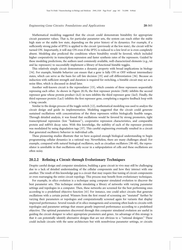
Mathematical modeling suggested that the circuit could demonstrate bistability for appropriate
circuit parameter values. That is, for particular parameter sets, the system can reach either the stable
high state or the stable low state, depending on the prior history of its dynamics. For example, if a
sufficiently strong pulse of IPTG is applied to the circuit (previously at the low state), the circuit will be
turned ON. Importantly, it will stay ON even if the IPTG is reduced to a low level or is even completely
absent. Modeling also predicted the conditions where bistability would be favored, which included
higher cooperativity in transcription repression and faster synthesis rates of the repressors. Guided by
these modeling predictions, the authors used commonly available, well-characterized elements (e.g., tet
and lac repressors) to successfully implement a library of functional bistable toggles.
This relatively simple circuit demonstrates a dynamic property with broad implications in biology
[34]. For example, bistable behavior can ensure that a gene is fully ON or OFF without intermediary
states, which can serve as the basis for cell fate decision [35] and cell differentiation [36]. Because an
induction with sufficient strength and duration is required for switching, a bistable circuit may act as a
noise filter, which is discussed in detail later.
Another well-known circuit is the repressilator [15], which consists of three repressors sequentially
repressing each other. As shown in Figure 20.1b, the first repressor protein (TetR) inhibits the second
repressor gene whose protein product (lcI) in turn inhibits the third repressor gene (lacI). Finally, the
third repressor protein (LacI) inhibits the first repressor gene, completing a negative feedback loop with
a long cascade.
Similar to the design process of the toggle switch [12], mathematical modeling was used to analyze the
circuit design and guide its implementation. Modeling suggested that the circuit could generate
sustained oscillations with concentrations of the three repressors within biologically feasible ranges.
Through detailed analysis, it was found that oscillations would be favored by strong promoters, tight
transcriptional repression (low ‘‘leakiness’’), cooperative repression characteristics, and comparable
protein and mRNA decay rates. With this knowledge, the stability of each of the repressor proteins
was modulated by using degradation tags [37]. This careful engineering eventually resulted in a circuit
that generated oscillatory behavior in individual cells.
These pioneering studies illustrate that we have acquired enough biological understanding to begin
programming cellular dynamics in a rational way. Nevertheless, there are many challenges ahead. For
example, compared with natural biological oscillators, such as circadian oscillators [38–40], the repres-
silator is unreliable in that oscillations only occur in a subpopulation of cells and these oscillations are
often noisy.
20.2.2 Refining a Circuit through Evolutionary Techniques
Despite careful design and computer simulation, building a gene circuit in vivo may still be challenging
due to a lack of detailed understanding of the cellular components and how they interact with one
another. The result of this knowledge gap is a circuit that may require fine tuning of circuit components
or even rearranging the entire circuit topology. This process may benefit from evolutionary techniques.
For example, in silico evolution is a technique using computer simulated evolution to discover the
best parameter sets. This technique entails simulating a library of networks with varying parameter
settings and topologies in a computer. Then, these networks are screened for the best performing ones
according to a predefined objective function [41] For instance, one could select circuits that generate
oscillations with a certain period. Winners from the first round of screening are ‘‘mutated’’ further by
varying their parameters or topologies and computationally screened again for variants that display
improved performance. Several rounds of in silico mutagenesis and screening often leads to circuits with
topologies and parameter settings that ensure greatly improved performance according to a predefined
objective. The optimal parameters discovered through this computer simulated evolution are useful in
guiding the circuit designer to select appropriate promoters and genes. An advantage of this strategy is
that it can potentially identify alternative designs that are not obvious to a ‘‘rational designer.’’ These
could include circuits with the same architecture but with nonobvious parameter settings, or circuits
Tuan Vo-Dinh / Nanotechnology in Biology and Medicine 2949_C020 Page Proof page 365 7.8.2006 8:49am
Engineering Gene Circuits: Foundations and Applications 20-365

with completely different network architecture and different parameter settings. However, the extent to
which this strategy can speed up circuit design and implementation is unclear. In fact, synthetic gene
circuits are yet to be experimentally implemented based on a design resulting from this approach.
Laboratory evolution can be realized by combining random mutagenesis and subsequent high-
throughput screening or selection for a desired function [42,43]. This ‘‘directed evolution’’ technique
has been used successfully for years to optimize enzymes by improving or altering their efficiency and
specificity [44]. In gene circuit design, directed evolution has been used to optimize metabolic pathways
to produce useful metabolites [45]. This optimization is accomplished by generating components such
as proteins and DNA regulatory elements with diverse kinetic properties that may not be naturally
available. In other words, it allows researchers to take advantage of a broader parameter space when
designing a gene circuit. In recent work, an evolved transcriptional activator (LuxR) [46] was used in a
synthetic pattern formation circuit [23] to optimize the final circuit function.
Another application of directed evolution is to optimize a partially functional or nonfunctional circuit
to be fully functional [47]. For example, this technique was used to evolve an initially nonfunctional
genetic inverter into a fully functional one [19]. Briefly, several rounds of mutagenesis by error-prone
PCR and subsequent high-throughput screening were able to achieve proper matching of the different
components that were assembled to make up the inverter, allowing for rapid optimization of the circuit
[19]. It is interesting to note that the functional circuit, created from directed evolution, ended up with
parameters tuned completely different from what was attempted by a rational approach [17]. This aspect
again highlights the advantage of evolution methods to explore a parameter space not readily accessible
by rational design.
More recently, this technique was further revised by combining mutagenesis with selection instead of
screening [48]. That is, the ability of a circuit to function properly is tied to the survival or death of the
host cell. This selection strategy, if properly established, can drastically improve the throughput of circuit
evolution. In this proof-of-concept work, Yokobayashi and Arnold were able to isolate a functional
inverter from a pool with more than 3 million fold background of nonfunctional circuits. Although this
work demonstrated a technique that was limited to optimization of a circuit with two output states (e.g.,
logic gates and toggle switches), more complex circuits may be broken down into such submodules for
individual optimization.
Random mutagenesis is unlikely to alter the connectivity of a circuit; rather, it largely results in
changes in one or more circuit parameters. Therefore, for directed evolution techniques to succeed,
they require a starting circuit with a topology that can in principle display the desired function with
proper parameter settings. Complementary to random mutagenesis, a combinatorial method will
enable the generation of a large number of circuits with diverse topologies from a small number of
elements. As a proof-of-concept, Guet and colleagues created 125 logic circuits using only three genes
and five promoters [18]. The resulting circuits displayed diverse phenotypes including NOR, NOT IF,
and NAND. Potentially, the same strategy can be applied to a larger number of elements and an even
greater diversity of circuit connectivity can be created. When coupled with efficient screening or
selection methods, this strategy can allow evolution of a circuit in both the topological space and the
parametric space.
20.3 Programming Robust Circuit Behavior
20.3.1 Cellular Noise
Another fundamental challenge in constructing gene circuits with reliable function is to deal with
cellular ‘‘noise’’ or stochastic fluctuations in cellular processes. Such noise can be due to a number of
factors, including small numbers of interacting molecules, heterogeneity of the cellular environment,
perturbations from the extracellular environment, and extended gene cascades [49–58].
Noise may benefit cells by providing phenotypic variations through which they adapt to changing
environments [59–62]. In most cases, however, noise is detrimental because it poses a significant
Tuan Vo-Dinh / Nanotechnology in Biology and Medicine 2949_C020 Page Proof page 366 7.8.2006 8:49am
20-366 Nanotechnology in Biology and Medicine
challenge for creating circuits with robust function. For example, the repressilator displays noisy
behavior. Only 40% of the cells in the population exhibited oscillatory behavior and these oscillations
are often out of phase with each other, even between sibling cells [15]. To a lesser degree, cellular noise
also cause variations in the oscillatory dynamics in a more sophisticated oscillator (metabolator)
recently implemented [16]. In contrast, natural oscillators function much more reliably. For example,
single-cell measurements in cyanobacteria revealed highly robust oscillations for days [63]. An intri-
guing question is how natural systems are able to function with such robustness despite cellular noise.
20.3.2 Basic Regulatory Motifs to Resist Noise
Extensive theoretical and experimental studies have been conducted to elucidate the underlying mech-
anisms by which biological systems achieve robustness [50,64–66]. One such mechanism is negative
feedback, where the output from a system reduces its own output. In a biological context, this occurs
when a protein limits its own accumulation by repressing its own synthesis [14,67] or by facilitating its
own turnover [68,69].
A well-studied example is bacterial chemotaxis, a phenomenon where bacteria move toward an
attractant by biasing their swimming motion [2]. This response is controlled by receptors that detect
the attractant gradient. When the bacterium reaches a steady concentration of attractant, adaptation
occurs. This ‘‘zeroes’’ the detector so that it can react to new gradients. This adaptation was found to be
resistant to internal and external perturbations [1,2]. In-depth theoretical analysis indicates that the
robust behavior of the circuit can be attributed to an integral negative feedback [70].
Over three decades ago, Savageau pioneered systems-level analysis of various regulatory motifs [71],
and found that negative feedback can confer stability in system output and improve system response
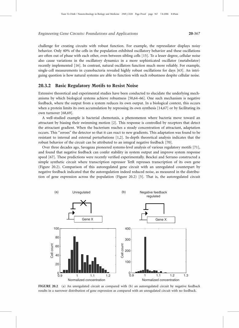
speed [67]. These predictions were recently verified experimentally. Besckei and Serrano constructed a
simple synthetic circuit where transcription repressor TetR represses transcription of its own gene
(Figure 20.2). Comparison of this autoregulated gene circuit with an unregulated counterpart by
negative feedback indicated that the autoregulation indeed reduced noise, as measured in the distribu-
tion of gene expression across the population (Figure 20.2) [5]. That is, the autoregulated circuit
(a) (b)Unregulated Negative feedbackregulated
Gene X Gene X
Normalized concentration Normalized concentration
Cel
l cou
nt
Cel
l cou
nt
0
20
40
60
80
100
0
100
200
300
400
0.9 1 1.1 1.2 0.9 1 1.1 1.2 1.3
FIGURE 20.2 (a) An unregulated circuit as compared with (b) an autoregulated circuit by negative feedback
results in a narrower distribution of gene expression as compared with an unregulated circuit with no feedback.
Tuan Vo-Dinh / Nanotechnology in Biology and Medicine 2949_C020 Page Proof page 367 7.8.2006 8:49am
Engineering Gene Circuits: Foundations and Applications 20-367

resulted in gene expression with a much narrower distribution compared to the unregulated circuit. To
further illustrate the importance of negative feedback, the autoregulatory loop was gradually removed by
adding an inducer. As the inducer concentration increased, the negative feedback circuit behavior
transitioned to that of the unregulated circuit, resulting in a wide distribution of gene expression [5].
Part of the mechanism behind this behavior may be an increase in the noise frequency. This frequency
shift is thought to enable the cell to more effectively filter the noise [72]. Using the same circuit, Alon
and colleagues demonstrated that negative feedback also significantly improved system response speed
[73].
An alternative approach to resisting noise and enhancing stability is increasing the rise-time in gene
expression. Rise-time is the time it takes for a gene circuit to reach a steady-state level of protein
expression [73]. An increased rise-time requires a longer-lasting input to fully induce a gene. This
behavior can be implemented through a coherent feed-forward loop, where transcription factor A
regulates B, and both jointly control the expression of C [6]. The latter stage of the feed-forward loop
causes an increase in rise-time, allowing the cell to filter noise from the input by withstanding transient
perturbations.
Another regulatory feature used to increase cellular robustness is positive feedback. Positive feedback
occurs when the output of a network causes an increase in the same output. It can be realized by
positive autoregulation [13,14], mutual activation [36,74,75] or mutual inhibition [12] (as detailed in
Figure 20.1a). Positive feedback loops often amplify noise [13,64]. On the other hand, when these
modules display bistable switching behavior with appropriate parameter sets, they may serve as a
noise-resistant device. This is because a sufficiently strong, long-lasting input signal is required to
change the state of the switch [76]. Using this feature as a noise filter to stabilize oscillations was
proposed on the basis of computational analysis [77]. Subsequent experimental efforts successfully
generated population-level oscillations, although these were damped oscillations [78]. In another
example of noise-resistant behavior, cellular ‘‘memory’’ is controlled by a positive feedback loop.
This memory allows the cell to maintain a state that depends on the initial input. Once the memory
is established, the state of the cell does not change with varying input as long as the memory is
sustained by the positive feedback loop [79].
20.3.3 Cell–Cell Communication
In recent years, scientists have discovered that bacteria do not exist merely as individuals, but can
coordinate their behavior as a group through quorum sensing [7,80,81]. Quorum sensing is a technique
by which bacteria use cell–cell communication to sense changes in cell population density. This ability
has been extensively studied for its role in regulating diverse physiological functions such as biolumin-
escence [80], virulence [82,83], biofilm formation [84], and programmed death [85–87].
In quorum sensing, signaling molecules are expressed by the cells and can freely diffuse through the
cell membrane. At a low population density, these molecules diffuse out of the cells into the extracellular
space with little accumulation of the signal within the cells. Thus, communication is weak and the cells
act independent of one another. As the population increases, the signaling molecule concentration
increases both inside and outside the cells. Upon reaching a threshold concentration, the signal will lead
to coordinated expression of genes responsible for various functions [7,81].
Intuitively, the large number of signaling molecules outside the cells can act as a buffer and temper the
fluctuations of the signaling molecule in each cell. That is, the fluctuation in the concentration of the
signaling molecule is likely much smaller compared with molecules of the same concentration, but
confined in individual cells. Consider a molecule with a concentration of 10 nM. If it is confined in the
cell, there will be about 10 molecules=cell (assuming a cell volume of 10�15 L). The fluctuation in its
concentration will be about 101=2, or a relative fluctuation of �0.3. However, for a freely diffusible
signaling molecule with the same concentration in a 1 mL culture, the molecular number is 1013, and the
relative fluctuation is around 10�6.5, which is considerably less noisy. Therefore, if a gene is under
control of such a signaling molecule, there will likely be less noise in its expression across the population.
Tuan Vo-Dinh / Nanotechnology in Biology and Medicine 2949_C020 Page Proof page 368 7.8.2006 8:49am
20-368 Nanotechnology in Biology and Medicine
Confirming this point, our simulations suggest that cell–cell communication can indeed reduce noise in
gene expression across a population of cells. The distribution of gene expression controlled by quorum
sensing is much tighter than the distribution found in the system without cell–cell communication
(unpublished data). Our simulations also suggest that this effect depends only on the cell density and
diffusion rate and not on the number of cells. With this evidence, quorum sensing may be a side effect of
batch culture cells detecting their environmental conditions through diffusion [88].
It has been proposed that cell–cell communication may be used to facilitate more robust oscillations.
Recent computer simulations have demonstrated that the repressilator oscillations can be synchronized
across an entire population of cells through cell–cell communication [89,90]. The same strategy was also
proposed to synchronize relaxation-type oscillators [91,92]. Although this synchronization strategy
remains to be demonstrated experimentally, there is evidence that suggests that interlocked oscillators
provide more robust behavior in nature [93].
In addition to synchronizing oscillators, cell–cell communication can serve as an effective mechanism
to achieve integrated and coordinated behavior among a population of cells. In a synthetic ‘‘population
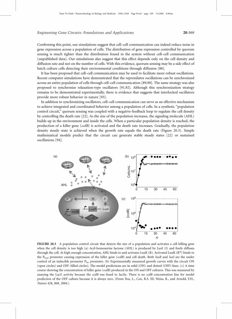
control circuit,’’ quorum sensing was coupled with a negative-feedback loop to regulate the cell density
by controlling the death rate [22]. As the size of the population increases, the signaling molecule (AHL)
builds up in the environment and inside the cells. When a particular population density is reached, the
production of a killer gene (ccdB) is activated and the death rate increases. Gradually, the population
density steady state is achieved when the growth rate equals the death rate (Figure 20.3). Simple
mathematical models predict that the circuit can generate stable steady states [22] or sustained
oscillations [94].
0 15 30 45 60
0 15 30 45 60
Cel
ls/m
L
109
107
105
104
106
108
H
[ccd
B]
AHL
(a) (b)
(c)R I
LuxILuxR
ccdB
ccdB
R*
PluxI
FIGURE 20.3 A population control circuit that detects the size of a population and activates a cell killing gene
when the cell density is too high (a) Acyl-homoserine lactone (AHL) is produced by LuxI (I) and freely diffuses
through the cell. At high enough concentration, AHL binds to and activates LuxR (R). Activated LuxR (R*) binds to
the PluxI promoter causing expression of the killer gene (ccdB) and cell death. Both luxR and luxI are the under
control of an inducible promoter Plac promoter. (b) Experimentally measured growth curves with the circuit ON
(open circles) and OFF (filled circles). The model predictions are in solid (ON) and dotted (OFF) lines. (c) A time
course showing the concentration of killer gene (ccdB) produced in the ON and OFF cultures. This was measured by
assaying the LacZ activity because the ccdB was fused to lacZa. There is no ccdB concentration line for model
prediction of the OFF culture because it is always zero. (From You, L., Cox, R.S. III, Weiss, R., and Arnold, F.H.,
Nature 428, 868, 2004.)
Tuan Vo-Dinh / Nanotechnology in Biology and Medicine 2949_C020 Page Proof page 369 7.8.2006 8:49am
Engineering Gene Circuits: Foundations and Applications 20-369

Experimentally, this population control circuit can be turned ON by inducing the quorum sensing
module. As expected, the uninduced (OFF) culture grew exponentially until reaching a stationary phase
at the maximum density the culture medium could sustain (Figure 20.3b). The ON culture initially grew
similarly as the OFF culture until a threshold was reached at 7 h after circuit activation. The culture then
underwent damped oscillations that were likely the result of a delay in the negative-feedback loop, which
can be caused by the synthesis and transport of the signal, activation of LuxR, expression of ccdB, and the
cascade leading to cell death by ccdB. Even though negative feedback is usually associated with increased
output stability, such long cascades can cause oscillations. In particular, recent measurements using a
microchemostat demonstrated that the circuit was able to generate sustained oscillations for up to
several hundred hours [94].
Importantly, this circuit also demonstrated that a certain degree of noise can be useful for program-
ming robust dynamics via cell–cell communication. Specifically, cell-to-cell variations are essential for
generating robust dynamics in the population control circuit. If cells performed identically, they would
all die upon reaching the critical density that leads to a sufficient level of the killer protein. Precisely
because of cell-to-cell variations, some cells died while others were left to survive and the overall
population dynamics could be robustly regulated.
In addition to auto-communication within a single population, cell–cell communication has also
been used to program communication between different populations and to coordinate pattern forma-
tion. Basu and colleagues created a pulse generator that is activated by a signal from nearby ‘‘sender’’
cells. An interesting aspect of this interaction is the spatiotemporal behavior. Specifically, the pulse-
generating cells were able to differentiate between communication from nearby and far-away sender cells
[95]. This spatiotemporal behavior led to the development of a pattern formation circuit. Similar to the
previous circuit, sender cells were used to produce an intercellular signal but this time the receiver cells
responded to the gradient by producing a bulls-eye pattern around the chemical source [23].
20.4 Applications
In addition to decoding the ‘‘design principles’’ of biological systems, synthetic gene circuits can also
find wide applications in diverse areas. These applications include innovative gene regulation systems
[11,20,24,26,96–99], gene therapy [100], drug development [101], cellular computation [102], stem cell
reprogramming [103], and bioremediation [104]. Here we discuss a few examples in more detail.
20.4.1 Gene Therapy
An important application area for synthetic biology is the development of innovative gene circuits for
therapeutic purposes. Recent years have witnessed encouraging advancements in the field of cancer gene
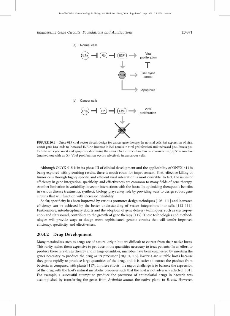
therapy in the creation of novel adenoviral vectors. For example, an oncolytic adenovirus dl1520, first
coined by its creators [105], has shown potential use in specific targeting of tumor cells. Pioneered by
McCormick and colleagues [106], ONYX-015 selectively propagates in cells with inactivated p53,
a common defect in tumor cells (Figure 20.4b). On the other hand, normal cells have active p53 and
ONYX-015 causes apoptosis (Figure 20.4a). Both pathways force the cell to enter the S-phase by increasing
the amount of free E2F. In normal cells, the combination of entering the S-phase and a viral infection results
in p53 inducing a cell cycle arrest or an apoptosis to prevent proliferation. Cancer cells with nonfunctional
p53, however, will allow replication of viral particles, leading to selective killing of these cells.
The concept of selective targeting is further expanded upon by Johnson and colleagues [107]. Taking
advantage of the mutations in the pRb pathway, common to most human cancers, they restrict
adenoviral infection to tumor cells with high efficiency and specificity. Their strategy is based on
identical dependence of both human cells and human adenovirus on the loss or mutation of Rb for
uncontrolled proliferation. The E2F promoter is known to be abnormally active in tumor cells. Thus, the
engineered human adenovirus ONYX-411, whose promoter of E1A or E1B has been replaced by the E2F
promoter, only replicates in human tumor cells with Rb-pathway defects [107].
Tuan Vo-Dinh / Nanotechnology in Biology and Medicine 2949_C020 Page Proof page 370 7.8.2006 8:49am
20-370 Nanotechnology in Biology and Medicine
Although ONYX-015 is in its phase III of clinical development and the applicability of ONYX-411 is
being explored with promising results, there is much room for improvement. First, effective killing of
tumor cells through highly specific and efficient viral integration is most desirable. In fact, the issues of
efficiency in gene integration, specificity, and effectiveness are common to many fields of gene therapy.
Another limitation is variability in vector interactions with the hosts. In optimizing therapeutic benefits
in various disease treatments, synthetic biology plays a key role by providing ways to design robust gene
circuits that will function with increased reliability.
So far, specificity has been improved by various promoter design techniques [108–111] and increased
efficiency can be achieved by the better understanding of vector integrations into cells [112–114].
Furthermore, interdisciplinary efforts and the adoption of gene delivery techniques, such as electropor-
ation and ultrasound, contribute to the growth of gene therapy [115]. These technologies and method-
ologies will provide ways to design more sophisticated genetic circuits that will confer improved
efficiency, specificity, and effectiveness.
20.4.2 Drug Development
Many metabolites such as drugs are of natural origin but are difficult to extract from their native hosts.
This rarity makes them expensive to produce in the quantities necessary to treat patients. In an effort to
produce these rare drugs cheaply and in large quantities, microbes have been engineered by inserting the
genes necessary to produce the drug or its precursor [20,101,116]. Bacteria are suitable hosts because
they grow rapidly to produce large quantities of the drug, and it is easier to extract the product from
bacteria as compared with plants [117]. In these efforts, the major challenge is to balance the expression
of the drug with the host’s natural metabolic processes such that the host is not adversely affected [101].
For example, a successful attempt to produce the precursor of antimalarial drugs in bacteria was
accomplished by transferring the genes from Artimisia annua, the native plant, to E. coli. However,
(a)
(b)
Normal cells
Cancer cells
Viralproliferation
Cell cyclearrest
Apoptosis
ViralproliferationE1a
E1a
Rb
Rb
E2F
E2F
p53
FIGURE 20.4 Onyx-015 viral vector circuit design for cancer gene therapy. In normal cells, (a) expression of viral
vector gene E1a leads to increased E2F. An increase in E2F results in viral proliferation and increased p53. Excess p53
leads to cell cycle arrest and apoptosis, destroying the virus. On the other hand, in cancerous cells (b) p53 is inactive
(marked out with an X). Viral proliferation occurs selectively in cancerous cells.
Tuan Vo-Dinh / Nanotechnology in Biology and Medicine 2949_C020 Page Proof page 371 7.8.2006 8:49am
Engineering Gene Circuits: Foundations and Applications 20-371

initial yields were limited by the synthase, a key enzyme in the production of the precursor. Conse-
quently, the production of the synthase and other upstream molecules were increased by optimizing
existing genes and introducing biosynthetic pathways from S. cerevisiae. Using a specialized pathway
from S. cerevisiae, rather than the existing pathway in E. coli, circumvented the inhibitory elements
found in yeast while also bypassing those in E. coli. In the final engineered E. coli strain that incorporated
genes from different organisms, final yields of the artemisinin precursor amorphadiene were increased
>10,000-fold with room for further optimization [117]. The benefits of being able to produce large
quantities of rare drugs that are in high demand are quite obvious. The success of this work has recently
attracted major investment from the industry [118], which will encourage broader efforts to engineer
bacteria for drug and metabolite production.
20.4.3 Biotechnology Applications
By bringing discovery and utilization of novel genetic components together with a rational design
approach, synthetic biology opens up tremendous possibilities for environmental engineering and
biomedical discovery [119] However, use of recombinant DNA techniques invites risks commonly
associated with conventional genetic engineering such as horizontal gene transfer to other species and
introduction of new allergens and toxins to the environment [120,121]. Furthermore, the potential to
realize desired behavior in organisms through rational design might translate into greater risks that are
malevolent or inadvertent in nature [122]. A reasonable way to deal with these risks would be to
make the containment of the proposed genetic circuitry an essential part of the design process. This way,
the effectiveness of the containment could be made to match the estimated risk [123]. For example,
containment in bacteria can be achieved through a built-in safeguard mechanism that can be imple-
mented using the quorum sensing based population control discussed earlier [22].
One practical application area for synthetic biology, where aforementioned risks should be minim-
ized, is environmental biotechnology. Microorganisms are commonly used for pollution reduction
(bioremediation) and can provide safe and cost-effective alternatives to existing physicochemical
methods [124]. However, risks and concerns associated with the release of recombinant microorganisms
into the environment [125] currently limit their widespread use. Therefore, researchers are designing
containment systems that will minimize horizontal gene transfer and uncontrolled proliferation of
recombinant microorganisms (for a good review see Ref. [104]). In order to realize the full potential
of synthetic biology in this area and answer safety concerns, microorganisms should be equipped with
not only biodegradation pathways but also conditional lethality systems that will ensure control over the
genetic material being released. One striking example of coupling of a biodegradation pathway with a
containment system is achieved by engineering a streptavidin based lethality system that is controlled by
TOL plasmid for aromatic hydrocarbon metabolism [126] In this system, the TOL pathway, which is
capable of breaking down toxic aromatic compounds, is used to control the expression of a streptavidin
gene, which binds to biotin, an essential coenzyme for microorganisms. Depletion of the environmental
inducer of the TOL pathway, m-methylbenzoate, led to streptavidin expression and eventually cell death
with remarkable efficiencies around 10�7–10�8 mutant escape rates [126].
Containment is an important issue for synthetic constructs; moreover, synthetic biology can address
this issue and tackle environmental pollution problems by introducing new perspectives and techniques.
Although the use of genetic engineering for biodegradation is not novel [127], design ideas from basic
circuits to improve robustness against mutations, and attain control at the level of populations rather
than at the level of individuals [22,128] can improve killing efficiencies of containment systems.
20.5 Outlook
We have presented recent progress in the nascent field of synthetic biology and highlighted a few
application areas. We believe that synthetic biology has great potential for numerous applications that
go well beyond what we can envision today. Nevertheless, many challenges still remain. A major hurdle is
Tuan Vo-Dinh / Nanotechnology in Biology and Medicine 2949_C020 Page Proof page 372 7.8.2006 8:49am
20-372 Nanotechnology in Biology and Medicine

the elucidation of control mechanisms that can ensure robust function of synthetic gene circuits. To this
end, synthetic biologists will benefit from rapid advances in systems biology [65,129–132]. On one hand,
efforts in systems biology will continue to expand the list of building blocks that serve as the foundation
of synthetic gene circuits. On the other, they will likely generate substantial lessons for optimal circuit
design from naturally biological systems [133–135].
Another challenge lies in the measurement and monitoring of intracellular and intercellular dynamics
with high precision. For de novo engineering of gene circuits, it has become clear that precise
characterization of circuit dynamics critically depends on well-controlled experimental conditions and
distinct cellular markers. To address this issue, the marriage between biology and nanotechnology can be
particularly promising [34].
Fine control of growth environments can be achieved using novel microfluidics devices [136,137]. For
instance, Balagadde and colleagues recently devised a microchemostat [94], an automated and continu-
ously operated microfluidic device with a reactor volume of 16 nL, which is an order of magnitude
smaller than conventional culturing systems. When applied to the population control circuit described
above, the microchemostat significantly delayed the process of mutants arising and enabled measure-
ments of complex population dynamics for more than 500 h with single-cell resolution. This work has
focused on observation of cell morphology in addition to cell density, but it can be readily adapted to
measure gene expression dynamics reported by fluorescence or luminescence.
Complementary to culturing systems, a wide array of nanosensors has been developed to facilitate
analysis of single cells and protein dynamics inside the cell [138–141]. We anticipate that these diverse
micro- and nanoscale devices can be integrated for elucidating temporal–spatial dynamics of cellular
processes with ever-increasing resolution.
Acknowledgments
We thank Chee Meng Tan, Jun Ozaki, and Guang Yao for their comments and suggestions on the
chapter.
References
1. Barkai, N., and S. Leibler. 1997. Robustness in simple biochemical networks. Nature 387
(6636):913–917.
2. Alon, U., M.G. Surette, N. Barkai, and S. Leibler. 1999. Robustness in bacterial chemotaxis. Nature
397 (6715):168–171.
3. Hasty, J., D. McMillen, and J.J. Collins. 2002. Engineered gene circuits. Nature 420 (6912):224–230.
4. Little, J.W., D.P. Shepley, and D.W. Wert. 1999. Robustness of a gene regulatory circuit. EMBO J 18
(15):4299–4307.
5. Becskei, A., and L. Serrano. 2000. Engineering stability in gene networks by autoregulation. Nature
405 (6786):590–593.
6. Mangan, S., and U. Alon. 2003. Structure and function of the feed-forward loop network motif.
Proc Natl Acad Sci USA 100 (21):11980–11985.
7. Miller, M.B., and B.L. Bassler. 2001. Quorum sensing in bacteria. Annu Rev Microbiol 55:165–199.
8. Sprinzak, D., and M.B. Elowitz. 2005. Reconstruction of genetic circuits. Nature 438 (7067):443–448.
9. Benner, S.A., and A.M. Sismour. 2005. Synthetic biology. Nat Rev Genet 6 (7):533–543.
10. Ferber, D. 2004. Synthetic biology. Microbes made to order. Science 303 (5655):158–161.
11. Kramer, B.P., A.U. Viretta, M. Daoud-El-Baba, D. Aubel, W. Weber, and M. Fussenegger. 2004. An
engineered epigenetic transgene switch in mammalian cells. Nat Biotechnol 22 (7):867–870.
12. Gardner, T.S., C.R. Cantor, and J.J. Collins. 2000. Construction of a genetic toggle switch in
Escherichia coli. Nature 403 (6767):339–342.
13. Becskei, A., B. Seraphin, and L. Serrano. 2001. Positive feedback in eukaryotic gene networks: cell
differentiation by graded to binary response conversion. EMBO J 20 (10):2528–2535.
Tuan Vo-Dinh / Nanotechnology in Biology and Medicine 2949_C020 Page Proof page 373 7.8.2006 8:49am
Engineering Gene Circuits: Foundations and Applications 20-373
14. Isaacs, F.J., J. Hasty, C.R. Cantor, and J.J. Collins. Prediction and measurement of an autoregulatory
genetic module. Proc Natl Acad Sci USA 100 (13):7714–7719.
15. Elowitz, M.B., and S. Leibler. 2000. A synthetic oscillatory network of transcriptional regulators.
Nature 403 (6767):335–338.
16. Fung, E., W.W. Wong, J.K. Suen, T. Bulter, S.G. Lee, and J.C. Liao. 2005. A synthetic gene-metabolic
oscillator. Nature 435 (7038):118–122.
17. Weiss, R., G.E. Homsy, and T. Knight, Jr.1999. Toward in vivo digital circuits. In Dimacs workshop
on evolution as computation, 275–295. Princeton: Springer.
18. Guet, C.C., M.B. Elowitz, W. Hsing, and S. Leibler. 2002. Combinatorial synthesis of genetic
networks. Science 296 (5572):1466–1470.
19. Yokobayashi, Y., R. Weiss, and F.H. Arnold. 2002. Directed evolution of a genetic circuit. Proc Natl
Acad Sci USA 99 (26):16587–16591.
20. Farmer, W.R., and J.C. Liao. 2000. Improving lycopene production in Escherichia coli by engineer-
ing metabolic control. Nat Biotechnol 18 (5):533–537.
21. Rackham, O., and J. Chin. 2005. A network of orthogonal ribosome.mRNA pairs. Nat Chem Biol
1:159–166.
22. You, L., R.S. Cox, 3rd, R. Weiss, and F.H. Arnold. 2004. Programmed population control by cell–
cell communication and regulated killing. Nature 428 (6985):868–871.
23. Basu, S., Y. Gerchman, C.H. Collins, F.H. Arnold, and R.A. Weiss. A synthetic multicellular system
for programmed pattern formation. Nature 434 (7037):1130–1134.
24. Bulter, T., S.G. Lee, W.W. Wong, E. Fung, M.R. Connor, and J. C. Liao. 2004. Design of artificial cell–
cell communication using gene and metabolic networks. Proc Natl Acad Sci USA 101 (8):2299–2304.
25. Chan, L.Y., S. Kosuri, and D. Endy. 2005. Refactoring bacteriophage T7. Mol Syst Biol 1 (1):
msb4100025-E1.
26. Kobayashi, H., M. Kaern, M. Araki, K. Chung, T.S. Gardner, C.R. Cantor, and J. J. Collins. 2004.
Programmable cells: Interfacing natural and engineered gene networks. Proc Natl Acad Sci USA 101
(22):8414–8419.
27. Noireaux, V., R. Bar-Ziv, and A. Libchaber. 2003. Principles of cell-free genetic circuit assembly.
Proc Natl Acad Sci USA 100 (22):12672–12677.
28. Isalan, M., C. Lemerle, and L. Serrano. 2005. Engineering gene networks to emulate Drosophila
embryonic pattern formation. PLoS Biol 3 (3):e64.
29. Bader, G.D., D. Betel, and C.W. Hogue. 2003. BIND: the Biomolecular Interaction Network
Database. Nucleic Acids Res 31 (1):248–250.
30. Xenarios, I., L. Salwinski, X.J. Duan, P. Higney, S.M. Kim, and D. Eisenberg. 2002. DIP, the
Database of Interacting Proteins: A research tool for studying cellular networks of protein inter-
actions. Nucleic Acids Res 30 (1):303–305.
31. Keseler, I.M., J. Collado-Vides, S. Gama-Castro, J. Ingraham, S. Paley, I.T. Paulsen, M. Peralta-Gil,
and P.D. Karp. 2005. EcoCyc: A comprehensive database resource for Escherichia coli. Nucleic Acids
Res 33 (Database issue):D334–337.
32. Wixon, J., and D. Kell. 2000. The Kyoto encyclopedia of genes and genomes—KEGG. Yeast 17
(1):48–55.
33. Tian, J., H. Gong, N. Sheng, X. Zhou, E. Gulari, X. Gao, and G. Church. 2004. Accurate multiplex
gene synthesis from programmable DNA microchips. Nature 432 (7020):1050–1054.
34. Philip, B. 2005. Synthetic biology for nanotechnology. Nanotechnology 16 (1):R1.
35. Ptashne, M. 2004. A genetic switch: Phage lambda revisited. 3rd ed. Cold Spring Harbor, NY: Cold
Spring Harbor Laboratory Press.
36. Ferrell, J.E. Jr. 2002. Self-perpetuating states in signal transduction: Positive feedback, double-
negative feedback and bistability. Curr Opin Cell Biol 14 (2):140–148.
37. Andersen, J.B., C. Sternberg, L.K. Poulsen, S.P. Bjorn, M. Givskov, and S. Molin. 1998. New
unstable variants of green fluorescent protein for studies of transient gene expression in bacteria.
Appl Environ Microbiol 64 (6):2240–2246.
Tuan Vo-Dinh / Nanotechnology in Biology and Medicine 2949_C020 Page Proof page 374 7.8.2006 8:49am
20-374 Nanotechnology in Biology and Medicine
38. Novak, B., and J.J. Tyson. 2004. A model for restriction point control of the mammalian cell cycle. J
Theor Biol 230 (4):563–579.
39. Panda, S., J.B. Hogenesch, and S.A. Kay. 2002. Circadian rhythms from flies to human. Nature 417
(6886):329–335.
40. Harmer, S.L., S. Panda, and S.A. Kay. 2001. Molecular bases of circadian rhythms. Annu Rev Cell
Dev Biol 17:215–253.
41. Francois, P., and V. Hakim. 2004. Design of genetic networks with specified functions by evolution
in silico. Proc Natl Acad Sci USA 101 (2):580–585.
42. Arnold, F.H. 2001. Combinatorial and computational challenges for biocatalyst design. Nature 409
(6817):253–257.
43. Yuan, L., I. Kurek, J. English, and R. Keenan. 2005. Laboratory-directed protein evolution. Micro-
biol Mol Biol Rev 69 (3):373–392.
44. Farinas, E.T., T. Bulter, and F.H. Arnold. 2001. Directed enzyme evolution. Curr Opin Biotechnol 12
(6):545–551.
45. Umeno, D., A.V. Tobias, and F.H. Arnold. 2005. Diversifying carotenoid biosynthetic pathways by
directed evolution. Microbiol Mol Biol Rev 69 (1):51–78.
46. Collins, C.H., F.H. Arnold, and J.R. Leadbetter. 2005. Directed evolution of Vibrio fischeri LuxR for
increased sensitivity to a broad spectrum of acyl-homoserine lactones. Mol Microbiol 55 (3):712–
720.
47. Hasty, J. 2002. Design then mutate. Proc Natl Acad Sci USA 99 (26):16516–16518.
48. Yohei, Y., and H.A. Frances. 2005. A dual selection module for directed evolution of genetic
circuits. Nat Comput—Int J 4 (3):245–254.
49. Blake, W.J., M, KAErn, C.R. Cantor, and J.J. Collins. 2003. Noise in eukaryotic gene expression.
Nature 422 (6932):633–637.
50. Rao, C.V., D.M. Wolf, and A.P. Arkin. 2002. Control, exploitation and tolerance of intracellular
noise. Nature 420 (6912):231–237.
51. McAdams, H.H., and A. Arkin. 1999. It’s a noisy business! Genetic regulation at the nanomolar
scale. Trends Genet 15 (2):65–69.
52. McAdams, H.H., and A. Arkin. 1997. Stochastic mechanisms in gene expression. Proc Natl Acad Sci
USA 94 (3):814–819.
53. Thattai, M., and A. van Oudenaarden. 2001. Intrinsic noise in gene regulatory networks. Proc Natl
Acad Sci USA 98 (15):8614–8619.
54. Ozbudak, E.M., M. Thattai, I. Kurtser, A.D. Grossman, and A. van Oudenaarden. Regulation of
noise in the expression of a single gene. Nat Genet 31 (1):69–73.
55. Levsky, J.M., and R.H. Singer. 2003. Gene expression and the myth of the average cell. Trends Cell
Biol 13 (1):4–6.
56. Elowitz, M.B., A.J. Levine, E.D. Siggia, and P.S. Swain. 2002. Stochastic gene expression in a single
cell. Science 297 (5584):1183–1186.
57. Guptasarma, P. 1995. Does replication-induced transcription regulate synthesis of the myriad low
copy number proteins of Escherichia coli? Bioessays 17 (11):987–997.
58. Colman-Lerner, A., A. Gordon, E. Serra, T. Chin, O. Resnekov, D. Endy, C.G. Pesce, and R. Brent.
2005. Regulated cell-to-cell variation in a cell-fate decision system. Nature 437 (7059):699–706.
59. Raser, J.M., and E.K. O’Shea. 2004. Control of stochasticity in eukaryotic gene expression. Science
304 (5678):1811–1814.
60. Korobkova, E., T. Emonet, J.M. Vilar, T.S. Shimizu, and P. Cluzel. 2004. From molecular noise to
behavioural variability in a single bacterium. Nature 428 (6982):574–578.
61. Weinberger, L.S., J.C. Burnett, J.E. Toettcher, A.P. Arkin, and D.V. Schaffer. 2005. Stochastic gene
expression in a lentiviral positive-feedback loop: HIV-1 Tat fluctuations drive phenotypic diversity.
Cell 122 (2):169–182.
62. A. Arkin, J. Ross, and H.H. McAdams. 1998. Stochastic kinetic analysis of developmental pathway
bifurcation in phage lambda-infected Escherichia coli cells. Genetics 149 (4):1633–1648.
Tuan Vo-Dinh / Nanotechnology in Biology and Medicine 2949_C020 Page Proof page 375 7.8.2006 8:49am
Engineering Gene Circuits: Foundations and Applications 20-375
63. Mihalcescu, I., W. Hsing, and S. Leibler. 2004. Resilient circadian oscillator revealed in individual
cyanobacteria. Nature 430 (6995):81–85.
64. Rao, C.V., and A.P. Arkin. 2001. Control motifs for intracellular regulatory networks. Annu Rev
Biomed Eng 3:391–419.
65. You, L. 2004. Toward computational systems biology. Cell Biochem Biophys 40 (2):167–184.
66. Kaern, M., T.C. Elston, W.J. Blake, and J.J. Collins. 2005. Stochasticity in gene expression: From
theories to phenotypes. Nat Rev Genet 6 (6):451–464.
67. Savageau, M.A. 1974. Comparison of classical and autogenous systems of regulation in inducible
operons. Nature 252 (5484):546–549.
68. Hillen, W., and C. Berens. 1994. Mechanisms underlying expression of Tn10 encoded tetracycline
resistance. Annu Rev Microbiol 48:345–369.
69. Batchelor, E., T.J. Silhavy, and M. Goulian. 2004. Continuous control in bacterial regulatory
circuits. J Bacteriol 186 (22):7618–7625.
70. Yi, T.M., Y. Huang, M.I. Simon, and J. Doyle. 2000. Robust perfect adaptation in bacterial
chemotaxis through integral feedback control. Proc Natl Acad Sci USA 97 (9):4649–4653.
71. Savageau, M.A. 1976.Biochemical systems analysis. Reading, MA: Addison-Wesley.
72. Austin, D.W., M.S. Allen, J.M. McCollum, R.D. Dar, J.R. Wilgus, G.S. Sayler, N.F. Samatova,
C.D. Cox, and M.L. Simpson. 2006. Gene network shaping of inherent noise spectra. Nature 439
(7076):608–611.
73. Rosenfeld, N., M.B. Elowitz, and U. Alon. 2002. Negative autoregulation speeds the response times
of transcription networks. J Mol Biol 323 (5):785–793.
74. Ferrell, J.E. Jr., and E.M. Machleder. 1998. The biochemical basis of an all-or-none cell fate switch
in Xenopus oocytes. Science 280 (5365):895–898.
75. Bagowski, C.P., and J.E. Ferrell. Jr. 2001. Bistability in the JNK cascade. Curr Biol 11 (15):1176–1182.
76. Hasty, J., D. McMillen, F. Isaacs, and J.J. Collins. 2001. Computational studies of gene regulatory
networks: In numero molecular biology. Nat Rev Genet 2 (4):268–279.
77. Barkai, N., and S. Leibler. 2000. Circadian clocks limited by noise. Nature 403 (6767):267–268.
78. Atkinson, M.R., M.A. Savageau, J.T. Myers, and A.J. Ninfa. 2003. Development of genetic circuitry
exhibiting toggle switch or oscillatory behavior in Escherichia coli. Cell 113 (5):597–607.
79. Acar, M., A. Becskei, and A. van Oudenaarden. 2005. Enhancement of cellular memory by reducing
stochastic transitions. Nature 435 (7039):228–232.
80. Ruby, E.G., and M.J. McFall-Ngai. 1992. A squid that glows in the night: Development of an
animal-bacterial mutualism. J Bacteriol 174 (15):4865–4870.
81. Fuqua, C., M.R. Parsek, and E.P. Greenberg. 2001. Regulation of gene expression by cell-to-cell
communication: acyl-homoserine lactone quorum sensing. Annu Rev Genet 35:439–468.
82. Piper, K.R., S. Beck von Bodman, and S.K. Farrand. 1993. Conjugation factor of Agrobacterium
tumefaciens regulates Ti plasmid transfer by autoinduction. Nature 362 (6419):448–450.
83. Hinton, J.C., J.M. Sidebotham, L.J. Hyman, M.C. Perombelon, and G.P. Salmond. 1989. Isolation
and characterisation of transposon-induced mutants of Erwinia carotovora subsp. atroseptica
exhibiting reduced virulence. Mol Gen Genet 217 (1):141–148.
84. Davies, D.G., M.R. Parsek, J.P. Pearson, B.H. Iglewski, J.W. Costerton, and E.P. Greenberg. 1998.
The involvement of cell-to-cell signals in the development of a bacterial biofilm. Science 280
(5361):295–298.
85. Steinmoen, H., E. Knutsen, and L.S. Havarstein. 2002. Induction of natural competence in
Streptococcus pneumoniae triggers lysis and DNA release from a subfraction of the cell population.
Proc Natl Acad Sci USA 99 (11):7681–7686.
86. Whitehead, N.A., A.M. Barnard, H. Slater, N.J. Simpson, and G.P. Salmond. 2001. Quorum-
sensing in Gram-negative bacteria. FEMS Microbiol Rev 25 (4):365–404.
87. Lewis, K. 2000. Programmed death in bacteria. Microbiol Mol Biol Rev 64 (3):503–514.
88. Redfield, R.J. 2002. Is quorum sensing a side effect of diffusion sensing? Trends Microbiol 10
(8):365–370.
Tuan Vo-Dinh / Nanotechnology in Biology and Medicine 2949_C020 Page Proof page 376 7.8.2006 8:49am
20-376 Nanotechnology in Biology and Medicine
89. Garcia-Ojalvo, J., M.B. Elowitz, and S.H. Strogatz. 2004. Modeling a synthetic multicellular clock:
Repressilators coupled by quorum sensing. Proc Natl Acad Sci USA 101 (30):10955–10960.
90. Wang, R., and L. Chen. 2005. Synchronizing genetic oscillators by signaling molecules. J Biol
Rhythms 20 (3):257–269.
91. McMillen, D., N. Kopell, J. Hasty, and J.J. Collins. 2002. Synchronizing genetic relaxation oscillators
by intercell signaling. Proc Natl Acad Sci USA 99 (2):679–684.
92. Kuznetsov A., M. Kaem, and N. Kopell. 2004. Synchrony in a population of hysteresis-based genetic
oscillators. SIAM J Appl Math 65:392–342.
93. Wagner, A. 2005. Circuit topology and the evolution of robustness in two-gene circadian oscil-
lators. Proc Natl Acad Sci USA 102 (33):11775–11780.
94. Balagadde, F.K., L. You, C.L. Hansen, F.H. Arnold, and S.R. Quake. 2005. Long-term monitoring of
bacteria undergoing programmed population control in a microchemostat. Science 309
(5731):137–140.
95. Basu, S., R. Mehreja, S. Thiberge, M.T. Chen, and R. Weiss.2004. Spatiotemporal control of gene
expression with pulse-generating networks. Proc Natl Acad Sci USA 101 (17):6355–6360.
96. Chen, W., P.T. Kallio, and J.E. Bailey. 1993. Construction and characterization of a novel cross-
regulation system for regulating cloned gene expression in Escherichia coli. Gene 130 (1):15–22.
97. Khlebnikov, A., O. Risa, T. Skaug, T.A. Carrier, and J.D. Keasling. 2000. Regulatable arabinose-
inducible gene expression system with consistent control in all cells of a culture. J Bacteriol 182
(24):7029–7034.
98. Bayer, T.S., and C.D. Smolke. 2005. Programmable ligand-controlled riboregulators of eukaryotic
gene expression. Nat Biotechnol 23 (3):337–343.
99. Isaacs, F.J., D.J. Dwyer, C. Ding, D.D. Pervouchine, C.R. Cantor, and J.J. Collins. 2004. Engi-
neered riboregulators enable post-transcriptional control of gene expression. Nat Biotechnol 22
(7):841–847.
100. Bischoff, J.R., D.H. Kirn, A. Williams, C. Heise, S. Horn, M. Muna, L. Ng, J.A. Nye, A. Sampson-
Johannes, A. Fattaey, and F. McCormick. 1996. An adenovirus mutant that replicates selectively in
p53-deficient human tumor cells. Science 274 (5286):373–376.
101. Khosla, C., and J.D. Keasling. 2003. Metabolic engineering for drug discovery and development.
Nat Rev Drug Discov 2 (12):1019–1025.
102. Simpson, M.L., G.S. Sayler, J.T. Fleming, and B. Applegate. 2001. Whole-cell biocomputing. Trends
Biotechnol 19 (8):317–320.
103. Lemischka, I.R. 2005. Stem cell biology: A view toward the future. Ann NY Acad Sci 1044:132–138.
104. Paul, D., G. Pandey, and R.K. Jain. 2005. Suicidal genetically engineered microorganisms for
bioremediation: Need and perspectives. Bioessays 27 (5):563–573.
105. Barker, D.D., and A.J. Berk. 1987. Adenovirus proteins from both E1B reading frames are required for
transformation of rodent cells by viral-infection and DNA transfection. Virology 156 (1):107–121.
106. Bischoff, J.R., D.H. Kim, A. Williams, C. Heise, S. Horn, M. Muna, L. Ng, J. A. Nye, A. Sampson-
Johannes, A. Fattaey, and F. McCormick. 1996. An adenovirus mutant that replicates selectively in
p53-deficient human tumor cells. Science 274 (5286):373–376.
107. Johnson, L., A. Shen, L. Boyle, J. Kunich, K. Pandey, M. Lemmon, T. Hermiston, M. Giedlin,
F. McCormick, and A. Fattaey. 2002. Selectively replicating adenoviruses targeting deregulated E2F
activity are potent, systemic antitumor agents. Cancer Cell 1 (4):325–337.
108. Vile, R.G., and I.R. Hart. 1993. In vitro and in vivo targeting of gene expression to melanoma cells.
Cancer Res 53 (5):962–967.
109. Vile, R.G., R.M. Diaz, N. Miller, S. Mitchell, A. Tuszyanski, and S.J. Russell. 1995. Tissue-specific
gene expression from Mo-MLV retroviral vectors with hybrid LTRs containing the murine tyrosi-
nase enhancer=promoter. Virology 214 (1):307–313.
110. Hughes, B.W., A.H. Wells, Z. Bebok, V.K. Gadi, R.I. Garver Jr., W.B. Parker, and E.J. Sorscher. 1995.
Bystander killing of melanoma cells using the human tyrosinase promoter to express the Escher-
ichia coli purine nucleoside phosphorylase gene. Cancer Res 55 (15):3339–3345.
Tuan Vo-Dinh / Nanotechnology in Biology and Medicine 2949_C020 Page Proof page 377 7.8.2006 8:49am
Engineering Gene Circuits: Foundations and Applications 20-377
111. Diaz, R.M., T. Eisen, I.R. Hart, and R.G. Vile. 1998. Exchange of viral promoter=enhancer elements
with heterologous regulatory sequences generates targeted hybrid long terminal repeat vectors for
gene therapy of melanoma. J Virol 72 (1):789–795.
112. Flotte, T.R. 2004. Gene therapy progress and prospects: Recombinant adeno-associated virus
(rAAV) vectors. Gene Ther 11 (10):805–810.
113. Sinn, P.L., S.L. Sauter, and P.B. McCray. Jr. 2005. Gene therapy progress and prospects: develop-
ment of improved lentiviral and retroviral vectors—Design, biosafety, and production. Gene Ther
12 (14):1089–1098.
114. St George, J.A. 2003. Gene therapy progress and prospects: Adenoviral vectors. Gene Ther 10
(14):1135–1141.
115. Wells, D.J. 2004. Gene therapy progress and prospects: Electroporation and other physical
methods. Gene Ther 11 (18):1363–1369.
116. Bailey, J.E. 1991. Toward a science of metabolic engineering. Science 252 (5013):1668–1675.
117. Martin, V.J., D.J. Pitera, S.T. Withers, J.D. Newman, and J.D. Keasling. 2003. Engineering a
mevalonate pathway in Escherichia coli for production of terpenoids. Nat Biotechnol 21 (7):
796–802.
118. Herrera, S. 2005. Synthetic biology offers alternative pathways to natural products. Nat Biotechnol
23 (3):270–271.
119. McDaniel, RAQ1 ., and R. Weiss. Forthcoming. Advances in synthetic biology: On the path from
prototypes to applications. Curr Opin Biotechnol. Corrected Proof.
120. Berg, P., D. Baltimore, S. Brenner, R.O. Roblin III, and M.F. Singer. 1975. Asilomar conference on
recombinant DNA molecules. Science 188 (4192):991–994.
121. Ball, P. 2004. Synthetic biologystarting from scratch. Nature 431 (7009):624.
122. 2004. Futures of artificial life. Nature 431 (7009):613. AQ2
120. Berg, P., D. Baltimore, S. Brenner, R.O. Roblin, and M.F. Singer. 1975. Summary statement of the
Asilomar conference on recombinant DNA molecules. Proc Natl Acad Sci USA 72 (6):1981–1984.
124. Pieper, D.H., and W. Reineke. 2000. Engineering bacteria for bioremediation. Curr Opin Biotechnol
11 (3):262–270.
125. Wilson, M., and S.E. Lindow. 1993. Release of recombinant microorganisms. Annu Rev Microbiol
47:913–944.
126. Kaplan, D.L., C. Mello, T. Sano, C. Cantor, and C. Smith.1999. Streptavidin-based containment
systems for genetically engineered microorganisms. Biomol Eng 16 (1–4):135–140.
127. Ramos, J.L., A. Wasserfallen, K. Rose, and K.N. Timmis. 1987. Redesigning metabolic routes:
Manipulation of TOL plasmid pathway for catabolism of alkylbenzoates. Science 235 (4788):593–
596.
128. Andre, J.B., and G. Bernard. 2005. Multicellular organization in bacteria as a target for drug
therapy. Ecol Lett 8 (8):800–810.
129. Ideker, T., T. Galitski, and L. Hood. 2001. A new approach to decoding life: Systems biology. Annu
Rev Genomics Hum Genet 2:343–372.
130. Gilman, A., and A.P. Arkin. 2002. Genetic ‘‘code’’: Representations and dynamical models of
genetic components and networks. Annu Rev Genomics Hum Genet 3:341–369.
131. Palsson, B. 2000. The challenges of in silico biology. Nat Biotechnol 18 (11):1147–1150.
132. Endy, D. 2005. Foundations for engineering biology. Nature 438 (7067):449–453.
133. Tyson, J.J., K. Chen, and B. Novak. 2001. Network dynamics and cell physiology. Nat Rev Mol Cell
Biol 2 (12):908–916.
134. Kaern, M., W.J. Blake, and J.J. Collins. 2003. The engineering of gene regulatory networks. Annu
Rev Biomed Eng 5:179–206.
135. Wall, M.E., W.S. Hlavacek, and M.A. Savageau. 2004. Design of gene circuits: Lessons from
bacteria. Nat Rev Genet 5 (1):34–42.
136. Quake, S.R., and A. Scherer. 2000. From micro- to nanofabrication with soft materials. Science 290
(5496):1536–1540.
Tuan Vo-Dinh / Nanotechnology in Biology and Medicine 2949_C020 Page Proof page 378 7.8.2006 8:49am
20-378 Nanotechnology in Biology and Medicine

137. Hong, J.W., and S.R. Quake. 2003. Integrated nanoliter systems. Nat Biotechnol 21 (10):1179–1183.
138. Thorsen, T., S.J. Maerkl, and S.R. Quake. 2002. Microfluidic large-scale integration. Science 298
(5593):580–584.
139. Cullum, B.M., G.D. Griffin, G.H. Miller, and T. Vo-Dinh. 2000. Intracellular measurements in
mammary carcinoma cells using fiber-optic nanosensors. Anal Biochem 277 (1):25–32.
140. Vo-Dinh, T., and P. Kasili. 2005. Fiber-optic nanosensors for single-cell monitoring. Anal Bioanal
Chem 382 (4):918–925.
141. Vo-Dinh, T., J.P. Alarie, B.M. Cullum, and G.D. Griffin. 2000. Antibody-based nanoprobe for
measurement of a fluorescent analyte in a single cell. Nat Biotechnol 18 (7):764–767.
Author Queries
[AQ1] Kindly update reference 119.
[AQ2] Please supply author names.
Tuan Vo-Dinh / Nanotechnology in Biology and Medicine 2949_C020 Page Proof page 379 7.8.2006 8:49am
Engineering Gene Circuits: Foundations and Applications 20-379

Tuan Vo-Dinh / Nanotechnology in Biology and Medicine 2949_C020 Page Proof page 380 7.8.2006 8:49am