engineering genetic circuit interactions within and...
TRANSCRIPT
Engineering genetic circuit interactions within andbetween synthetic minimal cellsKatarzyna P. Adamala1†‡, Daniel A. Martin-Alarcon2‡, Katriona R. Guthrie-Honea1
and Edward S. Boyden1,2,3*
Genetic circuits and reaction cascades are of great importance for synthetic biology, biochemistry and bioengineering. Anopen question is how to maximize the modularity of their design to enable the integration of different reaction networksand to optimize their scalability and flexibility. One option is encapsulation within liposomes, which enables chemicalreactions to proceed in well-isolated environments. Here we adapt liposome encapsulation to enable the modular,controlled compartmentalization of genetic circuits and cascades. We demonstrate that it is possible to engineer geneticcircuit-containing synthetic minimal cells (synells) to contain multiple-part genetic cascades, and that these cascades canbe controlled by external signals as well as inter-liposomal communication without crosstalk. We also show that liposomesthat contain different cascades can be fused in a controlled way so that the products of incompatible reactions can bebrought together. Synells thus enable a more modular creation of synthetic biology cascades, an essential step towardstheir ultimate programmability.
Chemical systems capable of performing biochemical reactionsin the absence of live cells have been used extensively inresearch and industry to study and model biological
processes1,2, to produce small molecules3,4, to engineer proteins5,6,to characterize RNAs7, as biosensors8,9 and molecular diagnostictools10, and to extend the sensing abilities of natural cells11.Organisms from all three domains of life have been used toobtain transcription/translation (TX/TL) extracts for cell-free pro-duction of biochemical products from genetic codes12.Encapsulating cell-free TX/TL extracts into liposomes creates bio-reactors often referred to as synthetic minimal cells (here abbre-viated as synells)13–16. Although synells have been employed tomake functional proteins using encapsulated systems reconstitutedfrom recombinant cell-free translation factors17–19, as well as cell-free extracts from bacterial6,20 and eukaryotic cells21, work on lipo-somal synells has so far focused on the expression of single genes,with the goal of synthesizing a single-gene product, and within ahomogeneous population of liposomes.
Here we confront a key issue in synthetic biology—the modularityof multicomponent genetic circuits and cascades. We show thatby encapsulating genetic circuits and cascades within synells(Fig. 1a,b) and orchestrating the synells to either operate in parallel(Fig. 1c), communicate with one another (Fig. 1d) or fuse with oneanother in a controlled way (Fig. 1e), we can create genetic cascadesthat take advantage of the modularity enabled by liposomal com-partmentalization. Thus, our strategy enables genetic cascades toproceed in well-isolated environments while permitting thedesired degree of control and communication. We present designstrategies to construct and utilize such synell networks, and thusexpand the utility of liposome technology and improve the modu-larity of synthetic biology. Synell networks may support complexchemical reactions that would benefit from both the high-fidelityisolation of multiple reactions from one another, as well as
controlled communication and regulatory signal exchangebetween those reactions. We show, for example, the controlledfusion of two populations of synells that contain mammalian tran-scriptional and mammalian translational machinery, which are nor-mally incompatible when combined in the same compartment.
ResultsConfinement of genetic circuits in liposomes. Before exploring thecontrol of, and communication with, synells that contain geneticcascades, we first characterized the basic structural and functionalproperties of individual synells. To characterize the size andfunctionality of our liposomes, we labelled liposome membraneswith red dye (rhodamine functionalized with a lipid tail) andfilled the liposomes with cell-free TX/TL extract derived fromHeLa cells22–25, as well as DNA encoding either green fluorescentprotein (GFP) or split GFP. Structured illumination microscopy(SIM) images showed that GFP liposomes had a diameterbetween 100 nm and 1 µm (Fig. 2a), a measurement that weconfirmed with dynamic light scattering (DLS, SupplementaryFig. 1). We used flow cytometry to quantify the functionalexpression of genes by synells; 68.4% of the GFP liposomesexpressed fluorescence, along with 61.8% of those thatencapsulated split GFP (Fig. 2b–d; Supplementary Fig. 2 showscontrol flow-cytometry experiments). We characterized theenzymatic activity of several reporters in our liposomes(Supplementary Fig. 3) and used a western blot to provide anadditional non-enzymatic characterization of luciferase expression(Supplementary Fig. 4). We compared the performance ofmammalian (HeLa) and bacterial (E. coli) TX/TL systems in ourliposomes, and found the mammalian system to be slower andhave a lower protein yield (Supplementary Fig. 5).
Having established that the liposomes were of the proper size andfunctionality, we next sought to verify that a well-known advantage
1Media Lab, Massachusetts Institute of Technology, Cambridge, Massachusetts 02139, USA. 2Department of Biological Engineering, Massachusetts Instituteof Technology, Cambridge, Massachusetts 02139, USA. 3McGovern Institute for Brain Research, Department of Brain and Cognitive Sciences,Massachusetts Institute of Technology, Cambridge, Massachusetts 02139, USA. †Present address: Department of Genetics, Cell Biology, and Development,University of Minnesota, 5-128 MCB 420 Washington Ave SE, Minneapolis, Minnesota 55455, USA. ‡These authors contributed equally to this work.*e-mail: [email protected]
ARTICLESPUBLISHED ONLINE: 14 NOVEMBER 2016 | DOI: 10.1038/NCHEM.2644
NATURE CHEMISTRY | ADVANCE ONLINE PUBLICATION | www.nature.com/naturechemistry 1
© 2016 Macmillan Publishers Limited, part of Springer Nature. All rights reserved.

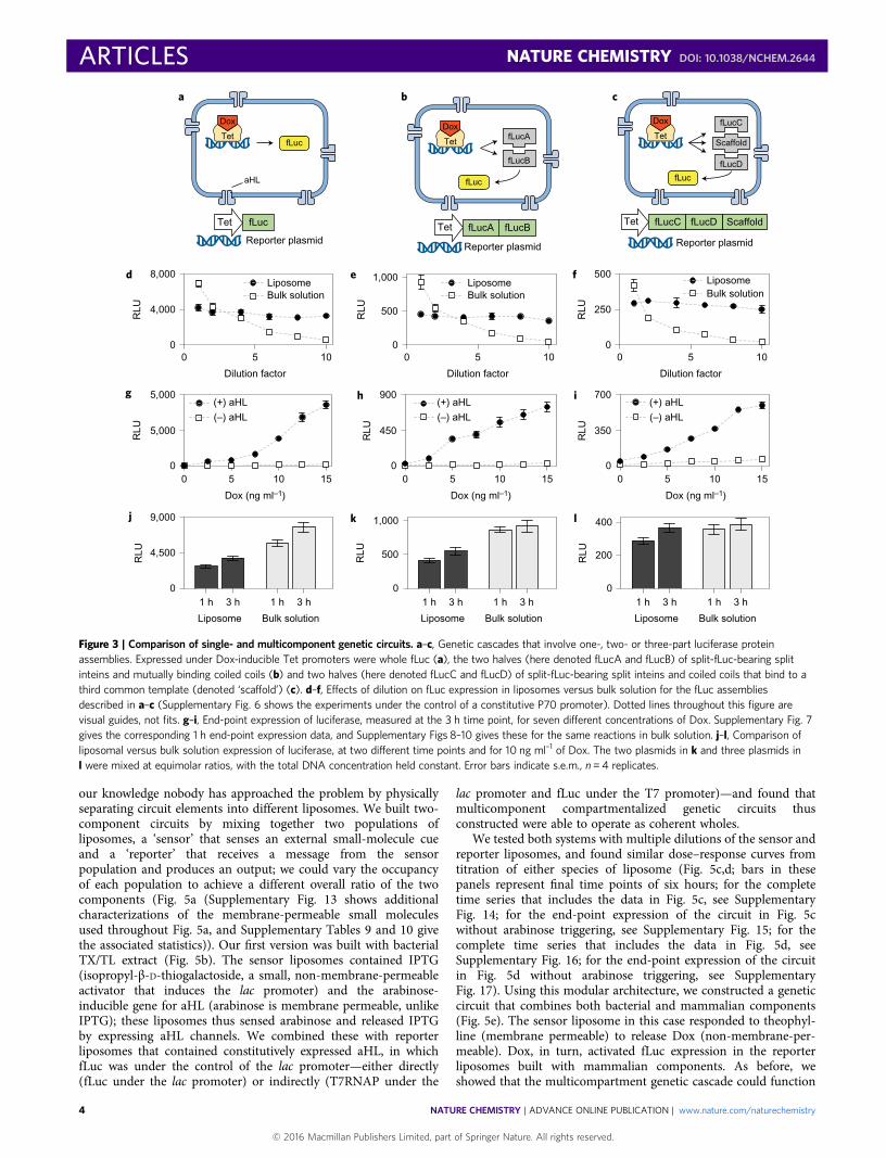
of liposomal compartmentalization—facilitated reaction efficacycaused by molecular confinement (encapsulating reactants withina liposome facilitates their interaction because of the smallvolume)26–29—can help support multicomponent genetic circuitsas well as chemical reactions of higher order. We compared cell-free TX/TL reactions that produce firefly luciferase (fLuc) fromone, two or three protein components, and tested them in bulk sol-ution versus synells. In this experiment, we used HeLa-cell extractthat constitutively expressed the ten–eleven translocation (Tet)protein to mediate small-molecule induction of the transcriptionof the one, two or three fLuc components, as well as alpha-haemo-lysin (aHL), which serves as a pore to admit doxycycline (Dox) totrigger Tet function20,30,31. The one-component luciferase wassimply conventional monolithic fLuc (Fig. 3a); the two-componentsystem (that is, to explore second-order reactions) comprised thetwo halves of a split fLuc, each attached to a coiled coil and a splitintein fragment to bring the halves together and covalently bridgethem (Fig. 3b)32; and the three-component system involved thehalves of split fLuc bearing coiled coils and split inteins, with thecoiled coils targeting a third protein, a scaffold (Fig. 3c)32.
For all three orders of luciferase-producing reactions, the effect ofdilution on fLuc expression was weaker for the liposomes than forthe bulk solution (Fig. 3d–f; P < 0.0001 for the interaction betweenfactors of encapsulation and dilution factor; analysis of variance
(ANOVA) with factors of encapsulation and dilution factor; seeSupplementary Tables 1–3 for the full statistics and SupplementaryFig. 6 for corresponding experiments under the control of a constitutiveP70 promoter). As expected, fLuc expression was proportional to theconcentration of Dox added to the external solution, and dependedon aHL (Fig. 3g–i show end-point expression after three hours(Supplementary Fig. 7 shows the corresponding expression at a onehour end point, and Supplementary Figs 8–10 for the same reactionsin bulk solution)). Liposomes produced lower amounts of fLuc thanthe same volume of TX/TL extract in bulk solution—probablybecause of the well-known property of stochastic loading of reagentsinto liposomes27,28 (P < 0.0001 for the factor of encapsulation inANOVA with factors of time, encapsulation and order(Supplementary Table 4 gives the full statistics)). For the third-orderreaction, we found that liposome encapsulation resulted in an efficacynearly equal to that of bulk solution (Fig. 3l; P = 0.1324 for the factor ofencapsulation in ANOVA with factors of time and encapsulation(Supplementary Table 7 gives the full statistics)), whereas for thefirst-order and second-order reactions the liposomes resulted inlower efficacies (Fig. 3j,k; P < 0.0001 for the factor of encapsulation inANOVAs for both analyses, each with factors of time and encapsulation(Supplementary Tables 5 and 6 give the full statistics)). Thus, molecularconfinement in liposomes may help facilitate higher-order reactionsthat require multiple chemical building blocks to be brought together,
IPTG
Dox Ara
Theo
RibosomeRNAP
fLucLac
P70 T7RNAP
ED
CBA
CBA ED
fLuc
Synell
aHL pores
Geneticcircuits
Non-permeableactivators
Permeableactivators
Cell extract
Phospholipids
B
A
C
D
E
fLucrLuc
a
b
c
d e
fLuc
A
B
D
fLuc
Time
B
A
C
SNAREs
Figure 1 | An overview of genetic circuit interactions within and between synells. a, Synells are semipermeable compartments made from a phospholipidbilayer membrane and various contents. The membrane can display a variety of proteins, including channel-forming proteins such as aHL (grey membranepores). The phospholipid membranes of synells are permeable to molecules such as theophylline (Theo) and arabinose (Ara), and are permeable to others,such as IPTG and Dox, when aHL channels are present; these molecules can be used to trigger activity within synells. Synells can encapsulate cell lysateswith transcriptional and/or translational activity, as well as DNA vectors that encode genes. In this Article, we demonstrate four novel competencies of synellsthat, together, can be used to create complex, modular genetic circuits. b, Synells can contain genetic circuits in which all the components and operations takeplace within the same liposome. fLuc, firefly luciferase. c, Two genetic circuits can work independently in separate liposome populations. rLuc, Renillaluciferase. d, Genetic circuits within two different liposome populations can interact. e, Genetic circuits can run in parallel in separate compartmentalizedreactions; if those reactions are encapsulated by liposomes that carry fusogenic peptides, such as SNAREs, the reaction products can be joined together in ahierarchical fashion. In panels b–e, the letters A, B, C, D and E represent inputs to genetic circuits (proteins, DNA, small molecules, and so on).
ARTICLES NATURE CHEMISTRY DOI: 10.1038/NCHEM.2644
NATURE CHEMISTRY | ADVANCE ONLINE PUBLICATION | www.nature.com/naturechemistry2
© 2016 Macmillan Publishers Limited, part of Springer Nature. All rights reserved.

because the restrictedmovement of reagents increases the probability ofthe requisite multiway interactions.
Insulation of genetic circuits that operate in parallel liposomepopulations. As a next step towards engineering sets of liposomesthat can communicate with one another, we set out to determinewhether liposomes could be used to insulate multiple andpotentially incompatible genetic circuits from each other, so thatthey could operate in the same bulk environment. This insulationwould enable modular design; each circuit could be optimizedindependently and deployed in the same environment as othercircuits without interference. These circuits could reuse the sameparts (proteins, DNA) for different purposes in differentliposomes, and thereby circumvent one limitation of geneticcircuits designed for all parts to operate within the same livingcell (where one must assume that all the circuit elements mightencounter each other and must therefore be inherentlyorthogonal). Different liposome populations could also containchemical microenvironments that are not mutually compatible(for example, bacterial and mammalian extracts, or mammaliantranscriptional and mammalian translational machinery)—thereare numerous examples throughout chemistry of reactions beingrun under specialized, and thus often isolated, reaction conditions33.
We first assessed whether multiple liposomal circuits couldoperate in parallel without crosstalk. To do this, we createdpopulations of liposomes that could respond differently to the sameexternal activator. We built two populations of liposomes carryingmammalian TX/TL extract and the same amount of Dox-inducibleluciferase DNA (either Renilla luciferase (rLuc) or fLuc), but variedthe amount of aHL DNA to result in high-aHL and low-aHL synellpopulations (Fig. 4a). High-aHL and low-aHL synells responded to
the non-membrane-permeable Dox in the external solution, doingso proportionally to their own aHL concentrations (Fig. 4b). Weobserved no evidence that Dox acting on one liposome populationaffected the expression of luciferase in the other population—specifi-cally, there was no significant difference in fLuc expression inhigh-aHL fLuc liposomes when the rLuc liposomes were high-aHLversus low-aHL, and the same held for the other combinations(Fig. 4b; Sidak’s multiple comparisons test after ANOVA withfactors of luciferase type and aHL combination (SupplementaryTable 8 gives the full statistics, and Supplementary Figs 11 and 12give the rLuc and fLuc expression data at different aHL plasmid con-centrations, for two different time points)). That is, luciferaseexpression from each liposome population depended only on theamount of aHL DNA present in that population, and not on thatof the other population (Fig. 4c–e). This experiment thus not onlyverifies the independent operation of multiple non-interactingliposomes, but also verifies that multiple liposome populations canbe programmed in advance to have varying response levels to agiven trigger and, subsequently, in the same internal solution, theycan be triggered to function simultaneously.
Communication between genetic circuits that operate in multipleliposome populations. Having established that genetic circuits inseparate populations of liposomes could operate independently,we next sought to begin to create controlled communicationpathways between populations of synells. In this way we couldcreate a compartmentalized genetic circuit—which, as notedabove, may need to be separated from others for reasons ofcontrol fidelity, toxicity or reagent tunability—and connect it toother compartmentalized circuits. Although previous work hasemphasized the importance of modularity in genetic circuits34, to
b dc
15,000
105
104
103
–103 103 104 105
Q4
0 –103 103 104 1050
102
0GFP
GFP Split GFP
GFP
Rhodamine
Rho
dam
ine
105
104
103
102
0
Rho
dam
ine
Split GFP
10,000
5,000
Flu
ores
cenc
e
0Q3
Q1 Q2
Q4Q3
Q1 Q2
v via
i ii
iii iv
Figure 2 | Molecular confinement of multicomponent genetic cascades. a, Images of liposomes that express GFP. i–iv, SIM images of representativeliposomes that express GFP and have membranes labelled with rhodamine. Every SIM image (i–iv) represents a separate liposome; all the liposomes wereimaged on the same day and all the liposomes came from the same sample, prepared 24 h before imaging. All SIM images in this figure are at the samescale. Scale bars, 1 µm (i and ii) and 200 nm (iii and iv). v,vi, Widefield epifluorescent images of liposomes that express GFP. The liposomes for this imagingsample were extruded through a 2 µm filter and dialysed with a 1 µm membrane; v shows sample after 6 h incubation and vi shows an aliquot of the samesample after 24 h incubation. Scale bars, 10 µm (v and vi). b–d, Fraction of synells that express GFP and split GFP, measured by flow cytometry(Supplementary Fig. 2 shows the control flow-cytometry experiments). b, Bulk expression of GFP and fluorescence measured on the sample prior to theflow-cytometry experiments. c, Analysis of samples that express GFP; 68.4% of the liposomes produced a measurable green signal. d, Analysis of samplesthat expressed split GFP; 61.8% of the liposomes produced a measurable green signal.
NATURE CHEMISTRY DOI: 10.1038/NCHEM.2644 ARTICLES
NATURE CHEMISTRY | ADVANCE ONLINE PUBLICATION | www.nature.com/naturechemistry 3
© 2016 Macmillan Publishers Limited, part of Springer Nature. All rights reserved.
our knowledge nobody has approached the problem by physicallyseparating circuit elements into different liposomes. We built two-component circuits by mixing together two populations ofliposomes, a ‘sensor’ that senses an external small-molecule cueand a ‘reporter’ that receives a message from the sensorpopulation and produces an output; we could vary the occupancyof each population to achieve a different overall ratio of the twocomponents (Fig. 5a (Supplementary Fig. 13 shows additionalcharacterizations of the membrane-permeable small moleculesused throughout Fig. 5a, and Supplementary Tables 9 and 10 givethe associated statistics)). Our first version was built with bacterialTX/TL extract (Fig. 5b). The sensor liposomes contained IPTG(isopropyl-β-D-thiogalactoside, a small, non-membrane-permeableactivator that induces the lac promoter) and the arabinose-inducible gene for aHL (arabinose is membrane permeable, unlikeIPTG); these liposomes thus sensed arabinose and released IPTGby expressing aHL channels. We combined these with reporterliposomes that contained constitutively expressed aHL, in whichfLuc was under the control of the lac promoter—either directly(fLuc under the lac promoter) or indirectly (T7RNAP under the
lac promoter and fLuc under the T7 promoter)—and found thatmulticomponent compartmentalized genetic circuits thusconstructed were able to operate as coherent wholes.
We tested both systems with multiple dilutions of the sensor andreporter liposomes, and found similar dose–response curves fromtitration of either species of liposome (Fig. 5c,d; bars in thesepanels represent final time points of six hours; for the completetime series that includes the data in Fig. 5c, see SupplementaryFig. 14; for the end-point expression of the circuit in Fig. 5cwithout arabinose triggering, see Supplementary Fig. 15; for thecomplete time series that includes the data in Fig. 5d, seeSupplementary Fig. 16; for the end-point expression of the circuitin Fig. 5d without arabinose triggering, see SupplementaryFig. 17). Using this modular architecture, we constructed a geneticcircuit that combines both bacterial and mammalian components(Fig. 5e). The sensor liposome in this case responded to theophyl-line (membrane permeable) to release Dox (non-membrane-per-meable). Dox, in turn, activated fLuc expression in the reporterliposomes built with mammalian components. As before, weshowed that the multicompartment genetic cascade could function
Dox
TetfLuc
aHL
Tet fLuc
Reporter plasmid
Dox
TetfLucA
fLucB
fLuc
Tet fLucA fLucB
Reporter plasmid
fLuc
Reporter plasmid
Dox
Tet
fLucC
fLucD
Scaffold
ScaffoldTet fLucC fLucD
8,000 1,000
500
0
500
250
0
RLU
RLU
RLU
RLU
RLU
RLU4,000
0
0 0
450
900
0
350
700
5,000
5,000
0
0 5 10
(+) aHL
(–) aHL
(+) aHL
(–) aHL
(+) aHL
(–) aHL
15
5
Dilution factor
Dox (ng ml–1)
0 5 10 15
Dox (ng ml–1)
0 5 10 15
Dox (ng ml–1)
LiposomeBulk solution
Liposome Bulk solution
LiposomeBulk solution
LiposomeBulk solution
10 0 5
Dilution factor
10 0 5
Dilution factor
10
1,000 400
500 200
0
9,000
4,500
01 h 3 h 1 h 3 h
Liposome Bulk solution
1 h 3 h 1 h 3 h
Liposome Bulk solution
1 h 3 h 1 h 3 h0
RLU
RLU
RLU
a
d
g h i
j k l
e f
b c
Figure 3 | Comparison of single- and multicomponent genetic circuits. a–c, Genetic cascades that involve one-, two- or three-part luciferase proteinassemblies. Expressed under Dox-inducible Tet promoters were whole fLuc (a), the two halves (here denoted fLucA and fLucB) of split-fLuc-bearing splitinteins and mutually binding coiled coils (b) and two halves (here denoted fLucC and fLucD) of split-fLuc-bearing split inteins and coiled coils that bind to athird common template (denoted ‘scaffold’) (c). d–f, Effects of dilution on fLuc expression in liposomes versus bulk solution for the fLuc assembliesdescribed in a–c (Supplementary Fig. 6 shows the experiments under the control of a constitutive P70 promoter). Dotted lines throughout this figure arevisual guides, not fits. g–i, End-point expression of luciferase, measured at the 3 h time point, for seven different concentrations of Dox. Supplementary Fig. 7gives the corresponding 1 h end-point expression data, and Supplementary Figs 8–10 gives these for the same reactions in bulk solution. j–l, Comparison ofliposomal versus bulk solution expression of luciferase, at two different time points and for 10 ng ml–1 of Dox. The two plasmids in k and three plasmids inl were mixed at equimolar ratios, with the total DNA concentration held constant. Error bars indicate s.e.m., n = 4 replicates.
ARTICLES NATURE CHEMISTRY DOI: 10.1038/NCHEM.2644
NATURE CHEMISTRY | ADVANCE ONLINE PUBLICATION | www.nature.com/naturechemistry4
© 2016 Macmillan Publishers Limited, part of Springer Nature. All rights reserved.

as designed, with similar fLuc expression dose–response curves ontitrating either the sensor or reporter liposome concentration(Fig. 5f; bars in this panel represent final time points of six hours;for the complete time series that includes the data in Fig. 5f, seeSupplementary Fig. 18; for the end-point expression of the circuitin Fig. 5f without theophylline triggering, see SupplementaryFig. 19). Thus, even multicomponent genetic circuits with differentchemical microenvironments (for example, made from bacterialversus mammalian cell extracts) can be assembled into coherentnetworks that comprise multiple modules.
Fusion of complementary genetic circuits. Finally, havingestablished that it is possible to maintain liposomes in high-integrity states despite being mixed, we sought to engineer synellsto fuse so that they could bring together two genetic cascades into
the same environment in a programmable fashion. Twoprecursors might require synthesis in different milieus, butultimately need to be reacted with one another. One prominentexample is that of mammalian transcription and translation.Functionally, mixed mammalian transcription and translationcell-free extracts are not able to result in the transcription of DNAinto RNA and then the translation of RNA into protein, perhapsbecause the microenvironments of the mammalian nucleus andcytoplasm are quite different, which makes their cell-free extractsincompatible (Supplementary Fig. 20). Rather than mixing thetwo cell-free extracts into a single non-functioning mixture, itmight be preferable to use synells to compartmentalize thereactions. Once nuclear-extract synells have completedtranscription, it might be desirable to fuse them with cytoplasmic-extract synells for the translation to take place.
Population A:High aHL concentration createshigh expression of firefly luciferase
Population B:Low aHL concentration createslow expression of Renilla luciferase
Reporter plasmid
aHL
Reporter plasmid
T7 TetaHL fLuc
T7 Tet
Tet Tet
Tet
TetrLuc
Time
Dox
Dox
DoxDox
fLuc
fLucfLuc
fLuc
Time
DoxDox
aHL rLuc
a
d e
0 5,0000
2,000
0 7000
14,000
0 50
500
0 50
13,0000 5
0 5
0
5,000
0
1,100
rLuc (0.1 nM aHL)
aHL (nM)(fLuc liposomes only)
aHL (nM)(rLuc liposomes only)
fLuc RLU fLuc RLU
fLucfLuc
(0.1 nM aHL)
rLuc
RLU
RLU
RLU
RLU
rLuc
RLU
rLuc
RLU
rLuc
RLU
rLuc RLU
b c
0 6,0000
15,000
0 5
0 5
rLuc
aHL (nM) fLuc RLU
fLuc0
6,000
0
15,000
RLU
RLU
fLucrLuc
0
6,000
aHL (fLuc)
aHL (rLuc) 0.1 nM 0.1 nM
0
14,000
0.1 nM 0.1 nM
5 nM
Two liposome populations withindependent aHL concentration
fLuc
RLU
5 nM
5 nM 5 nM
Figure 4 | Insulation of genetic circuits that operate in parallel liposome populations. a, Schematic of liposome populations designed to contain similargenetic components, but to respond differently to the same environmental concentration of the non-membrane-permeable small-molecule activator Dox, byexpressing different amounts of the aHL channel protein. These liposomes contain a measured amount of the plasmid for constitutively expressed aHL, andof a plasmid that drives either fLuc or rLuc from the Tet-inducible promoter (the luciferase plasmids were always held at the same concentration). For all thedata in this figure, the two populations were incubated together in the solution containing Dox and harvested after 6 h (Supplementary Figs 11 and 12 givethe rLuc and fLuc expression as a function of aHL plasmid concentration after 2 and 6 h, respectively). b, Each liposome contains either 0.1 or 5 nM of theaHL plasmid. c, Luciferase expression in symmetrical populations in which the amount of aHL DNA is the same across the two populations; the amount offLuc and rLuc expression is shown in the graphs with respect to aHL plasmid concentration and to each other. d,e, Luciferase expression in asymmetricalpopulations. d, Luciferase expression when Renilla liposomes have a constant aHL plasmid concentration (0.1 nM), but the concentration of that plasmid isvaried in the firefly liposomes. The expressions of rLuc and fLuc are shown in the graphs against the plasmid concentration in firefly liposomes and againsteach other. e, Luciferase expression as in d, but with constant aHL plasmid concentration in firefly liposomes and variable concentration in Renilla liposomes.Error bars indicate s.e.m., n = 4 replicates.
NATURE CHEMISTRY DOI: 10.1038/NCHEM.2644 ARTICLES
NATURE CHEMISTRY | ADVANCE ONLINE PUBLICATION | www.nature.com/naturechemistry 5
© 2016 Macmillan Publishers Limited, part of Springer Nature. All rights reserved.

Thus, we sought to make liposomes capable of controlled fusion(Fig. 6a). Fusing liposomes of opposite charge was previously demon-strated to activate gene expression in liposomes35. Our system usesonly one kind of membrane composition (POPC (1-palmitoyl-2-oleoyl-sn-glycero-3-phosphorylcholine) cholesterol membranes,known to be a good environment for membrane channels such asaHL), so to achieve fusion between liposomes we used SNARE(SNAP receptor (SNAP, soluble N-ethylmaleimide-sensitive factorattachment protein))/coiled-coil hybrid proteins (here the hybridproteins are called SNAREs for short), which can be generated incomplementary pairs that are specific in their fusion properties36,37.We could thus fuse together complementary circuit elements byencapsulating them in separate populations of SNARE-fusableliposomes. We confirmed that SNAREs mediated liposome fusionthrough SIM imaging (Fig. 6a) by observing fluorescence resonanceenergy transfer (FRET) signals from lipid dyes added to theliposome membranes (FRET signals showed that the fusion process
takes place within minutes (Supplementary Figs 21 and 22)) and byobserving mixing of the liposome content, reported as dequenchingof a molecular beacon encapsulated in one population of liposomesby a complementary target encapsulated in the other population(Supplementary Fig. 23). We observed large liposomes and alsoliposome aggregates (presumably in the process of fusing) of sizeson the order of 5–10 µm, and measured a minimal amount ofleakage from the liposomes during the process of fusion(Supplementary Fig. 24).
We tried several combinations of complementary circuitelements: the gene for T7RNAP and a T7-driven fLuc (Fig. 6b); anon-membrane-permeable small-molecule trigger (IPTG) and anIPTG-triggered (lac-promoter-driven) fLuc (Fig. 6c); genes for amembrane pore (aHL) and a lac-promoter-driven fLuc in anIPTG-containing ambient (Fig. 6d); and two different genes encod-ing for parts of split luciferase using the same fLucA and fLucB as inFig. 3b (Fig. 6e). For one final test, liposomes that carried
Occ
upan
cy
Sensor liposomes Final mixture Reporter liposomes
a
b
c d f
e
1.0
0.66
0.33
0.0
Occupancy
1.0
0.66
0.33
0.0
IPTGIPTG
IPTG
IPTG
PBAD
Sensor
Reporter
Lac
aHL
fLucLacprotein
Lacprotein
IPTGIPTG
IPTG IPTG IPTG
IPTG
IPTG IPTG IPTG
IPTG
IPTG
IPTGAra
Time
Ara
TimefLuc
Theo
Sensor
Reporter
Tet
Tet
Tet
aHL
fLuc
DoxDox
Dox
Dox
Dox DoxDox
Dox Dox
Theo TheoDox
Dox
Dox
DoxDox
Dox Dox
fLuc
Time
Time
0
15,000
0.00.250.500.751.0
0.0 0.25 0.50 0.75 1.0
Reporter liposome occupancy
Two-stagereporter circuit
Sensor liposomeoccupancy
RLU
Lac
T7 fLuc
T7RNAP
10,000
00.0 0.2 0.4 0.6 0.8 1.0
Reporter liposome occupancy
Reporter circuitSensor liposomeoccupancy
RLU
Tet fLuc
0.00.20.40.60.81.0
0.0
0.00
20,000
0.25 0.50 0.75 1.0
0.250.500.751.0
Reporter circuit
Reporter liposome occupancy
Sensor liposomeoccupancy
RLU
Lac fLuc
Figure 5 | Communication between genetic circuits that operate in multiple liposome populations. a, Scheme for mixing two populations of liposomes atdifferent ratios of their components while maintaining a constant lipid concentration of 10 mM (the same scheme was used throughout this figure and inFig. 6). Each population contains the same amount of liposomes, but the liposome occupancy can vary between 0 (all liposomes are empty) and 1 (themaximum fraction of the liposomes contain reagents). b–d, Externally activated two-part circuits, with bacterial TX/TL. b, Scheme of interacting populations,denoted sensor and reporter. Sensor liposomes contain the aHL gene and are filled with IPTG; reporter liposomes contain machinery for fLuc expression.During activation, arabinose (Ara) diffuses through the sensor liposome membrane and induces aHL expression, which releases IPTG, which induces fLucexpression in the reporter. c, Expression of fLuc for varying ratios of occupancy (as in a) for the sensor and reporter liposomes with the indicated contents.c represents the 6 h time point (Supplementary Fig. 14 shows the complete time series and Supplementary Fig. 15 shows this circuit without arabinose).d, Expression of fLuc for a circuit in which the reporter liposomes contain DNA for a multicomponent genetic cascade, as indicated. d represents the 6 htime point (Supplementary Fig. 16 shows the complete time series and Supplementary Fig. 17 shows this circuit without arabinose). e,f, Externally activatedtwo-part circuits that contain both bacterial and mammalian TX/TL components. e, Sensor vesicles contain the Theo-triggered aHL gene and Dox; reporterliposomes contain constitutively expressed aHL and Tet, and Dox/Tet-driven fLuc. During activation, Theo diffuses through the membrane of the activatorliposomes and induces aHL expression, which creates pores that release Dox from the activator. Dox induces fLuc expression in the reporter liposomes.f, Expression of fLuc for varying ratios of sensor and reporter liposomes (f represents the 6 h time point; Supplementary Fig. 18 gives the complete timeseries and shows this circuit without Theo). Error bars indicate s.e.m., n = 4 replicates.
ARTICLES NATURE CHEMISTRY DOI: 10.1038/NCHEM.2644
NATURE CHEMISTRY | ADVANCE ONLINE PUBLICATION | www.nature.com/naturechemistry6
© 2016 Macmillan Publishers Limited, part of Springer Nature. All rights reserved.

i ii iii
iv v vi
vii viii
ix
T7 RNAP
T7 RNAP
T7 RNAP
Liposomepopulation Awith SNARE A
Liposomepopulation Bwith SNARE B
Tim
e
a
c
e f
d
b
fLuc
Tim
e
Complementary SNAREs
T7RNAPP70 fLucT7
Population A Population B
2,000
0
0.0 B0.5 B1.0 B
0 A 1 A0.5 A
RLU
Same SNAREs1,000
1000 B
0 A 1 A
1 B
RLU
10
No SNAREs
0 A 1 A
0 B1 B
1,000
100
RLU
10
fLucLacIPTG
Population A Population B
RLU
0 A 1 A
0 A 1 A
Same SNAREs
0 A 1 A
0 B1 B
1,000
100
RLU
10
No SNAREs
0 A 1 A
0 B1 B
1,000
100
RLU
10
Complementary SNAREs
0
800 0.0 B0.5 B1.0 B
0 A 1 A0.5 A
No SNAREs
0 B1 B
0 B1 B
Same SNAREs
0 A 1 A
0 A 1 A
RLU
RLU
1,000
100
10
No SNAREs
0 B1 B
0 B1 B
1,000
100
10
RLU
RLU
1,000
100
10
1,000
100
10
0 B1 B
0 B1 B
No SNAREs
0 A 1 A
0 A 1 A
1,000
100
RLU
10
1,000
100
RLU
10
RLU
RLU
Complementary SNAREs
0
6000.0 B0.5 B1.0 B
0 A 1 A0.5 A
P70 aHL fLucLac
Population A Population B Same SNAREs
Same SNAREs
P70 fLucBP70 fLucA
RLU
1,000Complementary SNAREs
0
0.0 B0.5 B1.0 B
0 A 1 A0.5 A
Population A Population B
UBC fLuc
1,200
Complementary SNAREs
0
0.0 B0.5 B1.0 B
0 A 1 A0.5 A
Population A Population B
Transcriptionmachinery
Translationmachinery
Figure 6 | Fusion of complementary genetic circuits. a, General scheme for SNARE-mediated liposome fusion. We created two populations of liposomes,A and B, decorated with complementary SNARE protein mimics in their outer leaflet. The images to the right, in i–ix, are maximum-intensity projections ofSIM z-stacks of liposome membrane labelled with rhodamine, bearing complementary SNARE pairs and fused for 4 h. All the images (i–ix) represent separatefields of view. Scale bars, 5 µm. All liposomes in this figure, except f, contained bacterial TX/TL components. b–f, Five different types of the liposome fusionconcept, exploring several ways to distribute genetic circuits across fusable liposomes, with two different populations of liposomes at three occupancy levels foreach case. b, Mixing of constitutively expressed T7 RNA polymerase with fLuc under T7 promoter. c, Mixing of a non-membrane-permeable small-moleculeactivator IPTG with its inducible promoter driving fLuc production. d, Mixing of a constitutively expressed membrane channel with an inducible promoter drivingfLuc production in the background of the small molecule that induces the promoter (IPTG). e, Mixing liposomes with genes that encode split protein. f, Mixingliposomes that contain a mammalian transcription (HeLa) and translation (HeLa) system, producing fLuc. For all five systems in b–f, the large graph showsexperiments in which the two liposome populations had matching SNAREs, the top small panel is when both liposomes had the same SNARE, and the bottomone when neither population had any SNAREs. In both small graphs of b–f, the y axis is a logarithmic scale to show the near-zero values for non-fusingliposomes. Switching which liposome contained which SNARE had no effect on the results (Supplementary Fig. 25), whereas the absence of SNARE proteins orthe presence of identical SNAREs on both populations hindered fusions (small graphs in b–f). Error bars indicate s.e.m., n =4 replicates.
NATURE CHEMISTRY DOI: 10.1038/NCHEM.2644 ARTICLES
NATURE CHEMISTRY | ADVANCE ONLINE PUBLICATION | www.nature.com/naturechemistry 7
© 2016 Macmillan Publishers Limited, part of Springer Nature. All rights reserved.
mammalian nuclear (transcription) extract and the gene for fLuc,incubated for 12 hours, were then mixed with liposomes that con-tained cytoplasmic (translation) extract, and further incubated for12 hours (Fig. 6f ). We were able to observe the production offLuc protein, even though a direct combination of transcriptionaland translational machinery produced no fLuc above backgroundlevels (Supplementary Fig. 20). Throughout all these cases, weobserved production of the final output of the genetic cascadeonly when the two liposome populations were equipped withSNAREs, and only when they were a SNARE cognate pair (P <0.0001 for the factor of SNARE compatibility, ANOVA withfactors of mechanism, occupancy and SNARE compatibility(Supplementary Table 11 gives the full statistics; for systems inthis figure, switching which liposome contained which SNAREhad no effect on the results, as shown in Supplementary Fig. 25)).
Discussion. Liposomes are key in chemistry and chemical biologyfor compartmentalizing chemical reactions that require differentenvironments or act on different samples. In this work, we showhow synells—liposomes containing genes as well as transcriptionaland/or translational machinery—enable a great level of modularityfor genetic circuit design and execution. We showed that circuitscould be designed to run in synell populations in the samecontainer, independent of each other because of the insulationprovided by the liposomal membrane. Genetic circuits could alsobe connected to communicate with one another through small-molecule messengers. This communication was possible evenacross liposomes that contained incompatible microenvironments,as we showed by constructing the first genetic circuit to containbacterial and mammalian cell-free extracts and genetic elements.Finally, we explored the use of SNARE mimics to fuse synellstogether, enabling the direct union of separately synthesizedreaction components. Using this strategy, we were able to produceRNA encoding for fLuc in one population of liposomes thatcontained mammalian transcriptional extract, which on fusionwith liposomes that contained mammalian translational extractresulted in protein production—an outcome that does not occur ifthe gene is simply added to a mixture of the two extracts.
Synells thus enable a new level of modularity for syntheticbiology. Modularity is key in engineering, because breaking acomplex synthetic biology system into parts that can be indepen-dently controlled or regulated, without crosstalk, and that commu-nicate only in well-defined ways, enables each part to be optimizedindividually while supporting their incorporation into an emergentwhole. Our technologies will enable a large number of differentsynthetic biology problems to be made modular, even those thatinvolve genetic cascades that might interfere with each other (orpose toxicity issues) if they were to all occur in one pot. As ourmethod of compartmentalization is liposomal, there is no needfor specialized hardware to mediate the communication andcontrol of multiple interacting reaction systems. Precise temporalcontrol of synell networks could be enhanced even further byusing light to trigger optogenetic signalling cascades, which inturn can trigger downstream effects38,39. We also show that themolecular confinement of liposomes can facilitate multicomponentprotein–protein interactions.
Our synells, in addition to the power they offer to syntheticbiology, may also enable the simulation of various complex beha-viours that have been proposed as characteristics of early lifeforms. Controlled communication between cells, the fusion ofgenetic elements across cells and the assembly of complex geneticcascades towards defined cellular behaviours are all traits thatarose in the course of early evolution. Synells have been widelyused as models for studying the origin and earliest evolution oflife40–44. For example, one of us has previously shown that liposomesencapsulating a simple catalyst can be used to model early
Darwinian competition mechanisms41. Interacting encapsulatedgenetic circuits will hopefully enable the study of the more-complexcharacteristics that have been proposed for the last universalcommon ancestor45–47, and perhaps help to reveal the dynamicand boundary conditions that underlie the mechanisms ofDarwinian evolution48,49.
MaterialsCloning of expression constructs. The P70 (OR2-OR1-Pr (ref. 50)) and lac (Llac-0-1(ref. 51)) promoter constructs were used in a modified pCI vector (Promega). Theoriginal promoter region of the vector was replaced by the appropriate promoter tomake our constructs51. For bacterial expression, the previously described transcriptionterminator T500 was added at the end of each ORF (open reading frame). The originaluntranslated region (UTR) was also removed and replaced with the previously describedUTR1 (ref. 50). The mammalian Tet constructs were built into the Tet-On 3Gbidirectional vector (Clontech) by cloning the genes intoMCS1. The araBAD constructswere built using a PBAD vector52 (Thermo). We used PBAD–hisB and removed theHis-tag and the enterokinase recognition site prior to inserting the genes used inthis study.
Flow cytometry with GFP and split GFP. The fluorescence signal from these GFPliposomes was measured after 12 h of incubation for the experiments in Fig. 2b–d.Membranes (red fluorescence) were labelled with Lissamine rhodamine B1,2-dihexadecanoyl-sn-glycero-3-phosphoethanolamine triethylammonium salt(rhodamine DHPE), used at 0.2 molar percentage of the POPC concentration.GFP was expressed from a plasmid with the T7 promoter. The halves of split GFPwere fused to complementary coiled coils and expressed from two differentplasmids (both with the T7 promoter). For the flow-cytometry analysis, events intwo fluorescent channels were analysed: GFP and red fluorescence. Each datasetconsists of a minimum of 19,000 events. Figure 2c shows an analysis of liposomesthat expressed GFP and Fig. 2d shows an analysis of liposomes that expressed splitGFP. The percentage of liposomes that expressed protein was calculated as thepercentage of events in the quadrant positive in both the green and red channels(Q2 on both plots). The flow cytometer was not calibrated using size standards, andtherefore all the information about the size of the particles in the experiment isapproximate. For the detailed size measurements of the liposomes in this work,Supplementary Fig. 1 gives data from the DLS experiments. The flow-cytometryanalysis was performed on a FACSCanto II, and the data analysis was performedusing a FACSDiva 8.0.
fLuc assays. fLuc activity was assayed using the Steady-Glo Luciferase Assay System(Promega). The protein analysis was performed according to the manufacturer’sinstructions. The cell lysis protocol was replaced with a modified procedure forlysing liposome-encapsulated expression reactions. The 50 µl liposome reactionswere quenched by 10 µl of Quench Mix that contained 0.3% v/v Triton-X100 (todisrupt the vesicles), TURBO DNAse (Thermo; final concentration ∼2U per 60 µl;1 µl used), TURBO DNAse buffer (final concentration ∼0.5×, 2.5 µl 10× stock used),RNase Cocktail Enzyme Mix (mixture of RNAse A and RNAse T1, 3 µl per 60 µlreaction (Thermo)). The samples were incubated with the Quench Mix for 15 min at37 °C. The resulting sample was used directly with the Steady-Glo luciferase assay,according to the manufacturer’s instructions.
The result is given in relative light units (RLU) with a 10 s integration time.
Enzyme activity assays. Renilla, NanoLuc luciferase, beta-lactamase, beta-galactosidase and chloramphenicol acetyltransferase activity were assayed usingcommercially available kits, according to the manufacturer’s instructions(Supplementary Information gives the detailed procedures).
E. coli cell-free TX/TL extract. Our E. coli cell-free extract was prepared accordingto the Noireaux Lab protocol, from Rosetta 2 BL21 cells (Novagen)50,53. The entireextract preparation was performed in a cold room (4 °C).
HeLa cell-free extract. The HeLa cell-free translation extract was preparedaccording to a previously published protocol24. The entire extract preparation wasperformed in a cold room (4 °C). For the mammalian in vitro transcription,we used the HeLa cell-free nuclear fraction transcription system HeLaScribe(Promega).
SNARE protein mimics. The SNARE protein mimics were chemically synthesizedby solid-phase protein synthesis (Genscript). SNARE-A was a fusion of the E3coiled-coil motif and the transmembrane region of the VAMP2 protein(residues 85–116). SNARE-B was a fusion of the K3 coiled-coil motif with atransmembrane region from the syntaxin-1A protein (residues 258–288), asdescribed before36. The SNARE peptide-to-lipid molar ratio used in all theexperiments was 1:500.
Liposomes that undergo SNARE-mediated fusion form large aggregates madefrom multiple starter liposomes36,37; this does not affect the results in Fig. 6, but itwould probably reduce the molecular confinement effects observed in Fig. 3.
ARTICLES NATURE CHEMISTRY DOI: 10.1038/NCHEM.2644
NATURE CHEMISTRY | ADVANCE ONLINE PUBLICATION | www.nature.com/naturechemistry8
© 2016 Macmillan Publishers Limited, part of Springer Nature. All rights reserved.

Received 6 December 2015; accepted 12 September 2016;published online 14 November 2016
References1. Carlson, E. D., Gan, R., Hodgman, C. E. & Jewett, M. C. Cell-free protein
synthesis: applications come of age. Biotechnol. Adv. 30, 1185–1194 (2012).2. Smith, M. T., Wilding, K. M., Hunt, J. M., Bennett, A. M. & Bundy, B. C. The
emerging age of cell-free synthetic biology. FEBS Lett. 588, 2755–2761 (2014).3. Hodgman, C. E. & Jewett, M. C. Cell-free synthetic biology: thinking outside the
cell. Metab. Eng. 14, 261–269 (2012).4. Miller, D. & Gulbis, J. Engineering protocells: prospects for self-assembly and
nanoscale production-lines. Life 5, 1019–1053 (2015).5. Shimizu, Y., Kuruma, Y., Ying, B.-W., Umekage, S. & Ueda, T. Cell-free
translation systems for protein engineering. FEBS J. 273, 4133–4140 (2006).6. Shin, J. & Noireaux, V. An E. coli cell-free expression toolbox: application to
synthetic gene circuits and artificial cells. ACS Synth. Biol. 1, 29–41 (2012).7. Takahashi, M. K. et al. Rapidly characterizing the fast dynamics of RNA genetic
circuitry with cell-free transcription–translation (TX-TL) systems. ACS Synth.Biol. 4, 503–515 (2015).
8. Michener, J. K., Thodey, K., Liang, J. C. & Smolke, C. D. Applications ofgenetically-encoded biosensors for the construction and control of biosyntheticpathways. Metab. Eng. 14, 212–222 (2012).
9. Vamvakaki, V. & Chaniotakis, N. A. Pesticide detection with a liposome-basednano-biosensor. Biosens. Bioelectron. 22, 2848–2853 (2007).
10. Pardee, K. et al. Paper-based synthetic gene networks. Cell 159, 940–954 (2014).11. Lentini, R. et al. Integrating artificial with natural cells to translate chemical
messages that direct E. coli behaviour. Nat. Commun. 5, 4012 (2014).12. Zemella, A., Thoring, L., Hoffmeister, C. & Kubick, S. Cell-free protein synthesis:
pros and cons of prokaryotic and eukaryotic systems. Chem. Bio. Chem. 16,2420–31 (2015).
13. Forster, A. C. & Church, G. M. Towards synthesis of a minimal cell. Mol. Syst.Biol. 2, 45 (2006).
14. Brea, R. J., Hardy, M. D. & Devaraj, N. K. Towards self-assembled hybridartificial cells: novel bottom-up approaches to functional synthetic membranes.Chem. Pub. Soc. Euro. 21, 12564–12570 (2015).
15. Luisi, P. L., Ferri, F. & Stano, P. Approaches to semi-synthetic minimal cells: areview. Naturwissenschaften 93, 1–13 (2006).
16. Stano, P. & Luisi, P. L. Semi-synthetic minimal cells: origin and recentdevelopments. Curr. Opin. Biotechnol. 24, 633–638 (2013).
17. Murtas, G., Kuruma, Y., Bianchini, P., Diaspro, A. & Luisi, P. L. Protein synthesisin liposomes with a minimal set of enzymes. Biochem. Biophys. Res. Commun.363, 12–17 (2007).
18. Yu, W. et al. Synthesis of functional protein in liposome. J. Biosci. Bioeng. 92,590–593 (2001).
19. Oberholzer, T., Nierhaus, K. H. & Luisi, P. L. Protein expression in liposomes.Biochem. Biophys. Res. Commun. 261, 238–241 (1999).
20. Noireaux, V. & Libchaber, A. A vesicle bioreactor as a step toward an artificialcell assembly. Proc. Natl Acad. Sci. USA 101, 17669–17674 (2004).
21. Stech, M. et al. Production of functional antibody fragments in a vesicle-basedeukaryotic cell-free translation system. J. Biotechnol. 164, 220–231 (2012).
22. Weber, L. A, Feman, E. R. & Baglioni, C. A cell free system fromHeLa cells activein initiation of protein synthesis. Biochemistry 14, 5315–5321 (1975).
23. Wimmer, E. Cell-free, de novo synthesis of poliovirus. Science. 254, 1647–1651,(1991).
24. Mikami, S., Masutani, M., Sonenberg, N., Yokoyama, S. & Imataka, H. Anefficient mammalian cell-free translation system supplemented with translationfactors. Protein Expr. Purif. 46, 348–357 (2006).
25. Mikami, S., Kobayashi, T., Masutani, M., Yokoyama, S. & Imataka, H. A humancell-derived in vitro coupled transcription/translation system optimized forproduction of recombinant proteins. Protein Expr. Purif. 62, 190–198 (2008).
26. Tan, C., Saurabh, S., Bruchez, M. P., Schwartz, R. & Leduc, P. Molecularcrowding shapes gene expression in synthetic cellular nanosystems.Nat. Nanotechnol. 8, 602–608 (2013).
27. de Souza, T. P. et al. Encapsulation of ferritin, ribosomes, and ribo-peptidiccomplexes inside liposomes: insights into the origin of metabolism. Orig. LifeEvol. Biosph. 42, 421–428 (2012).
28. de Souza, T. P., Fahr, A., Luisi, P. L. & Stano, P. Spontaneous encapsulation andconcentration of biological macromolecules in liposomes: an intriguingphenomenon and its relevance in origins of life. J. Mol. Evol. 79, 179–192 (2014).
29. Caschera, F. & Noireaux, V. Integration of biological parts toward the synthesisof a minimal cell. Curr. Opin. Chem. Biol. 22, 85–91 (2014).
30. Stefureac, R., Long, Y. T., Kraatz, H. B., Howard, P. & Lee, J. S. Transport ofα-helical peptides through α-hemolysin and aerolysin pores. Biochemistry 45,9172–9179 (2006).
31. Gouaux, E., Hobaugh, M. & Song, L. α-Hemolysin, γ-hemolysin, and leukocidinfrom Staphylococcus aureus: distant in sequence but similar in structure.Protein Sci. 6, 2631–2635 (1997).
32. Selgrade, D. F., Lohmueller, J. J., Lienert, F. & Silver, P. A. Protein scaffold-activated protein trans-splicing in mammalian cells. 135, 7713–7719 (2013).
33. Tu, Y. et al. Mimicking the cell: bio-inspired functions of supramolecularassemblies. Chem. Rev. 116, 2023–2078 (2016).
34. Del Vecchio, D., Ninfa, A. J. & Sontag, E. D. Modular cell biology: retroactivityand insulation. Mol. Syst. Biol. 4, 161 (2008).
35. Caschera, F. et al. Programmed vesicle fusion triggers gene expression. Langmuir27, 13082–13090 (2011).
36. Meyenberg, K., Lygina, A. S., van den Bogaart, G., Jahn, R. & Diederichsen, U.SNARE derived peptide mimic inducing membrane fusion. Chem. Commun. 47,9405–9407 (2011).
37. Robson Marsden, H., Korobko, A. V., Zheng, T., Voskuhl, J. & Kros, A.Controlled liposome fusion mediated by SNARE protein mimics. Biomater. Sci.1, 1046–1054 (2013).
38. Inglés-Prieto, Á. et al. Light-assisted small-molecule screening against proteinkinases. Nat. Chem. Biol. 11, 952–954 (2015).
39. Boyden, E. S. A history of optogenetics: the development of tools for controllingbrain circuits with light. F1000 Biol. Rep. 3, 11 (2011).
40. Hanczyc, M. M., Fujikawa, S. M. & Szostak, J. W. Experimental models ofprimitive cellular compartments: encapsulation, growth, and division.Science 302, 618–622 (2003).
41. Adamala, K. & Szostak, J. W. Competition between model protocells driven byan encapsulated catalyst. Nat. Chem. 5, 495–501 (2013).
42. Balaram, P. Synthesizing life. Curr. Sci. 85, 1509–1510 (2003).43. Adamala, K. et al. Open questions in origin of life: experimental studies on the
origin of nucleic acids and proteins with specific and functional sequences by achemical synthetic biology approach. Comput. Struct. Biotechnol. J. 9,e201402004 (2014).
44. Ruiz-Mirazo, K., Briones, C. & de la Escosura, A. Prebiotic systems chemistry:new perspectives for the origins of life. Chem. Rev. 114, 285–366 (2014).
45. Glansdorff, N., Xu, Y. & Labedan, B. The last universal common ancestor:emergence, constitution and genetic legacy of an elusive forerunner. Biol. Direct3, 29 (2008).
46. Woese, C. The universal ancestor. Proc. Natl Acad. Sci. USA 95, 6854–6859(1998).
47. Theobald, D. L. A formal test of the theory of universal common ancestry.Nature 465, 219–222 (2010).
48. Spencer, A. C., Torre, P. & Mansy, S. S. The encapsulation of cell-freetranscription and translation machinery in vesicles for the construction ofcellular mimics. J. Vis. Exp. 80, e51304 (2013).
49. Adamala, K., Engelhart, A. E., Kamat, N. P., Jin, L. & Szostak, J. W. Constructionof a liposome dialyzer for the preparation of high-value, small-volume liposomeformulations. Nat. Protoc. 10, 927–938 (2015).
50. Shin, J. & Noireaux, V. Efficient cell-free expression with the endogenous E. coliRNA polymerase and sigma factor 70. J. Biol. Eng. 4, 8 (2010).
51. Lutz, R. & Bujard, H. Independent and tight regulation of transcriptional units inEscherichia coli via the LacR/O, the TetR/O and AraC/I1-I2 regulatory elements.Nucleic Acids Res. 25, 1203–1210 (1997).
52. Guzman, L. M., Belin, D., Carson, M. J. & Beckwith, J. Tight regulation,modulation, and high-level expression by vectors containing the arabinosePBAD promoter. J. Bacteriol. 177, 4121–4130 (1995).
53. Sun, Z. Z. et al. Protocols for implementing an Escherichia coli based TX-TL cell-free expression system for synthetic biology. J. Vis. Exp. 79, e50762 (2013).
AcknowledgementsWe thank E. Vasile and F. Chen for help with the SIM microscopy, and G. Paradis andK. Piatkevich for help with the flow-cytometry experiments. We thank N. Kamat and L. Jinfor help with troubleshooting the DLS machine. We thank J. Szostak for sharing theliposome encapsulation formula. We thank V. Noireaux, A. Mershin and A. Engelhart forhelpful discussions about cell-free TX/TL systems. E.S.B. acknowledges, for funding, theNational Institutes of Health (NIH) 1U01MH106011, Jeremy and Joyce Wertheimer, NIH1RM1HG008525, the Picower Institute Innovation Fund, NIH 1R01MH103910, NIH1R01NS075421, National Science Foundation CBET 1053233, New York Stem CellFoundation-Robertson Award and NIH Director’s Pioneer Award 1DP1NS087724.D.A.M.-A. acknowledges support from the Janet and Sheldon Razin (1959) Fellowship.
Author contributionsK.P.A. and D.A.M.-A. contributed equally to this work. K.P.A., D.A.M.-A. and K.R.G.-H.performed the experiments. K.P.A., D.A.M.-A. and E.S.B. designed experiments, analysedthe data and wrote the manuscript.
Additional informationSupplementary information is available in the online version of the paper. Reprints andpermissions information is available online at www.nature.com/reprints. Correspondence andrequests for materials should be addressed to E.S.B.
Competing financial interestsK.P.A., D.A.M.-A. and E.S.B. submitted a provisional patent application based on thiswork.
NATURE CHEMISTRY DOI: 10.1038/NCHEM.2644 ARTICLES
NATURE CHEMISTRY | ADVANCE ONLINE PUBLICATION | www.nature.com/naturechemistry 9
© 2016 Macmillan Publishers Limited, part of Springer Nature. All rights reserved.