entry of botulinum neurotoxin subtypes a1 and a2 into...
TRANSCRIPT
Entry of Botulinum Neurotoxin SubtypesA1 and A2 into Neurons
Abby R. Kroken,* Faith C. Blum,* Madison Zuverink, Joseph T. BarbieriDepartment of Microbiology and Molecular Genetics, Medical College of Wisconsin, Milwaukee, Wisconsin,USA
ABSTRACT Botulinum neurotoxins (BoNTs) are the most toxic proteins for humansbut also are common therapies for neurological diseases. BoNTs are dichain toxins,comprising an N-terminal catalytic domain (LC) disulfide bond linked to a C-terminalheavy chain (HC) which includes a translocation domain (HN) and a receptor bindingdomain (HC). Recently, the BoNT serotype A (BoNT/A) subtypes A1 and A2 were re-ported to possess similar potencies but different rates of cellular intoxication andpathology in a mouse model of botulism. The current study measured HCA1 andHCA2 entry into rat primary neurons and cultured Neuro2A cells. We found thatthere were two sequential steps during the association of BoNT/A with neurons. Theinitial step was ganglioside dependent, while the subsequent step involved associa-tion with synaptic vesicles. HCA1 and HCA2 entered the same population of synapticvesicles and entered cells at similar rates. The primary difference was that HCA2 hada higher degree of receptor occupancy for cells and neurons than HcA1. Thus, HCA2and HCA1 share receptors and entry pathway but differ in their affinity for receptor.The initial interaction of HCA1 and HCA2 with neurons may contribute to the uniquepathologies of BoNT/A1 and BoNT/A2 in mouse models.
KEYWORDS botulinum toxin, gangliosides, synaptic vesicles, synaptic vesicle protein2, TIRF microscopy, clostridial neurotoxins, Clostridium botulinum, toxins
Botulinum neurotoxins (BoNTs) are AB exotoxins secreted by several species of thegenus Clostridium. BoNTs are single-chain 150-kDa proteins cleaved by either
bacterial or host proteases to a dichain comprising a 50-kDa light chain (LC) and a100-kDa heavy chain (HC) linked by an interchain disulfide bond. The HC includes atranslocation domain (HN) and receptor binding domain (HC) (1). The HC includes anN-terminal subdomain (HCN) of limited known function and a C-terminal subdomain(HCC) that confers neuron specificity by binding dual neuron-specific receptors. Thereare seven BoNT serotypes (A to G) (2). BoNT serotype A (BoNT/A) binds a gangliosideand synaptic vesicle glycoprotein 2 (SV2) (3–6), which allows rapid toxin entry viasynaptic vesicles. Acidification of the synaptic vesicle lumen triggers HN to form achannel to facilitate LC translocation into the cytosol. Intracellular LC cleaves a SNARE(soluble N-ethylmaleimide-sensitive factor [NSF] attachment protein receptor) protein(7–12). SNARE protein cleavage inhibits exocytosis of cholinergic synaptic vesicles atneuromuscular junctions. BoNT LCs are long-lived proteases, sustaining paralysis inhumans for several months depending on the serotype (2), which led to the licensingof BoNT/A and BoNT/B for human therapies (13, 14).
BoNT serotypes include subtypes neutralized by serotype-specific antisera (15–17).Informatics has classified eight BoNT/A subtypes (A1 to A8), which vary by �10 to 15%in amino acid identity (15, 16, 18–21). While BoNT/A1 and BoNT/A2 cleave SNAP25 withsimilar kinetics (22–24) and possess similar potencies in the mouse model of botulism(BoNT/A1 and BoNT/A2 had specific activities of 1.25 � 108 and 1.27 � 108 50% lethaldoses [LD50] [in U/mg], respectively, in mice) (25, 26), BoNT/A1 and BoNT/A2 elicit
Received 22 September 2016 Accepted 5October 2016
Accepted manuscript posted online 17October 2016
Citation Kroken AR, Blum FC, Zuverink M,Barbieri JT. 2017. Entry of botulinumneurotoxin subtypes A1 and A2 into neurons.Infect Immun 85:e00795-16. https://doi.org/10.1128/IAI.00795-16.
Editor Steven R. Blanke, University of IllinoisUrbana
Copyright © 2016 American Society forMicrobiology. All Rights Reserved.
Address correspondence to Joseph T. Barbieri,[email protected].
* Present address: Abby R. Kroken, School ofOptometry, University of California, Berkeley,California, USA; Faith C. Blum, Department ofMicrobiology and Immunology, UniformedServices University of the Health Sciences,Bethesda, Maryland, USA.
CELLULAR MICROBIOLOGY:PATHOGEN-HOST CELL MOLECULAR INTERACTIONS
crossm
January 2017 Volume 85 Issue 1 e00795-16 iai.asm.org 1Infection and Immunity
on June 1, 2018 by guesthttp://iai.asm
.org/D
ownloaded from
different paralytic symptoms in mice (24). For example, BoNT/A1 appears to be morepotent than BoNT/A2 for grip strength on the contralateral side (27), and BoNT/A1shows more contralateral spread than BoNT/A2, which is prevented by colchicinetreatment in a rat model (26, 27). In addition, BoNT/A2 is more potent than BoNT/A1 incultured primary neurons (24, 28). Retrograde movement of BoNT/A1 has been ob-served in mice (29–31) and cultured neurons (32), but BoNT/A2 trafficking has not beenexamined. Thus, to identify potential differences between BoNT/A1 and BoNT/A2, entryof HCA1 and HCA2 into neurons was studied. This study identifies receptor occupancyas a primary difference in HCA1 and HCA2 binding to neurons, which may contribute tothe unique pathologies of BoNT/A1 and BoNT/A2 in mouse models.
RESULTSHCA1 and HCA2 binding to neurons. To analyze the initial membrane binding step,
rat cortical neurons were cooled to 4°C and incubated with 40 nM receptor bindingdomain (HC) of BoNT/A1 (HCA1), BoNT/A2 (HCA2), or tetanus neurotoxin (HCT) (Fig. 1).
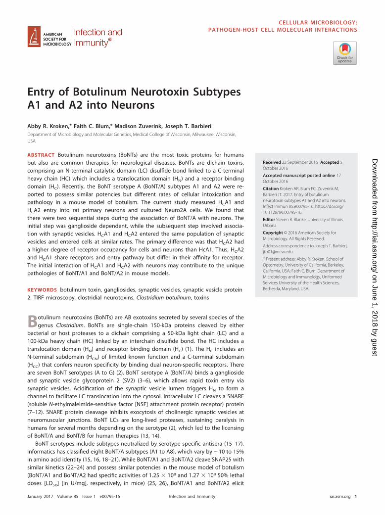
FIG 1 HCA2 and HCA1 binding to neurons. Rat primary cortical neurons were cooled to 4°C. Neurons wereincubated with 40 nM FLAG-HCA1, FLAG-HCA2, or FLAG-HCT for 30 min. Cells were washed, fixed, andstained for FLAG and synaptophysin 1 (Syp1). (A) Representative images of neurons stained for FLAG(red) and Syp1 (green), with an inset of the merged images below. (B) FLAG intensity was normalized toSyp1 intensity, and data from eight independent fields were averaged. (C) Colocalization of FLAG (HC)with Syp1 was measured, using Pearson’s correlation coefficients (ImageJ). Scale bar, 20 �m. The datasets shown are representative of replicate experiments and were evaluated using one-way ANOVA withGraphPad Prism version 5. For all figures, significance is expressed as follows: ns, not significant and P �0.05; *, P � 0.05; **, P � 0.01; ***, P � 0.001; ****, P � 0.0001.
Kroken et al. Infection and Immunity
January 2017 Volume 85 Issue 1 e00795-16 iai.asm.org 2
on June 1, 2018 by guesthttp://iai.asm
.org/D
ownloaded from
HCA2 bound neurons at �10-fold-higher efficiency than HCA1 (Fig. 1B). HC colocaliza-tion with synaptophysin 1 (Syp1) was measured using Pearson’s correlation coefficients.In this experimental system, Pearson’s coefficients of �0.6 indicate colocalization, thoseof 0.3 to 0.6 indicate partial colocalization, and those of �0.3 indicate segregation, aspreviously determined (33). Syp1 is a marker for synaptic vesicles (34–36). At 4°C,Pearson’s coefficients were �0.2 for HCA1 and Syp1 and �0.3 for HCA2 and Syp1 (Fig.1C), which indicated that both HCA1 and HCA2 were segregated from Syp1. In similarexperiments at 4°C, bound HCA1 and HCA2 also segregated from SV2a (Pearson’scoefficients of �0.3), the protein receptor for BoNT/A (data not shown) (4). Theobserved differences in Pearson’s coefficient between HCA1 and Syp1 and HCA2 andSyp1 were statistically significant but were not considered biologically significant. HCTwas a positive control for proteins that bind neurons at 4°C (33). Together, the datashow that the initial binding of HCA1 and HCA2 to neurons occurs independent ofsynaptic vesicle proteins and that HCA2 had a higher initial binding to neurons thanHCA1.
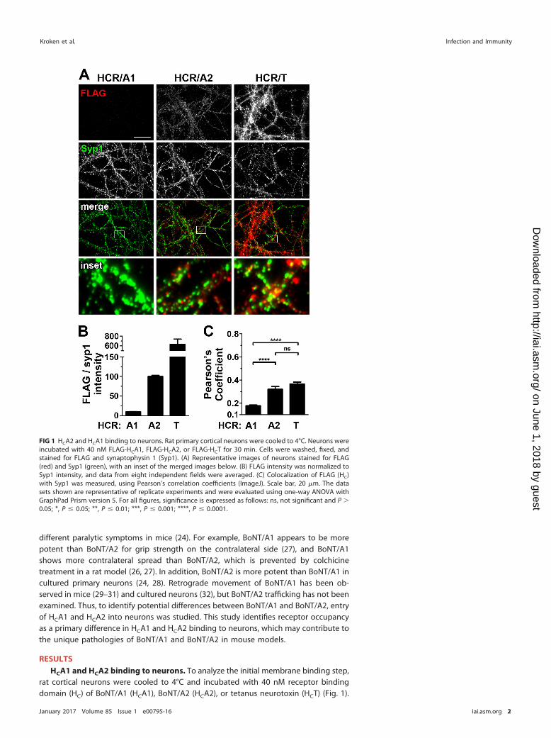
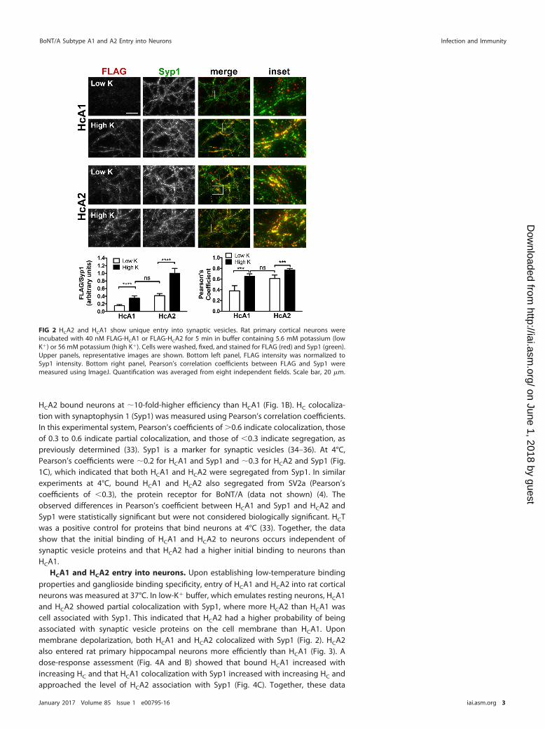
HCA1 and HCA2 entry into neurons. Upon establishing low-temperature bindingproperties and ganglioside binding specificity, entry of HCA1 and HCA2 into rat corticalneurons was measured at 37°C. In low-K� buffer, which emulates resting neurons, HCA1and HCA2 showed partial colocalization with Syp1, where more HCA2 than HCA1 wascell associated with Syp1. This indicated that HCA2 had a higher probability of beingassociated with synaptic vesicle proteins on the cell membrane than HCA1. Uponmembrane depolarization, both HCA1 and HCA2 colocalized with Syp1 (Fig. 2). HCA2also entered rat primary hippocampal neurons more efficiently than HCA1 (Fig. 3). Adose-response assessment (Fig. 4A and B) showed that bound HCA1 increased withincreasing HC and that HCA1 colocalization with Syp1 increased with increasing HC andapproached the level of HCA2 association with Syp1 (Fig. 4C). Together, these data
FIG 2 HCA2 and HCA1 show unique entry into synaptic vesicles. Rat primary cortical neurons wereincubated with 40 nM FLAG-HCA1 or FLAG-HCA2 for 5 min in buffer containing 5.6 mM potassium (lowK�) or 56 mM potassium (high K�). Cells were washed, fixed, and stained for FLAG (red) and Syp1 (green).Upper panels, representative images are shown. Bottom left panel, FLAG intensity was normalized toSyp1 intensity. Bottom right panel, Pearson’s correlation coefficients between FLAG and Syp1 weremeasured using ImageJ. Quantification was averaged from eight independent fields. Scale bar, 20 �m.
BoNT/A Subtype A1 and A2 Entry into Neurons Infection and Immunity
January 2017 Volume 85 Issue 1 e00795-16 iai.asm.org 3
on June 1, 2018 by guesthttp://iai.asm
.org/D
ownloaded from
show that HCA2 has higher affinity for synaptic vesicle proteins than HCA1 on restingneurons and that both HCA1 and HCA2 localize with synaptic vesicle proteins uponmembrane depolarization.
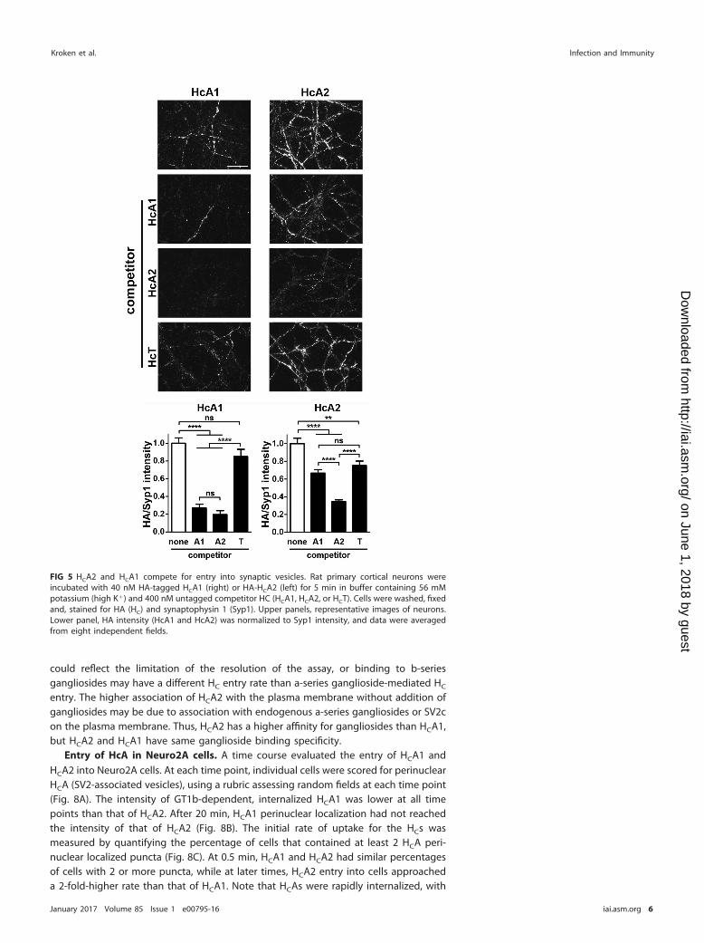
HCA1 and HCA2 compete for entry into neurons. A competition experimentdetermined if HCA1 and HCA2 used a similar pathway to enter neurons. Neurons wereincubated with 40 nM hemagglutinin (HA)-tagged HCA1 or HCA2, alone or with 400 nMuntagged HCA1 or HCA2 (Fig. 5). HCA1 and HCA2 effectively competed for HCA1association with neurons, while HCA2 was more effective than HCA1 as a competitor forHCA2 association with neurons. HCT of tetanus neurotoxin was a negative control forcompeting with HCA1 or HCA2 for cell association with synaptic vesicles, as wepreviously observed that HCT does not bind SV2 (33). HCT was less effective incompeting for HCA1 than either HCA1 or HCA2 (Fig. 5). The ability of HCA1 and HCA2 tocompete with the homologous and heterologous HCA subtype supports the entry ofboth HCs into similar synaptic vesicles. HCA2 also appears a more efficient competitorfor entry, possibly due to a higher affinity for gangliosides than HCA1.
Ganglioside interactions of HC with neurons and Neuro2A cells. HCA1 bindsgangliosides GT1b and GD1a via the terminal 4-galactose and the 5-sia sugars (5, 37).We previously showed that W1266, a component of the ganglioside binding pocket ofHCA, was required for ganglioside binding, as HCA1(W1266A) failed to bind ganglio-sides and did not have a detectable effect on HCA1 structure (PDB 4IQP) (38). SinceHCA1 and HCA2 are �90% identical, we predicted that W1266 in HCA2 would alsocontribute to ganglioside binding. HCA2 binding and entry into neurons were W1266
FIG 3 HCA2 and HCA1 entry into rat primary hippocampal neurons. Rat primary hippocampal neuronswere incubated with 40 nM FLAG-HCA1 or FLAG-HCA2 for 5 min in buffer containing 5.6 mM potassium(low K�) or 56 mM potassium (high K�). Cells were washed, fixed, and stained for FLAG-HCs andsynaptophysin 1. Upper panels, representative images are shown. Bottom left panel, FLAG intensity wasnormalized to Syp1 intensity. Bottom right panel, Pearson’s correlation coefficients between FLAG andsynaptophysin 1 were measured using ImageJ. Quantification was averaged from eight independentfields. Scale bar, 20 �m.
Kroken et al. Infection and Immunity
January 2017 Volume 85 Issue 1 e00795-16 iai.asm.org 4
on June 1, 2018 by guesthttp://iai.asm
.org/D
ownloaded from

dependent (Fig. 6). The similar binding of HCA2(W1266A) and HCA2 to resting neurons(low K�) at 37°C may be due to ganglioside-independent association with SV2 on thecell membrane, consistent with two independent receptor binding interactions, viagangliosides and SV2. Note that the inability of HCA2(W1266A) to enter synapticvesicles upon membrane depolarization is consistent with the need for dual receptorinteraction to facilitate intracellular entry, as previously observed with tetanus toxin(39).
The ganglioside binding specificities of HCA1 and HCA2 were tested in Neuro2Acells, a mouse neuroblastoma cell line. Neuro2A cells express synaptic vesicle proteins,including SV2c, the BoNT/A receptor, but do not express complex gangliosides at levelscomparable to those for primary neurons (40). Exogenous ganglioside-enrichedNeuro2A cells were incubated with HCA1 and HCA2 and analyzed for bound HC (Fig. 7).In ganglioside-enriched Neuro2A cells, both HCA2 and HCA1 localized to SV2c-positiveperinuclear vesicles, with �4-fold more HCA2 cell associated than HCA1. HCA2 also hada higher baseline entry into Neuro2A cells without addition of exogenous gangliosides,which may reflect HCA2’s greater affinity for gangliosides than HCA1. GT1b and GD1acontain sialic acid at the position 5 of the ganglioside, which appeared to target HCA1and HCA2 into the cells. Association with each ganglioside was sufficient to deliverHCA1 and HCA2 into SV2-containing vesicles. GD1b-mediated HC entry was low for bothHCA1 and HCA2, but the difference did not reach statistical significance for HCA1. This
FIG 4 Concentration-dependent entry of HCA1 into synaptic vesicles. Rat cortical neurons were incu-bated with HA-HCA1 (20 to 80 nM) or HA-HCA2 (20 nM) for 5 min in buffer containing 5.6 mM potassium(low K�) or 56 mM potassium (high K�). Cells were fixed and processed for immunofluorescence. (A)Representative images are shown. (B) The average intensity of HA (HC)/Syp1 was calculated from eightfields. (C) Pearson’s coefficients were calculated to determine colocalization between HA-tagged proteinand Syp1. The experiment was performed in triplicate, and the values were averaged. Scale bar, 20 �m.
BoNT/A Subtype A1 and A2 Entry into Neurons Infection and Immunity
January 2017 Volume 85 Issue 1 e00795-16 iai.asm.org 5
on June 1, 2018 by guesthttp://iai.asm
.org/D
ownloaded from
could reflect the limitation of the resolution of the assay, or binding to b-seriesgangliosides may have a different HC entry rate than a-series ganglioside-mediated HC
entry. The higher association of HCA2 with the plasma membrane without addition ofgangliosides may be due to association with endogenous a-series gangliosides or SV2con the plasma membrane. Thus, HCA2 has a higher affinity for gangliosides than HCA1,but HCA2 and HCA1 have same ganglioside binding specificity.
Entry of HcA in Neuro2A cells. A time course evaluated the entry of HCA1 andHCA2 into Neuro2A cells. At each time point, individual cells were scored for perinuclearHCA (SV2-associated vesicles), using a rubric assessing random fields at each time point(Fig. 8A). The intensity of GT1b-dependent, internalized HCA1 was lower at all timepoints than that of HCA2. After 20 min, HCA1 perinuclear localization had not reachedthe intensity of that of HCA2 (Fig. 8B). The initial rate of uptake for the HCs wasmeasured by quantifying the percentage of cells that contained at least 2 HCA peri-nuclear localized puncta (Fig. 8C). At 0.5 min, HCA1 and HCA2 had similar percentagesof cells with 2 or more puncta, while at later times, HCA2 entry into cells approacheda 2-fold-higher rate than that of HCA1. Note that HCAs were rapidly internalized, with
FIG 5 HCA2 and HCA1 compete for entry into synaptic vesicles. Rat primary cortical neurons wereincubated with 40 nM HA-tagged HCA1 (right) or HA-HCA2 (left) for 5 min in buffer containing 56 mMpotassium (high K�) and 400 nM untagged competitor HC (HCA1, HCA2, or HCT). Cells were washed, fixedand, stained for HA (HC) and synaptophysin 1 (Syp1). Upper panels, representative images of neurons.Lower panel, HA intensity (HcA1 and HcA2) was normalized to Syp1 intensity, and data were averagedfrom eight independent fields.
Kroken et al. Infection and Immunity
January 2017 Volume 85 Issue 1 e00795-16 iai.asm.org 6
on June 1, 2018 by guesthttp://iai.asm
.org/D
ownloaded from

�50% of cells containing internalized HCA at 0.5 min. Thus, the initial internalizationrates of HCA1 and HCA2 are similar, and HCA2 internalization is more efficient than thatof HCA1 due to a greater receptor occupancy.
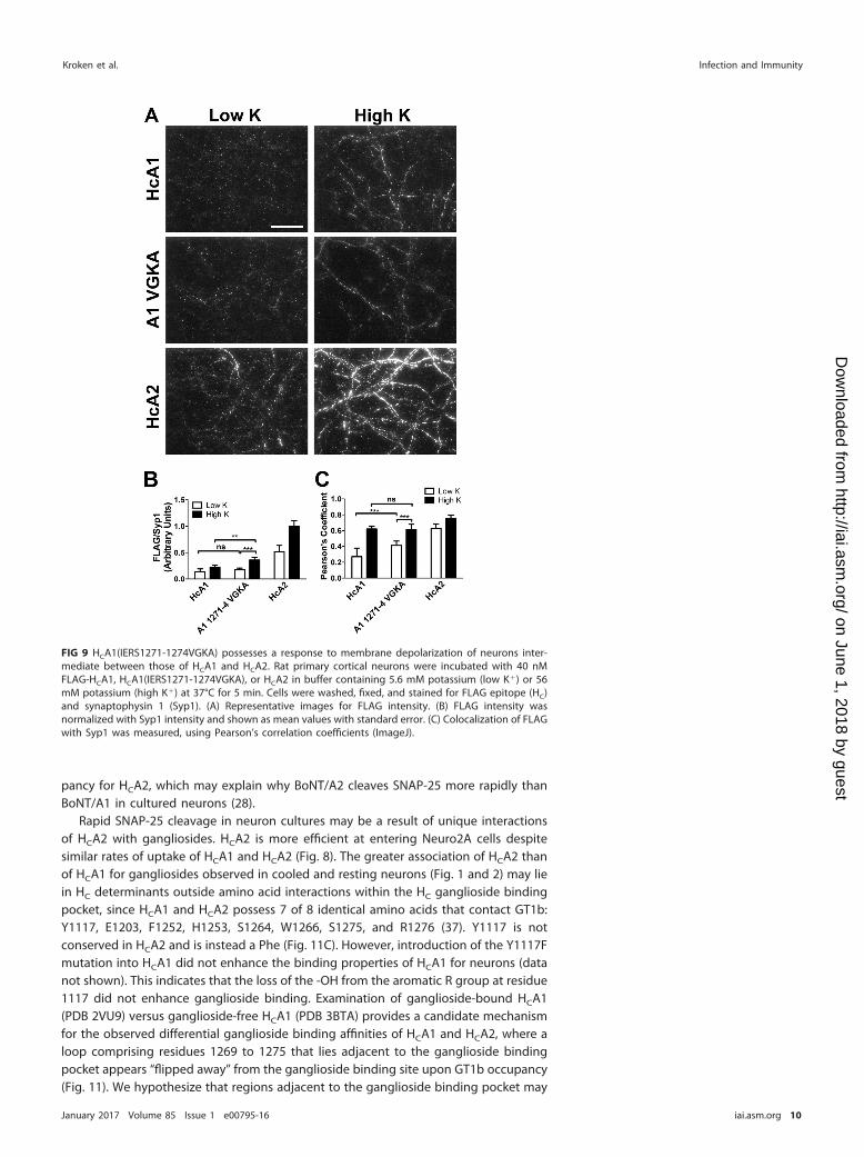
Entry of site-mutated HCA1 into neurons. An informatics assessment of the aminoacids surrounding the ganglioside showed that residues 1271 to 1274 were uniquebetween HCA1 and HCA2 (37). Gain-of-function mutations were used to assess a role forthese residues in HC entry. Mutations were engineered into HCA1 at a loop adjacent tothe ganglioside binding site which included four amino acid substitutions betweenHCA1 and HCA2, HCA1(IERS1271-1274VGKA). Resting and upon membrane depolariza-tion, HCA1(IERS1271-1274VGKA) possessed entry properties intermediate betweenthose of HCA1 and HCA2 (Fig. 9). This supports a role for the loop from residue 1271 to1274 in HC entry.
Entry of an HC chimera into neurons. The ganglioside binding site resides in theC-terminal subdomain of the HC (residues 1090 to 1296), while the SV2 binding siteresides within the C-terminal subdomain on an adjacent face of the HC and close to theN-terminal jellyroll subdomain (residues 870 to 1090) (41–43). Contributions of sub-domains in neuron entry were examined with HCA1 and HCA2 chimeras, fusing theN-terminal subdomain with the C-terminal subdomain of reciprocal HC. HCA1A2 con-tained the N-terminal subdomain of HCA1 and the C-terminal subdomain of HCA2,while HCA2A1 contained the N-terminal subdomain of HCA2 and the C-terminal sub-
FIG 6 W1266 mediates HCA2 binding to neurons. Rat primary cortical neurons were cooled to 4°C andincubated with 40 nM FLAG-tagged HCA2(W1266A) or HCA2 for 20 min. Cells were washed, fixed, stainedfor FLAG epitope and synaptophysin 1 (Syp1), processed, and analyzed. (A and B) Representative imagesof neurons stained for FLAG. (C) FLAG intensity was normalized with Syp1 intensity. (D) Neurons weremaintained at 37°C and incubated with 40 nM HCA2(W1266A) or HCA2 for 5 min in buffer containing 5.6mM potassium (low K�) or 56 mM potassium (high K�).
BoNT/A Subtype A1 and A2 Entry into Neurons Infection and Immunity
January 2017 Volume 85 Issue 1 e00795-16 iai.asm.org 7
on June 1, 2018 by guesthttp://iai.asm
.org/D
ownloaded from

domain of HCA1. HCA2A1 could not be purified to sufficient homology and was notutilized further in this study (Fig. 10A). In resting neurons, HCA1A2 binding to neuronswas similar to that of HCA2 (Fig. 10B and C), and upon membrane depolarization,HCA1A2 showed increased association, which approached that of HCA2 (Fig. 10D). Thisindicated that the C-terminal subdomain of HCA appears to be sufficient for synapticvesicle entry.
DISCUSSION
BoNT/A1 and BoNT/A2 possess similar potencies but have unique rates of entry intoneurons and unique pathologies in mouse models of botulism (28). The current studyresolved the sequential entry of HCA1 and HCA2 entry into neurons, identifying aninitial ganglioside-dependent step and a subsequent step involving association withsynaptic vesicles. Competition experiments in neurons supported HCA1 and HCA2 entryinto the same population of synaptic vesicles. The rates of cell entry were similar for thetwo subtypes, while HCA2 had a higher degree of receptor occupancy than HcA1. Thus,HCA2 and HCA1 share receptors and entry pathways but differ in their affinity for theirreceptor.
The current studies resolved the sequential entry of HCA1 and HCA2 into neuronswith an initial association which was dependent upon ganglioside binding. HCA2bound primary neurons with greater affinity than HCA1, independent of the associationwith synaptic vesicle marker proteins. At 37°C, in resting neurons HCA2 had a greaterassociation with synaptic vesicle proteins than HCA1, but upon membrane depolariza-tion, both HCs colocalized with synaptic vesicles. HCA2 and HCA1 entered into similarpopulations of synaptic vesicles, with HCA2 competing more efficiently for entry thanHCA1. Assessment of HCA1A2 implicated the C-terminal subdomain of the HC (HCC) asthe primary determinant for neuron binding and synaptic vesicle association. Thus, theunique entry of HCA1 and HCA2 into synaptic vesicles may contribute to the uniquerates of neuron intoxication and pathologies elicited by BoNT/A1 and BoNT/A2.
While early studies implicated gangliosides as host receptors for the clostridial
FIG 7 HCA2 and HCA1 share ganglioside binding specificity. Neuro2A cells were loaded with 20 �g/mlpurified gangliosides (GT1b, GD1b, GD1a, or GM1a) for 4 h. Cells were incubated with 40 nM HA-HCA1or HA-HCA2 for 20 min at 37°C. Cells were fixed and stained for HA and SV2c. Upper panel, representativeimages. Bottom left panel, HA intensity (HC) was normalized to SV2c intensity. Bottom right panel,schematic of the four gangliosides used in this experiment, with the 5-sialic acid-galactose bound by HCAhighlighted in gangliosides GT1b and GD1a. Scale bar, 50 �m.
Kroken et al. Infection and Immunity
January 2017 Volume 85 Issue 1 e00795-16 iai.asm.org 8
on June 1, 2018 by guesthttp://iai.asm
.org/D
ownloaded from

neurotoxins (44, 45), Montecucco and Schiavo proposed that clostridial neurotoxinsutilized dual host receptors, “a protein and a lipid,” to generate high-affinity binding toneurons (46). Subsequently, synaptotagmin was identified as the BoNT/B proteinreceptor (47, 48), and SV2 was identified as the BoNT/A protein receptor (4, 6, 41),where stimulation of synaptic vesicle cycling enhanced the efficiency of HC entry. Thecurrent study supports a sequential utilization of the dual BoNT host receptors. At lowtemperature, HCs bound neurons but segregated from synaptic vesicle marker proteins,while at 37°C, HCs associated with synaptic vesicle proteins, which increased uponmembrane depolarization. The greater association of HCA2 with synaptic vesicle pro-teins in resting neurons, relative to that of HCA1, implicates a greater receptor occu-
FIG 8 HCA1 and HCA2 entry into Neuro2A cells. Neuro2A cells were loaded with 20 �g/ml purified GT1bor left unloaded (none) for 4 h. Cells were washed and incubated with 40 nM FLAG-HCA1 or FLAG-HCA2for 1.25, 2.5, 5, 10, or 20 min at 37°C. Cells were fixed and stained for FLAG and SV2c. (A) Representativeimages of FLAG-reactive cell-associated material at 1.25- and 20-min incubations. (B) Individual cellswithin random fields were scored for internalized perinuclear FLAG intensity and assigned an arbitrarynumber (0, 1, or 2). Representative cells are shown by score, with cells scored as 0 containing no intensity,1 containing intermediate intensity, and 2 containing the most intensity. The cumulative score wasdivided by the total number of cells per field and plotted. At least 10 fields, or �100 cells per condition,in three replicates were counted. (C) The rate of endocytosis for HCA1 and HCA2 was assessed byincubation of a 40 nM concentration of either protein with Neuro2A cells for 0.5, 1, or 2 min. Individualcells were scored as positive for internalized signal if at least 2 FLAG-reactive puncta were present withinthe perinuclear region (SV2c marker) of a cell. Data are presented as percentage of cells containinginternalized signal. Linear regression was performed and slopes plotted for HCA1 and HCA2. An unpairedStudent t test between HCA1 and HCA2 in GT1b-loaded Neuro2A cells was performed at each time point.Scale bar, 50 �m.
BoNT/A Subtype A1 and A2 Entry into Neurons Infection and Immunity
January 2017 Volume 85 Issue 1 e00795-16 iai.asm.org 9
on June 1, 2018 by guesthttp://iai.asm
.org/D
ownloaded from
pancy for HCA2, which may explain why BoNT/A2 cleaves SNAP-25 more rapidly thanBoNT/A1 in cultured neurons (28).
Rapid SNAP-25 cleavage in neuron cultures may be a result of unique interactionsof HCA2 with gangliosides. HCA2 is more efficient at entering Neuro2A cells despitesimilar rates of uptake of HCA1 and HCA2 (Fig. 8). The greater association of HCA2 thanof HCA1 for gangliosides observed in cooled and resting neurons (Fig. 1 and 2) may liein HC determinants outside amino acid interactions within the HC ganglioside bindingpocket, since HCA1 and HCA2 possess 7 of 8 identical amino acids that contact GT1b:Y1117, E1203, F1252, H1253, S1264, W1266, S1275, and R1276 (37). Y1117 is notconserved in HCA2 and is instead a Phe (Fig. 11C). However, introduction of the Y1117Fmutation into HCA1 did not enhance the binding properties of HCA1 for neurons (datanot shown). This indicates that the loss of the -OH from the aromatic R group at residue1117 did not enhance ganglioside binding. Examination of ganglioside-bound HCA1(PDB 2VU9) versus ganglioside-free HCA1 (PDB 3BTA) provides a candidate mechanismfor the observed differential ganglioside binding affinities of HCA1 and HCA2, where aloop comprising residues 1269 to 1275 that lies adjacent to the ganglioside bindingpocket appears “flipped away” from the ganglioside binding site upon GT1b occupancy(Fig. 11). We hypothesize that regions adjacent to the ganglioside binding pocket may
FIG 9 HCA1(IERS1271-1274VGKA) possesses a response to membrane depolarization of neurons inter-mediate between those of HCA1 and HCA2. Rat primary cortical neurons were incubated with 40 nMFLAG-HCA1, HCA1(IERS1271-1274VGKA), or HCA2 in buffer containing 5.6 mM potassium (low K�) or 56mM potassium (high K�) at 37°C for 5 min. Cells were washed, fixed, and stained for FLAG epitope (HC)and synaptophysin 1 (Syp1). (A) Representative images for FLAG intensity. (B) FLAG intensity wasnormalized with Syp1 intensity and shown as mean values with standard error. (C) Colocalization of FLAGwith Syp1 was measured, using Pearson’s correlation coefficients (ImageJ).
Kroken et al. Infection and Immunity
January 2017 Volume 85 Issue 1 e00795-16 iai.asm.org 10
on June 1, 2018 by guesthttp://iai.asm
.org/D
ownloaded from

contribute to the greater affinity of HCA2 for gangliosides than HCA1, without influ-encing the ganglioside specificity of the HCs. The observation that HCA1(IERS1271-1274VGKA) possessed an intermediate association with neurons, relative to those ofHCA1 and HCA2 (Fig. 10), supports this hypothesis. Additional distanced amino acids,not yet identified, may also contribute to the observed affinity of HCA2 for gangliosides.
Recently, two reports identified residues of BoNT/A1 involved in SV2 binding. Benoitand colleagues (43) crystalized HCA1 complexed with the fourth luminal loop (L4) ofSV2c, which formed a right-handed quadrilateral �-helix and interacted with the HCA1�-sheets 40 and 41 (residues 1140 to 1153; secondary structure assignment as definedby Lacy and Stevens [49]) (43). This SV2 interaction site is located in the C-terminalsubdomain of HCA1 yet distant from the ganglioside binding pocket. Although severalof the HC-SV2 contacts were via main-chain interactions, four residues at the interfaceof the interaction site when mutated resulted in decreased binding between HCA1 andSV2c (43). This suggests that structural packing contributes to the HC-SV2 interaction,rather than the R-group composition, and is consistent with regions outside the SV2-L4contributing to SV2 affinity (Fig. 11). Strotmeier and colleagues used site-directedmutagenesis to determine residues contributing to the interaction of BoNT/A1 with
FIG 10 The C-terminal subdomain contributes to binding and entry of HC into synaptic vesicles. (A)Schematic of HC domains of BoNT/A1 (white) and BoNT/A2 (black), with the constructed HCA1A2 chimera.Rat primary cortical neurons were incubated with 20 nM HA-HCA1, HA-HCA2, or HA-HCA1A2 for 5 min inbuffer containing 5.6 mM potassium (open, low K�) or 56 mM potassium (solid, high K�). Cells werewashed, fixed, and stained for HA epitope (HC) and synaptophysin 1 Syp1. (B) Representative images. (C)HA intensity was normalized to Syp1 intensity and shown as mean values with standard error. (D)Colocalization between HA and Syp1 was measured, using Pearson’s coefficients. Scale bar, 40 �m.
BoNT/A Subtype A1 and A2 Entry into Neurons Infection and Immunity
January 2017 Volume 85 Issue 1 e00795-16 iai.asm.org 11
on June 1, 2018 by guesthttp://iai.asm
.org/D
ownloaded from

SV2c-L4 (42). SV2c-interacting residues were identified at the interface of the N and Csubdomains of HCA1, which partially overlapped the region crystalized with the SV2-L4loop. While making substantial contributions toward deciphering HC-SV2 interactions,neither study explains the subtype specificity of BoNT/A1 and BoNT/A2 with SV2. Thecurrent study showed HCA1A2 responded to membrane depolarization, which supportsthe localization of the SV2 binding site within the C-terminal subdomain. Our hypoth-esis is that the primary interaction between the HC and SV2 is mediated by theC-terminal subdomain of the HC. The determination that main-chain interactionscontribute to HCA1-SV2-L4 association (43) is consistent with a region(s) outside theHCA1-SV2-L4 also contributing to BoNT/A-synaptic vesicle interactions (Fig. 11). Theseinteractions may reside within other regions of SV2, including N-linked glycans, or mayinvolve other synaptic vesicles proteins, since synaptic vesicles proteins form higher-order complexes and in synaptic vesicle lysates (50), and earlier studies showed thatHCA1 bound synaptic vesicle protein complexes (51). Current studies are assessing therole of the N-terminal and C-terminal subdomain interfaces of HCA1 and HCA2 to testfor potential sites of interaction with SV2.
Despite the dissimilarities in occupancy between HCA1 and HCA2 observed inneurons and Neuro2A cells, the potencies in the mouse model of BoNT/A1 and
FIG 11 Molecular organization of the dual receptors of BoNT/A. (A) Schematic of BoNT/A1 (PDB 3BTA)light chain (LC) and heavy chain (HC). (B) BoNT/A1-HC-GT1b complex (PDB 2VU9) with the bound SV2-L4loop (blue) (PDB 4JRA) binding site. Residues 1143 to 1149 of HCA1 interacting with SV2-L4 are indicated(orange). The ganglioside binding pocket is shown (magenta). (C) HcA1 ganglioside binding site (GT1b),including H1251, S1264, W1266, and Y1267 (magenta). Adjacent to the GT1b site is Y1117 (cyan). (D) Theloop from residue 1271 to 1274 from the BoNT/A structure with side chains and surrounding main chainis overlaid in red. This loop opens upon GT1b occupancy (black) as opposed to unbound (red).
Kroken et al. Infection and Immunity
January 2017 Volume 85 Issue 1 e00795-16 iai.asm.org 12
on June 1, 2018 by guesthttp://iai.asm
.org/D
ownloaded from

BoNT/A2 are similar (26). Thus, the differential properties of the HC reported in thecurrent study may contribute to the rate of neuronal cell intoxication and unique BoNTpathologies associated with the various BoNT/A subtypes (25, 52). This implies thatabsolute neurotoxin potency can be uncoupled from the observed unique pathologiesof the BoNT/A subtypes in mouse models of botulism. The symptoms associated withintoxication are different for BoNT/A1 and BoNT/A2 in mice, where intravenous injec-tion of BoNT/A1 caused ruffled fur, labored breathing, descending paralysis, andspasticity before death (52), while intravenous injection of BoNT/A2 caused paralysis ofthe front legs, hind legs, and whole body, followed by death (24). Further, BoNT/A1diffuses more readily than BoNT/A2 to the contralateral side due to retrograde move-ment (27, 29, 53); whether retrograde trafficking of BoNT plays a role in the symptomselicited by the toxins remains unknown. The symptoms of other BoNT subtypes appearto be related to their similarity. BoNT/A3 shares 93% amino acid identity with BoNT/A2,and both cause similar symptoms in mice (52). BoNT/A5 shares 97% identity withBoNT/A1 and causes paralysis analogous to BoNT/A1 in mice (24). Further elucidationof the molecular mechanisms that differentiate BoNT/A1 and BoNT/A2 will contributeto our understanding of how clostridial neurotoxins elicit paralysis and how to mod-ulate these neurotoxins to enhance therapeutic potency.
MATERIALS AND METHODSSubcloning and production of epitope tagged-HCs. Escherichia coli codon-optimized DNA encod-
ing HCA1 (S877 to L1296) and HCA2 (K871 to L1296), also referred to as HC/A1 and HC/A2, weresynthesized by EZBiolab and GenScript, respectively. DNA encoding each HCA was subcloned into amodified pET-28a (Novagen), which introduced 6-His and 3-FLAG epitopes N-terminal to the HC. DNAsencoding HCA1 and HCA2 were also subcloned into a modified pET-28a vector to replace the 3-FLAGepitope with three repeats of the human influenza virus hemagglutinin epitope (HA) (YPYDVPDYA),which is recognized by a monoclonal rat antibody (clone 3F10; Roche). The pET-28a vector containingeach HC was transformed into E. coli BL21(DE3) (Stratagene) for protein expression. Bacterial cell culturingand protein purification by sequential column chromatography were performed as previously described(54). Proteins were resolved and normalized by SDS-PAGE. FLAG-HC and HA-HC showed similar propertieswith regard to binding and entry into neurons (A. R. Kroken and J. T. Barbieri, unpublished data). Mutatedforms of HCA1 were generated, using a QuikChange site-directed mutagenesis kit (Stratagene), andchimeras between HCA1 and HCA2 were generated by overlap PCR. Point mutations and chimeras wereconfirmed by DNA sequencing.
Cell culturing of primary neurons. E18 rat primary cortical, spinal, or hippocampal neurons(BrainBits, LLC) were cultured on 24-well glass-bottom dishes (MatTek) coated with poly-D-lysine (Sigma)and laminin (Life Technologies) in Neurobasal medium (Life Technologies) supplemented with B27,Glutamax (Life Technologies), and Primocin (InvivoGen) according to supplier directions. On day 7, halfof the culture medium was changed, and cells were analyzed on days 10 to 14. Neuro2A cells (ATCC)were plated on glass coverslips coated with collagen I and cultured in minimal essential medium (MEM)supplemented with 10% fetal bovine serum (FBS), 0.1% sodium bicarbonate, 1� nonessential aminoacids, 1 mM sodium pyruvate, 50 units/ml penicillin, and 50 �g/ml streptomycin (Life Technologies).
Entry of HCs into neurons and Neuro2A cells. Cells were washed twice with Dulbecco’s phosphate-buffered saline (DPBS), incubated with HCs in buffer for 5 min at 37°C or chilled at 4°C for 10 min, andincubated with HCs. Buffers were either high potassium (high K�) to stimulate membrane depolarizationand Ca2�-dependent synaptic vesicle cycling (56 mM KCl, 2.2 mM CaCl2, 15 mM HEPES, 145 mM NaCl, 0.5mM MgCl2, pH 7.4) or low potassium (low K�) to emulate resting membranes (5.6 mM KCl, 2.2 mM CaCl2,15 mM HEPES, 145 mM NaCl, 0.5 mM MgCl2, pH 7.4). Cells were washed twice and fixed in 4% (wt/vol)paraformaldehyde in DPBS for 15 min. Neuro2A cells were loaded with 10 �g gangliosides (GT1b, GD1b,GD1a, or GM1a; Matreya) in 0.5 ml (20 �g/ml) of complete MEM containing 0.5% FBS for 4 h prior toincubation with HCs in low-potassium buffer. Cells were washed twice and fixed in 4% (wt/vol)paraformaldehyde in DPBS for 15 min.
Immunofluorescence of fixed neurons or Neuro2A cells. Cells were permeabilized with 0.1%Triton X-100 – 4% formaldehyde in DPBS for 15 min, washed twice, incubated with 150 mM glycinefor 15 min, washed twice, and then incubated in 10% (vol/vol) FBS–2.5% (wt/vol) fish skin gelatin(Sigma)– 0.1% Triton X-100 – 0.05% Tween 20 (blocking solution) for 1 h. Fixed cells were incubatedwith primary antibodies (mouse anti-FLAG, 1:10,000 [Sigma]; rat anti-HA, 1:2,000 [Roche]; guinea piganti-synaptophysin 1, 1:2,000 [Synaptic Systems], or rabbit anti-SV2C, 1:2,000 [Synaptic Systems]) at4°C overnight. Cells were then washed 3 times for 5 min, and Alexa-labeled secondary antibodies(1:1,000; Life Technologies) were incubated at room temperature for 1 h. Cells were washed 4 timesfor 5 min, with the final wash containing DAPI (4=,6=-diamidino-2-phenylindole) stain, and fixed with4% paraformaldehyde. Neurons were preserved in CitiFluor AF-3 (Electron Microscopy Sciences), andNeuro2A cells were mounted on slides containing ProLong Gold (Life Technologies) and sealed withnail polish.
BoNT/A Subtype A1 and A2 Entry into Neurons Infection and Immunity
January 2017 Volume 85 Issue 1 e00795-16 iai.asm.org 13
on June 1, 2018 by guesthttp://iai.asm
.org/D
ownloaded from
Imaging and analysis of stained cells and neurons. Micrographs were acquired with a NikonTE2000 inverted total internal reflection fluorescence (TIRF) microscope equipped with a Nikon Apo TIRF100�/1.49 numerical aperture (NA) oil objective or a Plan Apo VC 60�/1.40 NA oil objective andCoolSnap HQ2 camera (Photometrics) with Nikon Elements or Metamorph acquisition software. At least8 fields were imaged for each experimental replicate, and fields were chosen while observing synapto-physin 1 (Syp1) or SV2c staining to limit bias. Intensity and Pearson’s correlation coefficients weremeasured using ImageJ (NIH) and the Intensity Correlation Analysis plugin (Wright Cell Imaging Facility).The numerical values for FLAG/Syp1 and other normalized intensity are expressed as arbitrary units (AU).Data sets shown are representative of replicate experiments and were evaluated by one-way analysis ofvariance (ANOVA) statistics, using GraphPad Prism version 5: ns, not significant (P � 0.05); *, P � 0.05; **,P � 0.01; ***, P � 0.001; and ****, P � 0.0001. Data sets for HCA1 and HCA2 uptake in Neuro2A cells wereevaluated by the unpaired Student t test, using GraphPad Prism version 5; significance cutoffs were asdescribed above.
ACKNOWLEDGMENTSThis study was supported by funds from the NIH (grants AI118389 and AI030162).We thank Amanda Przedpelski for technical support and Jung-Ja Kim (Medical
College of Wisconsin, Biochemistry) and Chen Chen (Medical College of Wisconsin,Barbieri laboratory) for helpful comments.
REFERENCES1. Lacy DB, Tepp W, Cohen AC, DasGupta BR, Stevens RC. 1998. Crystal
structure of botulinum neurotoxin type A and implications for toxicity.Nat Struct Biol 5:898 –902. https://doi.org/10.1038/2338.
2. Davletov B, Bajohrs M, Binz T. 2005. Beyond BOTOX: advantages andlimitations of individual botulinum neurotoxins. Trends Neurosci 28:446 – 452. https://doi.org/10.1016/j.tins.2005.06.001.
3. Yowler BC, Kensinger RD, Schengrund CL. 2002. Botulinum neurotoxin Aactivity is dependent upon the presence of specific gangliosides inneuroblastoma cells expressing synaptotagmin I. J Biol Chem 277:32815–32819. https://doi.org/10.1074/jbc.M205258200.
4. Dong M, Yeh F, Tepp WH, Dean C, Johnson EA, Janz R, Chapman ER.2006. SV2 is the protein receptor for botulinum neurotoxin A. Science312:592–596. https://doi.org/10.1126/science.1123654.
5. Rummel A, Mahrhold S, Bigalke H, Binz T. 2004. The HCC-domain ofbotulinum neurotoxins A and B exhibits a singular ganglioside bindingsite displaying serotype specific carbohydrate interaction. Mol Microbiol51:631– 643.
6. Mahrhold S, Rummel A, Bigalke H, Davletov B, Binz T. 2006. The synapticvesicle protein 2C mediates the uptake of botulinum neurotoxin A intophrenic nerves. FEBS Lett 580:2011–2014. https://doi.org/10.1016/j.febslet.2006.02.074.
7. Schiavo G, Benfenati F, Poulain B, Rossetto O, Polverino de Laureto P,DasGupta BR, Montecucco C. 1992. Tetanus and botulinum-B neurotox-ins block neurotransmitter release by proteolytic cleavage of synapto-brevin. Nature 359:832– 835. https://doi.org/10.1038/359832a0.
8. Schiavo G, Rossetto O, Catsicas S, Polverino de Laureto P, DasGupta BR,Benfenati F, Montecucco C. 1993. Identification of the nerve terminaltargets of botulinum neurotoxin serotypes A, D, and E. J Biol Chem268:23784 –23787.
9. Schiavo G, Shone CC, Rossetto O, Alexander FC, Montecucco C. 1993.Botulinum neurotoxin serotype F is a zinc endopeptidase specific forVAMP/synaptobrevin. J Biol Chem 268:11516 –11519.
10. Yamasaki S, Baumeister A, Binz T, Blasi J, Link E, Cornille F, Roques B,Fykse EM, Sudhof TC, Jahn R, Heiner Niemann. 1994. Cleavage of mem-bers of the synaptobrevin/VAMP family by types D and F botulinalneurotoxins and tetanus toxin. J Biol Chem 269:12764 –12772.
11. Yamasaki S, Binz T, Hayashi T, Szabo E, Yamasaki N, Eklund M, Jahn R,Niemann H. 1994. Botulinum neurotoxin type G proteolyses the Ala81-Ala82 bond of rat synaptobrevin 2. Biochem Biophys Res Commun200:829 – 835. https://doi.org/10.1006/bbrc.1994.1526.
12. Blasi J, Chapman ER, Yamasaki S, Binz T, Niemann H, Jahn R. 1993.Botulinum neurotoxin C1 blocks neurotransmitter release by means ofcleaving HPC-1/syntaxin. EMBO J 12:4821– 4828.
13. Schantz EJ, Johnson EA. 1992. Properties and use of botulinum toxin andother microbial neurotoxins in medicine. Microbiol Rev 56:80 –99.
14. Guntinas-Lichius O. 2004. First use of botulinum toxin type B in ENTpatients with secondary therapy failure of botulinum toxin type A. HNO52:53–56. https://doi.org/10.1007/s00106-003-0844-8.
15. Smith TJ, Lou J, Geren IN, Forsyth CM, Tsai R, Laporte SL, Tepp WH,
Bradshaw M, Johnson EA, Smith LA, Marks JD. 2005. Sequence variationwithin botulinum neurotoxin serotypes impacts antibody binding andneutralization. Infect Immun 73:5450 –5457. https://doi.org/10.1128/IAI.73.9.5450-5457.2005.
16. Kull S, Schulz KM, Weisemann J, Kirchner S, Schreiber T, Bollenbach A,Dabrowski PW, Nitsche A, Kalb SR, Dorner MB, Barr JR, Rummel A, DornerBG. 2015. Isolation and functional characterization of the novel Clostrid-ium botulinum neurotoxin A8 subtype. PLoS One 10:e0116381. https://doi.org/10.1371/journal.pone.0116381.
17. Jacobson MJ, Lin G, Tepp W, Dupuy J, Stenmark P, Stevens RC, JohnsonEA. 2011. Purification, modeling, and analysis of botulinum neurotoxinsubtype A5 (BoNT/A5) from Clostridium botulinum strain A661222. ApplEnviron Microbiol 77:4217– 4222. https://doi.org/10.1128/AEM.00201-11.
18. Hill KK, Smith TJ, Helma CH, Ticknor LO, Foley BT, Svensson RT, Brown JL,Johnson EA, Smith LA, Okinaka RT, Jackson PJ, Marks JD. 2007. Geneticdiversity among botulinum neurotoxin-producing clostridial strains. JBacteriol 189:818 – 832. https://doi.org/10.1128/JB.01180-06.
19. Carter AT, Paul CJ, Mason DR, Twine SM, Alston MJ, Logan SM, Austin JW,Peck MW. 2009. Independent evolution of neurotoxin and flagellargenetic loci in proteolytic Clostridium botulinum. BMC Genomics 10:115.https://doi.org/10.1186/1471-2164-10-115.
20. Luquez C, Raphael BH, Maslanka SE. 2009. Neurotoxin gene clusters inClostridium botulinum type Ab strains. Appl Environ Microbiol 75:6094 – 6101. https://doi.org/10.1128/AEM.01009-09.
21. Mazuet C, Ezan E, Volland H, Popoff MR, Becher F. 2012. Toxindetection in patients’ sera by mass spectrometry during two out-breaks of type A botulism in France. J Clin Microbiol 50:4091– 4094.https://doi.org/10.1128/JCM.02392-12.
22. Arndt JW, Jacobson MJ, Abola EE, Forsyth CM, Tepp WH, Marks JD,Johnson EA, Stevens RC. 2006. A structural perspective of the sequencevariability within botulinum neurotoxin subtypes A1-A4. J Mol Biol362:733–742. https://doi.org/10.1016/j.jmb.2006.07.040.
23. Henkel JS, Jacobson M, Tepp W, Pier C, Johnson EA, Barbieri JT. 2009.Catalytic properties of botulinum neurotoxin subtypes A3 and A4. Bio-chemistry 48:2522–2528. https://doi.org/10.1021/bi801686b.
24. Whitemarsh RC, Tepp WH, Bradshaw M, Lin G, Pier CL, Scherf JM,Johnson EA, Pellett S. 2013. Characterization of botulinum neurotoxin Asubtypes 1 through 5 by investigation of activities in mice, in neuronalcell cultures, and in vitro. Infect Immun 81:3894 –3902. https://doi.org/10.1128/IAI.00536-13.
25. Pellett S, Tepp WH, Whitemarsh RC, Bradshaw M, Johnson EA. 2015. In vivoonset and duration of action varies for botulinum neurotoxin A subtypes1-5. Toxicon 107:37–42. https://doi.org/10.1016/j.toxicon.2015.06.021.
26. Torii Y, Kiyota N, Sugimoto N, Mori Y, Goto Y, Harakawa T, Nakahira S,Kaji R, Kozaki S, Ginnaga A. 2011. Comparison of effects of botulinumtoxin subtype A1 and A2 using twitch tension assay and rat grip strengthtest. Toxicon 57:93–99. https://doi.org/10.1016/j.toxicon.2010.10.009.
27. Torii Y, Akaike N, Harakawa T, Kato K, Sugimoto N, Goto Y, Nakahira S,Kohda T, Kozaki S, Kaji R, Ginnaga A. 2011. Type A1 but not type A2
Kroken et al. Infection and Immunity
January 2017 Volume 85 Issue 1 e00795-16 iai.asm.org 14
on June 1, 2018 by guesthttp://iai.asm
.org/D
ownloaded from
botulinum toxin decreases the grip strength of the contralateral forelegthrough axonal transport from the toxin-treated foreleg of rats. J Phar-macol Sci 117:275–285. https://doi.org/10.1254/jphs.11121FP.
28. Pier CL, Chen C, Tepp WH, Lin G, Janda KD, Barbieri JT, Pellett S,Johnson EA. 2011. Botulinum neurotoxin subtype A2 enters neuronalcells faster than subtype A1. FEBS Lett 585:199 –206. https://doi.org/10.1016/j.febslet.2010.11.045.
29. Antonucci F, Rossi C, Gianfranceschi L, Rossetto O, Caleo M. 2008.Long-distance retrograde effects of botulinum neurotoxin A. J Neurosci28:3689 –3696. https://doi.org/10.1523/JNEUROSCI.0375-08.2008.
30. Restani L, Antonucci F, Gianfranceschi L, Rossi C, Rossetto O, Caleo M.2011. Evidence for anterograde transport and transcytosis of botulinumneurotoxin A (BoNT/A). J Neurosci 31:15650 –15659. https://doi.org/10.1523/JNEUROSCI.2618-11.2011.
31. Restani L, Novelli E, Bottari D, Leone P, Barone I, Galli-Resta L, Strettoi E,Caleo M. 2012. Botulinum neurotoxin A impairs neurotransmission fol-lowing retrograde transynaptic transport. Traffic 13:1083–1089. https://doi.org/10.1111/j.1600-0854.2012.01369.x.
32. Restani L, Giribaldi F, Manich M, Bercsenyi K, Menendez G, Rossetto O,Caleo M, Schiavo G. 2012. Botulinum neurotoxins A and E undergoretrograde axonal transport in primary motor neurons. PLoS Pathog8:e1003087. https://doi.org/10.1371/journal.ppat.1003087.
33. Blum FC, Chen C, Kroken AR, Barbieri JT. 2012. Tetanus toxin andbotulinum toxin a utilize unique mechanisms to enter neurons of thecentral nervous system. Infect Immun 80:1662–1669. https://doi.org/10.1128/IAI.00057-12.
34. Takamori S, Holt M, Stenius K, Lemke EA, Grønborg M, Riedel D, UrlaubH, Schenck S, Brügger B, Ringler P, Müller SA, Rammner B, Gräter F, HubJS, De Groot BL, Mieskes G, Moriyama Y, Klingauf J, Grubmüller H, HeuserJ, Wieland F, Jahn R. 2006. Molecular anatomy of a trafficking organelle.Cell 127:831– 846. https://doi.org/10.1016/j.cell.2006.10.030.
35. Bonanomi D, Benfenati F, Valtorta F. 2006. Protein sorting in the synapticvesicle life cycle. Prog Neurobiol 80:177–217. https://doi.org/10.1016/j.pneurobio.2006.09.002.
36. Rizzoli SO. 2014. Synaptic vesicle recycling: steps and principles. EMBO J33:788 – 822. https://doi.org/10.1002/embj.201386357.
37. Stenmark P, Dupuy J, Imamura A, Kiso M, Stevens RC. 2008. Crystalstructure of botulinum neurotoxin type A in complex with the cellsurface co-receptor GT1b-insight into the toxin-neuron interaction. PLoSPathog 4:e1000129. https://doi.org/10.1371/journal.ppat.1000129.
38. Przedpelski A, Tepp WH, Kroken AR, Fu Z, Kim JJ, Johnson EA, Barbieri JT.2013. Enhancing the protective immune response against botulism.Infect Immun 81:2638 –2644. https://doi.org/10.1128/IAI.00382-13.
39. Chen C, Fu Z, Kim J-JP, Barbieri JT, Baldwin MR. 2009. Gangliosides ashigh affinity receptors for tetanus neurotoxin. J Biol Chem 284:26569 –26577. https://doi.org/10.1074/jbc.M109.027391.
40. Blum FC, Przedpelski A, Tepp WH, Johnson EA, Barbieri JT. 2014. Entry of
a recombinant, full-length, atoxic tetanus neurotoxin into Neuro-2a cells.Infect Immun 82:873– 881. https://doi.org/10.1128/IAI.01539-13.
41. Mahrhold S, Strotmeier J, Garcia-Rodriguez C, Lou J, Marks JD, RummelA, Binz T. 2013. Identification of the SV2-protein receptor binding site ofbotulinum neurotoxin type E. Biochem J 453:37– 47. https://doi.org/10.1042/BJ20130391.
42. Strotmeier J, Mahrhold S, Krez N, Janzen C, Lou J, Marks JD, Binz T,Rummel A. 2014. Identification of the synaptic vesicle glycoprotein 2receptor binding site in botulinum neurotoxin A. FEBS Lett 588:1087–1093. https://doi.org/10.1016/j.febslet.2014.02.034.
43. Benoit RM, Frey D, Hilbert M, Kevenaar JT, Wieser MM, Stirnimann CU,McMillan D, Ceska T, Lebon F, Jaussi R, Steinmetz MO, Schertler GF,Hoogenraad CC, Capitani G, Kammerer RA. 2014. Structural basis forrecognition of synaptic vesicle protein 2C by botulinum neurotoxin A.Nature 505:108 –111. https://doi.org/10.1038/nature12732.
44. Habermann E, Dreyer F. 1986. Clostridial neurotoxins: handling andaction at the cellular and molecular level. Curr Top Microbiol Immunol129:93–179.
45. Halpern JL, Neale EA. 1995. Neurospecific binding, internalization, andretrograde axonal transport. Curr Top Microbiol Immunol 195:221–241.
46. Montecucco C, Schiavo G. 1994. Mechanism of action of tetanus andbotulinum neurotoxins. Mol Microbiol 13:1– 8. https://doi.org/10.1111/j.1365-2958.1994.tb00396.x.
47. Nishiki T, Kamata Y, Nemoto Y, Omori A, Ito T, Takahashi M, Kozaki S.1994. Identification of protein receptor for Clostridium botulinum type Bneurotoxin in rat brain synaptosomes. J Biol Chem 269:10498 –10503.
48. Dong M, Richards DA, Goodnough MC, Tepp WH, Johnson EA, ChapmanER. 2003. Synaptotagmins I and II mediate entry of botulinum neuro-toxin B into cells. J Cell Biol 162:1293–1303. https://doi.org/10.1083/jcb.200305098.
49. Lacy DB, Stevens RC. 1999. Sequence homology and structural analysisof the clostridial neurotoxins. J Mol Biol 291:1091–1104. https://doi.org/10.1006/jmbi.1999.2945.
50. Bennett MK, Calakos N, Kreiner T, Scheller RH. 1992. Synaptic vesiclemembrane proteins interact to form a multimeric complex. J Cell Biol116:761–775. https://doi.org/10.1083/jcb.116.3.761.
51. Baldwin MR, Barbieri JT. 2007. Association of botulinum neurotoxinserotypes A and B with synaptic vesicle protein complexes. Biochemistry46:3200 –3210. https://doi.org/10.1021/bi602396x.
52. Tepp WH, Lin G, Johnson EA. 2012. Purification and characterization ofa novel subtype a3 botulinum neurotoxin. Appl Environ Microbiol 78:3108 –3113. https://doi.org/10.1128/AEM.07967-11.
53. Matak I, Riederer P, Lackovic Z. 2012. Botulinum toxin’s axonal transportfrom periphery to the spinal cord. Neurochem Int 61:236 –239. https://doi.org/10.1016/j.neuint.2012.05.001.
54. Baldwin MR, Tepp WH, Przedpelski A, Pier CL, Bradshaw M, Johnson EA,Barbieri JT. 2008. Subunit vaccine against the seven serotypes of botu-lism. Infect Immun 76:1314 –1318. https://doi.org/10.1128/IAI.01025-07.
BoNT/A Subtype A1 and A2 Entry into Neurons Infection and Immunity
January 2017 Volume 85 Issue 1 e00795-16 iai.asm.org 15
on June 1, 2018 by guesthttp://iai.asm
.org/D
ownloaded from