epifix clinical case book - lifehealthcare · sb187.001 ep270.001 selected case studies of purion®...
TRANSCRIPT

SB187.001
EP270.001
Selected Case Studies of PURION® Processed Dehydrated Human
Amnion/Chorion Allografts (dHACM)
EpiFix® Clinical Case Book


EP270.001 3
INTRODUCTION
MiMedx® is an integrated developer, processor, and marketer of patent protected biomaterial products and tissues, including EpiFix® dehydrated Human Amnion/Chorion Membrane (dHACM) allografts. EpiFix® reduces inflammation and scar tissue formation, and provides growth factors that regulate host cells for enhanced healing.1,2,3
The PURION® Process
EpiFix® is processed using the proprietary PURION® Process, a unique approach that provides an easy to use allograft stored at ambient conditions. PURION® Processed allografts contain both amnion and chorion layers. The allografts are easy to handle and apply, and they have a 5 year shelf life at ambient conditions. PURION® Processed allografts are minimally manipulated and more than 300,000 grafts have been distributed to date with zero FDA reportable adverse reactions.
MiMedx® offers these case studies as supporting clinical data to our published randomized controlled trials and other published clinical studies.
EpiFix® is a dehydrated human amnion/chorion membrane allograft for acute and chronic wound care.
1. Koob TJ, Rennert R, Zabek N, Massee M, Lim JJ, Temenoff JS, Li WW, Gurtner G. “Biological properties of dehydrated human amnion/chorion composite graft: implications for chronic wound healing.” International Wound Journal. 2013;10(5):493-500.
2. Koob TJ, Lim JJ, Massee M, Zabek N, Denozière G. “Properties of dehydrated human amnion/chorion composite grafts: Implications for wound repair and soft tissue regeneration.” Journal of Biomedical Materials Research – Part B: Applied Biomaterials. 2014; 102(6):1353-1362.
3. Koob TJ, Lim JJ, Massee M, Zabek N, Rennert R, Gurtner G, Li WW. “Angiogenic properties of dehydrated human amnion/chorion allografts: therapeutic potential for soft tissue repair and regeneration.” Vascular Cell. 6:10 (1 May 2014).

EP
IFIX
®
4 EP270.001
FEATURES OF EPIFIX® DEHYDRATED HUMAN AMNION/CHORION ALLOGRAFT• EpiFix® contains growth factors and extracellular matrix components to enhance healing of soft tissue.
• May be used on acute or chronic, full or partial thickness wounds.
EpiFix® Application Guidelines
WOUND BED PREPARATIONEnsure the wound is free from clinical signs of infection. Sharp debridement of the wound to a viable wound base prior to graft implantation is recommended.
GRAFT SELECTIONMeasure the wound and select the appropriate size graft to minimize waste of graft material. EpiFix® is available in multiple sizes.
GRAFT PREPARATION Use sterile dry scissors to cut to fit within the wound margin.
GRAFT ORIENTATION
Embossed logo ensures correct placement to maximize the benefits of the amnion and chorion layers. The chorion layer containing stromal collagen is applied down against the wound bed; the amnion layer containing the epithelial layer is on top.
PRIMARY DRESSINGWound should be covered with a non-adherent contact layer. The graft should not be disturbed, if possible, until follow up.
SECONDARY DRESSINGUse an appropriate moisture management dressing for the wound type and treatment ideology.
SUPPORT THERAPIESEpiFix® is compatible with off-loading/compression/negative pressure therapies; EpiFix® can be used in conjunction with hyperbaric oxygen therapy.
EXPECTATION
Noticeable wound healing changes, such as wound area and/or depth, may vary depending on the wound being treated. In a randomized clinical trial using EpiFix® on diabetic foot ulcers, 50% of patients healed completely within 2 weeks1.
APPLICATION OF ADDITIONAL GRAFTS
It may take multiple graft applications to close the wound based on wound size, wound depth and desired rate of closure. Apply additional grafts weekly or biweekly, according to physician discretion.
1. Zelen CM, Serena TE, Snyder RJ. “A prospective, randomised comparative study of weekly versus biweekly application of dehydrated human amnion/chorion membrane allograft in the management of diabetic foot ulcers.” Int Wound J. 2014;11(2):122-8.

EP
IFIX®
AP
PLIC
ATION
5EP270.001
Size graft using sterile instruments and dry gloves
Graft will self adhere to wound site, reposition if necessary
APPLICATION OF dHACM MEMBRANE
Apply non-adhering dressing
Apply moist wound dressing for effective exudate management Bandage and off-load wound site
Trim graft to cover wound and overlap wound margins by 1-2 mm

DIA
BETI
C U
LCER
6 EP270.001
Diabetic Foot Ulcer: Case 134 year old male patient with a history of poorly controlled insulin independent diabetes mellitus.
Initial Examination: Patient was admitted through the emergency department for an infected diabetic foot wound that encompassed his entire midfoot. The wound extended from just proximal to the 1st metatarsophalangeal joint to the talar navicular joint of the right foot. The total surface area of the wound was 18.76 cm2. The wound was extended to the muscles but no bone was exposed. The wound revealed a foul odor and approximately 8cc of pus were expressed from the wound, collected in a syringe and sent out for microbiological analysis for culture and sensitivity studies. The patient had intact vascular supply to the foot with biphasic dorsalis pedis, posterior tibial, perforating peroneal waveforms via Doppler examination. His wound was graded as Grade IIB according to the University of Texas Classification System.
Treatment: The patient was taken to the operating room for an incision and debridement. After the initial debridement, a negative pressure wound closure system was applied to serve as an active drainage system to the wound. The patient was placed on gentamicin IV for 14 days. Ten days later the wound was debrided once again, and no nidus or active infection could be found. Two days later, the EpiFix® allograft was placed on the wound. The graft was not secured with sutures or staples, and non-adherent dressing and wet gauze were applied to the wound with a bolster suture.
Outcome:The graft was left intact for 7 days then the dressings were removed. The wound appeared to have reduced in size by 30%. The wound was redressed with sterile wet to moist gauze dressings. At day 14, the wound had reduced by another 15%. A second graft was applied. At day 28 the wound was extremely superficial and essentially healed with two pin point areas that went on to heal successfully. At 3 months, patient remains fully healed walking in a custom molded shoe.
After Initial Debridement
Result at 28 days
Prior to Treatment
Prior to Treatment

MO
HS
SUR
GER
Y
7EP270.001
Mohs Surgery Patient: Case 269 year old female with previous history of skin cancer, arthritis and hypertension.
Patient History: The patient is a moderate drinker and smokes one pack per day.
Initial Examination: Patient presented with a subcutaneous nodule inferior to her right eye. A 2.0 mm punch biopsy was performed. The path report showed an invasive carcinoma, favoring Basal cell carcinoma.
Treatment: Mohs surgery was scheduled to remove the lesion and it was elected to apply an EpiFix® 2 cm x 3 cm allograft. The patient was instructed not to remove the cover dressing for 48 hours and then to remove the dressing on a daily basis and reapply a sterile saline soaked gauze and redress. The patient continued with moisture retentive dressings and healed with no complications and very little scarring. Follow up at 10 months shows virtually zero scarring.
Application of EpiFix® Day 1
Week 4 Ten Months
Day 1 Ten Months
Week 2 Week 3

KELO
ID S
CA
RR
EV
ISIO
N
8 EP270.001
Keloid Scar Revision:Case 3Patient History: Patient presented with keloid scar after Caesarean section procedure.
Treatment: In order to evaluate the effectiveness of EpiFix® in keloid scar reduction, one-third of the scar was treated with EpiFix® in revision surgery. EpiFix®
was placed within the incision site before suturing.
Outcome:Scarring was greatly reduced in height and color. Subsequent revision surgery treated remainder of keloid scar with EpiFix®.
Pre-Op
Post-scar revision using EpiFix® on 1/3 portion of original scar
Scar after EpiFix® use

DIA
BETIC
ULC
ER
9EP270.001
Diabetic Foot Ulcer:Case 474 year old non-insulin-dependent diabetic male with non-healing neuropathic ulcer of the right great toe.
Patient History: Previous medical treatment included over-the-counter topical medications and oral antibiotics.
Initial Examination: On presentation, the wound was a Wagner Grade 2 full thickness plantar right hallux wound that had been present for four months. The size of the wound was 0.7 cm x 0.6 cm x 0.2 cm on initial presentation.
Treatment: The patient underwent the standard protocol with initial debridement and application of the EpiFix® allograft.
Outcome:After 4 weeks post-op, the patient showed gradual healing with eventual complete resolution of the wound. Hematoxylin and eosin (H&E) stain showed initial inflammatory cells moving to subsequent deposition of dense collagen and graft incorporation with minimal inflammatory response at the site.
Prior to Treatment
After Graft Placement
Result at 4 weeks

DIA
BETI
C U
LCER
10 EP270.001
Diabetic Foot Ulcer: Case 570 year old insulin-dependent diabetic female with non-healing neuropathic ulcer of the plantar left heel.
Patient History: Previous treatment included over-the-counter topical medication and oral antibiotics. The patient presented with a past medical history of insulin-dependent diabetes, hypertension and peripheral vascular disease. Ulcer present for 7-8 months secondary to callus formation.
Initial Examination: Objective findings included no signs of infection, pain or drainage. Weak palpable pulses were present but audible on Doppler with a biphasic sound. The wound presented as a Wagner Grade 2 full thickness left heel wound and had been present for seven to eight months. Size of the wound was 1.9 cm x 1.8 cm x 0.3 cm on initial presentation.
Treatment: Following irrigation and hemostasis, a 4 cm x 4 cm EpiFix® allograft was applied to the wound site. The wound was dressed and instructions were given to leave the dressing intact and to use an off-loading shoe during ambulation.
Outcome:At the first postoperative week, observation showed evidence of full graft uptake with healthy granulation tissue within the wound bed. Decrease of the wound margins was noted as well. At the fourth postoperative week, wound measurements were taken to be 1.0 cm x 0.8 cm x 0.1 cm with a resulting 50% reduction in overall wound volume and marked wound healing progression.
After Initial Debridement
After Graft Placement
Result at 4 weeks

DIA
BETIC
ULC
ER
11EP270.001
Diabetic Foot Ulcer:Case 654 year old female patient with insulin independent diabetes mellitus, controlled with an A1c of 8.1.
Initial Examination: Patient had chronic Charcot disease of the right foot with a plantar lateral ulcer, non-infected, non-ischemic graded as a IB according to Texas Classification. Patient had the wound for approximately 1 year. The wound was being treated with felted foam dressings for off-loading and weekly debridement.
Treatment: Patient was taken to the operating room for Charcot reconstruction with external fixation. The wound was debrided radically at time of reconstruction and EpiFix® was applied.
Outcome:Total wound closure achieved at 4 weeks.
After Initial Debridement
Result at 4 weeks

DIA
BETI
C U
LCER
12 EP270.001
Diabetic Foot Ulcer: Case 761 year old male patient with insulin indepen-dent diabetes, well controlled with a A1c of 7.3, no other comorbidities.
Initial Examination: Patient presented with a right foot, Grade IIA ulcer to the distal plantar aspect, below the 5th metatarsal head. After serial debridements, the patient developed a Grade IIIB ulcer. The patient was taken to the OR for debridement of osteomyelitis infected bone and soft tissue.
Treatment: After IV antibiotics for treatment of MRSA, and negative cultures, an EpiFix® amniotic membrane allograft was applied.
Outcome:The patient was kept non-weightbearing during this time. Total wound closure was obtained in 6 weeks.
Prior to Treatment
Prior to EpiFix® Placement
Result at 6 weeks
DIA
BETIC
ULC
ER
13EP270.001
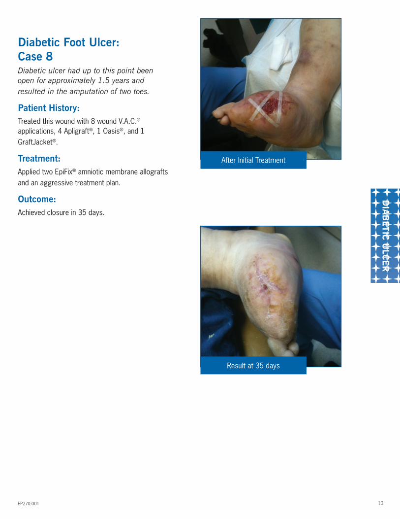
Diabetic Foot Ulcer: Case 8Diabetic ulcer had up to this point been open for approximately 1.5 years and resulted in the amputation of two toes.
Patient History:Treated this wound with 8 wound V.A.C.® applications, 4 Apligraft®, 1 Oasis®, and 1 GraftJacket®.
Treatment: Applied two EpiFix® amniotic membrane allografts and an aggressive treatment plan.
Outcome:Achieved closure in 35 days.
After Initial Treatment
Result at 35 days

PO
ST-T
RA
UM
ATIC
W
OU
ND
14 EP270.001
Post-Traumatic Wounds: Case 959 year old female
Patient History:Motor vehicle accident with open left ankle fracture treated with Open Reduction Internal Fixation (ORIF) with exposed bone.
Initial Examination:Treated with subsequent intravenous antibiotics, hardware removal, negative pressure therapy, Oasis® application, bone stimulator, and hyperbaric oxygen treatment.
Treatment: With worsening measurements, EpiFix® amniotic membrane was applied at three and half months.
Outcome:Due to unavailable product, only one application was utilized. Subsequent Medihoney® dressing changes and office debridements were associated with a gradual decrease in wound size to closure at ten months.
Healing rate of Patient
Patient four months after ankle ORIF
Patient at date of EpiFix® application
Patient 8 weeks after EpiFix® application
5
0
10
15
EpiFix® Applied Area (sq. cm) Volume (cm3)
1Months 2 3 4 5 6

15
PO
ST-SUR
GIC
AL
WO
UN
D
EP270.001
Post-Surgical Wounds: Case 1031 year old female
Patient History:
Crush injury to left calf
Initial Examination:Patient developed seroma necrosis that required inpatient surgical debridement.
Treatment: EpiFix® applied after debridement surgery 4 weeks after admission.
Outcome:Patient had significant epithelialization 4 weeks after EpiFix® application.
On admission
4 weeks later: Debridement and EpiFix® application
4 weeks post EpiFix® application

PO
ST-S
UR
GIC
AL
WO
UN
D
16 EP270.001
Post-Surgical Wounds: Case 11Right-hand dominant 53 year old female smoker with polyarticular rheumatoid and osteoarthritis who was dependent upon forearm crutches for ambulation.
Patient History:After undergoing left olecranon bursa excision, she developed post-operative wound dehiscence at three weeks. This was initially treated with in-office wound debridement, topical medicinal honey dressings and elbow splinting.
Initial Examination:Initial wound measured 2.7 cm x 1.8 cm x 0.1 cm prior to initial debridement and 2.8 cm x 2.8 cm x 0.3 cm thereafter. No joint exposure was noted. At five weeks postoperatively, the wound had retracted but remained open and negative pressure therapy was begun to enhance secondary intention closure of the wound. The wound healing progressed, only to plateau again despite eight weeks of therapy.
Treatment: The wound was then addressed with EpiFix® application at five weeks post-op with concomitant termination of negative pressure application. A single application of EpiFix® was used, resulting in wound closure at three weeks post application. A non-adherent dressing over the membrane was replaced after one week.
Outcome:The patient missed a second-week follow-up appointment, but was noted upon the third-week time point post-EpiFix® application to have the wound healed with re-epithelialization. The patient was discharged. No recurrence was reported at one month after follow-up inquiry by phone and there were no further follow-up visits. Wound measurement evaluation indicated a plateau in the effectiveness of more conservative therapies which was overcome by the subsequent application of EpiFix®.
5 weeks post-op, EpiFix® Applied
8 weeks post-op, 3 weeks post EpiFix® application
Wound age 10 weeks

17
PO
ST-SUR
GIC
AL
WO
UN
D
EP270.001
Post-Surgical Wounds: Case 1256 year old non-diabetic patient; neuropathic. Dehisced surgical wound after removal of ganglion cyst.
Treatment: Previously treated with negative pressure and Xeroform before initiating use of EpiFix® dehydrated Human Amnion/Chorion Membrane (dHACM) allograft.
OutcomeClosure achieved in 4 weeks with 4 applications of EpiFix®.
2nd application
4th application & subsequent closure 3rd application
Post-surgery
First application

OTH
ER
WO
UN
D
18 EP270.001
Other Wounds: Case 1368 year old male with chronic arterial insufficiency of the right ankle
Patient History:Patient’s wound showed no improvement after revascularization.
Treatment:
One application of EpiFix®.
Outcome:Patient’s wound was slow to heal until application of EpiFix®. Wound healed in 2 weeks after EpiFix® application.
Chronological improvement in wound size over time
On admission
Day 20: graft applied
Last visit, one month after closure/ 6 weeks post-EpiFix® application
0
0.5
1
1.5
2
2.5
1 2 3 4 5 6 7 8 9 10 11 12 13 14 15 16 17 18 19 20 21 22 23 24 25 26 27 28 29 30 31 32 33 34
Wou
nd A
rea
sq c
m
Days
Admitted
GraftApplied
Wound closedin 2 weeks


1775 West Oak Commons Court NE, Marietta, GA 30062866.477.4219 • www.mimedx.com All other cited products are registered trademarks of their respective owners.Patents and patents pending see: www.mimedx.com/patentsEpiFix®, PURION®, and MiMedx® are registered trademarks of MiMedx Group, Inc.©2014 MiMedx Group, Inc. All rights reserved. EP270.001