erk1/2 signaling pathway with uoride treatment map kinase
TRANSCRIPT
Page 1/19
MAP kinase phosphatase MKP-1 regulates p-ERK1/2 signaling pathway with �uoride treatmentLin Zhao
Ningxia Medical University, Department of Oral PathologyJiali Su
Yinchuan Stomatology HospitalSijia Liu
Ningxia Medical University, Department of Oral PathologyYang Li
Ningxia Medical University, Department of Oral PathologyTao Li
Ningxia Medical University, Department of Oral PathologyJianping Ruan
Xi'an Jiaotong UniversityKristina Xiao Liang
Universitetet i Bergen https://orcid.org/0000-0002-3586-4218Ruizhe Huang ( [email protected] )
https://orcid.org/0000-0001-8494-9077
Research article
Keywords: MKP-1, ERK1/2, NaF, LS8, Dental Fluorosis
Posted Date: July 21st, 2020
DOI: https://doi.org/10.21203/rs.3.rs-38922/v1
License: This work is licensed under a Creative Commons Attribution 4.0 International License. Read Full License
Version of Record: A version of this preprint was published on December 1st, 2020. See the publishedversion at https://doi.org/10.1016/j.bbrc.2020.12.100.
Page 2/19
Abstract
BackgroundDental �uorosis is characterized by hypomineralization of tooth enamel caused by ingestion of excessive�uoride during enamel formation. Excess �uoride could have effects on the ERK signaling, which isessential for the ameloblasts differentiation and tooth development. MAP kinase phosphatase-1 (MKP-1)plays a critical role in regulating ERK related kinases. However, the role of MKP-1 in ameloblast and themechanisms of MKP-1/ERK signaling in the pathogenesis of dental �uorosis are incompletelyunderstood.
ResultsHere, we adopted an in vitro �uorosis cell model using murine ameloblasts-like LS8 cells by employingsodium �uoride (NaF) as inducer. Using this system, we demonstrated that �uoride exposure led to aninhibition of p- MEK and p-ERK1/2 with a subsequent increase in MKP-1 expression in a dose-dependentmanner. We further identi�ed, under high dose �uoride, MKP-1 acted as a negative regulator of the�uoride-induced p-ERK1/2 signaling, leading to downregulation of CREB, c-myc, and Elk-1.
ConclusionOur results identify a novel MKP-1/ERK signaling mechanism that regulates dental �uorosis and providea framework for studying the molecular mechanisms of intervention and �uorosis remodeling undernormal and pathological conditions. MKP-1 inhibitors may prove to be a bene�t therapeutic strategy fordental �uorosis treatment.
BackgroundFluoride plays a dual role in tooth development. Fluoride at low concentrations can strengthen enameland prevent tooth decay. When permanent teeth are under development, high exposure to �uoride leads todental �uorosis, as referred to a condition characterized by staining and pitting of the teeth that affectsmillions of people worldwide. Although much research has been conducted, the mechanisms underlyingits onset and progression remains unknown.
The signaling networks responsible for properly building the dentition have been heavily investigated andthe extracellular signal-regulated kinase/mitogen-activated protein kinase (ERK-MAPK) pathway. Thismolecular cascade is initiated by binding of a growth factor to a receptor tyrosine kinase (RTK), leading tothe increased phosphorylation of successive kinases, activated effector kinases and the transcription oftarget genes [1]. Previous research has reported a probable link between �uoride exposure and ERK-MAPKpathway [2]. Investigation of the effects of �uoride on enamel-forming cells isolated from rats (primary
Page 3/19
enamel cells) revealed ERK pathway as an important regulator during tooth development [3]. Moreover,phosphorylated ERK1/2 are highly expressed in ameloblasts and odontoblasts in mandibular molars andincisors [3]. The �nding from a mouse-derived enamel cell line known as LS8 provided an in vitro modelfor understanding the molecular basis of dental �uorosis due to its relative ease of handling in the labcompared with primary enamel cells. Through our previous study of �uoride-treated LS8 cell, we identi�eda �uoride-induced downregulation of p-ERK1/2 which is further involved in apoptosis [4][1].
MAPK phosphatases (MKPs) is a negative regulator for Mitogen-activated protein kinase (MAPK) activityvia dual-speci�city phosphatases (DUSPs). Lately, MAPK phosphatase 1 (MKP-1) has emerged as themain counter-regulator of MAPK signaling [2]. MKP-1 locates in the nuclear region and controls geneexpression by inactivating the subcellular group of MAPKs [3]. MKP-1 is the original member of a familyof dual-speci�city phosphatases that can remove phosphates from tyrosine and threonine in ERK andrelated kinases [4]. MKP-1 activity can manifest positively or negatively the signaling outcomes through aparticular pathway, which varies in different cell types either as a function of the relative activities of thevarious MAPKs and/or abundance of the MAPK substrate [5]. However, the effect of MKP-1 in ameloblastand how MKP-1 regulates ERK signaling together with their downstream regulation of transcriptionfactors in dental �uorosis are unclear.
In the present study, we apply an established in vitro �uorosis system by using murine ameloblasts-likeLS8 cells and employed NaF as an inducer for dental �uorosis. We show that �uoride exposureinactivates both MEK and ERK1/2 pathways with a subsequent active in MKP-1, which negativelymediates the downstream regulation of transcription factors cAMP-response element-binding protein(CREB), c- myelocytomatosis oncogene cellular homolog (c-myc) and Elk-1. Moreover, blocking orenhancing either the ERK pathway attenuates the changes of MKP-1 in response to high dose �uorideexposure. Together, these data provide evidence MKP-1/ERK mediated pathways contribute to dental�uorosis pathogenesis and, importantly, indicating that MKP-1 inhibitors may prove to be bene�ttherapeutic strategy for dental �uorosis treatment.
ResultsFluoride exposure inhibits phosphorylation of MEK and ERK1/2 with a subsequent increase in MKP-1expression via a dose-dependent manner.
Fluoride is an environmental toxicant and induces dental �uorosis. NaF is one of the most commoninorganic �uorides, which is frequently used in the research of �uoride toxicity. The ERK-MAPK pathwayplays a vital role in the developmental processes of the dental epithelium and tooth growth [3]. Previously,our group has established the in vitro dental �uorisis model by treating NaF in murine ameloblasts-likeLS8 cells [5]. Brie�y, LS8 cells were incubated with NaF at a serial concentration of 0 mmol/L, 1 mmol/Land 2 mmol/L for 48 hours. We observed that NaF treatment induced a signi�cant decrease on cellnumber with a dosage-depended manner (Figure A1). However, the cell morphology was not changedafter the NaF treatment (Figure A1).
Page 4/19
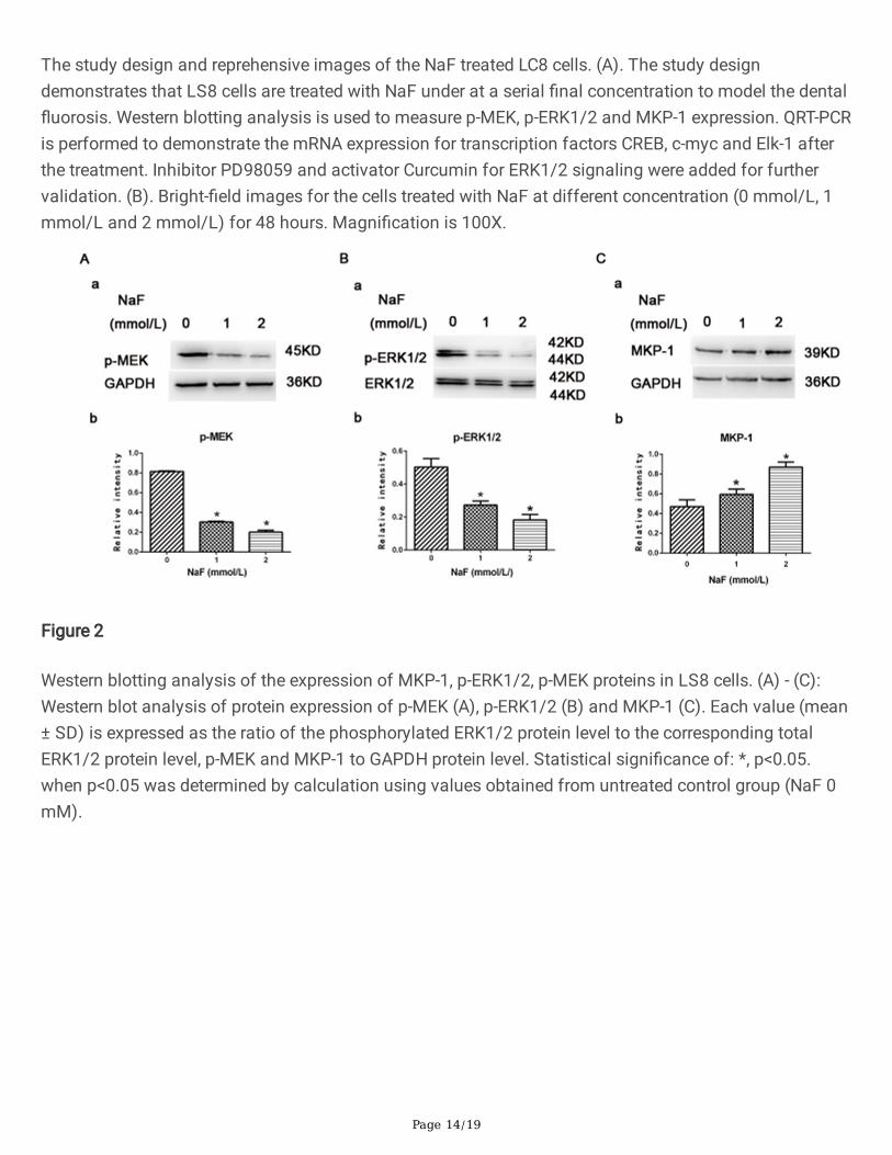
By using this model, we examined whether �uoride treatment in our cell system leads to MEK and ERKactivation. LS8 cells were treated with NaF for 48 hours with various concentrations (0 mmol/L,1 mmol/L, 2 mmol/L), and total cell lysates were subjected to western blot analysis using an antibodyagainst the phosphorylated form of MEK (p-MEK) and antibodies against phosphorylated forms ofERK1/2 (p-ERK1/2). As shown in Fig. 1, NaF treatment led to a signi�cant downregulated expression levelof p-MEK (Fig. 2A, a&b) and p-ERK1/2 (Fig. 2B, a&b) in NaF-treated cells compared to un-treated group ina dose-dependent manner. MAPKs are deactivated by members of the MAPK phosphatases such asMKP-1, which is vital in ERK and related kinases activation and plays a critical role in regulation of MAPKsignaling in various peripheral tissues. Therefore, we investigated the MKP-1 expression in the cells in thepresent of NaF. In contrast, we showed that the phosphorylation of MKP-1 (p-MKP-1, Fig. 2C, a&b) wasstrongly activated in �uoride-treated cells than those in control via a dose-dependent manner, suggestingthe negative feedback regulation of the MAPK/ ERK pathway. These results indicate that �uorideexposure inhibits phosphorylation of MEK and ERK1/2 with a subsequent increase in MKP-1 expressionvia a dose-dependent manner.
High dose �uoride exposure activates MKP-1 gene transcription and induces downregulation ofdownstream transcription factors.
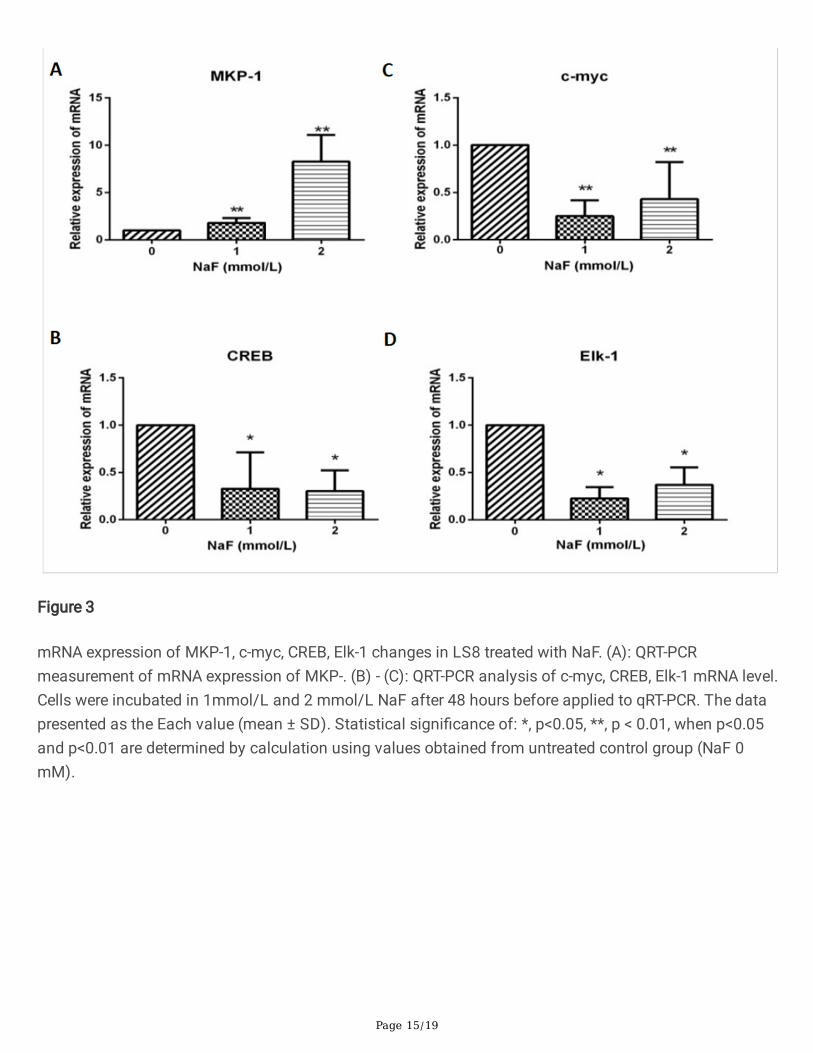
MKP-1 is an important kinase as the downstream regulators of both p38 and ERK1/2 [6]. Activation ofMKP-1 is regulated through phosphorylation of several transcription factors, including cAMP-responseelement-binding protein (CREB), c-myc and Elk-1. CREB acts as an indirect mediated of ERK [7]. The C-myelocytomatosis oncogene cellular homolog (c-Myc) family is one transcription factors that modulatesthe genes responsible for cellular homeostasis [8] and its gene transcription and protein stabilization andaccumulation [9] are sustained by the hyper-activated RAS/MEK/ERK pathway. Transcription factor Elk-1is a downstream substrate, as a feedback control mechanism to activate ERK l/2. The severity of dental�uorosis is dependent upon �uoride dose and the timing and duration of �uoride exposure. Therefore, weused the LS8 cells with �uoride treatment under a high dose (2 mmol/L) to mimic the in vivo dental�uorosis and investigated whether this condition activates MKP-1 gene expression and their downstreamtranscription factors c-myc, CREB and Elk-1. QRT-PCR measurement demonstrated a signi�cantlyincreased mRNA expression of MKP-1 (Fig. 3A), con�rming the positive feedback control mechanisminduced by �uoride treatment as demonstrated above in Fig. 2C. Quanti�cation of mRNA expression ofthe downstream molecule CREB displayed a downregulation of CREB gene level in �uoride treated LS8cells versus control cells, with similar change in both concentration (Fig. 3B). Furthermore, a signi�cantlydecreased level of c-myc (Fig. 3C) and Elk-1 (Fig. 3D) transcription expression was identi�ed in both NaF-treated groups compared to un-treated cells with a dosage-dependent pattern. These data suggest anegative feedback between MKP-1 signaling and downstream regulation of transcription factors CREB, c-myc and Elk-1.
PD98059 and curcumin treatment attenuate the changes of MKP-1 protein expression in response to highdose �uoride exposure.
Page 5/19
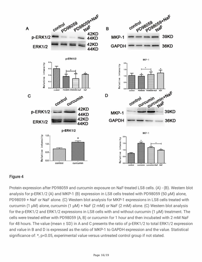
PD98059 is a potent and selective inhibitor of MAPK kinases via binding to the inactive form of MAPKand prevents activation by upstream activators, such as c-Raf [10]. Curcumin, one of the main bioactivecomponents extracted from a traditional Chinese medicinal herb, induces ERK/MARK activation with up-regulating phospholortated ERK1/2 signaling [11]. To further con�rm the linkage between MARK and ERKsignaling in dental �uorosis, the LS8 cells were treated with 50 µM PD98059 for 1 hour and thenincubated with 2 mM NaF for 48 hours. Western blot assessment was then applied to reveal the proteinexpression changes of p-ERK1/2 and MKP-1 for the cells under different combinations of the treatment.Our results showed, compared to the control and NaF group, a decreased expression of p-ERK1/2 inPD98059, PD98059 and NaF treated groups, suggesting the treatment of PD98059 were able to inhibit p-ERK1/2. In addition, we demonstrated that PD98059 and NaF treated groups showed signi�cantly lowerlevels of p-ERK1/2 than that with PD98059 alone (Fig. 4A). In contrast, the protein expression level ofMKP-1 increased gradually from the cells treated with PD98059 alone, PD98059 and NaF or NaF alone(Fig. 4B), where PD98059 and NaF exposed cells demonstrated a signi�cantly increased level of MKP-1than that exposed in NaF alone (Fig. 4B). To validate this observation, we used 1 µM curcumin to treat theLS8 cells for 1 hours before NaF treatment and we found that, curcumin treated cells exhibitedsigni�cantly higher p-ERK1/2 expression when normalized with total expression of ERK1/2 thanuntreated cells (Fig. 4C), suggesting treatment of curcumin actives the ERK1/2 signaling in LS8 cells.When the cells treated with curcumin alone, MKP-1 expression elevated in the all three treated groups(Fig. 4D) with a gradual decrease from the cells treated with curcumin alone, curcumin and NaF or NaFalone. In addition, curcumin and NaF treated groups showed signi�cantly higher level of MKP-1 than thatwith NaF alone (Fig. 4D). These �nding further con�rm the effect of the MKP-1 on negative regulationphosphorylation ERK signaling pathway in the cells with �uoride exposure, as described above (Fig. 2).
D98059 and curcumin treatment convert the gene expression changes of transcript factors in response tohigh dose �uoride exposure.
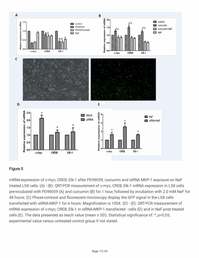
Our �ndings in Fig. 2 suggested a negative feedback between MKP-1 signaling and downstreamregulation of transcription factors CREB, c-myc and Elk-1. To further validate this observation, qRT-PCRmeasurement was applied to measure the of mRNA expression of c-myc, CREB, Elk-1 in LS8 cells pre-incubated with PD98059 (50 µM) or curcumin (1 µM) for 1 hour, followed by incubation with 2.0 mM NaFfor 24 hours. Our data showed that mRNA expression of c-myc and Elk-1 decreased in the cells treatedwith PD98059, PD98059 and NaF than NaF group and control group, whereas the mRNA expression ofCREB dropped in the group in the present of PD98059, PD98059 and NaF, and NaF than control group(Fig. 5A). In contrast, curcumin treated cells showed a signi�cantly highest transcriptional expressionlevel for c-myc, CREB and Elk-1 among the cells treated with control, curcumin and NaF, and NaF(Fig. 5B). To further con�rm whether lack of MKP-1 protein expression in response to �uoride treatment isregulated through a transcriptional mechanism via modulating downstream transcription factors c-myc,CREB and Elk-1, we then transfected the LS8 cells with siRNA targeting MKP-1 to know-out the MKP-1expression before the qRT-PCR was performed. MOCK siRNAs with the sequences that do not target anygene product was used for negative control. After transfection of GFP siRNA-MKP-1 for 6 hours, wedetected a bright green �uorescent signal, indicating the cells expressed MKP-1 after transfection
Page 6/19
(Fig. 5C). We demonstrated that mRNA expression of c-myc, CREB and Elk-1 increased in LS8 cellstreated with siRNA-MKP-1 than all the other groups (Fig. 5C). Similarly, a signi�cantly upregulated mRNAexpression of c-myc, CREB and Elk-1 was identi�ed in LS8 cells transfected with siRNA-MKP-1 comparedto that in MOCK control group (Fig. 5C). More importantly, curcumin and NaF treated cells showed asigni�cant increase of c-myc, CREB and Elk-1 mRNA expression than NaF treat cells (Fig. 5E). Theseevidences suggest that MKP-1 can facilitate gene transcription through modulating downstreamtranscription factors c-myc, CREB and Elk-1 in �uoride treated cells. Therefore, the previous observationsin the qRT-PCR have been con�rmed on this point.
DiscussionOur previous evidence showed the activation of ERK1/2 and other MAP Kinase in dental pulp cells [12]and excess �uoride have effects on the ERK signaling [1]. MKP-1, as an inhibitor of MAPKs, plays anessential role in regulating ERK related kinases. However, it remains unclear on how MKP-1 is regulated indental �uorosis. Here, we present compelling evidence that treatment with �uoride in vitro at themillimolar concentrations markedly activated MKP-1 expression and decreased MEK and ERK1/2phosphorylation level in a dose-dependent manner in mouse ameloblasts-like LS8 cells [1], indicating anegative feedback between MKP-1 and MEK/ERK signaling. We further use high dose �uoride to inducethe dental �uorosis and demonstrated a negative regulatorily role of MKP-1 in the �uoride-induced p-ERK1/2 signaling. We also found that MKP-1 downregulated ERK/MAPK-mediated CREB, c-myc, and Elk-1 transcriptions. Our study clearly demonstrates the effect of MKP-1 on dental �uorosis and itstherapeutic potential for the treatment of dental �uorosis.
In our previous work, we reported a positive expression of p-ERK, p-JNK, p-p38 and �uoride-inducedapoptosis led to the deceased phosphorylation and deactivation of ERK and JNK signaling cascade in aconcentration- and time-dependent manner in ameloblasts cells, indicating a linkage of MAPK signalingon dental �uorosis [1]. In present study, we con�rm the MEK and ERK1/2 phosphorylation in ameloblastsand their downregulation induced by �uoride exposure.
MAPK is a serine/threonine protein kinase that is widely present in eukaryotic cells. Previous studiesfound four different MAPKs, including ERK, c- JNK, ERK5 and p38 MAPK (p38) [13]. ERK cascade reactioncan be activated by various stimuli, such as RTK and G protein-coupled receptors. After activation, it canregulate the proliferation, differentiation and apoptosis. Ras/Raf/MEK/ERK cascade reaction is animportant signaling pathway in MAPKs. Various stimuli can activate the corresponding cell surfacereceptors that, in turn, activates the signal transduction pathway and produce an appropriate biologicalresponse. The Ras/Raf/MEK/ERK cascade reaction is the key factor in integrating the signal transductionpathway. Previous observation of individuals with orofacial and craniofacial disorders identi�ed anassociation of Ras/Raf/MEK/ERK pathway and dental malformations [14]. MKP-1 has the capacity tobind and dephosphorylate ERK, and subsequently p38 and JNK kinases. MKP-1 exerts dual effects underdifferent physiological conditions. Previous studies have suggested that MKP-1 at relatively low level hasa weak binding a�nity for ERK kinases and exerts little effect on their function, whereas MKP-1 is
Page 7/19
activated and shows a higher binding a�nity for p38 and JNK kinases than ERK kinases under otherphysiological conditions [10, 11]. When MKP-1 levels are high, it also inhibits ERK kinases, although to alesser extent than MARK/p38 and JNK, and thus provides a negative feedback loop on many cellularprocesses. Consistent with these �ndings, here, we report a gradual upregulation of MKP-1 and inhibitionof MEK and ERK expression from low to high �uoride exposure. Furthermore, we show that the relativehigh phosphorylation level of MKP-1 is associated with the negative feedback regulation of theMAPK/ERK pathway, which may contribute to the pathogenesis in dental �uorosis.
ERK-induced Elk-1 phosphorylation leads to enhanced DNA-binding and TCF-mediated transcriptionalactivation. Several early genes, such as c-jun, c-fos, and c-myc, are regulated upstream by Ras/ERK andp38. They were found to be involved in controlling the cell growth and metabolism [15]. Elk-1, afterphosphorylation by ERK, binds to the SRE cis-acting element in the promoter region of c-fos and inducesits transcription. Transcription factor Elk-1 is a part of the ternary complex which can be combined withserum effect factors (SRE) to regulate gene activity in order to reply to the serum and growth factors [16].The activation of ERKl/2 is transferred from the cytoplasmic to nuclear and activates its downstreamsubstrate EIk-1, and the phosphorylation of EIk-1 will promote the function of cell differentiation,proliferation and apoptosis [17–19]. In addition, CREB and c-myc are also recognized as putative activespeci�c transcriptional factor substrates which mediate indirectly by extracellular signal-regulated kinasesuch as ERK [7]. Activated phosphorylation of CREB is reported in human molar odontoblasts andcementoblasts in vivo [20]. Study in osteoblast cells reports that ERK/CREB signaling can inhibit cellularoxidative stress [21]. However, some studies reported that MEK/ERK/CREB signaling pathway may not bethe solely mediator in the signal transduction pathways in dental pulp cells [22]. The myelocytomatosisoncogene cellular homolog c-myc family consists of transcription factors which modulates genesresponsible for cellular homeostasis [8]. C-myc gene transcription and protein stabilization andaccumulation [9] are sustained by the hyper-activated RAS/MEK/ERK pathway. In this study, wedemonstrate that MKP-1 reduction results in the deactivation of ERK-induced Elk-1 phosphorylation incytoplasm, which consequently leads to downregulate the downstream substrates CREB and c-myc.
In this study, we used PD98059 and curcumin to inhibit and agitate the phosphorylation of MEK/ERKpathway, respectively, and then detected the trend of the expression of MKP-1 protein and expression ofdownstream transcription factors of ERK, and found that there was a interaction between thephosphorylation level of ERK1/2 and MKP-1. Silencing MKP-1 at the gene expression level and detectingthe expression of ERK downstream transcription factors con�rmed this interaction. As an exogenousstimulus, NaF reduced the phosphorylation of ERK in LS8 cells, which was caused by the decrease ofMKP-1. The negative feedback regulation mechanism of the formation of MEK/ERK-MKP-1 was involvedin the of dental �uorosis.
MethodsCell culture and treatments
Page 8/19
The mouse ameloblast-like cell line (LS8) was kindly donated by Malcolm L. Snead (Department ofBiomedical Sciences, University of Southern California) cultured in DMEM supplemented with 10% FBSand 100 units/ml penicillin, and 100 mg/ml streptomycin (Invitrogen, CA, USA). The incubatoratmosphere was humidi�ed and adjusted at 5% CO2 and 95% air at 37°C. When reached 70–80%con�uence, the cells were incubated with serum-free medium containing the indicated concentrations(0~2 mM) of NaF. After the treatment, the cells were incubated for 24 hours or 48 hours at 37°C.
Chemicals and antibodies and antibodies
Rabbit Anti-phospho-p44/42 ERK (#9101), rabbit Anti-phospho-MEK (#9154), Anti-ERK (#9102), andGAPDH (#97166) were purchased from Cell Signaling (Beverly, MA, USA). MKP-1 (#373841) waspurchased from Santa Cruz Biotechnology (Santa Cruz, CA, USA). All antibodies were used at a dilutionratio of 1:500 1:1000 for western blot analysis. Inhibitor PD98059 was purchased from Cell Signaling(Boston, MA, USA). Activator curcumin was purchased from Sigma Aldrich Fluka (St. Louis., MO, USA).The inhibitor and activator were dissolved separately in dime thylsulfoxide (DMSO, Sigma, MO, USA)immediately before use. Anti-rabbit and anti-mouse secondary antibody were purchased from ZhongshanBiological Manufacture (Zhongshan Co., Ltd, Beijing, China). Dulbecco’s modi�ed Eagle’s medium(DMEM) was supplied by Thermo Scienti�c Company (Logan, Utah, USA). Fetal bovine serum (FBS) wassupplied by Gibco (GIBCO, Invitrogen, CA, USA), Lipofectamine 2000 Transfection Reagent was purchasedfrom Invitrogen (Carlsbad, CA, USA). Trizol Reagent was purchased from Invitrogen (Carlsbad, CA, USA).MKP-1-mus-773 siRNA that speci�cally target mouse MKP-1 and control siRNA, or FAM-labeled negativecontrol siRNA were purchased from Gene Pharma (Gene Pharma Co., Ltd, Shanghai, China). Allprocedures were conducted with approval from the Ethics Committee at Xi’an Jiaotong University, Xi’an,China.
Transient transfection siRNA
SiRNA-MKP-1 purchased form Shanghai jima pharmaceutical technology co. LTD. At 40% con�uence,LS8 cells were transfected with silencing RNA (siRNA) against MKP-1, using Lipofectamine 2000transfection reagent (Invitrogen, CA, USA) according to the manufacturer’s instruction. Brie�y, MKP-1-siRNA was diluted in serum-free culture medium with the transfection reagent, mixed by vertexing andincubated for 20 minutes at room temperature to allow the formation of the transfection complex. Thenthe MKP-1-siRNA was added to the cells for 24 hours. The effectiveness of gene silencing was monitoredby measuring the MKP-1 levels in relation to GAPDH, as analyzed by qRT-PCR for mRNA expression level.Cells transfected with MKP-1-siRNA, were further exposed to NaF for 24 hours. The mRNA expressionlevel of c-myc, CREB, Elk-1 was determined by using qRT-PCR.
Protein extraction
Cells were washed with chilled PBS and lysed in ice-cold RIPA buffer as previously described [5],consisting of 50 mM Tris-HCl, pH 7.5, 50 mM NaCl, 5 mM EDTA, 10 mM EGTA, 2 mM sodiumpyrophosphate, 4 mM paranitrophenyl phosphate, 1 mM sodium orthovanadate, 1 mM
Page 9/19
phenylmethylsulfonyl �uoride, 2 µg/ml aprotinin, 2 µg/ml leupeptin and 2 µg/ml pepstatin. Cell lysatewas collected using a cells scraper (Corning, Acton, MA) and the homogenates sonicated on ice for 30min. The lysate was collected by centrifugation at 12,000×g for 15 min at 4°C. The protein content wasdetermined using a BCA protein assay kit (Pierce, Rockford, IL) by extrapolation to dye binding for astandard series of known protein concentration using spectrophotometry.
Western blotting
A volume of supernatant corresponding to an equal mass of protein for each experimental condition wasmixed with loading buffer (5-sodium dodecyl sulfate, 5% v/v) and denatured by heating the samples at95°C for 5 min. Lysate proteins were resolved to size by electrophoresis using 10-12% sodium dodecylsulfate polyacrylamide gel electrophoresis (SDS-PAGE) and transferred to 0.22 µm polyvinylidene �uoride(PVDF) membrane (Millipore, Bedford, MA, USA) using a semi-dry blotting system (Bio-Rad, Hercules, CA,USA). Non-speci�c absorption by the membranes was blocked by incubation with 5g% (w/v) skin milk inTris-buffered saline (TBS, 500 mM NaCl, 20 mM Tris-HCl pH 7.5) with 0.05% (v/v) Tween-20 for 2 hours.Samples were incubated overnight with one of the following primary antibodies at 4°C: Anti-phospho-ERKand total ERK (1:1000), Anti-phospho-MEK (1:1000), Anti-MKP-1 (1:500), GAPDH (1:1000), each diluted inTBS with 5% (v/v) bovine serum containing 0.1% Tween-20 for 24 hours at room temperature with gentleshaking. The membranes were washed using 0.1% Tween-20 TBS three times for 10 min each andincubated with horseradish peroxidase conjugated anti-rabbit or anti-mouse secondary antibody(1:10000), as appropriate for each primary antibody, for 1 hour at room temperature. After washing inTBS-0.1% Tween-20 three times, an enhanced chemiluminescence kit (Millipore, MA, USA) was used todetect immunoreactive protein bands. Blots were immuno-detected with an anti-GAPDH antibody(1:1000) to con�rm equal mass of protein loaded among samples. The intensity for eachimmunoreactive protein band was quanti�ed using a Quantity One densitometer (BioRad, Hercules, USA).
RNA Extraction and quantitative real-time polymerase chain reaction (qRT-PCR)
The total RNA of the cells was extracted using Trizol reagent (Carlsbad, CA, USA) according to themanufacturer's instruction. The quality and quantity of the isolated RNA was examined using aNanoDrop 2000/2000C spectrophotometer with measuring absorbance at 260/280nm. First-strand c-DNA synthesis was performed on 2 µg of total RNA using reverse transcription with Real Master Mix(Thermo Fisher Scienti�c, Logan, Utah, USA). QRT-PCR was performed in a 10 µL reaction mixture systemusing an Applied Bio systems 7500 Real-Time PCR System (Thermo Fisher, Waltham, MA, USA) with aninitial denaturation of 5 min at 95°C, followed by 40 cycles of 95°C for 5 s, 60°C for 30 s, and 72°C for 30s. Primers used in the ampli�cation were designed and synthesized by Gene Pharma (Gene Pharma Inc.,Shanghai, China). The sequences of the PCR primers were as follow: MKP-1 F:5'CCCCTGAGTACTAGTGTGCCTGAC 3', MKP-1 R:5' AGCTGAAGTTC GGGGAGATGATAC 3';C-myc F:5' GCTGCA TGA GGA GAC ACC 3', c-myc R:5' GTG CGG AGG TTT GCT GTG 3'; CREB F:5' ACA GAT TGC CACATT AGC 3', CREB R:5' GGACTTGTGGAGACTGGA 3' ; Elk-1 F:5' ATATCATCCGCAAGGTG AGC3', Elk-1 R:5'ATGGCCGAGGTTACAGACAC3' ; GAPDH F:5' GCTGA GTATGTCGTGGAGT3, 'GAPDH R:5'
Page 10/19
GTTCACACCCATCACAAAC3; were used as the internal control. A melting curve analysis was performedon each amplicon to ensure ampli�cation of a single PCR product. The relative expression levels werecalculated using the comparative threshold cycle (△△CT) method.
Statistical analysis
Statistical analyses were performed using SPSS software, Version 18.0 (SPSS Inc; Chicago, IL). All datawere expressed as mean ± standard deviation (SD) with each experiment performed in triplicates.Differences among groups were tested by one-way ANOVA or two-way ANOVA, and the T test was usedfor two individual comparisons. For all analyses, two-tailed p values of less than 0.05 were consideredsigni�cant.
ConclusionsIn summary, we determine that upregulation of MKP-1 induced by �uoride treatment via negativelyregulating cellular p-MEK1/2 levels following feedback-regulated p-ERK1/2 signaling, which consequentlyfacilitates gene transcription through modulating speci�c transcription factor substrates c-myc, CREBand Elk-1 in �uoride treated cells (Fig. 6). These �ndings provide novel insights into the role of MKP-1 inthe pathogenesis of dental �uorosis and its potential as a new target for dental �uorosis therapy. These�ndings together with our previous evidence on �uoride induced apoptosis through p-ERK and p-JNK [5],suggest the MEK-ERK-JNK signaling cascade controlling the mechanisms involved in ERK1/2-mediatedcell apoptosis in dental �uorosis.
DeclarationsConsent for publication
All authors have read and approved the �nal transcript
Acknowledgements
We thank Professor Malcolm L. Snead (Department of Biomedical Sciences, University of SouthernCalifornia) for donation of LS8 cells.
Author's contributions:
L.Z, J.S, S.L, Y.L, T. X and M. L.S contribute to the experimental investigation; L.Z and K.L contribute to thewriting original draft; all the authors contribute to review and edit; R.H and L.Z contribute the resources;R.H and K.L contribute to supervision and funding acquisition. All authors read and approved the �nalmanuscript.
Funding
Page 11/19
This research was funded by special talent project of Ningxia Medical University [No. XT2017020]. Thefunding bodies played no role in the design of the study and collection, analysis, and interpretation ofdata and in writing the manuscript.
Competing interests
The authors declare no competing interests.
Availability of data and materials
The datasets during and/or analyzed during the current study available from the corresponding author onreasonable request
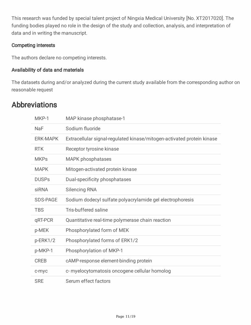
Abbreviations
MKP-1 MAP kinase phosphatase-1
NaF Sodium �uoride
ERK-MAPK Extracellular signal-regulated kinase/mitogen-activated protein kinase
RTK Receptor tyrosine kinase
MKPs MAPK phosphatases
MAPK Mitogen-activated protein kinase
DUSPs Dual-speci�city phosphatases
siRNA Silencing RNA
SDS-PAGE Sodium dodecyl sulfate polyacrylamide gel electrophoresis
TBS Tris-buffered saline
qRT-PCR Quantitative real-time polymerase chain reaction
p-MEK Phosphorylated form of MEK
p-ERK1/2 Phosphorylated forms of ERK1/2
p-MKP-1 Phosphorylation of MKP-1
CREB cAMP-response element-binding protein
c-myc c- myelocytomatosis oncogene cellular homolog
SRE Serum effect factors
Page 12/19
References1. Zhao L, et al. LS8 cell apoptosis induced by NaF through p-ERK and p-JNK - a mechanism study of
dental �uorosis. Acta Odontol Scand. 2016;74(7):539–49.
2. Kim HS, Asmis R. Mitogen-activated protein kinase phosphatase 1 (MKP-1) in macrophage biologyand cardiovascular disease. A redox-regulated master controller of monocyte function andmacrophage phenotype. Free Radic Biol Med. 2017;109:75–83.
3. Patterson KI, et al. Dual-speci�city phosphatases: critical regulators with diverse cellular targets.Biochem J. 2009;418(3):475–89.
4. Kondoh K, Nishida E. Regulation of MAP kinases by MAP kinase phosphatases. Biochim BiophysActa. 2007;1773(8):1227–37.
5. Roth Flach RJ, Bennett AM, [12]. Expert Opin Ther Targets, 2010. 14(12): p. 1323-32.
�. Cho KW, et al. ERK activation is involved in tooth development via FGF10 signaling. J Exp Zool B MolDev Evol. 2009;312(8):901–11.
7. Otani T, et al. Osteocalcin triggers Fas/FasL-mediated necroptosis in adipocytes via activation ofp300. Cell Death Dis. 2018;9(12):1194.
�. Ciccarelli C, et al. Disruption of MEK/ERK/c-Myc signaling radiosensitizes prostate cancer cells invitro and in vivo. J Cancer Res Clin Oncol. 2018;144(9):1685–99.
9. Sears RC. The life cycle of C-myc: from synthesis to degradation. Cell Cycle. 2004;3(9):1133–7.
10. Alessi DR, et al. PD 098059 is a speci�c inhibitor of the activation of mitogen-activated proteinkinase kinase in vitro and in vivo. J Biol Chem. 1995;270(46):27489–94.
11. Li X, et al., Curcumin Inhibits Apoptosis of Chondrocytes through Activation ERK1/2 SignalingPathways Induced Autophagy. Nutrients, 2017. 9(4).
12. Lin SI, et al. [13]: Role of TAK1 and MEK/ERK signaling. J Formos Med Assoc. 2018;117(8):697–704.
13. Li L, et al. The Ras/Raf/MEK/ERK signaling pathway and its role in the occurrence and developmentof HCC. Oncol Lett. 2016;12(5):3045–50.
14. Vogiatzi A, Mavrothalassitis G. Craniofacial, orofacial and dental disorders: the role of the RAS/ERKpathway. Expert Rev Mol Med. 2019;21:e2.
15. Chan CP, et al. Thrombin activates Ras-CREB/ATF-1 signaling and stimulates c-fos, c-jun, and c-mycexpression in human gingival �broblasts. J Periodontol. 2008;79(7):1248–54.
1�. Shi L, et al. Oral administration of Lentinus edodes beta-glucans ameliorates DSS-induced ulcerativecolitis in mice via MAPK-Elk-1 and MAPK-PPARgamma pathways. Food Funct. 2016;7(11):4614–27.
17. Cavigelli M, et al. Induction of c-fos expression through JNK-mediated TCF/Elk-1 phosphorylation.EMBO J. 1995;14(23):5957–64.
1�. Tresini M, et al. Lack of Elk-1 phosphorylation and dysregulation of the extracellular regulated kinasesignaling pathway in senescent human �broblast. Exp Cell Res. 2001;269(2):287–300.
Page 13/19
19. Goetze S, et al. TNFalpha induces expression of transcription factors c-fos, Egr-1, and Ets-1 invascular lesions through extracellular signal-regulated kinases 1/2. Atherosclerosis. 2001;159(1):93–101.
20. Klinz FJ, et al. Transcription factor CREB is phosphorylated in human molar odontoblasts andcementoblasts in vivo. Histochem Cell Biol. 2013;139(4):615–20.
21. Ding G, Zhao J, Jiang D. Allicin inhibits oxidative stress-induced mitochondrial dysfunction andapoptosis by promoting PI3K/AKT and CREB/ERK signaling in osteoblast cells. Exp Ther Med.2016;11(6):2553–60.
22. Chang MC, et al. Prostaglandin F(2alpha) stimulates MEK-ERK signalling but decreases theexpression of alkaline phosphatase in dental pulp cells. Int Endod J. 2010;43(6):461–8.
Figures
Figure 1
Page 14/19
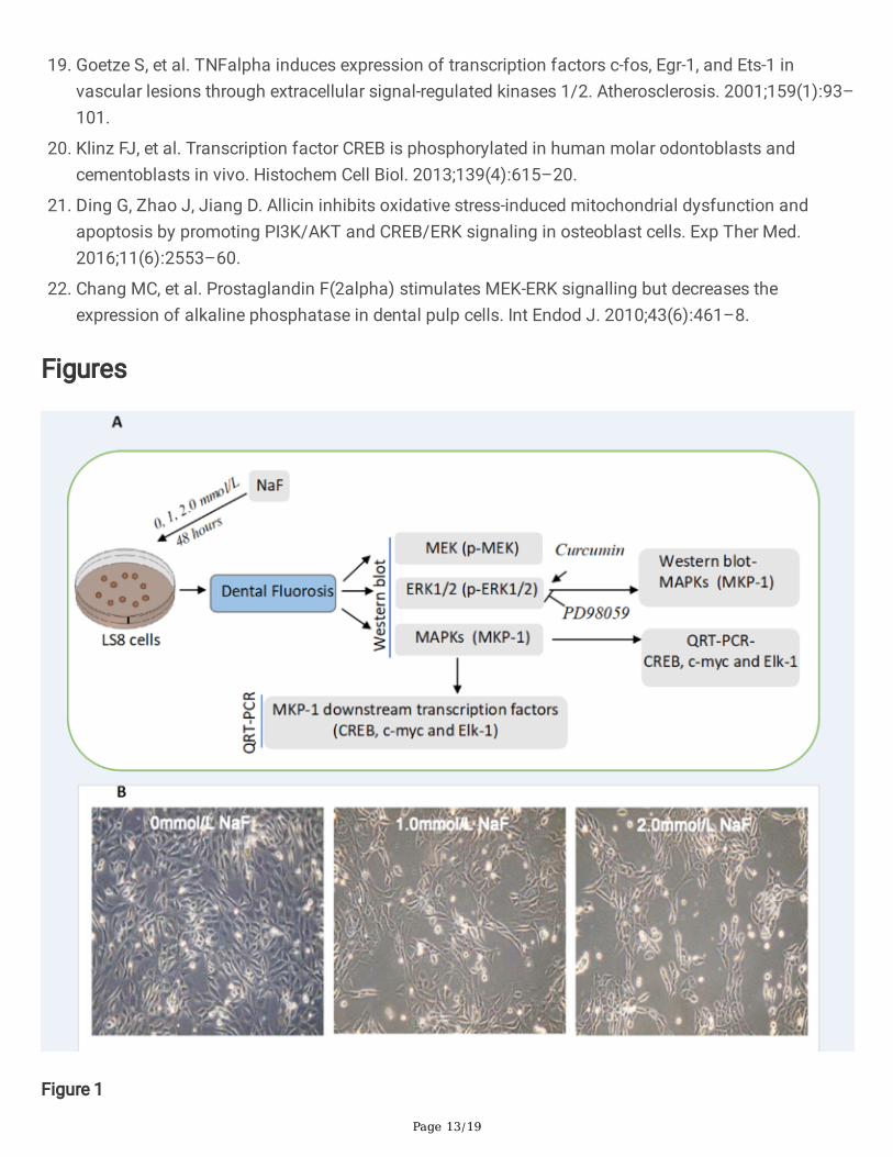
The study design and reprehensive images of the NaF treated LC8 cells. (A). The study designdemonstrates that LS8 cells are treated with NaF under at a serial �nal concentration to model the dental�uorosis. Western blotting analysis is used to measure p-MEK, p-ERK1/2 and MKP-1 expression. QRT-PCRis performed to demonstrate the mRNA expression for transcription factors CREB, c-myc and Elk-1 afterthe treatment. Inhibitor PD98059 and activator Curcumin for ERK1/2 signaling were added for furthervalidation. (B). Bright-�eld images for the cells treated with NaF at different concentration (0 mmol/L, 1mmol/L and 2 mmol/L) for 48 hours. Magni�cation is 100X.
Figure 2
Western blotting analysis of the expression of MKP-1, p-ERK1/2, p-MEK proteins in LS8 cells. (A) - (C):Western blot analysis of protein expression of p-MEK (A), p-ERK1/2 (B) and MKP-1 (C). Each value (mean± SD) is expressed as the ratio of the phosphorylated ERK1/2 protein level to the corresponding totalERK1/2 protein level, p-MEK and MKP-1 to GAPDH protein level. Statistical signi�cance of: *, p<0.05.when p<0.05 was determined by calculation using values obtained from untreated control group (NaF 0mM).
Page 15/19
Figure 3
mRNA expression of MKP-1, c-myc, CREB, Elk-1 changes in LS8 treated with NaF. (A): QRT-PCRmeasurement of mRNA expression of MKP-. (B) - (C): QRT-PCR analysis of c-myc, CREB, Elk-1 mRNA level.Cells were incubated in 1mmol/L and 2 mmol/L NaF after 48 hours before applied to qRT-PCR. The datapresented as the Each value (mean ± SD). Statistical signi�cance of: *, p<0.05, **, p < 0.01, when p<0.05and p<0.01 are determined by calculation using values obtained from untreated control group (NaF 0mM).
Page 16/19
Figure 4
Protein expression after PD98059 and curcumin exposure on NaF-treated LS8 cells. (A) - (B). Western blotanalysis for p-ERK1/2 (A) and MKP-1 (B) expression in LS8 cells treated with PD98059 (50 µM) alone,PD98059 + NaF or NaF alone. (C) Western blot analysis for MKP-1 expressions in LS8 cells treated withcurcumin (1 µM) alone, curcumin (1 µM) + NaF (2 mM) or NaF (2 mM) alone. (C) Western blot analysisfor the p-ERK1/2 and ERK1/2 expressions in LS8 cells with and without curcumin (1 µM) treatment. Thecells were treated either with PD98059 (A, B) or curcumin for 1 hour and then incubated with 2 mM NaFfor 48 hours. The value (mean ± SD) in A and C presents the ratio of p-ERK1/2 to total ERK1/2 expressionand value in B and D is expressed as the ratio of MKP-1 to GAPDH expression and the value. Statisticalsigni�cance of: *, p<0.05, experimental value versus untreated control group if not stated.
Page 17/19
Figure 5
mRNA-expression of c-myc, CREB, Elk-1 after PD98059, curcumin and siRNA MKP-1 exposure on NaFtreated LS8 cells. (A) - (B): QRT-PCR measurement of c-myc, CREB, Elk-1 mRNA expression in LS8 cellspre-incubated with PD98059 (A) and curcumin (B) for 1 hour, followed by incubation with 2.0 mM NaF for48 hours. (C) Phase-contrast and �uorescent microscopy display the GFP signal in the LS8 cellstransfected with siRNA-MKP-1 for 6 hours. Magni�cation is 100X. (D) - (E): QRT-PCR measurement ofmRNA expression of c-myc, CREB, Elk-1 in siRNA-MKP-1 transfected - cells (D) and in NaF-post treatedcells (E). The data presented as teach value (mean ± SD). Statistical signi�cance of: *, p<0.05,experimental value versus untreated control group if not stated.
Page 18/19
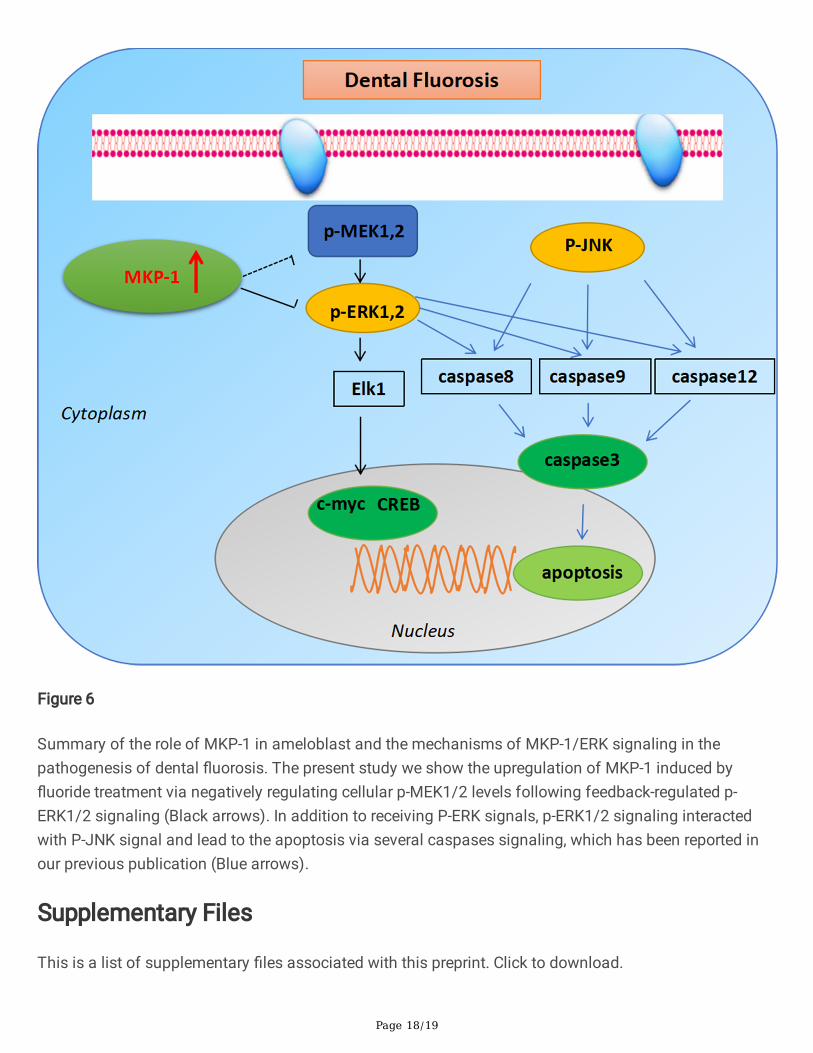
Figure 6
Summary of the role of MKP-1 in ameloblast and the mechanisms of MKP-1/ERK signaling in thepathogenesis of dental �uorosis. The present study we show the upregulation of MKP-1 induced by�uoride treatment via negatively regulating cellular p-MEK1/2 levels following feedback-regulated p-ERK1/2 signaling (Black arrows). In addition to receiving P-ERK signals, p-ERK1/2 signaling interactedwith P-JNK signal and lead to the apoptosis via several caspases signaling, which has been reported inour previous publication (Blue arrows).
Supplementary Files
This is a list of supplementary �les associated with this preprint. Click to download.