essential role of neutrophils in germ cell-specific
TRANSCRIPT
718
BIOLOGY OF REPRODUCTION 65, 718–725 (2001)
Essential Role of Neutrophils in Germ Cell-Specific Apoptosis FollowingIschemia/Reperfusion Injury of the Mouse Testis1
Jeffrey J. Lysiak,2,3 Stephen D. Turner,3 Quoc An T. Nguyen,3 Kai Singbartl,5 Klaus Ley,5and Terry T. Turner3,4
Departments of Urology,3 Cell Biology,4 and Biomedical Engineering,5 The University of Virginia Health Science Center,Charlottesville, Virginia 22908
ABSTRACT
This study investigates the role of neutrophils in ischemia-induced aspermatogenesis in the mouse. Previous studies in therat have demonstrated that ischemia-inducing testicular torsionfollowed by torsion repair and reperfusion resulted in germ cell-specific apoptosis. This was correlated with an increase in neu-trophil adhesion to subtunical venules, an increase in reactiveoxygen species, and increased expression of several apoptosis-associated molecules. In the present investigation, wild-typeC57BL/6 mice were subjected to various degrees and durationof testicular torsion. A torsion of 7208 for 2 h caused disruptionof the seminiferous epithelium and significantly reduced testisweight and daily sperm production. An immunohistochemicalmethod specific for apoptotic nuclei indicated that these effectswere due to germ cell-specific apoptosis. An increase in mye-loperoxidase (MPO) activity and an increase in the number ofneutrophils adhering to testicular subtunical venules after tor-sion repair/reperfusion demonstrated an increase in neutrophilrecruitment to the testis. In contrast, E-selectin knockout miceand wild-type mice rendered neutropenic showed a significantdecrease in neutrophil recruitment as evidenced by MPO activ-ity and microscopic examination of subtunical venules. Impor-tantly, germ cell-specific apoptosis was also reduced. Thus, germcell-specific apoptosis is observed after ischemia/reperfusion ofthe murine testis, and this apoptosis is directly linked to therecruitment of neutrophils to subtunical venules. Endothelialcell adhesion molecules, particularly E-selectin, play an impor-tant role in mediating this pathology.
apoptosis, testis
INTRODUCTION
Testicular torsion is a pathologic condition in humansthat renders the testis ischemic, and surgical intervention isusually required to reestablish blood flow. Even with suc-cessful surgical repair, however, testicular atrophy is a com-mon clinical outcome and is a significant urological issue[1]. In a rat model of testicular torsion, permanent loss ofspermatogenesis is observed after torsion repair despite thereturn of blood flow [2] and the maintenance of Leydig [3]and Sertoli [4, 5] cell function. This loss of spermatogenesishas been shown to be due to germ cell-specific apoptosis[6, 7]. Previous work using the in situ TUNEL technique
1Supported by the National Institutes of Health, grants R01-DK-53072 toT.T.T and HL 54136 to K.L. This work was supported in part by a grantfrom the A.F.U.D./AUA Research Scholar Program to J.J.L.2Correspondence: Jeffrey J. Lysiak, Department of Urology, Box 800422,University of Virginia Health Science Center, Charlottesville, VA 22908.FAX: 804 924 8311; e-mail: [email protected]
Received: 26 January 2001.First decision: 23 February 2001.Accepted: 12 April 2001.Q 2001 by the Society for the Study of Reproduction, Inc.ISSN: 0006-3363. http://www.biolreprod.org
has demonstrated that spermatogonia are the primary celltypes undergoing apoptosis after a 1-h, 7208 testicular tor-sion followed by torsion repair [6]. This observation hasrecently been confirmed using the same rat model systembut employing an immunohistochemical method of detect-ing apoptotic cells in situ [7]. Spermatogonia in stages IIand III of the seminiferous tubule epithelial cycle were theprimary cells targeted by this injury [7]. Germ cell-specificapoptosis coincided with an increase in polymorphonuclear(PMN) cells adhering to subtunical venules and a corre-sponding increase in interstitial reactive oxygen species(ROS) as assessed by thiobarbituric acid reactive substanc-es (TBARS) assay [6].
Recent studies from this laboratory have focused on themolecular mechanisms of the induction of germ cell-spe-cific apoptosis after torsion-associated testicular ischemiain the rat. These studies have demonstrated that inductionof germ cell apoptosis in this model is most likely throughan intrinsic pathway involving the mitochondria, as indi-cated by increases in Bax, Bcl-XS, and cytoplasmic cyto-chrome c; however, stimulation of the Fas/FasL, or extrin-isic pathway is also a potential contributor to the initiationof germ cell apoptosis [7]. The upstream events responsiblefor stimulating these two apoptotic pathways have not beenaddressed, but it has been hypothesized that PMN recruit-ment to subtunical venules of the testis is involved in theinitial steps of the pathway to germ cell death [6, 7].
An increase in adhesion of PMNs to venules has beennoted after ischemia/reperfusion in several organs [8] andis believed to be involved in tissue damage subsequent toreperfusion [9, 10]. This pathologic response is commonlyreferred to as ischemia/reperfusion injury and resembles aninflammatory response [11, 12]. Damage to endothelialcells following ischemia/reperfusion of other tissues resultsin the cell surface expression of cell adhesion moleculesinvolved in PMN recruitment [13]. One class of endothelialcell adhesion molecules are the selectins [14]. The selectinfamily consists of three members; L, P, and E selectins. L-Selectin is found on leukocytes, P-selectin can be expressedon platelets and endothelial cells, and E-selectin is foundexclusively on endothelial cells [15]. E-selectin is not pre-sent on the surface of unstimulated endothelial cells, how-ever, upon stimulation with interleukin-1 (IL-1), tumor ne-crosis factor a (TNFa), or endotoxin it is transcriptionallyup-regulated. Expression is maximal 3–8 h after stimulationand gradually decreases within 12–24 h [16].
The aim of the present study was to investigate the roleof neutrophil recruitment in the pathway to germ cell-spe-cific apoptosis after ischemia/reperfusion of the testis. De-termining the involvement of neutrophils and factors re-quired for their recruitment may lead to the design of newtherapies to protect spermatogenesis as well as to new in-sights into ischemia/reperfusion injury in other organs.
719INVOLVEMENT OF NEUTROPHILS IN GERM CELL APOPTOSIS
MATERIALS AND METHODS
AnimalsThis work was conducted in accordance with the Guiding Principals
of the Care and Use of Research Animals promulgated by the Society forthe Study of Reproduction. Male adult, C57BL/6 mice (wild-type) wereobtained from the University of Virginia’s vivarium sources. Adult maleE-selectin knockout homozygous mice [17] are maintained in colonies atthe Center for Comparative Medicine at the University of Virginia. E-selectin heterozygous mice were achieved by mating E-selectin homozy-gous knockout males with C57BL/6 females. Male E-selectin heterozy-gotes derived from these matings were housed until 5–6 wk of age, afterwhich they were considered mature and were used in experiments. Wild-type C57BL/6 mice were rendered neutropenic by an i.p. injection of a1:20 dilution of antineutrophil antiserum (Inter-Cell Technologies, Inc.,Hopewell, NJ). Mice were bled prior to and 24 h after the injection, andblood neutrophil counts were performed to ensure neutropenia. Briefly, 5ml of blood was collected from the tail vein, blood cells were diluted andstained in Kimura stain, and neutrophils were counted with the aid of ahemocytometer. Pilot experiments revealed that mice remained neutrope-nic for approximately 45 h after the administration of the antiserum. Allmice were maintained on a 12L:12D cycle with food and water ad libitum.
Experimental Testicular TorsionAdult male C57BL/6 mice were anesthetized with an i.p. injection of
0.01 mg/g sodium pentobarbital, and the testis was rotated as describedby Turner et al. [6]. Briefly, the testis was exteriorized through a lowmidline laparotomy, the gubernaculum was divided, and the testis wasfreed from the epididymo-testicular membrane. To determine the appro-priate degree and duration of torsion, mice were divided into 7 groups: 1)control (nonoperated), 2) 3608 sham, 3) 7208 sham, 4) 3608/1 h, 5) 7208/1 h, 6) 3608/2 h, and 7) 7208/2 h. During the sham or torsion period thetestis remained in the abdomen with a closed incision. At the appropriatetime the incision was reopened, the testis was counter-rotated to the naturalposition, the gubernaculum was rejoined, and the testis was reinserted intothe scrotum via the inguinal canal. At the time of repair testes were ex-amined and scored for apparent degree of ischemia and of reperfusion,respectively. Testes were collected 15 days after the repair of torsion, fixedin Bouin solution, embedded in paraffin, sectioned, and stained with he-matoxylin and eosin (H&E) for histological examination. Sham-operatedanimals were treated identically except that upon completion of the torsionmaneuver, the testis was immediately counter-rotated.
After histological assessment, the degree and duration of torsion thatdemonstrated the most consistent disruption of spermatogenesis was thenapplied to another group of wild-type mice as well as a sham control group(n 5 5 per group). Animals were killed 30 days after the repair of torsionand the testes were examined for atrophy and daily sperm production(DSP).
Measurement of Testicular Blood FlowA laser-Doppler flow meter and flow probe (ALF-21, Transonic Sys-
tems Inc., Ithaca, NY) attached to a micromanipulator were used to mon-itor testicular microvascular flow in real time. The flow probe (1-mm di-ameter) is capable of monitoring microvascular perfusion in a tissue vol-ume of approximately 1 mm3. The principles of laser-Doppler flowmetryhave been described elsewhere [18, 19]. Briefly, the fiber-optic flow probedirects monochromatic light to a microvascular bed where the light issubjected to a Doppler shift by moving red cells. Reflected light travelsback through the fiber-optic cable to a photo detector, and the output signalis processed to report blood flow as relative perfusion units (PUs).
The laser-Doppler flow probe was carefully positioned to monitor flowover microvascular fields only. Measurements of blood flow were recordedover 1 min and averaged. A computer interface and FlowTrace software(Transonic Systems) were used to analyze the flowmeter output. Back-ground noise was determined in each experiment by positioning the probeover the testis 10 min after cessation of a heartbeat. This value was sub-tracted from all blood flow data before any subsequent calculation. Bloodflow was recorded prior to torsion, 110 min after torsion during the is-chemic period, and 5 min after the repair of a 2-h torsion.
Histological Assessment of Testes after the Repairof Testicular Torsion
Histological sections of testes from the above-mentioned seven groupswere qualitatively scored for disruption of spermatogenesis by two inde-
pendent investigators. Mid-testis cross-sections were given a score from 0to 3 on basis of the following criterion: 0 5 normal seminiferous histology,no disruption of spermatogenesis; 1 5 slight effect, few tubule cross-sections show disruption of seminiferous epithelium; 2 5 moderate effect,many tubule cross-sections show disruption of seminiferous epithelium.Disruptions are largely limited to loss of spermatids and spermatozoa, butsome tubules can exhibit complete loss of germinal elements. The majorityof tubules remain apparently normal; and 3 5 severely affected, the ma-jority of tubule cross-sections show disruption of seminiferous epitheliumand complete loss of germinal elements.
Evaluation of Sperm Production
Thirty days after the repair of testicular torsion or sham operation,testes were removed and immediately weighed to assess torsion-inducedatrophy. DSP was also determined according to the method of Robb et al.[20] as reported previously from this laboratory [6]. Briefly, testes wereweighed and then homogenized in 50 ml of 0.154 M NaCl, 0.5% TritonX-100 (v:v), 2% sodium azide, and eosin Y. The concentration of spermnuclei was calculated using a hemocytometer and expressed as DSP(sperm 3 106/g testis per day). Testicular sperm output (TSO; sperm 3106/testis per day) was also calculated as an indicator of the net effect onthe individual testis.
Evaluation of Germ Cell Apoptosis
Germ cell apoptosis was examined immunohistochemically with themonoclonal antibody F7-26 (Apostain; Alexis Corporation, San Diego,CA) directed against single-stranded DNA (ssDNA). At the specified timepoints after torsion, repair testes were removed from the scrotum, rinsedin saline, immersed in Bouin fixative for 6 h, and paraffin-embedded. TheApostain technique was performed according to the manufacturer’s pro-tocol. Briefly, sections were deparaffinized, rehydrated, rinsed in 5 mMMgCl2 in PBS, rinsed in distilled H2O, and incubated for 15 min in ice-cold 0.1 N HCl. Subsequently, sections were rinsed in distilled H2O andincubated for 5 min in 5 mM MgCl2 and 0.2% Triton X-100 in PBS. Theslides were then placed into 50-ml centrifuge tubes containing 30 ml of50% formamide, and the tubes were immersed for 20 min in water pre-heated to 568C. After heating the slides were immediately removed andtransferred into ice-cold PBS for 10 min. Slides were then immersed in3% H2O2 to block endogenous peroxidases, immersed in 0.1% BSA, 1%nonfat dry milk to block nonspecific binding of antibody, rinsed in PBS,and incubated overnight with a 1:100 dilution of F7-26. Slides werewashed, incubated for 1 h with biotinylated rat anti-mouse antibody (Zym-ed, San Francisco, CA), and washed. The biotinylated secondary antibodywas visualized with avidin-biotin-peroxidase complex (Elite ABC Kit;Vector Laboratories, Burlingame, CA) and diaminobenzidine (DAB; Sig-ma Chemical Co., St. Louis, MO) as the chromogen. Sections were coun-terstained with hematoxylin, dehydrated, and mounted.
The number of apoptotic cells was evaluated by counting the positivelystained nuclei in 30 circular seminiferous tubule cross-sections per testissection. Data were averaged for each testis and expressed as apoptoticcells per tubule cross-section. Statistical evaluations were by analysis ofvariance followed by the Tukey range test (P , 0.05) after evaluation ofeach data set for homogeneity by Chauvenet’s criterion [21].
Evaluation of TesticularNeutrophil Content
Testicular neutrophil content was determined by myeloperoxidase(MPO) assay. MPO is stored in primary granules of neutrophils and theenzyme activity is a common measure of neutrophil accumulation [22].Following testicular torsion and repair, testes were removed and snap-frozen at 2808C. Tissues were homogenized in 2.0 ml of ice-cold 20 mMKPO4 buffer pH 7.4, centrifuged at 17 000 3 g at 48C for 30 min, andthe pellets were resuspended in ice-cold KPO4 buffer pH 7.4. Suspensionswere then spun two more times and 0.5% (w/v) hexacyltrimethylamoniumbromide, 10 mM EDTA in 50 mM KPO4 pH 6.0 was added to the re-maining pellet. Suspensions were sonicated five times for 1 sec each onice, freeze-thawed three times, incubated at 48C for 20 min, and centri-fuged at 17 000 3 g for 15 min at 48C. The resulting supernatant wasassayed for protein concentration using bicinchoninic acid assay (BCAProtein Assay, Pierce, Rockford, IL) to ensure equal amounts of proteinin the resulting supernatants were assayed. Duplicate samples of the su-pernatant were incubated with 0.2 mg/ml o-dianisidine, 158 mM H2O2 in50 mM KPO4 pH 6.0 at a ratio of 4:1, and changes in absorbance were
720 LYSIAK ET AL.
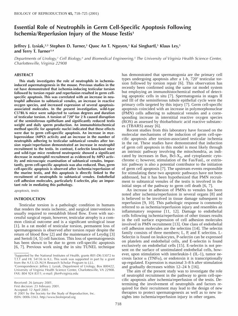
FIG. 1. Testis sections from wild-type C57BL/6 mice 15 days after A)sham operation, B) after 1-h 7208 torsion, C) 2-h 7208 torsion; stainedwith H&E. Arrows point to multinucleated cells and asterisk indicatesseminiferous tubules with severe disruption of the seminiferous epitheli-um. Magnification 360.
TABLE 1. Qualitative histological scores* (mean 6 SEM) of mouse testesafter various times (h) and degrees (8) of testicular torsion.
Group Score Group Score Group Score
Control3608/sham7208/sham
0.0 6 0.00.2 6 0.20.5 6 0.3
—3608/1h7208/1h
—0.7 6 0.50.7 6 0.2
—3608/2h7208/2h
—1.8 6 0.52.5 6 0.3
* Mid-testis cross sections scored independently by two investigators foroverall disruption of spermatogenesis. 0 5 Normal seminiferous histology,no disruption. 1 5 Slight effect, few tubule cross-sections show disruptionof seminiferous epithelium. 2 5 Moderate effect, many tubule cross-sec-tions show disruption of seminiferous epithelium; disruptions are largelylimited to loss of spermatids and spermatozoa but some tubules can ex-hibit complete loss of germinal elements; the majority of tubules remainapparently normal. 3 5 Severe effect, the majority of tubule cross-sectionsshow disruption of seminiferous epithelium; tubule cross-sections oftenexhibit complete loss of germinal elements.
detected at 460 nm using a microtiter photometric plate reader (Titer-Tek,Huntsville, AL).
Evaluation of Testicular Subtunical VenuleNeutrophil Margination
Subtunical venules were examined for the accumulation of neutrophilsfrom various groups 4 h after the repair of torsion as well as from shamcontrols. Briefly, H&E-stained sections of testes were examined using aNikon Microphot SA microscope with an attached CCD-camera and ISeesoftware (Inovision Corporation, Raleigh, NC). Images were captured andsubtunical venules were traced using ISee software. The intraluminal areaof each vessel was determined. Adherent neutrophils were then countedand the numbers expressed as neutrophils per mm2 of luminal area.
Statistical AnalysisAll statistical evaluations were either by ANOVA followed by the Tu-
key range test or the Student t-test (P , 0.05) after evaluation of eachdata set by Chauvenet’s criterion for homogeneity.
RESULTS
Evaluation of Testicular Blood Flow Duringand after Repair of Torsion
Testicular microvascular blood flow prior to testiculartorsion was 16.3 6 1.9 PU. One hundred ten minutes aftertesticular torsion, mean microvascular blood flow droppedto 0.2 6 0.2 PU. Five minutes after the repair of torsion,mean microvascular blood flow was 7.1 6 1.0 PU (n 5 4,for each group).
Evaluation of Experimental Testicular Torsion
Sections of testes from control (nonoperated) wild-typeC57BL/6 mice; or from the same strain mice 15 days aftersham operation; or after torsion of 3608 for 1 h, 7208 for 1h, 3608 for 2 h, or 7208 for 2 h were examined for disruptionof spermatogenesis. Figure 1 illustrates sections from testeshaving received a sham operation, a 1-h, 7208; or a 2-h,7208 torsion. Sham-operated mice demonstrated normalseminiferous tubule morphology. Mice that received a 7208testicular torsion had moderate-to-severe disruptions of theseminiferous epithelium, depending on the duration of thetorsion. Mice that underwent a 7208 testicular torsion for 2h displayed the most severe disruption of the seminiferousepithelium compared with the other torsion groups (Table1).
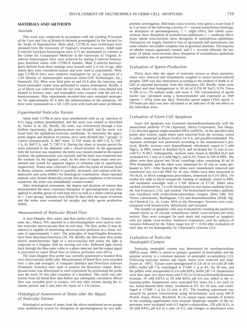
A 2-h, 7208 testicular torsion demonstrated a severe phe-notype by histology; thus, this degree and duration of tor-sion was applied to other groups of wild-type mice thatwere killed 30 days later. A significant decrease in testisweight, DSP, and TSO was observed in this group com-pared with control and sham-operated mice (Fig. 2). Thus,a 2-h, 7208 torsion causes a morphological disruption to theseminiferous epithelium, testicular atrophy, and a decreasein DSP. A 2-h, 7208 testicular torsion was employed in allsubsequent torsion experiments.
Evaluation of Germ Cell Apoptosis in Wild-TypeC57BL/6 Mice
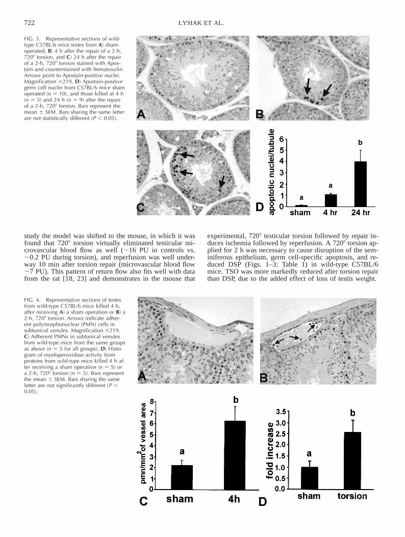
Tissue sections from testis from wild-type, sham-oper-ated animals revealed very few nuclei detected by theApostain technique (Fig. 3A). Sections from mice 4 h afterthe repair of torsion demonstrated a trend toward an in-crease in the number of stained germ cell nuclei (Fig. 3, Band D), which became significantly different 24 h after therepair of torsion (Fig. 3, C and D). Spermatogonial nucleiwere the predominant cell type undergoing apoptosis fol-lowing the repair of testicular torsion.
721INVOLVEMENT OF NEUTROPHILS IN GERM CELL APOPTOSIS
FIG. 2. Histograms of A) testis weight, B) daily sperm production (DSP),and C) testicular sperm output (TSO) from wild-type C57BL/6 mice 30days after subjection to control (no procedure; n 5 6), sham-operated (n5 7), and 2-h, 7208 torsion followed by repair (n 5 5). Bars represent themean 6 SEM. Bars sharing the same letter are not statistically different (P, 0.05).
Neutrophil Recruitment Following Testicular Torsionin Wild-Type C57BL/6 Mice
Sections from sham-operated, wild-type C57BL/6 micerevealed that very few neutrophils adhered to subtunicalvenules (Fig. 4, A and C), whereas sections from wild-typemice 4 h after the repair of torsion displayed a significantincrease in the presence of neutrophils in subtunical venules(Fig. 4, B and C). Results from MPO assays revealed asignificant increase in MPO activity from wild-type testes4 h after the repair of torsion (Fig. 4D). This increase inMPO activity indirectly indicates a recruitment of neutro-phils to the testis after torsion repair.
Evaluation of Germ Cell Apoptosis in E-Selectin-Deficientand Neutropenic Mice Following Testicular Torsion
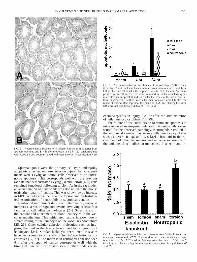
As in wild-type mice, tissue sections from sham-oper-ated, E-selectin knockout mice displayed very few apo-ptotic nuclei (Fig. 5A). In contrast to wild-type testes, E-selectin knockout mice testes did not show histological ev-idence of increased apoptosis at either 4 (Fig. 5B) or 24(not shown) h after repair of torsion. In fact, a significantdecrease in apoptotic germ cells was noted in E-selectinknockout mice at both 4 and 24 h after the repair of torsioncompared with wild-type mice (Fig. 6). E-Selectin hetero-zygous animals displayed values of germ apoptosis inter-mediate to E-selectin knockout mice and wild-type micewhen assessed 4 h after the repair of torsion (Fig. 6).
Wild-type mice were rendered neutropenic by the ad-ministration of antineutrophil antiserum. The concentrationof neutrophils in wild-type mice prior to antiserum admin-istration was 690 6 77.5 neutrophils/ml, and this value de-creased to 290 6 28.5 neutrophils/ml, 24 h after the ad-ministration of the antineutrophil antiserum. Neutropenicmice killed 4 h after the repair of torsion revealed a de-crease in germ cell apoptosis compared with normal wild-type mice (Fig. 6).
Evaluation of Neutrophil Recruitmentin E-Selectin-Deficient and Neutropenic MiceFollowing Testicular Torsion
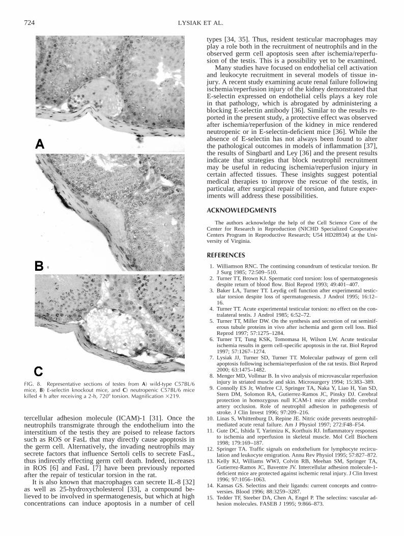
In contrast to the data obtained from wild-type mice(Fig. 4), MPO assays performed on testes from E-selectinknockout mice revealed no significant increase in MPO ac-tivity 4 h after the repair of torsion (Fig. 7). MPO assaysperformed on testes from mice rendered neutropenic re-vealed an increase in MPO activity 4 h after the repair oftorsion (Fig. 7); however, this response to ischemia/reper-fusion is reduced compared with that observed in wild-typemice (Fig. 4). Consistent with the MPO data above, norecruitment of neutrophils to subtunical venules was ob-served in the E-selectin knockout animals, whereas fewneutrophils could be observed in the subtunical venules ofthe neutropenic mice (Fig. 8).
DISCUSSION
In ischemia/reperfusion injury of the rat testis germ cell-specific apoptosis has been observed contemporaneouslywith an increase in leukocyte margination and diapedesis[6]. An increase in ROS as evident by an increase in tes-ticular lipid peroxidation and by the ability of certain re-active oxygen scavengers to partially abrogate the torsion-induced apoptosis has also been observed [6]; thus, it hasbeen hypothesized that the pathology seen after testiculartorsion is a classical ischemia/reperfusion injury, and that
neutrophils were the initiator cells for the molecular eventsleading to germ cell apoptosis [6]. However, the recruitmentand involvement of neutrophils in torsion-induced germcell apoptosis has not been directly linked. Results from thepresent study demonstrate an essential role of neutrophilsin the germ cell death observed after testicular ischemia/reperfusion.
This is the first report of germ cell-specific apoptosis ina mouse model of testicular torsion. Previous studies in arat model of testicular torsion found that a 7208 torsion issufficient to virtually eliminate testicular blood flow [2, 18],and that reperfusion begins immediately after repair of a 1-h or 2-h torsion with mean blood flow returning to controlvalues within 1 h [18, 23]. In the rat model, a 1-h, 7208torsion is sufficient to cause germ cell apoptosis and renderthe animal permanently azoospermic [6]. In the present
722 LYSIAK ET AL.
FIG. 3. Representative sections of wild-type C57BL/6 mice testes from A) sham-operated, B) 4 h after the repair of a 2-h,7208 torsion, and C) 24 h after the repairof a 2-h, 7208 torsion stained with Apos-tain and counterstained with hematoxylin.Arrows point to Apostain-positive nuclei.Magnification 3219. D) Apostain-positivegerm cell nuclei from C57BL/6 mice shamoperated (n 5 10), and those killed at 4 h(n 5 5) and 24 h (n 5 9) after the repairof a 2-h, 7208 torsion. Bars represent themean 6 SEM. Bars sharing the same letterare not statistically different (P , 0.05).
FIG. 4. Representative sections of testesfrom wild-type C57BL/6 mice killed 4 h,after receiving A) a sham operation or B) a2-h, 7208 torsion. Arrows indicate adher-ent polymorphonuclear (PMN) cells insubtunical venules. Magnification 3219.C) Adherent PMNs in subtunical venulesfrom wild-type mice from the same groupsas above (n 5 5 for all groups). D) Histo-gram of myeloperoxidase activity fromproteins from wild-type mice killed 4 h af-ter receiving a sham operation (n 5 5) ora 2-h, 7208 torsion (n 5 5). Bars representthe mean 6 SEM. Bars sharing the sameletter are not significantly different (P ,0.05).
study the model was shifted to the mouse, in which it wasfound that 7208 torsion virtually eliminated testicular mi-crovascular blood flow as well (;16 PU in controls vs.;0.2 PU during torsion), and reperfusion was well under-way 10 min after torsion repair (microvascular blood flow;7 PU). This pattern of return flow also fits well with datafrom the rat [18, 23] and demonstrates in the mouse that
experimental, 7208 testicular torsion followed by repair in-duces ischemia followed by reperfusion. A 7208 torsion ap-plied for 2 h was necessary to cause disruption of the sem-iniferous epithelium, germ cell-specific apoptosis, and re-duced DSP (Figs. 1–3; Table 1) in wild-type C57BL/6mice. TSO was more markedly reduced after torsion repairthan DSP, due to the added effect of loss of testis weight.
723INVOLVEMENT OF NEUTROPHILS IN GERM CELL APOPTOSIS
FIG. 5. Representative sections of E-selectin knockout mice testes fromA) sham-operated and B) 4 h after the repair of a 2-h, 7208 torsion stainedwith Apostain and counterstained with hematoxylin. Magnification 360.
FIG. 6. Apostain-positive germ cell nuclei from wild-type C57BL/6 mice(from Fig. 3) and E-selectin knockout mice from sham-operated, and thosekilled at 4 and 24 h after the repair of a 2-h, 7208 torsion. Apostain-positive germ cell nuclei were also examined in E-selectin heterozygousmice after sham-operated and 24 h after the repair of torsion as well asfrom neutropenic C57BL/6 mice after sham-operated and 4 h after therepair of torsion. Bars represent the mean 6 SEM. Bars sharing the sameletter are not significantly different (P , 0.05).
FIG. 7. Myeloperoxidase activity from proteins from E-selectin knockoutmice and neutropenic C57BL/6 mice killed 4 h after receiving a shamoperation or a 2-h, 7208 torsion. Bars represent the mean 6 SEM; n 5 5for all groups. Bars sharing the same letter are not statistically different (P# 0.05).
Spermatogonia were the primary cell type undergoingapoptosis after ischemia/reperfusion injury. In no experi-ments were Leydig or Sertoli cells observed to be under-going apoptosis. This corresponds well with the previousrat data that demonstrated Leydig [3] and Sertoli [4, 5] cellsremained functional following torsion. As in the rat model,an accumulation of neutrophils was also noted in the mousetestis after repair of torsion. This was shown by an increasein MPO activity after the repair of torsion and by histolog-ical examination of neutrophils in subtunical venules.
Neutrophil recruitment during an inflammatory responseinvolves a series of regulated events involving at least fourfamilies of cell adhesion molecules [24]. Selectins aid inthe capture and attachment of blood leukocytes to the vas-cular endothelium. This initial step results in slow, down-stream rolling of the leukocyte on the vascular endothelium[25, 26]. Other cellular adhesion molecules, such as inte-grins, then aid in the firm adhesion and transmigration ofleukocytes [24]. Similar leukocyte recruitment cascadeshave been shown to occur after ischemia/reperfusion in oth-er tissues [12, 27]. The increase in neutrophil adhesion seen4 h after the repair of torsion corresponds well with thetiming of E-selectin expression seen in other models of is-
chemia/reperfusion injury [28] or after the administrationof inflammatory cytokines [16, 29].
The failure of testicular torsion to stimulate apoptosis inmice rendered neutropenic indicates that neutrophils are es-sential for the observed pathology. Neutrophils recruited tothe subtunical venules may secrete inflammatory cytokinessuch as TNFa, IL-1b, and IL-8 [30]. These aid in the re-cruitment of other leukocytes and enhance expression ofthe endothelial cell adhesion molecules, E-selectin and in-
724 LYSIAK ET AL.
FIG. 8. Representative sections of testes from A) wild-type C57BL/6mice, B) E-selectin knockout mice, and C) neutropenic C57BL/6 micekilled 4 h after receiving a 2-h, 7208 torsion. Magnification 3219.
tercellular adhesion molecule (ICAM)-1 [31]. Once theneutrophils transmigrate through the endothelium into theinterstitium of the testis they are poised to release factorssuch as ROS or FasL that may directly cause apoptosis inthe germ cell. Alternatively, the invading neutrophils maysecrete factors that influence Sertoli cells to secrete FasL,thus indirectly effecting germ cell death. Indeed, increasesin ROS [6] and FasL [7] have been previously reportedafter the repair of testicular torsion in the rat.
It is also known that macrophages can secrete IL-8 [32]as well as 25-hydroxycholesterol [33], a compound be-lieved to be involved in spermatogenesis, but which at highconcentrations can induce apoptosis in a number of cell
types [34, 35]. Thus, resident testicular macrophages mayplay a role both in the recruitment of neutrophils and in theobserved germ cell apoptosis seen after ischemia/reperfu-sion of the testis. This is a possibility yet to be examined.
Many studies have focused on endothelial cell activationand leukocyte recruitment in several models of tissue in-jury. A recent study examining acute renal failure followingischemia/reperfusion injury of the kidney demonstrated thatE-selectin expressed on endothelial cells plays a key rolein that pathology, which is abrogated by administering ablocking E-selectin antibody [36]. Similar to the results re-ported in the present study, a protective effect was observedafter ischemia/reperfusion of the kidney in mice renderedneutropenic or in E-selectin-deficient mice [36]. While theabsence of E-selectin has not always been found to alterthe pathological outcomes in models of inflammation [37],the results of Singbartl and Ley [36] and the present resultsindicate that strategies that block neutrophil recruitmentmay be useful in reducing ischemia/reperfusion injury incertain affected tissues. These insights suggest potentialmedical therapies to improve the rescue of the testis, inparticular, after surgical repair of torsion, and future exper-iments will address these possibilities.
ACKNOWLEDGMENTS
The authors acknowledge the help of the Cell Science Core of theCenter for Research in Reproduction (NICHD Specialized CooperativeCenters Program in Reproductive Research; U54 HD28934) at the Uni-versity of Virginia.
REFERENCES
1. Williamson RNC. The continuing conundrum of testicular torsion. BrJ Surg 1985; 72:509–510.
2. Turner TT, Brown KJ. Spermatic cord torsion: loss of spermatogenesisdespite return of blood flow. Biol Reprod 1993; 49:401–407.
3. Baker LA, Turner TT. Leydig cell function after experimental testic-ular torsion despite loss of spermatogenesis. J Androl 1995; 16:12–16.
4. Turner TT. Acute experimental testicular torsion: no effect on the con-tralateral testis. J Androl 1985; 6:52–72.
5. Turner TT, Miller DW. On the synthesis and secretion of rat seminif-erous tubule proteins in vivo after ischemia and germ cell loss. BiolReprod 1997; 57:1275–1284.
6. Turner TT, Tung KSK, Tomomasa H, Wilson LW. Acute testicularischemia results in germ cell-specific apoptosis in the rat. Biol Reprod1997; 57:1267–1274.
7. Lysiak JJ, Turner SD, Turner TT. Molecular pathway of germ cellapoptosis following ischemia/reperfusion of the rat testis. Biol Reprod2000; 63:1475–1482.
8. Menger MD, Vollmar B. In vivo analysis of microvascular reperfusioninjury in striated muscle and skin. Microsurgery 1994; 15:383–389.
9. Connolly ES Jr, Winfree CJ, Springer TA, Naka Y, Liao H, Yan SD,Stern DM, Solomon RA, Gutierrez-Ramos JC, Pinsky DJ. Cerebralprotection in homozygous null ICAM-1 mice after middle cerebralartery occlusion. Role of neutrophil adhesion in pathogenesis ofstroke. J Clin Invest 1996; 97:209–216.
10. Linas S, Whittenburg D, Repine JE. Nitric oxide prevents neutrophil-mediated acute renal failure. Am J Physiol 1997; 272:F48–F54.
11. Gute DC, Ishida T, Yarimizu K, Korthuis RJ. Inflammatory responsesto ischemia and reperfusion in skeletal muscle. Mol Cell Biochem1998; 179:169–187.
12. Springer TA. Traffic signals on endothelium for lymphocyte recircu-lation and leukocyte emigration. Annu Rev Physiol 1995; 57:827–872.
13. Kelly KJ, Williams WWJ, Colvin RB, Meehan SM, Springer TA,Gutierrez-Ramos JC, Baventre JV. Intercellular adhesion molecule-1-deficient mice are protected against ischemic renal injury. J Clin Invest1996; 97:1056–1063.
14. Kansas GS. Selectins and their ligands: current concepts and contro-versies. Blood 1996; 88:3259–3287.
15. Tedder TF, Steeber DA, Chen A, Engel P. The selectins: vascular ad-hesion molecules. FASEB J 1995; 9:866–873.
725INVOLVEMENT OF NEUTROPHILS IN GERM CELL APOPTOSIS
16. Weller A, Isenmann S, Vestweber D. Cloning of the mouse endothelialselectins. Expression of both E- and P-selectin is inducible by tumornecrosis factor alpha. J Biol Chem 1992; 267:15176–15183.
17. Bullard DC, Kunkel EJ, Kubo H, Hicks MJ, Lorenzo I, Doyle NA,Doerschuk CM, Ley K, Beaudet AL. Infectious susceptibility and se-vere deficiency of leukocyte rolling and recruitment in E-selectin andP-selectin double mutant mice. J Exp Med 1996; 183:2329–2336.
18. Becker EJ, Prillaman HM, Turner TT. Microvascular blood flow isaltered after repair of testicular torsion in the rat. J Urol 1997; 157:1493–1498.
19. Bonner RF, Clem TR, Bowen PD, Bowman RL. Laser-Doppler con-tinuous real time monitor of pulsatile and mean blood flow tissuemicrocirculation. In: Chen S, Chu B, Nossal R (eds.), Scattering Tech-niques Applied to Supramolecular and Nonequilibrium Systems. NewYork: Plenum Press; 1981.
20. Robb GW, Amann RP, Killian GJ. Daily sperm production and epi-didymal sperm reserves of pubertal and adult rats. J Reprod Fertil1978; 54:103–107.
21. Worthington AG, Jeffner J. Treatment of Experimental Data. NewYork: Wiley and Sons; 1943: 76–80.
22. Grisham MB, Benoit JN, Granger DN. Assessment of leukocyte in-volvement during ischemia and reperfusion of intestine. Methods En-zymol 1990; 186:729–742.
23. Lysiak JJ, Nguyen QAT, Turner TT. Testicular vasomotion correlateswith interstitial oxygen tensions: persistence after repair of torsion.Biol Reprod 2000; 63:1383–1389.
24. Ley K. Gene-targeted mice in leukocyte adhesion research. Microcir-culation 1995; 2:141–150.
25. Kunkel EJ, Ley K. Distinct phenotype of E-selectin-deficient mice: E-selectin is required for slow leukocyte rolling in vivo. Circ Res 1996;79:1196–1204.
26. Jung U, Ramos C, Bullard DC, Ley K. Gene-targeted mice revealimportance of L-selectin-dependent rolling for neutrophil adhesion.Am J Physiol 1998; 274:H1785–H1791.
27. Weyrich AS, Xin-liang M, Lefer DJ, Albertine KH, Lefer AM. Invivo neutralization of P-selectin protects feline heart and endotheliumin myocardial ischemia and reperfusion injury. J Clin Invest 1993; 91:2620–2629.
28. Takada M, Nadeau KC, Shaw GD, Marquette KA, Tilney NL. The
cytokine-adhesion molecule cascade in ischemia/reperfusion injury ofthe rat kidney: inhibition by a soluble P-selectin ligand. J Clin Invest1997; 99:2682–2690.
29. Keelan ETM, Licence ST, Peters AM, Binns RM, Haskard DO. Char-acterization of E-selectin expression in vivo with use of a radiolabeledmonoclonal antibody. Am J Physiol 1994; 266(Heart Circ Physiol 35):H278–H290.
30. Derevianko A, D’Amico R, Simms H. Polymorphonuclear leucocyte(PMN)-derived inflammatory cytokines—regulation by oxygen ten-sion and extracellular matrix. Clin Exp Immunol 1996; 106:560–567.
31. Fassbender K, Kaptur S, Becker P, Groschl J, Hennerici M. Adhesionmolecules in tissue injury: kinetics of expression and shedding andassociation with cytokine release in humans. Clin Immunol Immu-nopathol 1998; 89:54–60.
32. Carre PC, Mortenson RL, King TE Jr, Noble PW, Sable CL, RichesDWH. Increased expression of the Interleukin-8 gene by alveolar mac-rophages in idiopathic pulmonary fibrosis. J Clin Invest 1991; 88:1802–1810.
33. Nes WD, Lukyanenko YO, Jia ZH, Quideau S, Howald WN, ThomasK, West RR, Hutson JC. Identification of the lipophilic factor pro-duced by macrophages that stimulates steroidogenesis. Endocrinology2000; 141:953–958.
34. Yin J, Chaufour X, McLachlan C, McGuire M, White G, King N,Hambly B. Apoptosis of vascular smooth muscle cells induced bycholesterol and its oxides in vitro and in vivo. Atherosclerosis 2000;148:365–374.
35. Astruc M, Roussillon S, Defay R, Descomps B, Crastes De Paulet A.DNA and cholesterol biosynthesis in synchronized embryonic rat fi-broblasts II. Effects of sterol biosynthesis inhibitors on cell division.Biochim Biophys Acta 1983; 763:11–18.
36. Singbartl K, Ley K. Protection from ischemia-reperfusion induced se-vere acute renal failure by blocking E-selectin. Crit Care Med 2000;28:2507–2514.
37. Labow MA, Norton CR, Rumberger JM, Lombard-Gillooly KM,Shuster DJ, Hubbard J, Bertko R, Knaack PA, Terry RW, HarbisonML, Kontgen F, Stewart CL, McIntyre KW, Will PC, Burns DK, Wol-itzky BA. Characterization of E-selectin-deficient mice: demonstrationof overlapping function of the endothelial selectins. Immunity 1994;1:709–720.