essentials in protein electrophoresis
TRANSCRIPT
Protein Electrophoresis
ByDr Abdullah A.Hashish
Lecturer of Clinical Pathology Dept.SCU-FOM
Objectives
•Definitions.
•Basic theory.
•Components.
• Types.
•Applications.
Definitions
1. Electrophoresis:
migration of charged particles of any size in a liquid medium under the influence of an electrical field.
2. Iontophoresis or isotachophoresis:
specifically to the migration of small ions.
3. Zone electrophoresis:
each protein zone is sharply separated from neighboring zones by a protein-free area.
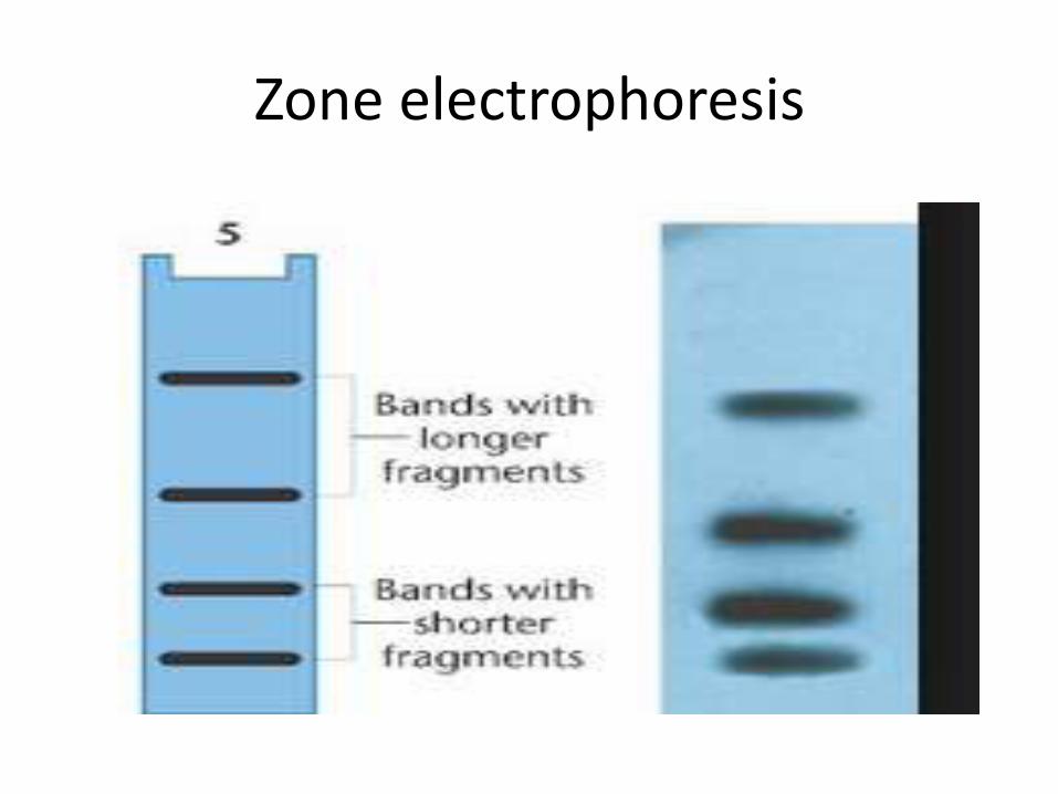
Zone electrophoresis

Basic Theory
• Depending on the charge they carry, ionized solutes movetoward either the cathode(negative electrode) or the anode (positive electrode) in an electrophoresis system.
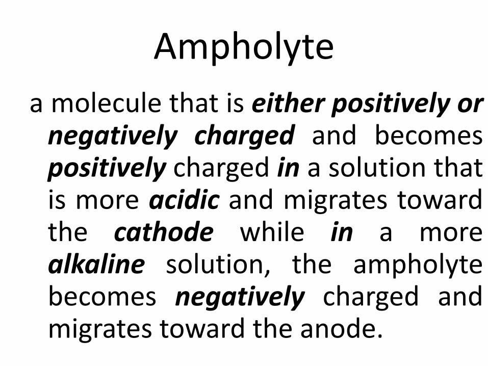
Ampholyte
a molecule that is either positively or negatively charged and becomes positively charged in a solution that is more acidic and migrates toward the cathode while in a more alkaline solution, the ampholytebecomes negatively charged and migrates toward the anode.
How to remember:
•Anions Anode.
•Cations Cathode.

Factors Affecting the Motilityof Ions in an Electrophoretic System
• Net charge of the molecules.
• Size and shape of the molecules.
• Strength of the electrical field.
• Support medium properties.
• Ionic strength of the buffer.
• Temperature.
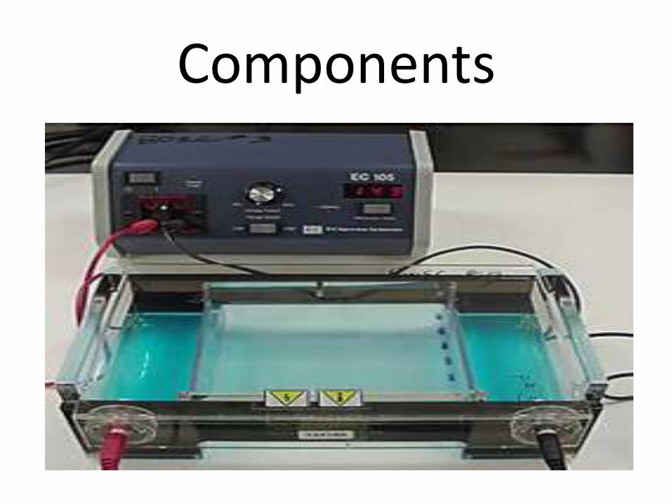
Components

Components
1. Power supply:
1. (Voltage, ampere, time).
2. Constant vs. pulsee-power.
2. Buffer:
1. pH. (application & direction of migration).
2. Ionic strength:
1. rate of migration.
2. sharpness of the zone.
3. High ionic strength high resolution separation.
3. Commonly used: barbital EDTA or Tri-boric EDTA.
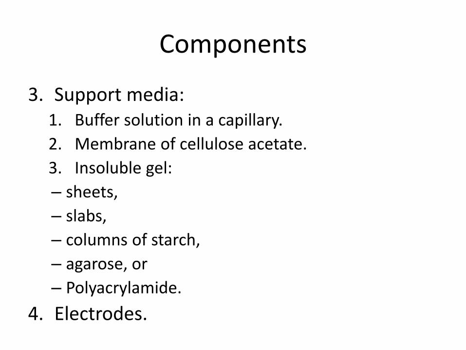
Components
3. Support media:1. Buffer solution in a capillary.
2. Membrane of cellulose acetate.
3. Insoluble gel:
– sheets,
– slabs,
– columns of starch,
– agarose, or
– Polyacrylamide.
4. Electrodes.
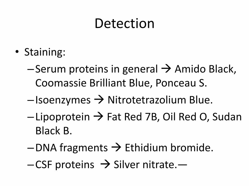
Detection
• Staining:
–Serum proteins in general Amido Black, Coomassie Brilliant Blue, Ponceau S.
– Isoenzymes Nitrotetrazolium Blue.
–Lipoprotein Fat Red 7B, Oil Red O, Sudan Black B.
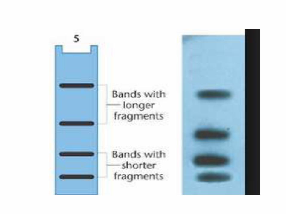
–DNA fragments Ethidium bromide.
–CSF proteins Silver nitrate.—
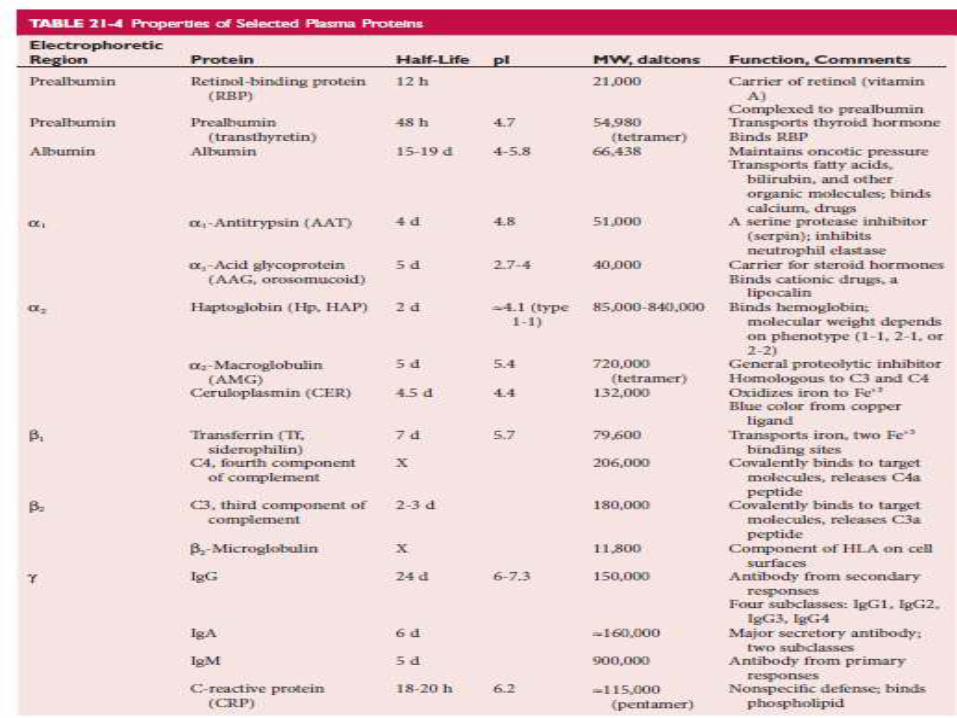
What is important to know in the previous table?
1.Electrophoretic regions.
2.Included proteins.
3.Common function of the protein.

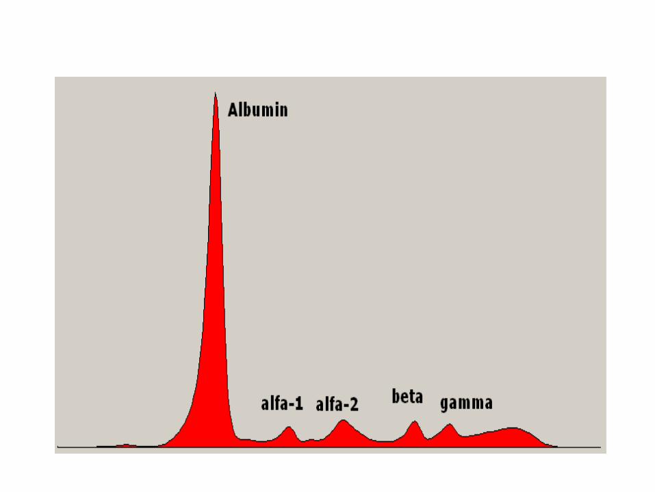
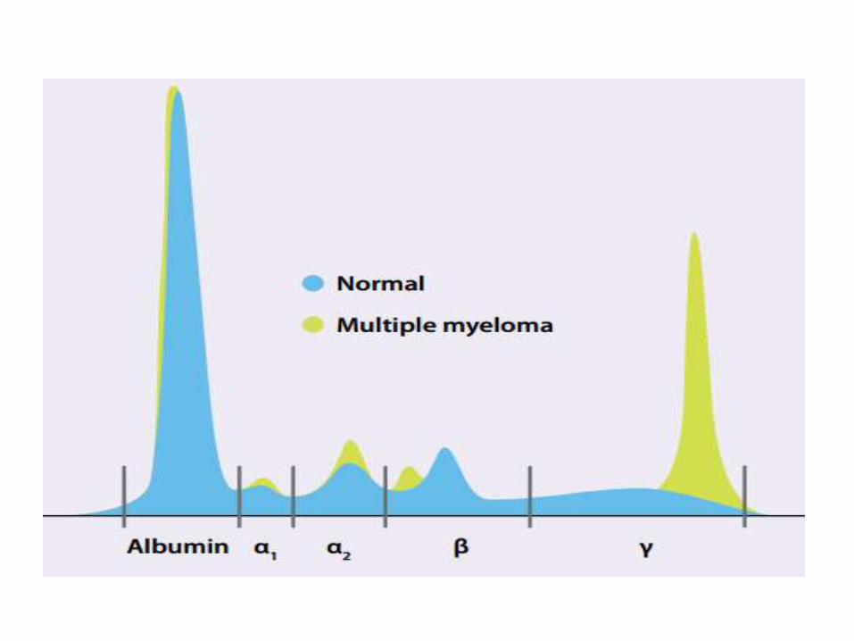
Applications
• used to separate proteins in:
–Serum,
–Urine,
–Cerebrospinal fluid (CSF),
–Other physiologic fluids,
–Erythrocytes and,
–Nucleic acids

THANK YOU