establishment of stable and secretable tatκ-gfp ... zariyantey abd hamid.pdf · terkini,...
TRANSCRIPT
Sains Malaysiana 47(10)(2018): 2473–2480 http://dx.doi.org/10.17576/jsm-2018-4710-24
Establishment of Stable and Secretable Tatκ-GFP Recombinant Protein: A Preliminary Report of Promoter Methylation in 293t Cell Line(Pembangunan Protein Rekombinan Stabil dan Terembes TATκ-GFP: Laporan Awalan
Proses Pemetilan Promoter pada Sel Selanjar 293T)
ZARIYANTEY ABD HAMID, FAZLINA NORDIN*, RAJA NORAZIREEN RAJA AHMAD, BALQIS MAT RASHID & UBASHINI VIJAKUMARAN
ABSTRACT
Induced pluripotent stem cells (iPSC) is a novel technology useful for therapeutic and research applications. To date, iPSCs is produced through genetic modification that can promote mutation; making it harmful for therapeutic use. Therefore, application of non-genetic modification through direct delivery of recombinant proteins aided by protein transduction domain (PTD) enable a safer production of iPSC. This study is aimed to establish a stable production of secretable recombinant protein via recombination of green fluorescence protein (GFP) and a novel PTD peptide, namely TATκ-GFP. 293Tcell line was transfected with 20 µg/ml of TATκ-GFP plasmid and the stably transfected 293T cells were then cultured for 54 days to determine the stability of expression and secretion of TATκ-GFP recombinant protein in prolonged culture. Methylation at the CMV promoter of the TATκ-GFP plasmid was investigated following treatment of transfected cells with 3 µM/mL of demethylation agent, namely 5-Azacytidine for 72 h in three cycles. Flow cytometry analysis demonstrated a transfection efficiency of 9.33% and successful secretion of TATκ-GFP proteins into the culture medium as analysed by Western blot at 72 h post-transfection. However, the transfected cells exhibited a decreasing level of GFP expression and secretion following prolonged culture with notable stability that only sustained for two weeks. 5-Azacytidine-treated cells showed a slight increase of GFP expression compared to non-treated control, suggesting possible promoter methylation which could cause instability of TATκ-GFP expression. Conclusively, promoter methylation should be considered for future establishment of iPSCs as it could inhibit stable expression and secretion of recombinant proteins.
Keywords: Induced pluripotent stem cell (iPSC); methylation; protein transduction domain (PTD); trans-activator of transcription (TAT); transcription factors
ABSTRAK
Sel asal aruhan pluripoten (iPSC) adalah teknologi terkini yang bermanfaat bagi aplikasi perubatan dan penyelidikan. Terkini, penghasilan iPSC adalah melalui kaedah pengubahsuaian genetik yang berupaya merangsang mutasi, menjadikan ia berbahaya bagi kegunaan perubatan. Justeru, penggunaan kaedah tanpa pengubahsuaian genetik melalui penghantaran terus protein rekombinan dengan bantuan domain pentransduksi protein (PTD) membolehkan penghasilan iPSC yang lebih selamat. Kajian ini bertujuan untuk membangunkan penghasilan protein rekombinan yang stabil dan terembes melalui rekombinasi protein fluoresen hijau (GFP) dan peptida baru PTD iaitu TATκ. Sel selanjar 293T ditransfeksi dengan 20 µg/mL plasmid TATκ-GFP dan populasi sel dengan transfeksi stabil seterusnya dikultur selama 54 hari untuk mengenal pasti kestabilan pengekspresan dan perembesan protein TATκ-GFP pada tempoh pengkulturan yang berpanjangan. Kehadiran proses pemetilan pada promoter CMV plasmid TATκ-GFP dikaji melalui rawatan ke atas sel ditransfeksi plasmid dengan 3 µM/mL agen pengenyahmetilan 5-Azasitidin selama 72 jam bagi tiga kitaran. Analisis sitometri aliran menunjukkan kecekapan transfeksi sebanyak 9.33% dan kejayaan protein TATκ-GFP dirembeskan ke dalam media pengkulturan berdasarkan analisis Western Blot selepas 72 jam transfeksi. Namun, sel ditransfeksi mempamerkan penurunan pengekspresan dan perembesan protein GFP pada tempoh pengkulturan yang berpanjangan dengan kestabilannya didapati hanya bertahan selama dua minggu. Sel terawat 5-Azasitidin menunjukkan terdapatnya sedikit peningkatan pengekspresan GFP berbanding sel kawalan tanpa rawatan yang mencadangkan kemungkinan kehadiran pemetilan pada promoter yang boleh menyebabkan ketidakstabilan pengekspresan TATκ-GFP. Kesimpulannya, pemetilan promoter perlu diberi perhatian pada masa hadapan untuk penghasilan iPSC memandangkan ia berupaya merencat kestabilan pengekspresan dan perembesan protein rekombinan.
Kata kunci: Faktor transkripsi; domain transduksi protein (PTD); pemetilan; sel asal aruhan pluripoten (iPSC); transkripsi pengaktif-trans(TAT)
2482
INTRODUCTION
Pluripotent stem cells are master cells that can give rise to any cell type that makes up human body. One of the main sources for pluripotent stem cells is embryonic stem cells. First isolation of human pluripotent stem cell by Thomson et al. (1998) had opened a new avenue for the potential use of the pluripotent stem cell in medical applications especially for regeneration of cells and tissue. However, destruction of human embryos for the isolation of embryonic stem cells raises ethical issues (Deng 2010; Martinez et al. 2011). In 2006, Takahashi and Yamanaka successfully reprogrammed somatic mouse fibroblast cells into pluripotent stem cells. They have introduced four types of transcription factors which are known as Yamanaka factors or the magical factors (Oct 3/4, Sox2, c-Myc and Klf4) into mouse fibroblast cells via retroviral transduction. This method had triggered the mouse fibroblast cells to undergo cellular reprogramming into pluripotent stem cells, or widely regardless induced pluripotent stem cells (iPSC). Subsequently, establishment of iPSC from human fibroblast was also successfully achieved (Takahashi et al. 2007). This technology has become a breakthrough as it overcomes the need to use the human embryos which is unethical. Furthermore, immune rejection is preventable since iPSC created using patient’s own adult stem cells. In addition to the therapeutic potential, iPSC is also beneficial as a research tool to study various aspects of diseases, drug development and toxicology testing (Deng 2010; Han et al. 2010; Leeper et al. 2010; Martinez et al. 2011; Stadfeld & Hochedlinger 2010; Yoshida & Yamanaka 2010). However, iPSC technology faces few constraints due to genetic manipulation. It is believed that insertion of virus vector that used to deliver the four transcription factors could permanently modify the host genome and subsequently lead to genetic mutation, which can increase the risk of tumorigenicity (Cho et al. 2010; Fazlina Nordin 2014; Han et al. 2010; Martinez et al. 2011; Okano et al. 2013; Yamanaka 2009; Yoshida & Yamanaka 2010). Therefore, this strategy remains unacceptable for clinical trials due to safety reasons. One of the alternative ways to overcome this concern is to deliver fusion proteins of these transcription factors using protein transduction domain (PTD) (Kim et al. 2009; Zhang et al. 2012; Zhou & Zhang 2013). Protein transduction is defined as the introduction of proteins exogenously into target cells owing to their ability to penetrate the cell membrane and further uptake by the target cells (Ford et al. 2001). Translocation property of a protein is determined by the presence of short amino acid sequences within the proteins, namely protein transduction domains (PTDs); which is fundamental in mediating cellular entry (Beerens et al. 2003). The most widely studied PTDs are the HIV TAT protein, homeodomain of Drosophila antennapeptide (AntpHD) and the herpes simplex virus VP22 protein (Beerens et al. 2003; Ford et al. 2001; Noguchi & Matsumoto 2006b). PTDs possess a number of features that making them a promising candidate in mediating delivery of therapeutic proteins in a safe manner. These features include
their ability to deliver macromolecules including siRNA (Katas et al. 2014) efficiently into target cells independently of cell types, provide stable transduction, allow precise time and dose management as well as confer minimal risk of genetic modification of target cells. Among PTDs, the HIV TAT has been the most widely studied and a number of modifications of its amino acid sequence have been done to enhance its transduction efficacy and stability. This has led to a number of reports of which the improved synthetic TAT demonstrated greater transduction efficiency and stability in mediating delivery of several peptides and proteins into target cells as compared to its original TAT sequence (Noguchi & Matsumoto 2004). In the present study, a novel synthetic TAT sequence, namely TATκ was studied. TATκ was established by introducing mutations to displace the two furin cleavage sites (RQRR and RKKR) within the native TAT peptide to enhance the bioavailability of the cargo (Flinterman et al. 2009). In the context of IPSC, recombinant protein that resulted from conjugation of PTD and reprogramming factors enhances transduction and translocation of protein into target cell. This new technique yields non-genetically modified, IPSC. Furthermore, Kim et al. (2009), has produced human IPSC by using arginine as PTD to deliver Yamanaka factors into human newborn fibroblast (HNF) cells. Then, Zhang et al. (2012) further exploited TAT-PTD and 11 Arginine (R)-PTD to reprogramme human fibroblast into iPSC. In this present research, the 293T cell line was selected as mammalian expression system for the establishment and secretion of combined protein TATκ-PTD and Green Fluorescent Protein (GFP). The novelty of current study is relying on the technology of recombinant protein production utilizing eukaryotic cells as producer cells for expression and secretion of recombinant protein. This approach can be considered novel as previous effort for recombinant protein technology in iPSCs only limited to protein produced by prokaryotic system which require purification prior to application. To date, there is no study concerning the efficiency of TATκ recombinant proteins to reprogramme differentiated cells into iPSC has been reported. Thus, the use of newly synthesized PTD peptide namely TATκ which claimed to have better PTD property (Flinterman et al. 2009; Nordin et al. 2014) is another novelty factor. Although recombinant protein technology is safer than genetic modification strategy, it has a number of limitations including stability and yield. Therefore, the stability of production and secretion of recombinant protein TATκ-GFP in 293T cell will be studied in terms of cultivation period and methylation activity of transcription promoter.
MATERIALS AND METHODS
CELL CULTURE
293T cell line were cultured and maintained in DMEM (Invitrogen) with 25 mM glucose, 10% fetal bovine serum (FBS, Invitrogen) and 1 mM sodium pyruvate under 5%
2483
CO2 at 37°C. A different density of cells was used as optimized depending on the size of the culture plate in each experiment.
GENERATION OF TATΚ-GFP STABLE CELL LINE
293T cells (1.0 × 10⁶) were seeded in 10 mL DMEM complete medium in 100 mm diameter culture dish. A standard protocol for calcium phosphate (CaCl2) was performed with modifications (Fazlina et al. 2016). On day 3 of culture, a total of 1 mL of transfection mixture containing 20 µg of plasmid DNA, 0.5 M CaCl2 and 2× HEBSS (HEPES Buffered Saline Solution) at pH6.7 were incubated at room temperature for 30 min and added dropwise to the plated 293T cells. The culture medium was replaced with fresh complete medium on the following day and the cells were further incubated at 37°C for 72 h. Following 72 h post-transfection, the transfected cells were further treated with Puromycin (2.5 µg/mL). Complete growth media with or without puromycin was changed every 3-4 days and clones were picked after 3 to 4 weeks. Non-transfected 293T cell line will be used as negative control for each experiment.
LONG TERM CULTURE
Stable cell lines obtained were cultured in 6 well culture dish with 5.0 × 10⁵ cells per well. Cells were maintained and complete medium with or without puromycin (DMEM with 10% FBS) were changed every 3-4 days. Cells were split after they are 80% confluent and culture were continued for 54 days before the experiment was terminated. The second round of transfection will be performed at Day 36 to enhance the GFP expression. Whole cell lysate and culture supernatant were collected at day 32, 43 and 50 and subjected to immunoblotting.
IMMUNOBLOTTING
For immunoblotting, cells were washed and lysed in RIPA buffer (Sigma Aldrich) and 1× protease inhibitor. Lysates were further clarified with centrifugation at 10,000 rpm for 10 min. Gel electrophoresis was done using Bolt Bis-Tris plus 4-12% (Invitrogen) before transfered on PVDF membrane (GE). Immunoblotting were performed with 5% skim milk powder in 0.1% Tween PBS. For sample preparations from cell supernatants, cell culture medium was collected and secreted proteins were concentrated using Viva Spin (GE) for 15 min at 3,000 rpm. A standard protocol for antibody staining was performed with slight modifications (Fazlina et al. 2016). The following primary antibodies were used: 1/1000 dilution of anti-rabbit GFP polyclonal antibody (Cell Signalling Technology, US) and 1/1000 dilution of anti-mouse α-Tubulin monoclonal antibody (Sigma, Aldrich). The following secondary antibodies were used; 0.2 µg/mL goat anti-mouse IgG (Sigma) horseradish peroxidase and 0.2 µg/mL goat anti-rabbit polyclonal horseradish peroxidase (Santa Cruz Biotech.).
DNA DEMETHYLATION
After 54 days of culture, stable cell lines were cultured in 6-well plate (1.0 × 10⁶ cells per well) and 3 cycles of 3 µmM of 5-Azacytidine (Sigma Aldrich) treatments were conducted. Each cycle of treatment consists of treatment with 3 µmM 5-Azacytidine for 72 h followed by 11 days of treatment-free culture for 6 weeks.
FLOW CYTOMETRY
5-Azacytidine treated and non-treated cells (1.0 × 10⁶ cells) were collected and washed in 1× PBS (Gibco) and the GFP expression was analysed using FACS Calibur.
STATISTICAL ANALYSIS
Sample means between control and treatment conditions were compared by paired t-test using Prism Graph software version 5. The data were confirmed to be normally distributed and had similar variance as estimated by SEM (n=3). Data were concluded to be significant with P value of p<0.05.
RESULTS
293T-TATΚ-GFP STABLE MIXED POPULATION SHOWS A DECREASED EXPRESSION OF TATΚ-GFP RECOMBINANT
PROTEIN IN PROLONGED CULTURE
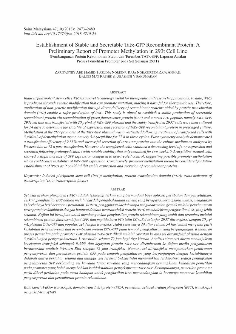
Figure 1 shows the stability expression study of 293T-TATκ-GFP. The transfected 293T were treated with puromycin at 72 h post-transfection to select for stable mixed population which are puromycin resistant. The transfected 293T were continuously cultured up to day 54 (D54) before the experiment was terminated. A second transfection was performed at day 36 (D36) on the stable mixed population to enhance the recombinant protein expression. FACS analysis showed that the expression level of GFP in transfected 293T cells decreases in prolonged culture (Figure 2). Data shown is an average percentage ± S.E.M of triplicate independent samples. The GFP expression was 9.33±0.295% at 72 h post-transfection (Day 3). A second round of transfection on Day 36 (D36) were able to restore the expression and TATκ-GFP protein was detected at day 43 (D43) with 39.16±0.384 percent, which was higher as compared to the first transfection. However, it was not statistically significant (p<0.05). The same population of the selected cells were further cultured and analysed on day 50 (D50) with decreased GFP expression, 2.19±0.140%. Fluorescence microscopy analysis shows reduce number of the GFP positive cells with agreement to FACS data (Figure 3). Visually, the numbers of GFP positive cells reduce periodically after two weeks of culture to only a few GFP positive cells up to Day 30, after first transfection (Figure 3(A)). A similar pattern of GFP expression as in the first transfection was observed up to Day 50, after second round of transfection (Figure 3(B)). Overall, the data shows that the selections of mixed population for establishment of

2484
The expression and secretion of TATκ-GFP recombinant protein was observed up to Day 54 before the experiment was terminated
FIGURE 1. Stability expression study of 293T-TATκ-GFP mixed population
FACS analysis showed that the expression level of GFP in transfected 293T cells decreases in prolonged culture. Notably the level of GFP expression was increased at day 43 after second round of transfection and decreased by day 50. Data were not statistically significant (p>0.05). A total of 1 × 10⁶ cells were cultured in a complete growth medium and further transfected with plasmid encoding TATκ-GFP sequence. The TATκ-GFP expression was determined at 72 h post-transfection following selection of stable mixed population with 2.5 μg/mL puromycin at day 43 and day 50. Data shown are the mean±SEM of triplicate from independent experiments
FIGURE 2. The level of TATκ-GFP expression in the transfected 293T-TATκ-GFP by FACS analysis
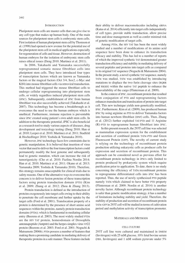
The number of GFP positive cells was reduced up to day 32 after first transfection (3A). A similar pattern of GFP positive cells was observed after second transfection (3B). These data are in line with FACS analysis as shown in Figure 2. Approximately 72 h (Day 3) post-transfection, cells were further treated with 2.5 μg/mL puromycin for selection of stable mixed population. A second transfection was performed at Day 36 following puromycin treatment. The expression of TATκ-GFP recombinant protein was observed up to day 54 before the experiment was terminated. A green fluorescence indicates the presence of GFP or TATκ-GFP recombinant protein in the transfected 293T cells. Magnifications at 10×
FIGURE 3. Fluorescence microscopy analysis of 293T-TATκ-GFP cells in prolonged culture
2485
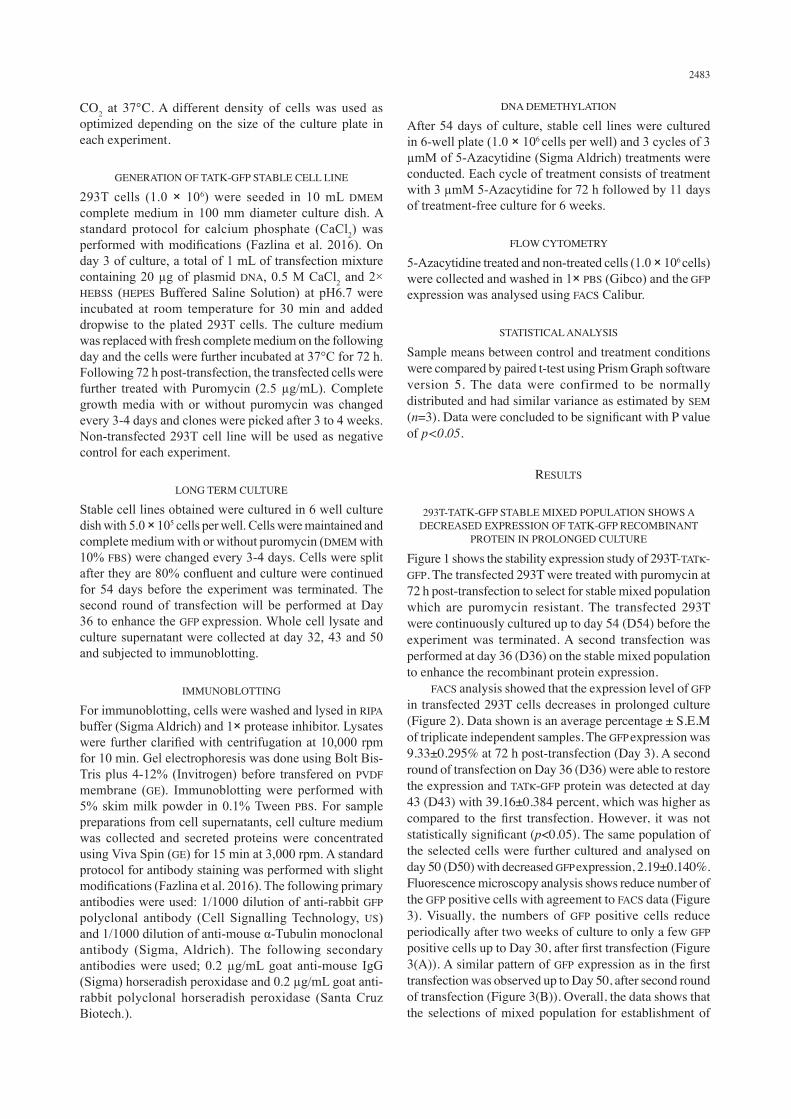
producer cell secreting TATκ-GFP are potentially achieved. 293T-TATκ-GFP Stable Mixed Population Has Lost the Ability to Secretes TATκ-GFP Recombinant Protein in Prolonged Culture Figure 4 shows the expression and secretion of TATκ-GFP recombinant protein at 72 h (Day 3) post-transfection by Western Blot analysis. Both cell lysate and culture supernatant (sup) of transfected 293T cells were further analysed at Day 3 post-transfection. TATκ-GFP recombinant protein was present in both samples as indicated by prominent bands at approximately 35 kD of protein weight as compared to non-transfected 293T (Control) (Figure 4). This finding is consistent with data by fluorescent microscopy analysis, whereby bundle of GFP positive cells were present at Day 3 post-transfection (Figure 3(A)).
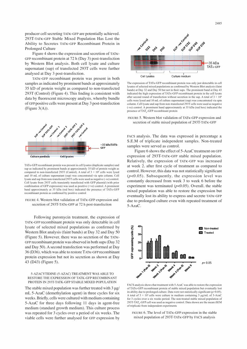
FACS analysis. The data was expressed in percentage ± S.E.M of triplicate independent samples. Non-treated samples were served as control. Figure 6 shows the effect of 5-AzaC treatment on GFP expression of 293T-TATκ-GFP stable mixed population. Relatively, the expression of TATκ-GFP was increased at week 2, after first cycle of treatment as compared to control. However, this data was not statistically significant (p<0.05). Subsequently, the expression level was constantly decreased from week 3 to week 6 before the experiment was terminated (p<0.05). Overall, the stable mixed population was able to restore the expression but eventually lost its ability to express and secrete TATκ-GFP due to prolonged culture even with repeated treatment of 5-AzaC.
TATκ-GFP recombinant protein was present in cell lysates (duplicate samples) and sup as indicated by prominent bands at approximately 35 kD of protein weight as compared to non-transfected 293T (Control). A total of 3 × 10⁶ cells were lysed and 10 mL of culture supernatant (sup) was concentrated via spin column. Cell lysate and sup from non-transfected 293T cells were used as negative (-ve) control. Cell lysate from 293T cells transiently transfected with GFP plasmid (with prior confirmation of GFP expression) was used as positive (+ve) control. A prominent band approximately at 35 kDa (red box) indicated the presence of TATκ-GFP recombinant protein as confirmed by positive control
FIGURE 4. Western blot validation of TATκ-GFP expression and secretion of 293T-TATκ-GFP at 72 h post-transfection
Following puromycin treatment, the expression of TATκ-GFP recombinant protein was only detectable in cell lysate of selected mixed populations as confirmed by Western Blot analysis (faint bands) at Day 32 and Day 50 (Figure 5). However, there was no secretion of the TATκ-GFP recombinant protein was observed in both sups (Day 32 and Day 50). A second transfection was performed at Day 36 (D36), which was able to restore TATκ-GFP recombinant protein expression but not its secretion as shown at Day 43 (D43) (Figure 5).
5-AZACYTIDINE (5-AZAC) TREATMENT WAS ABLE TO RESTORE THE EXPRESSION OF TATΚ-GFP RECOMBINANT PROTEIN IN 293T-TATΚ-GFP STABLE MIXED POPULATION
The stable mixed population was further treated with 3 μg/mL 5-AzaC (demethylation agent) in three cycles for six weeks. Briefly, cells were cultured with medium containing 5-AzaC for three days following 11 days in agent-free medium (standard growth medium). This culture process was repeated for 3 cycles over a period of six weeks. The viable cells were further analysed for GFP expression by
The expression of TATκ-GFP recombinant protein was only just detectable in cell lysates of selected mixed populations as confirmed by Western Blot analysis (faint bands) at Day 32 and Day 50 but not in their sups. The prominent band at Day 43 indicated the high expression of TATκ-GFP recombinant protein in the cell lysate after second round of transfection without secretion in the sup. A total of 3 × 10⁶ cells were lysed and 10 mL of culture supernatant (sup) was concentrated via spin column. Cell lysate and sup from non-transfected 293T cells were used as negative (-ve) control. A prominent band approximately at 35 kDa (red box) indicated the presence of TATƙ-GFP recombinant protein
FIGURE 5. Western blot validation of TATκ-GFP expression and secretion of stable mixed population of 293T-TATκ-GFP
FACS analysis shows that treatment with 5-AzaC was able to restore the expression of TATκ-GFP recombinant protein of stable mixed population but eventually lost its ability due to prolonged culture. Data were not statistically significant (p>0.05). A total of 5 × 10⁵ cells were culture in medium containing 3 µg/mL of 5-AzaC for 3 cycles over a six weeks period. The non-treated stable mixed population of 293T-TATƙ-GFP cell was used as negative control. Data shown are the mean±SEM of triplicate from independent experiments
FIGURE 6. The level of TATκ-GFP expression in the stable mixed population of 293T-TATκ-GFP by FACS analysis
2486
DISCUSSION
A previous study by the same group, Nordin et al. (2014), have demonstrated the establishment of Oct-3/4 and KLF4 proteins fused to TATκ domain producer cells. These producer cells were able to express and secrete recombinant proteins and successful uptake by the targeted cell lines. Another study also has reported the ability of novel TATκ fusion proteins to efficiently transduced target cells (Flinterman et al. 2009). A synthetic TATκ domain, which was developed by this group, has been shown to allow the secretion and subsequent uptake by target cells of GFP (protein marker) and apoptin (tumour-specific cytotoxicity protein) to induce cell death in tumour cells. In this present study, the transient expression and secretion of TATκ-GFP has been successfully confirmed. In addition, a stable mixed population (producer cells) secreting TATκ-GFP has been established. However, we discovered a reduction of GFP expression in these stable producer cells in prolonged culture, resulting in low secretion of TATκ-GFP as analysed by Western blot. Further investigation has demonstrated that treating these producer cells with a de-methylation agent, 5-azacytidine (5-AzaC) could restore GFP expression and secretion into culture medium, suggesting methylation of the cytomegaloviral (CMV) promoter being the cause of reduced expression of GFP in long-term culture. It has often been reported that in stably transfected cells, the CMV immediate early promoter can be silenced by methylation during continous in vitro culture (Grassi et al. 2003). In adult somatic tissues, methylation typically occurs at CpG nucleotides, where there is a high content of C and G bases (Grassi et al. 2003; Hsu et al. 2010; Mariati et al. 2014). DNA methylation is known to inhibit gene expression, resulting in decreased protein expression (Elias et al. 2013). The cytomegalovirus (CMV) promoter is one of the most widely used for gene expression and protein secretion in mammalian expression vectors. This promoter DNA sequence encoded 33 CpG nucleotides which is prone to methylation (Moritz et al. 2015). CMV promoter has been reported to give strong expression of target genes in a variety of mammalian cell types (Cheng et al. 2004). Nevertheless, protein expression driven by the CMV promoter in most transfected mammalian cell lines shows a continous decrease due to extensive methylation of this promoter (Brooks et al. 2004). However, this gene silencing can be re-activated by treating the cells with demethylating agents such as 5-AzaC. This agent causes demethylation of target genes by removing the methyl-group from the CMV promoter, allowing higher level expression of the target gene. Interestingly, this stable mixed population gradually lost its ability to express TATκ-GFP in continuous culture up to week six even after 5-AzaC treatment, thus suggesting that the treated cells has biologically tolerate the concentration of given 5-AzaC. A few factors may contribute to 5-AzaC tolerance which is: low level of cytidine kinase enzyme activation; high level of detoxification of cytidine
deaminase enzyme (Michalowsky & Jones 1987); and less integration of methylation agent into DNA. Biologically, 5-Azacytidine will undergo phosphorylation mediated by uridine-cytidine kinase enzymes (UCK1 and UCK2) to form 5-Aza-2’-deoxycytidine triphosphate by ribonucleotide reductase (RNR) (Saunthararajah 2013). Thus, less activity mediated by either UCK1 or UCK2 will affect the 5-AzaC methylation activity. Detoxification of 5-AzaC happens through biology reaction of cytidine deaminase produced by liver cells. In addition, incorrect site integration of 5-AzaC with nucleotide sites of CpG island within promoter sequence will also reduce methylation activity (Michalowsky& Jones 1987). This issue perhaps can be resolved by increase the concentration of the methylation agent used. Additionally, Western blot analysis of culture medium of 293T-TATκ-GFP stable mixed population at day 43 (after second transfection) failed to detect band which confirmed the absent of TATκ-GFP secretion. This is probably due to very low concentration of secreted protein in culture medium, or the recombinant protein could not be secreted. Post-translational modifications of protein in the eukaryotic/mammalian expression system can caused the cleavage of signal peptide which is important to enhance protein movement into endoplasmid reticulum for secretion (Hedge & Bernstein 2006). This could explain the absence of recombinant protein in the culture medium although it is being expressed at gene level. Contrary, Koutsokeras et al. (2014) reported that PTD fused to protein of interest is the culprit that inhibit the secretion of the protein itself. When taken together with previous reports demonstrating that 293T cells can be readily maintained in vitro for protein secretion, these data have confirmed previous study by Nordin et al. (2017) that such systems may be adaptable to human therapy. Moreover, current studies have confirmed the ability of TATκ fusion proteins to enter target cells and biologically functional in the case of TATκ-Oct-3/4 (Nordin et al. 2014). Fundamental understanding of immunogenicity, protein half-life, protein concentration and modes of delivery are important in order to be potentially considered for long-term and effective human applications (Nordin et al. 2017). Until now, it is unclear how proteins behave when interacting with the cell membrane to mediate entry. In theory, protein transduction across the cellular membrane results in partial unfolding of the protein which may differ from one protein to another (Schwarze et al. 2000). The transduced proteins become biologically functional, once refolding inside the target cells (Bonifaci et al. 1995). Also animal studies can be conducted to further investigate the phenotypic changes to determine how promising in vivo protein transduction to deliver factors for iPSC reprogramming. Generally, few improvements could be further explored in the future: to establish a producer cell from a single clone that could be stably secrete TATκ recombinant proteins in prolonged culture; to further investigate the DNA methylation profile of the CMV promoter that derived the TATκ recombinant protein expression; to consider a
2487
different methylation agent which is non-toxic to cells; and to produce sufficient of physiological levels of secreted proteins for large scale studies.
CONCLUSION
In conclusion, the establishment of 293T-TATκ-GFP stable mixed population as a producer cell for continuous secretion of iPSC reprogramming factors is possible. However, these stable mixed population demonstrated a reduce expression level of recombinant protein in prolonged culture. 5-AzaC treatment could help to restore the protein expression but not its secretion, suggesting DNA methylation of the CMV promoter. Therefore, understanding and improving the mechanisms involved in the direct delivery of TAT fusion proteins into target cells should provide an alternative and safe platform to generate iPSC which could benefits the therapeutic applications.
ACKNOWLEDGEMENTS
We would like to thank Professor Farzin Farzaneh from Cancer Clinical Academic Group, Department of Haemato-Oncology, King’s College London, UK for the permission to continue this research and providing the TATκ plasmid. Also, Universiti Kebangsaan Malaysia (UKM) and Ministry of Higher Education Malaysia (MOHE) for the awarded grant FRGS/1/2016/SKK08/UKM/03/2. The authors declare no conflict of interest.
REFERENCES
Beerens, A.M., Al Hadithy, A.F., Rots, M.G. & Haisma, H.J. 2003. Protein transduction domains and their utility in gene therapy. Curr. Gene Ther. 3: 486-494.
Bonifaci, N., Sitia, R. & Rubartelli, A. 1995. Nuclear translocation of an exogenous fusion protein containing HIV Tat requires unfolding. AIDS 9(9): 995-1000.
Brooks, A.R., Harkins, R.N., Wang, P., Qian, H.S., Liu, P. & Rubanyi, G.M. 2004. Transcriptional silencing is associated with extensive methylation of the CMV promoter following adenoviral gene delivery to muscle. The Journal of Gene Medicine 6: 395-404.
Cheng, T., Xu, C.Y., Wang, Y.B., Chen, M., Wu, T., Zhang, J. & Xia, N.S. 2004. A rapid and efficient method to express target genes in mammalian cells by baculovirus. World Journal of Gastroenterology: WJG 10(11): 1612-1618.
Cho, H.J., Lee, C.S., Kwon, Y.W., Paek, J.S., Lee, S.H., Hur, J. & Kim, Y. 2010. Induction of pluripotent stem cells from adult somatic cells by protein-based reprogramming without genetic manipulation. Blood 116(3): 386-395.
Deng, W. 2010. Induced pluripotent stem cells: Paths to new medicines. EMBO Reports 11(3): 161-165.
Elias, M.H., Baba, A.A., Husin, A., Sulong, S., Hassan, R., Sim, G.A., Abdul Wahid, S.F. & Ankathil, R. 2013. HOXA4 gene promoter hypermethylation as an epigenetic mechanism mediating resistance to imatinib mesylate in chronic myeloid leukemia patients. BioMed Research International 2013: 129715. doi: 10.1155/2013/129715.
Fazlina Nordin, Zariyantey Abdul Hamid, Lucas Chan, Farzin Farzaneh & Hamid, M.K.A.A. 2016. Transient expression
of green fluorescent protein in integrase-defective lentiviral vector-transduced 293T cell line. In Lentiviral Vectors and Exosomes as Gene and Protein Delivery Tools, Methods in Molecular Biology, edited by Maurizio Federico. New York, NY: Springer Science + Business Media. pp. 159-173.
Fazlina Nordin, Noralisa Abdul Karim & Wahid, S.F.A. 2014. Transgene expression is transient in non-integrating lentiviral-based transduction system: An alternative approach for safety gene therapy application. Regenerative Research 3(1): 1-7.
Flinterman, M., Farzaneh, F., Habib, N., Malik, F., Gaken, J. & Tavassoli, M. 2009. Delivery of therapeutic proteins as secretable TAT fusion products. Molecular Therapy 17(2): 334-342.
Ford, K.G., Souberbielle, B.E., Darling, D. & Farzaneh, F., 2001. Protein transduction: An alternative to genetic intervention? Gene Ther. 8: 1-4.
Grassi, G., Maccaroni, P., Meyer, R., Kaiser, H., D’Ambrosio, E., Pascale, E., Grassi, M., Kuhn, A., Di Nardo, P., Kandolf, R. & Kupper, J.H. 2003. Inhibitors of DNA methylation and histone deacetylation activate cytomegalovirus promoter-controlled reporter gene expression in human glioblastoma cell line U87. Carcinogenesis 24(10): 1625-1635.
Han, W., Zhao, Y. & Fu, X. 2010. Induced pluripotent stem cells: The dragon awakens. Bioscience 60(4): 278-285.
Hedge, R.S. & Bernstein, H.D. 2006. The surprising complexity of signal sequences. Trends in Biochemical Sciences 31(10): 563-571.
Hsu, C.C., Li, H.P., Hung, Y.H., Leu, Y.W., Wu, W.H., Wang, F.S. & Huang, T.H.M. 2010. Targated methylation of CMV and E1A viral promoters. Biochemical and Biophysical Research Communications 402(2): 228-234.
Katas, H., Abdul Ghafoor, R.M. & Ee, L.C. 2014. Comparative characterization and cytotoxicity study of TAT-peptide as potential vectors for siRNA and Dicer-substrate siRNA. Drug Dev. Ind. Pharm. 40(11): 1443-1450.
Kim, D., Kim, C.H., Moon, J.I., Chung, Y.G., Chang, M.Y., Han, B.S. & Kim, K.S. 2009. Generation of human induced pluripotent stem cells by direct delivery of reprogramming proteins. Cell Stem Cell 4(6): 472.
Koutsokeras, A., Purkayastha, N., Rigby, A., Subang, M.C., Sclanders, M., Vessillier, S. & Gould, D. 2014. Generation of an efficiently secreted, cell penetrating NF-κB inhibitor. The FASEB Journal 28(1): 373.
Leeper, N.J., Hunter, A.L. & Cooke, J.P. 2010. Stem cell therapy for vascular regeneration adult embryonic, and induced pluripotent stem cells. Circulation 122(5): 517-526.
Okano, H., Nakamura, M., Yoshida, K., Okana, Y., Tsuji, O., Nori, Sa. & Miura, K. 2013. Steps toward safe cell therapy using induced pluripotent stem cell. Circulation Research 112(3): 523-533.
Mariati, M., Koh, E.Y., Yeo, J.H., Ho, S.C. & Yang, Y. 2014. Toward stable gene expression in CHO cells. Bioengineered 5(5): 340-345.
Martinez, S.I., Nivet, E. & Belmonte, J.C.I. 2011. The labyrinth of nuclear reprogramming. Journal of Molecular Cell Biology 3(6): 327-329.
Michalowsky, L.A. & Jones, P.A. 1987. Differential nuclear protein binding to 5-azacytosine-containing DNA as a potential mechanism for 5-aza-2‘-deoxycytidine resistance. Molecular and Celullar Biology 7(9): 3076-3083.
Moritz, B., Becker, P.B. & Gopfert, U. 2015. CMV promoter mutants with a reduced propensity to productivity loss in CHO cells. Scientific Reports 5: 16952. doi:10.1038/srep16952.
2488
Noguchi, H. & Matsumoto, S. 2006b. Protein transduction technology: A novel therapeutic perspective. Acta Med. Okayama 60: 1-11.
Nordin, F., Tye, G.J, Gaken, J. & Farzaneh, F. 2014. TATκ fusion protein of OCT-3/4 and KLF-4: Stable mixed population cell lines capable of delivering fusion proteins to target cells. Journal of Cell Science & Therapy 5(2): 158.
Nordin., F., Raja Ahmad, R.N. & Farzaneh, F. 2017. Transctivator protein: An alternative for delivery of recombinant proteins for safer reprogramming of induced pluripotent stem cell. Virus Research 235: 106-114.
Saunthararajah, Y. 2013. Key clinical observations after 5-azacytidine and decitabine treatment of myelodplastic syndromes suggest practical solutions for better outcomes. Hematology ASH Education Program 2013(1): 511-521.
Schwarze, S.R., Hruska, K.A. & Dowdy, S.F. 2000. Protein transduction: Unrestricted delivery into all cells? Trends in Cell Biology 10(7): 290-295.
Stadfeld, M. & Hochedlinger, K. 2010. Induced pluripotency: History, mechanism, and applications. Genes & Development 24(20): 2239-2263.
Takahashi, K. & Yamanaka, S. 2006. Induction of pluripotent stem cells from mouse embryonic and adult fibroblast cultures by defined factors. Cell 126(4): 663-676.
Takahashi, K., Tanabe, K., Ohnuki, M., Narita, M., Ichisaka, T., Tomoda, K. & Yamanaka, S. 2007. Induction of pluripotent stem cells from adult human fibroblast by defined factors. Cell 131(5): 861-872.
Thomson, J.A., Itskovitz-Eldor, J., Shapiro, S.S., Waknitz, M.A., Swiergiel, J.J., Marshall, V.S. & Jones, J.M. 1998. Embryonic stem cell lines derived from human blastocyst. Science 282(5391): 1145-1147.
Yamanaka, S. 2009. A fresh look at iPS cells. Cell 137(1): 13-17.Yoshida, Y. & Yamanaka, S. 2010. Recent stem cell advances:
Induced pluripotent stem cells for disease modelling and stem cell-based regeneration. Circulation 122(1): 80-87.
Zhang, H., Ma, Y., Gu, J., Liao, B., Li, J. & Jin, Y. 2012. Reprogramming of somatic cells via TAT-mediated protein transduction of recombinant factors. Biomaterials 33(20): 5047-5055.
Zhou, Y.Y. & Zheng, F. 2013. Integration-free methods for generating induced pluripotent stem cells. Genomics, Proteomics & Bioinformatics 11(5): 284-287.
Zariyantey Abd Hamid & Balqis Mat Rashid Biomedical Science Programme & Centre of Health & Applied SciencesUniversiti Kebangsaan Malaysia (UKM)Jalan Raja Muda Abdul Aziz50300 Kuala Lumpur, Federal TerritoryMalaysia
Fazlina Nordin* & Ubashini VijakumaranCell Therapy Centre (CTC)Universiti Kebangsaan Malaysia Medical Centre (UKMMC) Jalan Yaakob Latif, Bandar Tun Razak 56000 Cheras, Kuala Lumpur, Federal TerritoryMalaysia
Raja Norazireen Raja Ahmad Department of Molecular Physiology Faculty of Life SciencesKumamoto UniversityKumamoto 860-8556Japan
*Corresponding author; email: [email protected]
Received: 6 March 2018Accepted: 20 June 2018