Étude de la réponse à l'ennoyage chez le chêne sessile (quercus
TRANSCRIPT

HAL Id: tel-00367796https://tel.archives-ouvertes.fr/tel-00367796
Submitted on 12 Mar 2009
HAL is a multi-disciplinary open accessarchive for the deposit and dissemination of sci-entific research documents, whether they are pub-lished or not. The documents may come fromteaching and research institutions in France orabroad, or from public or private research centers.
L’archive ouverte pluridisciplinaire HAL, estdestinée au dépôt et à la diffusion de documentsscientifiques de niveau recherche, publiés ou non,émanant des établissements d’enseignement et derecherche français ou étrangers, des laboratoirespublics ou privés.
Étude de la réponse à l’ennoyage chez le chêne sessile(Quercus petraea) et le chêne pédonculé (Quercus
robur): Implication de l’hémoglobine non-symbiotiqueClaire Parent
To cite this version:Claire Parent. Étude de la réponse à l’ennoyage chez le chêne sessile (Quercus petraea) et le chêne pé-donculé (Quercus robur): Implication de l’hémoglobine non-symbiotique. Biologie végétale. Universitéde Franche-Comté, 2008. Français. <tel-00367796>


Année 2008
UNIVERSITÉ DE FRANCHE-COMTÉ U.F.R. SCIENCES, TECHNIQUES
Laboratoire de Chrono-environnement (UMR UFC/CNRS 6249 USC INRA)
THÈSE
Présentée en vue de l’obtention du grade de DOCTEUR DE L’UNIVERSITÉ DE FRANCHE-COMTÉ
Spécialité : Sciences de la vie École doctorale : Homme, Environnement, Santé
Étude de la réponse à l’ennoyage chez le chêne sessile
(Quercus petraea) et le chêne pédonculé (Quercus robur):
Implication de l’hémoglobine non-symbiotique
par
Claire PARENT
Soutenue le 05 décembre 2008 devant le jury composé de : Jean-Louis JULIEN (Professeur, Université de Clermont-Ferrand) Rapporteur David WENDEHENNE (Professeur, Université de Dijon) Rapporteur Michèle CREVECOEUR (Chargée de cours, Université de Genève) Examinateur Pierre-Marie BADOT Professeur, Université de Franche-Comté) Directeur Nicolas CAPELLI (Maître de Conférences HDR, Université de Franche-Comté) Encadrant James DAT (Professeur, Université d’Angers) Encadrant


to my sailor

Je voudrais tout d’abord remercier mes deux encadrants, James et Nicolas. Quand on
prend la décision de s’engager pour 3 ans dans une thèse, on ne sait pas forcément à quel
point les personnes qui vont vous encadrer sont importantes. Je ne les connaissais pas avant
de commencer mon DEA mais si j’avais pu les choisir, il est certain que c’est ces deux-là que
j’aurais choisi. Ils ont toujours été là, ont su m’encourager quand certaines choses allaient à
vau l’eau et aussi m’offrir une bière (ou deux…) pour fêter les bonnes nouvelles. Je pourrais
en écrire long pour les remercier mais je pense qu’ils savent déjà tout le bien que je pense et
toute la reconnaissance que j’ai pour eux.
Je voudrais remercier le Professeur Pierre-Marie Badot, mon directeur de thèse, de
m’avoir acceptée dans son laboratoire et sans qui tout ce travail n’aurait pas pu exister. Je le
remercie également avec l’actuel et l’ancien directeur, Hervé Richard et Patrick Giraudoux de
m’avoir accueillie au laboratoire de Biologie Environnementale maintenant devenu l’UMR
Chrono-Environnement et de m’avoir donné les moyens tout au long de ces trois années de
réaliser mon travail de recherche dans les meilleures conditions.
Je remercie vivement messieurs les Professeurs Jean-Louis Julien et David
Wendehenne pour avoir accepté d’être mes rapporteurs de thèse et de juger mon travail. C’est
un grand honneur pour moi qu’ils fassent partie de mon jury.
Un grand merci à Michèle Crèvecoeur, d’abord pour sa participation en tant
qu’examinatrice de mon travail de thèse mais surtout pour sa présence tout au long de ces 3
ans. En plus, de m’avoir fait bénéficier de sa grande expérience en histologie et cytologie, de
m’avoir accueillie plusieurs fois dans son laboratoire, d’avoir persévéré malgré les problèmes
que posait le « Quercus », de m’avoir transmis ses connaissances en fixation, fixateurs,
enrobage, en hybridation in situ, et j’en passe, elle a souvent été là pour me soutenir,
m’encourager, discuter de recherche et autre…
Hélène Folzer est partie depuis deux ans déjà mais elle a passé une bonne partie de sa
fin de thèse, quand je suis arrivée au laboratoire en DEA, à m’apprendre les différentes
techniques de biologie moléculaire mais aussi la culture des chênes.
Merci à Audrey pour tout son travail de master mais surtout pour m’avoir accueillie et
supportée chez elle lors de mes passages à Genève, et aussi merci à sa maman pour le vélo ;-)

Merci à Anne Utz, pour son accueil, sa bonne humeur, ses expressions suisses et pour
m’avoir appris à faire de longs rubans et à Christophe pour les soirées tapas genevoises…
Merci à Angélique Besson-Bard et encore une fois à David Wendehenne pour leur
accueil à l’INRA de Dijon, qui m’a permis de participer à une expérience de Biotin-switch et
pour m’avoir fait partager leurs connaissances sur la S-nitrosylation et le NO.
Merci à Fabienne Tatin-Froux pour m’avoir expliqué tous les rouages du Li-Cor et des
mesures de photosynthèse. Merci à Dom pour son aide à différents moments de mon travail,
notamment sur les coupes technovit et les subtilités des ultra-microtomes du labo.
Merci également aux secrétaires du labo, Françoise et Brigitte, mais aussi Mariette
Jobard pour leur travail permanent pour le bon fonctionnement dans toutes nos commandes,
démarches et paperasse administrative...
Merci, merci, merci à p’tiote Coline, ma super collègue/copine de bureau, merci de
m’avoir écouté parler de l’hémoglobine du chêne et de sonde ARN, merci d’avoir cliqué sur
les cellules, merci pour ton enthousiasme dans les stats sur la physio du chêne, merci pour le
Time Is Up, merci pour Jules, merci pour Joe Dassin, …
« Allo Micka, c’est moi, ouais, tu sais comment on fait dans Endnote pour… ? Ah, ok,
merci… A t’al, j’t’appelle, p’tet y’aura un magret à -50% ». MERCI pour tout ce que tu es…
Merci au bureau du bas, Sam, Laure et Greg, pour l’ambiance, les pauses et les
hurlements à travers le couloir (A tes souhaits !), merci à Thomas (tu nous manques), Fred,
Clem, Amélie, Bastien, Dave, Francis, Olivier, Marc et tous les autres thésards, stagiaires et
occupants de passage ou permanents de la place Leclerc pour la bonne humeur, les petits mots
sympas et les échanges qui ont fait que venir travailler au labo était agréable.
Merci à tous mes amis, pour leur soutien, leur amitié, tout particulièrement mon Yo,
Marie, morue Claire Pu/Bellamy et morue Mathilde, frérot Marco (Joooooohn Ford aussi),
François, Aline, Jb, Jess, Gérald et toute l’équipe d’Aldebert (maintenant que j’ai mon
doctorat, c’est bon… ?).
Merci enfin à ma famille, mon papa et ma maman, pour leur amour et leur présence
rassurante même quand je suis trop trop chiante, à mon grand frère et ma p’tite sœur qui sont
toujours là pour m’écouter (même quand je suis trop trop chiante aussi) et à ma mèmère lulu
(la plus belle personne qui soit).


Sommaire

___________________________________________________________________________ Sommaire
I. Introduction ................................................................................... 1
II. Synthèse Bibliographique ............................................................. 6
A. Le chêne ............................................................................................... 6
1. Le chêne au sein de la forêt ......................................................................... 6
a) Importance de la forêt ................................................................................ 6
b) Importance du chêne .................................................................................. 7
2. Le chêne sessile et le chêne pédonculé ........................................................ 8
a) Ecologie ..................................................................................................... 8
b) Différenciation des deux espèces ............................................................... 9
B. Les réponses des plantes à l’excès d’eau
"An overview of plant responses to soil waterlogging" ...................................... 12
C. Stress hypoxique, signalisation et hémoglobine non symbiotique
"An overview of plant sensing and signaling during hypoxia" ......................... 21
III. Résultats
A. Caractérisation du gène QpHb1 et expression en réponse à un
stress hypoxique court
1. "A novel non-symbiotic hemoglobin from oak: cellular and tissue
specificity of gene expression" ............................................................................. 46
2. "A novel non-symbiotic hemoglobin from oak: roles in root signaling and development?" ............................................................................................... 60
B. Adaptation contrastée des deux espèces de chênes et expression de QpHb1 en réponse à un stress long
"Response of two oak species to flooding stress: involvement of non-symbiotic hemoglobin" ......................................................................................... 63

___________________________________________________________________________ Sommaire
IV. Discussion générale et Perspectives ............................................ 95
V. Références Bibliographiques .................................................... 102
VI. Annexes
Liste des abréviations ............................................................................... 124
Liste des figures et tableaux ..................................................................... 126
Liste des publications et communications .............................................. 129
Figures annexes ......................................................................................... 131
Chapitre d’ouvrage : « Hypoxia stress : Current Understanding and Perspectives” .............................................................................................. 132
Review : « Formes réactives de l’oxygène, stress et mort cellulaire chez les plantes » ....................................................................................... 144

Introduction

____________________________________________ Introduction
1
A l’échelle du globe, les sols, à hydromorphie temporaire, c'est-à-dire subissant un
excès d’eau (ennoyage), occupent des surfaces de plus en plus importantes. Ces sols, souvent
mal drainés se rencontrent fréquemment et le plus souvent dans nos régions tempérées, mais
pas seulement. Les régions relativement arides peuvent elles-mêmes présenter des excès d’eau
dans le sol lors de fortes précipitations occasionnelles.
On peut affirmer presque avec certitude, qu’au cours des prochaines décennies, les
inondations seront plus marquées et plus fréquentes en raison du réchauffement climatique.
En effet, les prévisions fournies par les climatologues regroupés au sein du GIEC (Groupe
d’experts Intergouvernemental sur l’Evolution du Climat) indiquent une augmentation des
épisodes de fortes précipitations même dans les régions évoluant globalement vers la
sécheresse (Fig 1; GIEC, 2007). Aux causes naturelles et aux modifications climatiques
globales, on peut ajouter l’anthropisation : l’imperméabilisation des sols due à l’urbanisation
croissante, le terrassement, l’absence de drainage et l’irrigation intensive des cultures. Ces
divers éléments génèrent des accumulations d’eau dans le sol. Ce sont des zones non
constructibles car référencées inondables, et difficilement valorisables par l’agriculture. Elles
sont donc le plus souvent laissées à l’exploitation forestière. En France, prés de deux millions
d’hectares de sols forestiers sont soumis à l’ennoyage temporaire (Lévy & Lefèvre, 2001). La
présence d’une nappe d’eau temporaire ou continue a des conséquences sur la végétation et
sur la diversité des écosystèmes forestiers. De plus, elle modifie la répartition des espèces et
leur régénération (Siebel & Blom 1998, Lopez & Kursar 1999, Lavabre & Andreassian 2000,
Parolin et al. 2004, Vreugdenhil et al. 2006). La tolérance des plantes ligneuses à l’ennoyage dépendra des caractéristiques du site
(type de sol, altitude, exposition, température…) mais surtout de l’espèce considérée, de ses
réserves glucidiques et de son stade de développement. En effet, les jeunes plants sont
Tableau.1. : Exemples des impacts dus aux changements climatiques basés sur des projections pour le milieu du 21ème siècle. Extrait de « Climate Change 2007: Synthesis Report. An Assessment of the Intergovernmental Panel on Climate Change »

___________________________________________ Introduction
2
particulièrement vulnérables à l’excès d’eau et les précipitations étant particulièrement
abondantes au printemps, au moment de la germination des graines, l’ennoyage exerce une
forte sélection intra et interspécifique sur les espèces ligneuses (Streng et al. 1989, Angelov et
al. 1996, Guo et al. 1998, Kozlowski & Pallardy 2002, Walls et al. 2005). Les conséquences
néfastes de l’excès d’eau ne s’appliquent pas seulement aux semis. En effet, la saturation de la
macroporosité du sol par l’eau a des conséquences très importantes puisqu’elle est l’un des
principaux facteurs limitant la productivité végétale (Kozlowski 1997).
La contrainte majeure liée à l'excès d'eau dans les sols est sans doute la réduction
drastique des échanges gazeux au niveau du compartiment racinaire (Drew 1997). Les pores
du sol, permettant l’aération de la rhizosphère, sont rapidement saturés par l’eau. Le peu
d’oxygène encore présent est rapidement consommé par les végétaux mais aussi les
microorganismes du sol. Pour les plantes, l’ennoyage représente donc principalement un
déficit en oxygène au niveau des racines (hypoxie racinaire). Les racines ne disposant plus
d’assez d’oxygène pour la respiration, elles doivent trouver d’autres voies alternatives pour
leur métabolisme énergétique cellulaire (Dat et al. 2004). Pour survivre à ces conditions de
stress, les plantes doivent mettre en place des stratégies de tolérance, soit en s’adaptant aux
conditions anaérobies et en favorisant le métabolisme fermentaire, soit en essayant de
maintenir le métabolisme aérobie en développant des adaptations morphologiques permettant
de fournir de l’oxygène aux racines ennoyées (Drew 1997, Vartapetian & Jackson 1997, Blom
1999, Gibbs & Greenway 2003). Parmi ces adaptations, on peut citer les aérenchymes,
réseaux de lacunes gazeuses se formant dans le cortex des tissus et qui offrent à la plante une
alternative pour suppléer le déficit en oxygène (Drew et al. 2000, Jackson & Ricard 2002). Au
niveau de la base de la tige, chez de nombreux ligneux, le développement de lenticelles
hypertrophiées favoriserait la diffusion de l’oxygène mais aussi l’évacuation des produits
phytotoxiques produits par le métabolisme anaérobie. Autre adaptation caractéristique de la
réponse à l’ennoyage, les racines d’adaptation ou racines adventives se développent à
proximité de l’interface sol/air, là où la concentration en oxygène est la plus élevée. Elles
permettent également l’absorption d’eau qui n’est plus assurée par le système racinaire initial
qui devient nécrosé.
A l'échelle cellulaire et moléculaire, plusieurs hypothèses sont avancées pour tenter
d'expliquer l’initiation de la cascade d'événements conduisant à une réponse adaptée. Les
changements de potentiel redox au niveau de la rhizosphère et/ou la réduction du pH
cytosolique comptent parmi les premiers signaux déclenchant la réponse à l’ennoyage (Dat et

___________________________________________ Introduction
3
al. 2004). Cependant l’hypothèse retenue par le plus grand nombre d’études est une baisse de
la teneur intracellulaire en oxygène qui pourrait être perçue rapidement grâce à certaines
molécules qui joueraient un rôle de senseur (Drew 1997). Cependant à ce jour, aucune
molécule fonctionnelle de ce type n'a été identifiée chez les plantes supérieures. Chez certains
micro-organismes, les variations de la concentration en oxygène du milieu sont perçues grâce
à des protéines hémiques dont l'hémoglobine fait partie (Drew 1997). Chez les plantes, on
distingue deux types d’hémoglobine dont l'hémoglobine non symbiotique de classe 1 qui
présente une forte affinité pour l’oxygène. Plusieurs travaux de recherche ont montré qu’elle
est synthétisée dans les racines en conditions d'hypoxie (Taylor et al. 1994, Trevaskis et al.
1997, Lira Ruan et al. 2001). Même si sa fonction n’a pas encore été clairement établie,
plusieurs rôles autres que celui de senseur d’oxygène ont été proposés. Elle pourrait jouer un
rôle très important dans la régulation du monoxyde d’azote et par réaction avec celui-ci
maintenir l’homéostasie énergétique de la cellule.

___________________________________________ Introduction
4
Objectifs généraux et plan de la thèse
L’objectif de mon travail de recherche a été de caractériser, par une approche intégrée,
la réponse à l’ennoyage de deux espèces de chênes, le chêne sessile (Quercus petraea L.) et le
chêne pédonculé (Quercus robur L.). Le chêne est l’espèce ligneuse la plus répandue en
Europe (IFN 2005) et les deux espèces étudiées, bien que génétiquement très proches,
présentent une différence de tolérance à l’ennoyage. En effet, le chêne sessile préfère les sols
bien drainés alors que le chêne pédonculé prospère sur des sols bien alimentés en eau.
L’étude du comportement de ces deux espèces en conditions d’ennoyage, vise à mieux
comprendre les mécanismes de la réponse aux conditions d’hypoxie racinaire mais aussi à
identifier certains éléments clés de l’adaptation à ce stress. Dans ce contexte, nous avons
choisi de nous intéresser à plusieurs réponses dans le cadre d’une étude intégrée : moléculaire,
cellulaire, physiologique et morphologique. Nous nous sommes ainsi penchés sur les
mécanismes intervenant rapidement lors de l’application du stress et en réponse à une durée
courte d’ennoyage, et ensuite, sur les adaptations mises en place lorsque l’ennoyage perdure.
Nous nous sommes tout d’abord proposés d’analyser quelles étaient les différences au
niveau de la croissance et du développement chez les deux espèces en réponse à l’ennoyage.
Pour cela, nous avons analysé différents paramètres de croissance (biomasse, longueur des
tiges, surface foliaire, nombre de feuilles…) afin de bien comprendre l’impact de l’ennoyage
sur le développement du chêne.
Ces différences sont la conséquence de modifications métaboliques, cellulaires et
moléculaires relatives au stress subi. Pour relier ces résultats aux différences observées à
d’autres niveaux, nous avons entrepris l‘étude des modifications de différents paramètres
physiologiques pendant l’hypoxie racinaire. Nous avons ainsi suivi la conductance
stomatique, la photosynthèse et le potentiel hydrique. D’autre part, nous nous sommes
demandé comment le système racinaire, premier organe à subir l’ennoyage, était affecté par
l’hypoxie. Pour cela, nous avons étudié l’augmentation de la porosité racinaire chez les deux
espèces au cours du traitement. Les résultats obtenus nous ont amenés à nous intéresser aux
modifications susceptibles d’expliquer la tolérance accrue du chêne pédonculé par rapport au
chêne sessile. L’hémoglobine non-symbiotique pourrait être un marqueur moléculaire de la
tolérance à l’hypoxie et semble jouer un rôle prépondérant dans la réponse à ce stress. Nous
avons donc entrepris le clonage de ce gène chez le chêne et ainsi confirmé sa présence dans
son génome. Ce gène, QpHb1, a été séquencé, comparé à celui d’autres espèces et ensuite
caractérisé en conditions témoins : nous avons analysé son expression par Northern blotting

___________________________________________ Introduction
5
dans les différents organes mais également sa distribution au niveau tissulaire dans les racines
par hybridation in situ. Ces expériences nous ont permis de démontrer une distribution
hétérogène des transcrits dans les différents tissus de la racine suggérant un rôle constitutif
pour l’hémoglobine non-symbiotique. Par la suite, nous avons cherché à mettre en relation la
différence de tolérance à l’ennoyage observée chez les deux espèces de chêne et une
éventuelle différence dans l’expression de QpHb1. Pour cela, nous avons réalisé les
expériences de Northern aux différents temps de traitement ainsi que l’hybridation in situ des
transcrits après 14 jours d’ennoyage. Enfin, nous avons comparé les adaptations
morphologiques mises en place par les deux espèces notamment le développement de racines
adventives ainsi que l’expression de l’hémoglobine dans ces racines d’adaptation.
Les résultats obtenus durant ces trois années de thèse ont été intégrés dans ce
manuscrit sous la forme d’articles de recherche dans la partie « Résultats ». La première
partie, composée de deux articles, se concentre sur la réponse rapide à l’ennoyage et la
caractérisation du gène d’hémoglobine non-symbiotique cloné chez le chêne sessile, QpHb1.
La deuxième partie, composée d’un article en préparation, porte sur l’analyse intégrée des
réponses lors d’un ennoyage prolongé de 28 jours mais également sur l’implication de
l’hémoglobine non-symbiotique dans la tolérance et la mise en place des adaptations
morphologiques (aérenchymes, racines adventives).
Le présent manuscrit est organisé en trois volets, comportant chacun plusieurs parties.
Le premier volet correspond à une synthèse bibliographique et à l’état des
connaissances actuelles relatives à notre sujet d’étude. Il comporte (i) une première partie sur
le chêne qui est suivie par deux autres parties traitant respectivement (ii) de la réponse des
plantes à l’excès d’eau et (iii) des voies de signalisation mises en place en réponse à ce stress
et notamment au rôle joué par l’hémoglobine. Ces parties ont fait l’objet de publications sous
forme de review et sont intégrées comme telles.
Le second volet, comme annoncé ci-dessus est présenté sous forme de publications
(acceptées ou en préparation). Il constitue le corps du manuscrit et présente en détail les
matériels et méthodes employés ainsi que les résultats des expérimentations réalisées, qui sont
ensuite discutés.
Le dernier volet reprend les principales connaissances tirées de notre étude afin de les
analyser dans une discussion générale et de présenter les différentes perspectives
envisageables.

Synthèse Bibliographique

A. Le chêne

_____________________________________ Synthèse Bibliographique
6
1. Le chêne au sein de la forêt
a) Importance de la forêt
Parmi les écosystèmes terrestres, les forêts sont ceux qui concentrent la diversité
biologique la plus riche. Elles participent ainsi au maintien de la biodiversité, de la qualité et
de la gestion des ressources en eau (Kremar et al. 2005). Elles constituent le refuge d’une
majorité d’espèces, face à l’emprise croissante des zones urbanisées et à la banalisation des
espaces agricoles. Le rôle des forêts dans les grands équilibres écologiques, en liaison avec le
cycle de l’eau, représente un enjeu majeur pour le développement durable des ressources
naturelles de la planète. Plus que jamais les eaux et forêts sont indissociables (Rapport de
développement durable, gestion 2006, ONF).
La forêt représente environ 30% des terres émergées du globe avec un peu moins de 4
milliards d’hectares (FAO 2007). En Europe, la superficie des forêts augmente dans la plupart
des pays et était de 193 millions d’hectares en 2005. En termes de surface, la France se place
à la 3ème position, derrière la Suède et la Finlande, avec 15 millions d’hectares de forêt soit
28,3% de son territoire (Fig.1). La forêt française est surtout composée de feuillus, environ
63%, et par sa diversité géographique, géologique et climatique, elle possède une biodiversité
particulièrement importante (Cinotti 1996).
Fig.1 Répartition de la surface forestière en Europe par pays. FAO, 2007.

____
L
angiosp
régions
C’est un
aux path
énergie
la tonne
Les esp
feuillag
en Fran
petraea
le chên
(Quercu
suber) e
Le chên
boisée
prépond
français
Fig.2 Répeuplem
_______
Les chênes
permes dico
du globe, n
n arbre à bo
hogènes. L
en bois de
ellerie, etc.
pèces de chê
e caduc, sem
nce. Après
) et le chên
e vert (Que
us pubescen
et le chêne é
ne est l’arbr
(Preney et
dérante (Fig
ses.
épartition dement sur l’en
autres3
autres feuil6%
pin sylvestr8%
sapin pec8%
a
_______
b) I
appartienne
otylédones d
notamment d
ois très dur
e bois de c
chauffage,
ênes sont n
mi-persistan
les deux e
ne pédonculé
ercus ilex),
ns), le chên
écarlate (Qu
re le plus ré
al. 1997).
g.2). A elle
e la surface bnsemble des
s chênes3%
llus
re
ctinéautres pins
7%
_______
Importance
ent à la fam
du genre Q
dans l’hémi
r et sa forte
chêne est trè
pour la con
nombreuses
nt ou persis
espèces les
é (Quercus
, le chêne d
ne rouge d’A
uercus cocc
épandu en F
Le chêne
es seules, c
boisée pour lforêts doma
pédonc10%
épicéa commun
5%
_______
7
du chêne
mille des Fag
Quercus. Ils
isphère nord
teneur en t
ès importan
nstruction m
(le genre Q
stant. Une d
plus coura
robur), on
des marais
Amérique (
inea).
France, ava
pédonculé
ces deux es
a productionaniales franç
culé%
autres rési10%
_______
gacées, de l
s sont prése
d (Kleinschm
tanins lui co
nt économiq
marine et da
Quercus com
dizaine d’esp
antes que so
trouve le ch
(Quercus p
(Quercus ru
ant le pin. Il
et le chên
spèces form
n par essenceçaises. IFN, 2
h2
ineux
____ Synt
’ordre des F
ents dans d
mit 1993, N
onfère une
quement : i
ans le bâtim
mpte 250 es
pèces seulem
ont le chên
hêne chevel
palustris), l
ubra), le ch
l représente
ne sessile o
ment la maj
e prépondér005
hêtre22%
sessile21%
thèse Bibliog
Fagales, ce
de très nom
Nixon 1993b
résistance n
l est utilisé
ment, la men
spèces) et v
ment sont p
ne sessile (
lu (Quercus
le chêne pu
hêne liège (
e 40% de la
occupent un
ajorité des c
rante du
graphique
sont des
mbreuses
b).
naturelle
é comme
nuiserie,
variées à
présentes
Quercus
s cerris),
ubescent
Quercus
a surface
ne place
chênaies

____
L
dévelop
climat e
occupe
Atlantiq
sessile
excepté
E
région m
les Pyré
présent
On le re
les Alpe
chêne e
grands e
espèces
Les chê
lumière
Fig.3 Ré
(a
_______
2.
Le chêne se
pper jusqu’à
est trop sec,
la plus gran
que et de l
est incluse
le Portugal
En France,
méditerrané
énées (Fig.4
partout en
encontre da
es du Nord
en France,
et ce sont c
de chênes,
ênes sessiles
, sol, nutrit
épartition du
a)
_______
Le chên
a) E
essile et le
à 1000m d’
, on peut le
nde partie d
’Espagne ju
dans celle
l et une gran
le chêne p
éenne. Il est
4a). Le chê
plaine, diss
ans tous les
et quasi abs
on pense a
eux exploit
cependant
s et pédoncu
ion et taux
u chêne pédo
_______
ne sessile et
Ecologie
chêne pédo
’altitude, et
es rencontre
de l’Europe
usqu’à la S
e du chêne
nde partie d
édonculé se
t présent en
êne sessile,
séminé dans
massifs mo
sent des Alp
avant tout a
tés pour le b
différencie
ulés n’ont e
d’humidité
onculé(a) et d
_______
8
le chêne pé
onculé sont
t à l’except
er sur l’ense
tempérée d
Scandinavie
pédonculé
de l’Espagne
e rencontre
n plaine, à l
lui, est ind
s le Sud-Ou
ontagneux j
pes du Sud e
aux chênes
bois d’œuvr
er les deux e
effectivemen
é et n’occup
du chêne ses
(b
_______
édonculé
présents en
tion de la b
emble de l’h
depuis l’Ou
e (Fig3a). L
et compren
e (Fig3b).
partout sau
l’étage colli
digène sur l
uest mais ra
usqu’à 1 60
et de la Cor
sessiles et
re. Il est fac
espèces entr
nt pas les m
pent par con
ssile(b) en Eu
)
____ Synt
n plaine ma
bordure méd
hexagone. L
ural et la Vo
L’aire de ré
nd toute l’
uf dans les
inéen et jus
l’ensemble
are en régio
00 m. Il est
rse (Fig.4b)
t pédonculé
cile de les d
re elles s’av
mêmes exige
nséquent pa
urope. IDF, 2
thèse Bibliog
ais peuvent
diterranéenn
Le chêne pé
olga jusqu’à
épartition d
Europe occ
Alpes du S
squ’à 1 300
du territoir
on méditerra
t toutefois r
. Quand on
és, ils sont
distinguer de
vère plus co
ences en ma
as les même
2004.
graphique
aussi se
ne où le
édonculé
à l’océan
du chêne
cidentale
Sud et la
0 m dans
re. Il est
anéenne.
rare dans
parle de
les plus
es autres
omplexe.
atière de
es niches

_____________________________________ Synthèse Bibliographique
9
écologiques. Le chêne pédonculé pousse préférentiellement sur des sols calcaires, et bien
alimentés en eau. Les stations humides, voire inondées pendant quelques mois, lui
conviennent, il est donc caractérisé comme espèce tolérante à l’inondation, alors qu’on trouve
le chêne sessile sur des sols profonds, bien drainés et plutôt acides (Lévy et al. 1992).
Différenciation des deux espèces
Ce sont les différences phénotypiques qui ont d’abord permis de distinguer les deux
espèces. Ainsi le tableau 1, nous montre qu’au niveau du fruit, la longueur du pédoncule,
supérieure à 18 mm chez le chêne pédonculé et inférieure à 6mm chez le chêne sessile, est un
caractère qui discrimine bien ces deux espèces, et c’est d’ailleurs ce pédoncule long qui a
donné son nom au chêne pédonculé. Pour les feuilles, c’est l’inverse, elles possèdent un
pétiole assez long et bien visible chez le chêne sessile alors qu’il est très court chez le chêne
pédonculé (Fig.5).
Leurs caractéristiques anatomiques et écologiques discriminantes leur octroient le nom
d’espèce, même si au niveau génotypique, le chêne sessile et le chêne pédonculé présentent
beaucoup de similitudes. En effet, la taille de leur génome est semblable, l’organisation
génomique est conservée et il n’existe que très peu de marqueurs moléculaires les
différenciant (Kelleher et al. 2005).
Fig.4 Répartition du chêne pédonculé (a) et du chêne sessile (b) en France. IFN, 2001.
(a) (b)

_____________________________________ Synthèse Bibliographique
10
Caractère Q. petraea Q. robur
Fruit Longueur du pédoncule Court ou absent (< 6mm) Long (entre 18 et 90mm) Stries du gland Pas de stries Strié Pubescence du pédoncule Poils regroupés Aucune Forme du gland Ovoïde et trapu Allongé
Feuilles Longueur du limbe Grand (approx. >10 cm) Petit (approx. < 10 cm) Longueur du pétiole Long (>12 mm) Court (< 7 mm) Lobes à la base de la feuille Absents Bien développés Pilosité de la feuille Importante Réduite Forme de la feuille Ovale - plus large au milieu Obovale - plus large au milieu Veines Aucunes Présentes Paires de lobe >6 <6 Largeur du lobe Etroit Large
Pollen Taille du pollen Grand Petit
Le développement, ces dernières années, de marqueurs moléculaires permet d’avoir
maintenant d’avoir accès à une partie de l’information génétique jusqu’alors inaccessible. Les
différentes approches ont permis d’obtenir des marqueurs spécifiques à chacune des deux
espèces, mais pas exclusifs à l’une ou l’autre. Plus récemment, les recherches se sont
orientées vers les marqueurs ciblés, c'est-à-dire des gènes qui pourraient différencier les deux
chênes étudiés. En l’occurrence, c’est la réponse à l’ennoyage des racines qui a été retenue
sachant qu’il s’agit d’un caractère adaptatif important que les deux chênes expriment
différemment (Bodénès et al. 1997a, Muir et al. 2000, Kremer et al. 2002).
Tableau.1. Principaux caractères morphologiques discriminant Quercus petraea et Quercus robur. Adapté de Bodénès et al. 1997b et Kelleher et al. 2004.

_____________________________________ Synthèse Bibliographique
11
(a) (b)
Fig.5 Planche illustrant la morphologie générale des feuilles et fruits du chêne pédonculé (a) et chêne sessile (b). Source : « Flora von Deutschland, & Ouml ; sterreich und der Schweiz », 1885.

Nous avons donc choisi d’étudier le chêne sessile et le chêne pédonculé, pour leur
importance écologique, économique et dans le but d’identifier des marqueurs de
discrimination entre les deux espèces lors de leurs réponses aux stress d’origine abiotique.
Cette étude vise à faire avancer nos connaissances fondamentales sur l’origine des
différences de tolérance à l’ennoyage entre les deux espèces mais aussi à caractériser les
réponses mises en place pendant l’hypoxie.
La partie bibliographique qui suit fait l’état des lieux des éléments déjà connus sur les
réponses des plantes à l’excès d’eau et apporte des réponses aux questions suivantes :
Quelles sont les modifications du milieu rhizosphérique consécutives à l’ennoyage ?
Quels changements métaboliques sont observés durant les conditions d’hypoxie racinaire ?
Quelles sont les réponses physiologiques, morphologiques et anatomiques mises en place par
la plante pour survivre dans ces conditions de stress ?

B. Les réponses
des plantes à l’excès d’eau

_____________________________________ Synthèse Bibliographique
12
An overview of plant responses to soil waterlogging
Cette partie a fait l’objet d’une publication acceptée le 13 avril 2008 dans la revue Plant
stress. Les auteurs sont C. Parent, N. Capelli, A. Berger, M. Crèvecoeur, J.F. Dat.
Résumé:
Dans leur milieu naturel, les plantes sont fréquemment soumises à l’ennoyage temporaire ou
permanent du sol. Les propriétés physico-chimiques du sol sont fortement modifiées par
l’ennoyage, plus particulièrement le potentiel redox, le pH et le taux d’oxygène. Les
conditions d’hypoxie voire d’anoxie qui en découlent sont fréquemment imposées au système
racinaire de la plante. Le manque d’oxygène se répercute sur la croissance, le développement
et la survie de la plante. Une des réponses les mieux connues durant les conditions
d’ennoyage est le changement de métabolisme, qui se traduit par le passage de la respiration
aérobie à la fermentation anaérobie. La plupart des protéines dont la synthèse est induite
durant l’hypoxie sont des enzymes impliquées dans la mise en place de ce métabolisme
fermentaire. Afin de pouvoir continuer à produire de l’ATP, d’autres accepteurs d’électrons
que l’oxygène sont utilisés. Par ailleurs, la mise en place de la glycolyse suivie de la
fermentation éthanolique est un élément indispensable à la survie de la cellule en conditions
d’ennoyage. Les réponses de la plante impliquent également une réduction de la conductance
stomatique et de la photosynthèse ainsi que de la conductivité hydraulique racinaire. Ces
modifications physiologiques se répercutent sur les réserves glucidiques et leur translocation à
travers la plante. D’ailleurs, une gestion efficace des réserves glucidiques s’avère très
importante car elle pourrait être impliquée dans la différence de tolérance entre les espèces.
Parmi les autres adaptations observées, des changements morphologiques apparaissent,
notamment le développement de lenticelles hypertrophiées, l’initiation de racines adventives
et la formation d’aérenchymes. Les approches génomiques et protéomiques développées
récemment ont amélioré nos connaissances sur les mécanismes d’adaptation des plantes en
réponse à l’ennoyage racinaire, cependant la diversité de ces réponses et la complexité de
leurs relations méritent d’être soulignées. Cette review actualise les connaissances sur les
réponses au niveau métaboliques, physiologiques et morphologiques et les adaptations des
plantes en réponse à l’ennoyage.

13
Plant Stress ©2008 Global Science Books
An Overview of Plant Responses to Soil Waterlogging
Claire Parent1† • Nicolas Capelli1† • Audrey Berger2 • Michèle Crèvecoeur2 • James F. Dat1*
1 Laboratory of Chrono-Environment, UMR UFC/CNRS 6249 USC INRA, University of Franche-Comté, F-25030 Besançon Cedex, France
2 Plant Biology Department, University of Geneva, Quai Ernest-Ansermet 30, CH 1211 Geneva 4, Switzerland Corresponding author: * [email protected] † These authors contributed equally to the work
ABSTRACT Under natural conditions, plants are frequently exposed to transient or permanent soil waterlogging. Flooding drastically influences the soil physico-chemical properties, most notably soil redox potential, pH and O2 level. Thus, conditions of hypoxia or anoxia are commonly encountered by plant root systems. These O2 restrictive conditions dramatically affect plant growth, development and survival. One of the best characterised plant responses to soil waterlogging is the metabolic switch from aerobic respiration to anaerobic fermentation. In fact, most proteins induced during hypoxic conditions are enzymes involved in the establishment of this fermentative pathway. Because the plant cells need to keep a continuous ATP supply, the use of alternative electron acceptors and/or alternative pathways may be key elements of survival under soil waterlogging. The plant response may also include a reduction in stomatal conductance and photosynthesis, as well as root hydraulic conductivity. These physiological modifications may in turn affect carbohydrate reserves and translocation. In fact, efficient use of carbohydrates may discriminate between tolerant and intolerant species. Other observed adaptations include morphological changes which comprise the formation of hypertrophied lenticels, the initiation of adventitious roots and/or the development of aerenchyma. Our knowledge of the basic adaptive mechanisms of plants to soil waterlogging has benefited from large scale genomic and proteomic approaches, however, the diversity of the adaptive responses involved underlines the difficulty when studying this stress. This update reviews our current comprehension of the metabolic, physiological and morphological responses and adaptations of plants to soil waterlogging. ___________________________________________________________________________
Keywords: anoxia, adaptation, hypoxia, roots, soil waterlogging Abbreviations: ABA, abscissic acid; ADH, alcohol dehydrogenase; ANPs, anaerobic proteins; Eh, redox potential; Hb, hemoglobin; IAA, auxin; LDH, lactate dehydrogenase; Lp, hydraulic conductivity; NO, nitric oxide; PDC, pyruvate decarboxylase; PIPs, plasma membrane intrinsic proteins
CONTENTS INTRODUCTION ................................................................................................................................................................................................................20 CHANGES IN THE ROOT ENVIRONMENT DURING SOIL WATERLOGGING ......................................................................................................21 METABOLIC RESPONSES AND ADAPTATIONS TO HYPOXIA AND ANOXIA .....................................................................................................22 PHYSIOLOGICAL RESPONSES TO SOIL WATERLOGGING .....................................................................................................................................22 MORPHOLOGICAL AND ANATOMICAL ADAPTATIONS TO SOIL WATERLOGGING ......................................................................................23 CONCLUSION .....................................................................................................................................................................................................................24 ACKNOWLEDGEMENTS ..................................................................................................................................................................................................25 REFERENCES......................................................................................................................................................................................................................25
INTRODUCTION Soil waterlogging has long been identified as a major abiotic stress and the constraints it imposes on roots have marked effects on plant growth and development. When such events take place in the spring, they can greatly reduce seed germination and seedling establishment. Thus, soil waterlogging is an important factor affecting the growth, development and survival of numerous plant species, not only in natural ecosystems but also in agricultural and horticultural systems (Dat et al. 2006).
Rapid changes in soil properties take place following soil waterlogging. As water saturates the soil pores, gases are displaced, a reduction in gas diffusion occurs and phytotoxic compounds accumulate as anaerobic conditions prevail. All these changes greatly affect the capacity of a plant to survive such conditions. In response, the stomatal resistance increases, photosynthesis and root hydraulic conductivity decline, and the translocation of photoassimilates is reduced.
However, one of the best characterised plant adaptations to hypoxia/anoxia includes a switch in biochemical and metabolic processes commonly observed when O2 availability becomes limiting (Dat et al. 2004). The selective synthesis of a set of about 20 anaerobic stress proteins (ANPs) enables oxygen-independent energy generating metabolic processes under conditions unfavourable for aerobic energy production (Subbaiah and Sachs 2003). Other observed adaptations include morphological and anatomical changes which comprise the formation of hypertrophied lenticels, the initiation of adventitious roots and the development of aerenchyma (Vartapetian and Jackson 1997; Jackson and Colmer 2005; Folzer et al. 2006).
This review details the different plant stress responses to hypoxia/anoxia, induced by soil waterlogging/flooding and examines some of the key metabolic, physiological and morphological adaptive features.
Received: 11 December, 2007. Accepted: 13 April, 2008. Invited Review

Plant responses to soil waterlogging. Parent et al.
14
CHANGES IN THE ROOT ENVIRONMENT DURING SOIL WATERLOGGING As water saturates the soil, air spaces are filled, leading to the modification of several soil physico-chemical characteristics (Kirk et al. 2003; Dat et al. 2004). The first event that takes place is in fact the increased presence of H2O: soil water saturation characterises flooding. Nevertheless, the mechanisms which trigger a plant response are often presumed by-products of root zone flooding (i.e. changes in soil redox and pH; a decline in O2 level …).
Soil redox potential (Eh) is often considered the most appropriate indicator of the chemical changes taking place during soil flooding (Pezeshki and DeLaune 1998). Eh generally declines during soil waterlogging (Pezeshki and DeLaune 1998; Pezeshki 2001; Boivin et al. 2002; Lu et al. 2004). It is not only an indicator of O2 level (Eh around +350 mV under anaerobic conditions) (Pezeshki and De- Laune 1998) as reducing conditions lead to a high competitive demand for O2, it also critically affects the availability and concentration of different plant nutrients (Pezeshki 2001). However, changes in Eh are influenced by the presence of organic matter as well as Fe and Mn (Lu et al. 2004). Soil reduction induces the release of cations and phosphorous through adsorption of ferrous ion and dissolution of oxides (Boivin et al. 2002). Soil reducing conditions also favour the production of ethanol, lactic acid, acetaldehyde and acetic and formic acid.
Another soil chemical characteristic which is strongly
affected by soil waterlogging conditions is soil pH, which is negatively correlated with Eh (Singh 2001; Zarate-Valde et al. 2006). The soil pH generally tends to increase towards neutrality upon waterlogging (Lu et al. 2004). The increase in pH may be explained by the dissolution of carbonate and bicarbonate early during waterlogging (Lu et al. 2004). Soil pH also affects the turnover of soil organic matter and processes such as mineralization, nitrification and urea hydrolysis (Probert and Keating 2000). Overall, however, one of the main effects of flooding is a lower pool of available O2 in the submerged plant part, as gases diffuse 10,000 faster in air than in water. The effect of O2 limitation on cellular metabolism is concentration dependent and the gradual decline in O2 availability in the root environment has varying effects on plant metabolism: i) normoxia allows aerobic respiration and metabolism to proceed normally and most of the ATP is generated via oxidative phosphorylation, ii) hypoxia occurs when the reduction in available O2 starts to be a limiting factor for ATP production through oxidative phosphorylation and, iii) anoxia when ATP is only produced through fermentative glycolysis, as no more O2 is available. Thus, as anaerobic conditions develop in the waterlogged soil, there is an increasing amount of by-products of fermentative metabolism accumulating in the root environment and the levels of CO2, methane and volatile fatty acids increase (Pezeshki 2001). The decline in available energy has dramatic consequences on cellular processes, leading to water and nutrient imbalances and/or
Fig. 1 Schematic diagram of the main metabolic pathways proposed during plant flooding stress. Hypoxia causes a decrease in mitochondrial respiration, which is partly compensated by increases in both the glycolytic flux and fermentation pathways. Nitrate has been proposed as an intermediate electron acceptor under low O2 tensions and may participate in NAD(P)H oxidation during hypoxia (Igamberdiev et al. 2005). NO can be oxygenated to nitrate with the tightly bound O2 of class-1 hemoglobin [Hb(Fe2+)O2], which is oxidized to metHb [Hb(Fe3+)]. The alanine aminotransferase enzyme which converts pyruvate to alanine is strongly induced in hypoxic conditions. However, unlike ethanol formation, there is no consumption of NAD(P)H in the process (Gibbs and Greenway 2003). MetHb-R: methemoglobin reductase; NO: nitric oxide.

Plant responses to soil waterlogging. Parent et al.
15
deficiency (Dat et al. 2006). In addition, these environmental changes may also make the plant more prone to other stresses, more particularly to pathogen infection (Munkvold and Yang 1995; Yanar et al. 1997; Balerdi et al. 2003). METABOLIC RESPONSES AND ADAPTATIONS TO HYPOXIA AND ANOXIA
The immediate consequence of soil waterlogging is a period of hypoxia, followed by a strong decline in O2 leading to anoxic conditions (Blom and Voesenek 1996). Indeed, cellular oxygen deficiency is termed “hypoxic” as soon as oxygen levels limit mitochondrial respiration and “anoxic” when respiration is completely inhibited. As respiration declines, the electron flow through the respiratory pathway is reduced, thus diminishing ATP production. Consequently, chemical oxidising power (i.e nicotinamide adenine dinucleotide, NAD+) must be generated via alternative pathways that do not use O2 as terminal electron acceptor (Roberts et al. 1984; Drew et al. 1994; Drew 1997; Summers et al. 2000). As adenosine diphosphate (ADP) oxidative phosphorylation becomes limiting, plants shift their metabolism from aerobic respiration to anaerobic fermentation (Fig. 1) (Peng et al. 2001; Fukao and Bailey-Serres 2004). The fermentative pathway serves as a metabolic safe route and includes two steps: carboxylation of pyruvate to acetaldehyde (catalysed by pyruvate decarboxylase, PDC) and the subsequent reduction of acetaldehyde to ethanol with concomitant oxidation of NAD(P)H to NAD(P)+, catalysed by alcohol dehydrogenase (ADH) (Vartapetian and Jackson 1997; Kingston-Smith and Theodorou 2000; Nakazono et al. 2000). The fermentative metabolic route allows the synthesis of only 2 moles of ATP against 36 per mole of glucose produced during aerobic respiration. To compensate the deficit in energy, glycolysis is accelerated, leading to the depletion of carbohydrate reserves (“Pasteur effect”). Not surprisingly, the enzymes that participate in the fermentation pathway (see above PDC and ADH) belong to a group of approximately 20 ANPs, selectively induced during hypoxic stress, whereas overall protein synthesis is reduced (Sachs et al. 1980; Chang et al. 2000). ANPs which are induced mainly under hypoxia include enzymes of glycolysis, ethanolic fermentation, processes related to carbohydrate metabolism but also others involved in aerenchyma formation (xyloglucans endotransglycosylase) and cytoplasmic pH control (Vartapetian 2006). Species tolerant to soil waterlogging are generally considered those able to maintain
their energy status via fermentation. In addition to their ability to keep an appropriate energy level, maintenance of cytosolic pH is critical. When hypoxia or anoxia occur the pH of the cytoplasm shows an early decrease that is attributed to an initial production of lactic acid by fermentation. According to the “Davies-Roberts pH-stat theory”, the decline in pH permits the switch from lactate to ethanol fermentation by inhibition of lactate dehydrogenase (LDH) and activation of ADH (Chang et al. 2000). Because acidosis can induce cell necrosis, the switch taking place maintains pH at approximately 6.8, thus allowing cell survival. Although this hypothesis has been verified in some cases, there are numerous reports which question this model (Tadege et al. 1998; Kato-Noguchi 2000b). Indeed, it is obvious today that the correlation between lactate and cytoplasmic acidification is not ubiquitous in all tissues and plants studied (Felle 2005). Because O2 is lacking under hypoxic conditions, it has to be substituted by alternative electron acceptors. In fact, nitrate has long been considered as a terminal electron acceptor for plant mitochondria under hypoxic or anoxic conditions (Vartapetian and Polyakova 1998; Vartapetian et al. 2003). More recently nitrate reduction has been investigated as an alternative respiratory pathway, and it could be crucial for the maintenance of redox and energy homeostasis of the cell under limiting oxygen conditions (Igamberdiev and Hill 2004). This sequence of reactions, referred to as the Hb/NO cycle, in which NO (nitric oxide) is oxidized to nitrate, involves a class 1 non-symbiotic hemoglobin which is induced under hypoxia (Fig. 1) (Dordas et al. 2003; 2004; Perazzolli et al. 2004; Parent et al. 2008a). The postulated Hb/NO cycle was very recently demonstrated in hypoxic roots and in addition to being important during the plant flooding response it could also play a role early during seed germination (Hebelstrup et al. 2007). PHYSIOLOGICAL RESPONSES TO SOIL WATERLOGGING
One of the earliest plant physiological responses to soil flooding is a reduction in stomatal conductance (Fig. 2) (Sena Gomes and Kozlowski 1980; Pezeshki and Chambers 1985; Folzer et al. 2006). Soil waterlogging may not only increase stomatal resistance but also limit water uptake, thus in term leading to internal water deficit (Jackson and Hall 1987; Ismail and Noor 1996; Pezeshki et al. 1996; Pezeshki 2001; Nicolas et al. 2005; Folzer et al. 2006; Parent et al. 2008a). Low O2 levels may also reduce hydraulic
Fig. 2 Main physico-chemical events taking place in the rhizosphere during soil waterlogging and the resulting modifications in plant metabolism and physiology followed by the initiation of adaptive responses.

Plant responses to soil waterlogging. Parent et al.
16
conductivity (Fig. 2; Lp) consequent to a decrease in root permeability (Clarkson et al. 2000; Else et al. 2001). The decrease in Lp may be linked to aquaporin gating by cytosolic pH (Tournaire- Roux et al. 2003). Evidence suggests that the regulation of plasma membrane intrinsic proteins (PIPs) by pH is especially relevant under anoxic conditions (Postaire et al. 2007), as a conserved histidine residue at position 197 in the intracellular Loop D has been identified to be the major pH-sensing site under physiological conditions (Tournaire- Roux et al. 2003; Kaldenhoff and Fischer 2006; Secchi et al. 2007). In fact, downregulation of aquaporin genes is commonly associated with a decline in root Lp as aquaporins control radial water movement in the roots (North et al. 2004; Vandeleur et al. 2005). Thus, it seems that the reduced Lp throughout the plant under soil waterlogging conditions is most probably linked to inhibition of water transport by aquaporins, though in depth studies on the effect of aquaporin on whole plant water regulation during soil waterlogging are still lacking. Furthermore, the reduction in radial water movement may in part be explained by the presence of cross-sectional oxygen gradients in the root tissue. Indeed, there is clear evidence that in flooded soils, an O2 gradient exists between the stele, which may be under anoxic conditions, and the cortical cells which may only be under hypoxic conditions (Thomson and Greenway 1991; Colmer 2003). Thus, these differences in tissue microenvironment may also contribute to cross-sectional differences in cellular energy levels and subsequent declines in root Lp.
O2 deficiency generally induces a rapid reduction in the rate of photosynthesis in flood-intolerant plants which is generally considered a result of reduced stomatal aperture (Huang et al. 1997; Gravatt and Kirby 1998; Pezeshki and DeLaune 1998; Malik et al. 2001). Other factors such as a decrease in leaf chlorophyll content, early leaf senescence and a reduction in leaf area may also contribute to inhibition of photosynthesis at a later stage (Sena Gomes and Kozlowski 1980; Cao and Conner 1999).
When the stress is prolonged it may lead to the inhibition of photosynthetic activity of the mesophyll (Huang et al. 1994; Liao and Lin 1994; Pezeshki et al. 1996), as well as reductions in the metabolic activity and the translocation of photoassimilates (Pezeshki 1994; Drew 1997; Pezeshki 2001; Sachs and Vartapetian 2007). The outcome of a decline in photosynthesis on plant growth and development may be dramatic and it may lead to concurrent physiological dysfunctions such as the inhibition of water transport and changes in hormone balance
(Vuylsteker et al. 1998; Kato-Noguchi 2000a; Else et al. 2001; Gunawardena et al. 2001). In order to maintain its metabolic activity, the plant has to draw on its carbohydrate reserves. As initial carbohydrate supply is correlated with the level of tolerance to hypoxia/anoxia in many species, presumably through its involvement in providing energy during anaerobic conditions, the level of carbohydrate reserves may be a crucial factor in the tolerance to long term flooding (Setter et al. 1997; Ram et al. 2002). For instance, an increased capacity to utilize sugars through the glycolytic pathway enables rice seedlings to survive longer periods of flooding (Ito et al. 1999).
Although a plant may have high sugar reserves, these must however be available and converted readily through an efficient glycolytic pathway. In fact, the availability of photoassimilates to the cells under anaerobiosis has been proposed as one of the limiting steps for survival under flooding conditions (Pezeshki 2001). Indeed, waterlogged soils tend to reduce the translocation of photosynthetic products from “source” leaves to “sink” roots (Barta and Sulc 2002; Yordanova et al. 2004). As a result, the maintenance of photosynthetic activity and accumulation of soluble sugars to roots is clearly an important adaptation to flooding (Chen et al. 2005). MORPHOLOGICAL AND ANATOMICAL ADAPTATIONS TO SOIL WATERLOGGING
The presence of hypertrophied lenticels is a common anatomical change observed in many woody species during flooding (Fig. 3) (Yamamoto et al. 1995; Kozlowski 1997). Hypertrophic growth appears as swelling of tissues at the stem base and is believed to result from radial cell division and expansion. It has long been associated with auxin (IAA) and ethylene production (Blake and Reid 1981; Kozlowski 1997). The development of hypertrophied lenticels is believed to facilitate the downward diffusion of O2 as well as the potential venting of compounds produced in the roots as by-products of anaerobic metabolism (ethanol, CH4, CO2). Although there is still no clear consensus on their actual physiological role, their number has been associated with increase tolerance to flooding in Quercus species (Colin-Belgrand et al. 1991; Parelle et al. 2006b). In addition, hypertrophied lenticels tend to be more developed under the water surface (Tang and Kozlowski 1982; Parelle et al. 2006a) which does not support a role as
Fig. 3 Anatomical and morphological adaptations taking place during plant flooding.

Plant responses to soil waterlogging. Parent et al.
17
important facilitators of O2 entry and delivery toward the root system, as commonly assumed. It is thus more probable that lenticels may in fact help maintain plant water homeostasis during flooding, by partially replacing the decaying root system and providing a means of water intake for the shoot. In support for such a role, lenticels are permeable to water (Groh et al. 2002), the tendency for stomatal conductance to return towards control levels after a transient decrease has generally been associated with their development (Pezeshki 1996; Gravatt and Kirby 1998; Folzer et al. 2006), and their presence is associated with maintenance of plant water status during flooding stress in Quercus species (Parent et al. 2008a). Thus, although their function is still not clearly established, it seems that lenticels may play a crucial role during adaptation to flooding conditions in some species by helping maintain shoot water homeostasis.
Another important morphological adaptation to flooding is the development of adventitious roots (Fig. 3), which functionally replace basal roots (Bacanamwo and Purcell 1999; Gibberd et al. 2001; Malik et al. 2001). The formation of these specialised roots takes place when the original root system becomes incapable of supplying the shoot with the required water and minerals (Mergemann and Sauter 2000). Furthermore, decay of the main root system may be considered as a sacrifice to allow a more efficient use of energy for the development of a more adapted root system (Dat et al. 2006).
Adventitious roots are commonly formed near the base of the stem or in the region where lenticels are abundant, and their growth is lateral, parallel to the water/soil surface. Their presence at the interface between the water saturated soil and atmosphere reflects their importance in replacing the normal root system both underwater and following retreat of the water table. Furthermore, the ability to produce adventitious roots is commonly associated with enhanced tolerance to flooding and their development has commonly been associated with ethylene production (Voesenek et al. 1993; Mergemann and Sauter 2000; Steffens et al. 2006). More recently, other molecules have been identified as key players in their initiation (Pagnussat et al. 2002; 2003; 2004). Indeed, recent data indicate that NO production works downstream of IAA in the control of adventitious root formation. However, the understanding of the role of NO in the regulation of adventitious roots is in its infancy and important findings on the crucial role of NO in flooding stress tolerance may lie ahead.
Finally, one of the most important responses to waterlogging is the development of lacunae gas spaces (aerenchyma) in the root cortex (Fig. 3). The development of aerenchyma may be a response to flooding in both flood tolerant and flood intolerant species (Vartapetian and Jackson 1997; Schussler and Longstreth 2000; Chen et al. 2002; Evans 2004). On the other hand, aerenchyma formation is an adaptive response in flood tolerant species only, specifically in bottomland woody species (Kludze et al. 1994; Pezeshki 1996). The increase in porosity may enhance venting toward the shoot and the atmosphere of phytotoxic compounds, produced in the roots (i.e., ethanol, methane) (Visser et al. 1997; Visser and Pierik 2007) and/or enhance the longitudinal diffusion of gases in the roots, thus increasing their aeration (Laan et al. 1991; Evans 2004). In fact, the proportion of aerenchyma is generally considered as a key discriminating factor between wetland and non-wetland plants (Vasellati et al. 2001).
The development of aerenchyma or lacunae tissues is not unique to roots. They are also observed in the leaf sheath following submergence, forming an interconnecting system of shoot-root ventilation (Jackson and Armstrong 1999; Fabbri et al. 2005). Aerenchyma increases tissue porosity which itself can be initiated as a result of osmotic dependant
changes in cell shape (Fig. 3) (Justin and Armstrong 1987; Folzer et al. 2006). The changes in cell shape and assemblage in the root cortex are most likely linked to enhanced cell wall loosening enzyme activity and with suberin deposition in the exodermis (Colmer 2003; De Simone et al. 2003; Armstrong and Armstrong 2005; Enstone and Peterson 2005).
The development of a suberized exodermis correlates with the development of aerenchyma in maize (Enstone and Peterson 2005) and is associated with a decline in radial loss of root O2 (Visser et al. 2000; Armstrong and Armstrong 2005). Such a barrier on the periphery of the cortex may not only reduce the loss of O2 to the rhizosphere but could also protect the plant from phytotoxins produced by microorganisms in the environment surrounding the roots (Soukup et al. 2002; Armstrong and Armstrong 2005; Soukup et al. 2007).
The development of aerenchyma has been investigated for many years and it is now clear that at least two types of developmental processes are involved. The first is the constitutive development of aerenchyma as it occurs whether or not the plant is under waterlogged conditions. It forms by cells separating during tissue development. The cell death type taking place through cell separating is termed schizogeny (formed by cell separation) and is developmentally regulated and independent of any external stimuli. It is the outcome of highly regulated tissue specific patterns of cell separation. The other type of cell death process is termed lysogeny (formed by partial breakdown of the cortex), resembles programmed cell death, typically observed during the hypersensitive response of plant pathogen interactions (Mittler et al. 1997; Parent et al. 2008b) and more recently identified during other abiotic stresses (Pellinen et al. 1999; Dat et al. 2001; Dat et al. 2003; Van Breusegem and Dat 2006). The active cell death process which takes place during aerenchyma development is genetically controlled and shows many similarities with apoptosis, though there is increasing evidence that it generally lacks several features of apoptotic cell death (Buckner et al. 2000). In Sagittaria lancifolia for example, nuclear changes (clumping of chromatin, fragmentation, disruption of the nuclear membrane), are the earliest events observed following flooding. These nuclear changes are followed by plasma membrane becoming crenulated, tonoplast disintegration, organelle swelling and disruption, loss of cytoplasmic contents and collapse of the cell (Schussler and Longstreth 2000). This sequence of events seems common to most species studied, though the timing of tonoplast disruption varies (Schussler and Longstreth 2000).
CONCLUSION
This short update reviews our current understanding of plant biochemical, physiological and morphological responses to soil waterlogging. The changes taking place in the root zone and their perception by the plant are clearly essential for the establishment of an appropriate response. The alteration in gas diffusion, the soil chemical environment (pH, Eh) and, the accumulation of toxic by-products of anaerobic processes coupled to the decline in O2 are clearly keys to the capacity of a plant to set up the right response. These adaptive features include changes in metabolism which may help preserve the plant cell integrity. Although less efficient than aerobic processes, the fermentative pathway can help maintain the cell pH but also ATP homeostasis. In addition to the glycolytic pathway to lactate and to ethanol, nitrate reduction could be used as an alternative respiratory pathway to help maintain redox and energy homeostasis under hypoxic and anoxic conditions. Other features such as higher carbohydrate reserves and/or their efficient use, maintenance of photosynthesis and plant water status through shoot elongation or aquaporin gating may greatly improve plant

Plant responses to soil waterlogging. Parent et al.
18
survival to submergence. Finally, morphological changes such as lenticels formation, aerenchyma development, adventitious roots initiation and/or root suberization can not only ameliorate the rate of O2 diffusion to the submerged growing parts but also help alleviate water and nutrient deficiencies. Most of these adaptive features have been well characterised in model species adapted to flooding conditions such as maize, rice and carex, however the exact role of lenticels as well as the molecular processes involved in aerenchyma formation still need further scrutiny. In addition, our understanding of the adaptive response of woody species making up forest ecosystems is still in its infancy. ACKNOWLEDGEMENTS The authors are indebted to the Conseil Régional de Franche-Comté for financial support. C Parent is the recipient of a doctoral fellowship from the «Ministère de l’Education Nationale, de la Recherche et de la Technologie». REFERENCES Armstrong J, Armstrong W (2005) Rice: Sulfide-induced barriers to root radial oxygen loss, Fe2+ and water uptake, and lateral root emergence. Annals of Botany 96, 625-638 Bacanamwo M, Purcell LC (1999) Soybean dry matter and N accumulation responses to flooding stress, N sources and hypoxia. Journal of Experimental Botany 50, 689-696 Balerdi CF, Crane JH, Schaffer B (2003) Managing your tropical fruit grove under changing water table levels. Fact Sheet HS 957, 1-5 Barta AL, Sulc RM (2002) Interaction between waterlogging injury and irradiance level in alfalfa. Crop Science 42, 1529-1534 Blake TJ, Reid DM (1981) Ethylene, water relations and tolerance to waterlogging of three Eucalyptus species. Australian Journal of Plant Physiology 8, 497-505 Blom CW, Voesenek LA (1996) Flooding: The survival strategies of plants. Tree Physiology 11, 290-295 Boivin P, Favre F, Hammecker C, Maeght JL, Delarivière J, Poussin JC, Wopereis MCS (2002) Processes driving soil solution chemistry in a flooded rice-cropped vertisol: Analysis of long-time monitoring data. Geoderma 110, 87-107 Buckner B, Johal GS, Janick-Buckner D (2000) Cell death in maize. Physiologia Plantarum 108, 231-239 Cao FL, Conner WH (1999) Selection of flood-tolerant Populus deltoides clones for reforestation projects in China. Forest Ecology and Management 117, 211-220 Chang WP, Huang L, Shen M, Webster C, Burlingame AL, Roberts JK (2000) Patterns of protein synthesis and tolerance of anoxia in root tips of maize seedlings acclimated to a low-oxygen environment, and identification of proteins by mass spectrometry. Plant Physiology 122, 295-318 Chen H, Qualls R, Blank R (2005) Effect of soil flooding on photosynthesis, carbohydrate partitioning and nutrient uptake in the invasive exotic Lepidium latifolium. Aquatic Botany 82, 250-268 Chen H, Qualls R, Miller G (2002) Adaptive responses of Lepidium latifolium to soil flooding: Biomass allocation, adventitious rooting, aerenchyma formation and ethylene production. Environmental and Experimental Botany 48, 119-128 Clarkson DT, Carvajal M, Henzler T, Waterhouse RN, Smyth AJ, Cooke DT, Steudle E (2000) Root hydraulic conductance: Diurnal aquaporin expression and the effects of nutrient stress. Journal of Experimental Botany 51, 61-70 Colin-Belgrand M, Dreyer E, Biron P (1991) Sensitivity of seedlings from different oak species to waterlogging: Effects on root growth and mineral nutrition. Annales des Sciences Forestieres 48, 193-204 Colmer TD (2003) Long-distance transport of gases in plants: A perspective on internal aeration and radial oxygen loss from roots. Plant, Cell and Environment 26, 17-36 Dat J, Capelli N, Folzer H, Bourgeade P, Badot P-M (2004) Sensing and signaling during plant flooding. Plant Physiology and Biochemistry. 42, 273-282 Dat J, Folzer H, Parent C, Badot P-M, Capelli N (2006) Hypoxia stress: Current Understanding and Perspectives. In: Teixeira da Silva JA (Ed) Floriculture, Ornamental and Plant Biotechnology: Advances and Topical Issues (Vol 3), Global Science Books, London, United Kingdom, pp 664-674 Dat JF, Inzé D, Van Breusegem F (2001) Catalase-deficient tobacco plants: Tools for in planta studies on the role of hydrogen peroxide. Redox Report 6, 37-42 Dat JF, Pellinen R, Beeckman T, Van De Cotte B, Langebartels C, Kangasjarvi J, Inzé D, Van Breusegem F (2003) Changes in hydrogen
peroxide homeostasis trigger an active cell death process in tobacco. Plant Journal 33, 621-632 De Simone O, Haase K, Muller E, Junk WJ, Hartmann K, Schreiber L, Schmidt W (2003) Apoplasmic barriers and oxygen transport properties of hypodermal cell walls in roots from four Amazonian tree species. Plant Physiology 132, 206-217 Dordas C, Hasinoff B, Rivoal J, Hill R (2004) Class-1 hemoglobins, nitrate and NO levels in anoxic maize cell-suspension cultures. Planta. 219, 66-72 Dordas C, Rivoal J, Hill R (2003) Plant haemoglobins, nitric oxide and hypoxic stress. Annals of Botany 91, 173-178 Drew M (1997) Oxygen deficiency and root metabolism: Injury and acclimation under hypoxia and anoxia. Annual Review Plant Physiology and Plant Molecular Biology 48, 223-250 Drew MC, Cobb BG, Johnson JR, Andrews D, Morgan PW, Jordan W, Jiu HC (1994) Metabolic acclimation of root tips to oxygen deficiency. Annals of Botany 74, 281-286 Else MA, Coupland D, Dutton L, Jackson MB (2001) Decreased root hydraulic conductivity reduces leaf water potential, initiates stomatal closure and slows leaf expansion in flooded plants of castor oil (Ricinus communis) despite diminished delivery of ABA from the roots to shoots in xylem sap. Physiologia Plantarum 111, 46-54 Enstone DE, Peterson CA (2005) Suberin lamella development in maize seedling roots grown in aerated and stagnant conditions. Plant, Cell and Environment 28, 444-455 Evans DE (2004) Aerenchyma formation. New Phytologist 161, 35-49 Fabbri LT, Rua GH, Bartoloni N (2005) Different patterns of aerenchyma formation in two hygrophytic species of Paspalum (Poaceae) as response to flooding. Flora: Morphology, Distribution, Functional Ecology of Plants 200, 354-360 Felle HH (2005) pH regulation in anoxic plants. Annals of Botany 96, 519-532 Folzer H, Dat J, Capelli N, Rieffel D, Badot P-M (2006) Response to flooding of sessile oak: An integrative study. Tree Physiology 26, 759–766 Fukao T, Bailey-Serres J (2004) Plant responses to hypoxia- is survival a balancing act? Trends in Plant Science. 9, 449-456 Gibberd MR, Gray JD, Cocks PS, Colmer TD (2001) Waterlogging tolerance among a diverse range of Trifolium accessions is related to root porosity, lateral root formation and 'aerotropic rooting'. Annals of Botany 88, 579-589 Gibbs J, Greenway H (2003) Mechanisms of anoxia tolerance in plants. I. Growth, survival and anaerobic catabolism. Functional Plant Biology 30, 1-47 Gravatt DA, Kirby CJ (1998) Patterns of photosynthesis and starch allocation in seedlings of four bottomland hardwood tree species subjected to flooding. Tree Physiology 18, 411-417 Groh B, Hubner C, Lendzian KJ (2002) Water and oxygen permeance of phellems isolated from trees: The role of waxes and lenticels. Planta 215, 794-801 Gunawardena A, Pearce D, Jackson M, Hawes C, Evans D (2001) Characterisation of programmed cell death during aerenchyma formation induced by ethylene or hypoxia in roots of maize (Zea mays L.). Planta 212, 205-214 Hebelstrup KH, Igamberdiev AU, Hill RD (2007) Metabolic effects of hemoglobin gene expression in plants. Gene 398, 86-93 Huang B, Johnson JW, NeSmith DS (1997) Responses to root-zone CO2 enrichment and hypoxia of wheat genotypes differing in waterlogging tolerance. Crop Science 37, 464-468 Huang B, Johnson JW, Nesmith S, Bridges DC (1994) Growth, physiological and anatomical responses of two wheat genotypes to waterlogging and nutrient supply. Journal of Experimental Botany 45, 193-202 Igamberdiev A, Hill R (2004) Nitrate, NO and haemoglobin in plant adaptation to hypoxia: an alternative to classic fermentation pathways. Journal of Experimental Botany. 55, 2473-2482 Igamberdiev AU, Baron K, Manac'h-Little N, Stoimenova M, Hill RD (2005) The Haemoglobin/Nitric oxide cycle: Involvement in flooding stress and effects on hormone signalling. Annals of Botany 96, 557-564 Ismail MR, Noor KM (1996) Growth and physiological processes of young starfruit (Averrhoa carambola L.) plants under soil flooding. Scientia Horticulturae 65, 229-238 Ito O, Ella E, Kawano N (1999) Physiological basis of submergence tolerance in rainfed lowland rice ecosystem. Field Crops Research 64, 75-90 Jackson MB, Armstrong W (1999) Formation of aerenchyma and the processes of plant ventilation in relation to soil flooding and submergence. Plant Biology 1, 274-287 Jackson MB, Colmer TD (2005) Response and adaptation by plants to flooding stress. Annals of Botany 96, 501-505 Jackson MB, Hall KC (1987) Early stomatal closure in waterlogged pea plants is mediated by abscisic acid in the absence of foliar water deficits. Plant, Cell and Environment 10, 121-130

Plant responses to soil waterlogging. Parent et al.
19
Justin SHFW, Armstrong W (1987) The anatomical characteristics of roots and plant response to soil flooding. New Phytologist 106, 465-495 Kaldenhoff R, Fischer M (2006) Functional aquaporin diversity in plants. Biochimica and Biophysica Acta - Biomembranes 1758, 1134-1141 Kato-Noguchi H (2000a) Abscisic acid and hypoxic induction of anoxia tolerance in roots of lettuce seedlings. Journal of Experimental Botany 51, 1939-1944 Kato-Noguchi H (2000b) Evaluation of the importance of lactate for the activation of ethanolic fermentation in lettuce roots in anoxia. Physiologia Plantarum 109, 28-33 Kingston-Smith AH, Theodorou MK (2000) Post-ingestion metabolism of fresh forage. New Phytologist 148, 37-55 Kirk GJD, Solivas JL, Alberto MC (2003) Effects of flooding and redox conditions on solute diffusion in soil. European Journal of Soil Science 54, 617-624 Kludze HK, Pezeshki SR, DeLaune RD (1994) Evaluation of root oxygenation and growth in baldcypress in response to short-term soil hypoxia. Canadian Journal of Forest Research 24, 804-809 Kozlowski T (1997) Responses of woody plants to flooding and salinity. Tree Physiology Monograph. 1, 1-29 Laan P, Clement JMAM, Blom CWPM (1991) Growth and development of Rumex roots as affected by hypoxic and anoxic conditions. Plant and Soil 136, 145-151 Liao CT, Lin CH (1994) Effect of flooding stress on photosynthetic activities of Momordica charantia. Plant Physiology and Biochemistry 32, 479-485 Lu Y, Watanabe A, Kimura M (2004) Contribution of plant photosynthates to dissolved organic carbon in a flooded rice soil. Biogeochemistry 71, 1-15 Malik AI, Colmer TD, Lambers H, Schortemeyer M (2001) Changes in physiological and morphological traits of roots and shoots of wheat in response to different depths of waterlogging. Australian Journal of Plant Physiology 28, 1121-1131 Mergemann H, Sauter M (2000) Ethylene induces epidermal cell death at the site of adventitious root emergence in rice. Plant Physiology 124, 609-614 Mittler R, Simon L, Lam E (1997) Pathogen-induced programmed cell death in tobacco. Journal of Cell Science 110, 1333-1344 Munkvold GP, Yang XB (1995) Crop damage and epidemics associated with 1993 floods in Iowa. Plant Disease 79, 95-101 Nakazono M, Tsuji H, Li Y, Saisho D, Arimura S-I, Tsutsumi N, Hirai A (2000) Expression of a gene encoding mitochondrial aldehyde dehydrogenase in rice increases under submerged conditions. Plant Physiology 124, 587-598 Nicolas E, Torrecillas A, Dell'Amico J, Alarcon JJ (2005) The effect of short-term flooding on the sap flow, gas exchange and hydraulic conductivity of young apricot trees. Trees - Structure and Function 19, 51-57 North GB, Martre P, Nobel PS (2004) Aquaporins account for variations in hydraulic conductance for metabolically active root regions of Agave deserti in wet, dry, and rewetted soil. Plant, Cell and Environment 27, 219-228 Pagnussat GC, Lanteri ML, Lamattina L (2003) Nitric oxide and cyclic GMP are messengers in the indole acetic acid-induced adventitious rooting process. Plant Physiology 132, 1241-1248 Pagnussat GC, Lanteri ML, Lombardo MC, Lamattina L (2004) Nitric oxide mediates the indole acetic acid induction activation of a mitogen-activated protein kinase cascade involved in adventitious root development. Plant Physiology 135, 279-286 Pagnussat GC, Simontacchi M, Puntarulo S, Lamattina L (2002) Nitric oxide is required for root organogenesis. Plant Physiology 129, 954-956 Parelle J, Brendel O, Bodenes C, Berveiller D, Dizengremel P, Jolivet Y, Dreyer E (2006a) Differences in morphological and physiological responses to water-logging between two sympatric oak species (Quercus petraea [Matt.] Liebl., Quercus robur L.). Annals of Forest Science 63, 849-859 Parelle J, Roudaut J-P, Ducrey M (2006b) Light acclimation and photosynthetic response of beech (Fagus sylvatica L.) saplings under artificial shading or natural Mediterranean conditions. Annals of Forest Science 63, 257-266 Parent C, Berger A, Folzer H, Dat J, Crèvecoeur M, Badot P-M, Capelli N (2008a) A novel nonsymbiotic hemoglobin from oak: Cellular and tissue specificity of gene expression. New Phytologist 177, 142-154 Parent C, Capelli N, Dat J (2008b) Reactive oxygen species, stress and cell death in plants. Comptes Rendus - Biologies 331, 255-261 Pellinen R, Palva T, Kangasjarvi J (1999) Subcellular localization of ozone-induced hydrogen peroxide production in birch (Betula pendula) leaf cells. Plant Journal 20, 349-356 Peng H-P, Chan C-S, Shih M-C, Yang SF (2001) Signaling events in the hypoxic induction of alcohol dehydrogenase gene in Arabidopsis. Plant Physiology 126, 742-749
Perazzolli M, Dominici P, Romero-Puertas M, Zago E, Zeier J, Sonoda M, Lamb C, Delledonne M (2004) Arabidopsis nonsymbiotic hemoglobin AHb1 modulates nitric oxide bioactivity. The Plant Cell. 16, 2785-2794 Pezeshki SR (1994) Responses of baldcypress (Taxodium distichum) seedlings to hypoxia: Leaf protein content, ribulose-1,5-bisphosphate carboxylase/oxygenase activity and photosynthesis. Photosynthetica 30, 59-68 Pezeshki SR (1996) Responses of three bottomland species with different flood tolerance capabilities to various flooding regimes. Wetlands Ecology and Management 4, 245-256 Pezeshki SR (2001) Wetland plant responses to soil flooding. Environmental and Experimental Botany 46, 299-312 Pezeshki SR, Chambers JL (1985) Stomatal and photosynthetic response of sweet gum (Liquidambar styraciflua) to flooding. Canadian Journal of Forest Research 15, 371-375 Pezeshki SR, DeLaune RD (1998) Responses of seedlings of selected woody species to soil oxidation-reduction conditions. Environmental and Experimental Botany 40, 123-133 Pezeshki SR, Pardue JH, Delaune RD (1996) Leaf gas exchange and growth of flood-tolerant and flood-sensitive tree species under low soil redox conditions. Tree Physiology 16, 453-458 Postaire O, Verdoucq L, Maurel C (2007) Aquaporins in plants: From molecular structure to integrated functions. Advances in Botanical Research 46, 75-136 Probert ME, Keating BA (2000) What soil constraints should be included in crop and forest models? Agriculture, Ecosystems and Environment 82, 273-281 Ram PC, Singh BB, Singh AK, Ram P, Singh PN, Singh HP, Boamfa I, Harren F, Santosa E, Jackson MB, Setter TL, Reuss J, Wade LJ, Pal Singh V, Singh RK (2002) Submergence tolerance in rainfed lowland rice: Physiological basis and prospects for cultivar improvement through marker-aided breeding. Field Crops Research 76, 131-152 Roberts JK, Callis J, Jardetzky O, Walbot V, Freeling M (1984) Cytoplasmic acidosis as a determinant of flooding intolerance in plants. Proceedings of the National Academy of Sciences USA 81, 6029-6033 Sachs M, Freeling M, Okimoto R (1980) The anaerobic proteins of maize. Cell 20, 761-767 Sachs M, Vartapetian B (2007) Plant anaerobic stress I. Metabolic adaptation to oxygen deficiency. Plant Stress 1, 123-135 Schussler EE, Longstreth DJ (2000) Changes in cell structure during the formation of root aerenchyma in Sagittaria lancifolia (Alismataceae). American Journal of Botany 87, 12-19 Secchi F, Lovisolo C, Uehlein N, Kaldenhoff R, Schubert A (2007) Isolation and functional characterization of three aquaporins from olive (Olea europaea L.). Planta 225, 381-392 Sena Gomes AR, Kozlowski TT (1980) Growth responses and adaptations of Fraxinus pennsylvanica seedlings to flooding. Plant Physiology 66, 267-271 Setter TL, Ellis M, Laureles EV, Ella ES, Senadhira D, Mishra SB, Sarkarung S, Datta S (1997) Physiology and genetics of submergence tolerance in rice. Annals of Botany 79, 67-77 Singh SN (2001) Exploring correlation between redox potential and other edaphic factors in field and laboratory conditions in relation to methane efflux. Environment International 27, 265-274 Soukup A, Armstrong W, Schreiber L, Franke R, Votrubova O (2007) Apoplastic barriers to radial oxygen loss and solute penetration: A chemical and functional comparison of the exodermis of two wetland species, Phragmites australis and Glyceria maxima. New Phytologist 173, 264-278 Soukup A, Votrubova O, Cizkova H (2002) Development of anatomical structure of roots of Phragmites australis. New Phytologist 153, 277-287 Steffens B, Wang J, Sauter M (2006) Interactions between ethylene, gibberellin and abscisic acid regulate emergence and growth rate of adventitious roots in deepwater rice. Planta 223, 604-612 Subbaiah C, Sachs M (2003) Molecular and cellular adaptations of maize to flooding stress. Annals of Botany 91, 119-127 Summers J, Ratcliffe R, Jackson M (2000) Anoxia tolerance in the aquatic monocot Potamogeton pectinatus: Absence of oxygen stimulates elongation in association with an unusually large Pasteur effect. Journal of Experimental Botany 51, 1413-1422 Tadege M, Brandle R, Kuhlemeier C (1998) Anoxia tolerance in tobacco roots: Effect of overexpression of pyruvate decarboxylase. Plant Journal 14, 327-335 Tang Z, Kozlowski T (1982) Some physiological and growth responses of Betula papyrifera seedlings to flooding. Physiologia Plantarum 55, 415-420 Thomson CJ, Greenway H (1991) Metabolic evidence for stelar anoxia in maize roots exposed to low O2 concentrations. Plant Physiology 96, 1294-1301 Tournaire-Roux C, Sutka M, Javot H, Gout E, Gerbeau P, Luu D-T, Bligny R, Maurel C (2003) Cytosolic pH regulates root water transport during anoxic stress through gating of aquaporins. Nature 425, 393-397 V

Plant responses to soil waterlogging. Parent et al.
20
an Breusegem F, Dat JF (2006) Reactive oxygen species in plant cell death. Plant Physiology 141, 384-390 Vandeleur R, Niemietz C, Tilbrook J, Tyerman SD (2005) Roles of aquaporins in root responses to irrigation. Plant and Soil 274, 141-161 Vartapetian BB (2006) Plant anaerobic stress as a novel trend in ecological physiology, biochemistry, and molecular biology: 2. Further development of the problem. Russian Journal of Plant Physiology 53, 711-738 Vartapetian BB, Andreeva IN, Generozova IP, Polyakova LI, Maslova IP, Dolgikh YI, Stepanova AY (2003) Functional electron microscopy in studies of plant response and adaptation to anaerobic stress. Annals of Botany 91, 155-172 Vartapetian BB, Jackson M (1997) Plant adaptations to anaerobic stress. Annals of Botany 79, 3-20 Vartapetian BB, Polyakova LI (1998) Protective effect of exogenous nitrate on the mitochondrial ultrastructure of Oryza sativa coleoptiles under strict anoxia. Protoplasma 206, 163-167 Vasellati V, Oesterheld M, Medan D, Loreti J (2001) Effects of flooding and drought on the anatomy of Paspalum dilatatum. Annals of Botany 88, 355-360 Visser E, Colmer T, Blom C, Voesenek L (2000) Changes in growth, porosity, and radial oxygen loss from adventitious roots of selected mono- and dicotyledonous wetland species with contrasting types of aerenchyma. Plant, Cell and Environment 23, 1237-1245
Visser E, Nabben R, Blom C, Voesenek L (1997) Elongation by primary lateral roots and adventitious roots during conditions of hypoxia and high ethylene concentrations. Plant, Cell and Environment 20, 647-653 Visser E, Pierik R (2007) Inhibition of root elongation by ethylene in wetland and non-wetland plant species and the impact of longitudinal ventilation. Plant, Cell and Environment 30, 31-38 Voesenek L, Banga M, Thier R, Mudde C, Harren F, Barendse G, Blom C (1993) Submergence-induced ethylene synthesis, entrapment, and growth in two plant species with contrasting flooding resistances. Plant Physiology 103, 783-791 Vuylsteker C, Dewaele E, Rambour S (1998) Auxin induced lateral root formation in chicory. Annals of Botany 81, 449-454 Yamamoto F, Sakata T, Terazawa K (1995) Physiological, morphological and anatomical response of Fraxinus mandshurica seedlings to flooding. Tree Physiology 15, 713-719 Yanar Y, Lipps PE, Deep IW (1997) Effect of soil saturation duration and soil water content on root rot of maize caused by Pythium arrhenomanes. Plant Disease 81, 475-480 Yordanova R, Christov K, Popova L (2004) Antioxidative enzymes in barley plants subjected to soil flooding. Environmental and Experimental Botany 51, 93-101 Zarate-Valde JL, Zdsoski RJ, Lauchli AE (2006) Short-term effect of moisture on soil solution pH and soil Eh. Soil Science 171, 423-431

Les plantes sont capables de s’acclimater à des conditions environnementales défavorables et
de mettre en place des adaptations métaboliques et morphologiques leur permettant de
survivre aux effets néfastes du stress. Afin de mettre en place une réponse appropriée, le
stress doit être détecté par la plante et permettre l’initiation d’une cascade de signalisation
spécifique.
La partie bibliographique suivante se penche sur la perception et les voies de
signalisation du stress par ennoyage. Cette revue de la littérature discute notamment des
modifications du milieu qui pourraient être détectées par la plante et propose différentes
voies de signalisation possibles qui permettaient la mise en place d’une réponse adaptée.

C. Stress hypoxique, signalisation et
hémoglobine non-symbiotique

_____________________________________ Synthèse Bibliographique
21
An overview of plant sensing and signaling during hypoxia
Cette partie fait l’objet d’une publication en préparation.
Résumé:
Conséquence du réchauffement global de la planète, l’intensité et la fréquence des
précipitations vont augmenter et favoriser les inondations. L’impact de ces événements
climatiques sur la croissance et le rendement des cultures agricoles risque d’être important.
Ces modifications environnementales vont également exercer une forte pression de sélection
sur les écosystèmes naturels. La biodiversité pourrait être menacée et la diversité génétique
intra et interspécifique des espèces sera un caractère essentiel pour l’adaptation et la survie
aux changements climatiques majeurs prédits. Une exposition plus fréquente à l’ennoyage va
entraîner des conséquences sur la croissance, le développement et la survie des végétaux.
Depuis une dizaine d’années, les connaissances sur la réponse des plantes à l’hypoxie se sont
élargies grâce aux nouvelles techniques génomiques et protéomiques associées à des
approches physiologiques et morphologiques. Ces recherches ont permis de caractériser les
modifications cellulaires et moléculaires clés impliquées dans la réponse à l’hypoxie. La
perception et la signalisation du stress ainsi que la rapidité de la mise en place de ces deux
mécanismes sont essentielles pour initier une réponse efficace permettant la survie de la
plante. Parmi les nouveaux candidats identifiés, l’hémoglobine non-symbiotique, le
monoxyde d’azote et les espèces réactives de l’oxygène semblent occuper une place
prépondérante dans la réponse à l’hypoxie. Cependant le calcium, l’éthylène et d’autres
molécules plus « classiques » de la réponse au stress sont aussi fortement impliquées dans
l’établissement d’une réponse adaptée. Cette review fait le point sur les connaissances
actuelles des voies de perception et de signalisation impliquées dans la réponse adaptative au
stress hypoxique.
.

_____________________________________ Synthèse Bibliographique
22
An overview of plant sensing and signaling during hypoxia
ABSTRACT
The predicted changes in the regional distribution and frequency of precipitations as a result
of climate fluctuations will increase the occurrence of flush floods in the near future. The
effect of such events on crop growth and yield will be substantial. Furthermore, these
environmental changes will also exert an increased selective pressure on natural ecosystems.
Biodiversity will be at risk and species intra and interspecific genetic diversity will be
essential in helping species adapt and survive to these global changes. Overall, vegetation will
become more frequently exposed to soil waterlogging thus altering growth, development and
survival. Over the last decade, our understanding of the response of plants to hypoxia has
greatly benefited from the use of large scale genomic and proteomic approaches coupled to
physiological and morphological studies. These studies have helped characterize key cellular
and molecular changes taking place during hypoxia. The importance of sensing and signaling
mechanisms has also been put forward, as the fast and efficient establishment of an
appropriate response seems essential for plant survival. Among the new identified players,
non-symbiotic hemoglobins, nitric oxide and reactive oxygen species seem to have gain
considerable importance in the flooding response, although calcium, ethylene and other more
“classical” molecules are still evidently crucial for an adequate response. This update reviews
our current understanding of the sensing and signaling networks involved in the adaptive
response to hypoxia.
Key words: Adaptation, flooding, hypoxia, nitric oxide; non-symbiotic hemoglobin; plant
response, plant signaling
List of abbreviations: ABA, Abscissic acid; ACC, 1-aminocyclopropane 1-carboxylate;
ADH, Alcohol dehydrogenase; ANP, Anaerobic protein; CK, Cytokinin; Eh, Redox potential;
GA, Gibberellin; GAPC, Glyceraldehyde 3-phosphate dehydrogenase; Hb, Hemoglobin; HIF,
Hypoxia-Inducible heterodimeric transcription Factor; IAA, Auxin; LDH, Lactate
dehydrogenase; Lp, Hydraulic conductance; PDC, Pyruvate decarboxylase; ROS, Reactive
oxygen species; SA, Salicylic acid; XET, Xyloglucan endo-transglycosylase;

_____________________________________ Synthèse Bibliographique
23
INTRODUCTION
There is increasing evidence that plants grown both in natural and agricultural ecosystems
will become increasingly exposed to periods of drought and soil waterlogging. Indeed,
climate change models predict substantial changes in regional precipitation distribution
patterns. Thus the occurrence of periods of drought followed by periods of intensive rain will
undoubtfully mark the climate of the coming decades. The impact of drought on plant growth
and development has long been studied, however, less is known of the impact of soil
waterlogging. In fact, little is known of the intra and interspecific variability of functional
traits essential for the establishment of an appropriate adaptive response to hypoxia. Thus, soil
waterlogging will become a major abiotic constraint and the strain it imposes on roots will
have marked effects on germination and seedling establishment. Furthermore, hypoxia affects
the growth, development and survival of numerous plant species, not only in natural
ecosystems but also in agricultural and horticultural systems (Dat et al, 2006).
One of the best characterized plant adaptations to hypoxia/anoxia includes a switch in
biochemical and metabolic processes commonly observed when O2 availability becomes
limiting. The selective synthesis of a set of about 20 anaerobic proteins (ANPs) will enable an
oxygen-independent energy-generating metabolic process under conditions unfavourable for
aerobic energy production (Subbaiah and Sachs 2003). Other observed adaptations include
morphological and anatomical changes which comprise the formation of hypertrophied
lenticels, the initiation of adventitious roots and the development of aerenchyma (Vartapetian
and Jackson 1997, Jackson and Colmer 2005, Folzer et al, 2006).
There is increasing evidence that one of the crucial steps in the response to any stress
condition is the kinetic of induction of the sensing and signaling cascades involved. Similarly,
during the hypoxia/anoxia, the sensing and signaling mechanisms involved during the early
stages of the response are vital. Although, such mechanisms have been extensively studied in
animals, they have been more rarely described in plants. This review details the potential
sensing mechanisms described during the hypoxia response and examines the signaling
network involved in the establishment of adaptive features.

_____________________________________ Synthèse Bibliographique
24
SENSING CHANGES IN THE ROOT ENVIRONMENT?
A slight change in environmental conditions, may affect many physico-chemical parameters.
Any of these may in fact be sensed by the plant and it is clear today that whichever the stress
or the species considered, the response is dependent on the kinetic of induction with which
plant “receptors” will adequately identify the stressful parameter(s) and transmit this
information for an appropriate genome switch to take place.
One of the first physical parameters affected during the early stage of soil flooding will be a
change in moisture content, as water saturates the soil. This change may be directly sensed by
the roots as evidenced by hydrotropic experiments. In a recent series of elegant experiments,
results obtained with an Arabidopsis mutant (miz1) impaired in hydrotropism confirmed that
hydrotropism was extremely important to explain root architecture and development
(Kobayashi et al, 2007). In addition, it was established that the early perception phase of
hydrotropism takes place in the root cap (Jaffe et al, 1985, Kiss 2007, Kobayashi et al, 2007).
However, in addition to direct hydro-sensing, soil water saturation will affect gravity. These
changes may in turn be sensed through gravi-sensing mechanisms in the root. Gravi-sensing is
essential for proper plant growth and development and it ensures that roots will grow
downwards into the soil to take up water and nutrients. However, the high water content in the
soil during soil flooding will alter the pressure component, thus potentially triggering a
gravity response from cells in the roots. In fact, in nature, plant roots generally grow upwards
when they come into contact with the water table thus supporting the idea that gravity sensing
may be involved in redirecting root growth. These changes in root growth orientation are also
sensed in the root cap and are believed to be regulated by lateral auxin gradients (Morita and
Tasaka 2004, Perrin et al, 2005). Numerous studies have investigated the gravity response in
various systems, however to date, the molecular identity of the mechano-sensing mechanism
remains unknown (Morita and Tasaka 2004, Perrin et al, 2005).
In addition to changes in soil moisture and gravity, as water saturates soil interstices, various
gases will be displaced. The displacement of this gaseous environment will lead to the
modification of soil physico-chemical characteristics, including an increase in soil
compaction.
One of the most important events taking place during soil waterlogging is the depletion of
oxygen in the rhizosphere. This will lead to hypoxic/anoxic conditions and a decrease in root
metabolism as a result of a reduction in ATP production. The decline in available energy will

_____________________________________ Synthèse Bibliographique
25
have dramatic consequences on cellular processes, thus leading to water and nutrient
imbalances and/or deficiency. These environmental changes may also make the plant more
prone to other stresses, more particularly to pathogen infection (Munkvold and Yang 1995,
Yanar et al, 1997, Balerdi et al, 2003).
In addition, the water saturation of the soil not only reduces the O2 concentration in the soil
but also increases the level of other volatiles such as CO2, methane, volatile fatty acids and
gaseous hormones (i.e. ethylene) which are produced during fermentation.
Under most soil waterlogging conditions, soil pH will approach neutrality. However the pH
changes will very much depend on the presence of organic matter and reducible-Fe contents
in the soil. pH itself may not directly affect plant growth, although it will lead to the release of
various compounds; phytotoxic and/or ions such as manganese and aluminium, and a
reduction in organic matter turnover and mineralization.
Soil redox potential (Eh) will generally decline during hypoxia as a result of alterations in soil
pH (Pezeshki and DeLaune 1998, Pezeshki 2001). This reduction in Eh may also be
compounded by the release in the rhizosphere of by-products of anaerobic metabolism
(i.e.CO2, N2, H+, methane). The presence of these compounds will alter not only the the
chemiostatic equilibrium but also existing symbiotic relationships between soil
microorganisms and the plant.
SENSING AND SIGNALLING NETWORKS DURING HYPOXIA/ANOXIA
In contrast to the wealth of data available on the molecular and cellular mechanisms of
hypoxia sensing and signalling in animals, such mechanisms are still poorly understood in
plants. In mammals, the Hypoxia-Inducible heterodimeric transcription Factor (HIF) is a key
regulatory element in the response to hypoxia (Giaccia et al, 2004, Semenza 2004). To date,
no such sensor has been identified in plants (Bailey-Serres and Chang 2005, Agarwal and
Grover 2006) even though, recent broad range approaches (i.e. DNA chip technology or
proteome analysis) have helped identify novel genes and proteins involved in plant responses
to soil waterlogging and anaerobiosis (Chang et al, 2000, Klok et al, 2002, Branco-price et al,
2005, Liu et al, 2005, Loreti et al, 2005, Agarwal and Grover 2006). As a result, novel
components of the signal transduction pathway leading to hypoxia-induced gene expression
have been documented (Lasanthi-Kudahettige et al, 2007, Miyashita et al, 2007).

_____________________________________ Synthèse Bibliographique
26
CALCIUM AND CALCIUM BINDING PROTEINS
Plants respond to changes in environmental conditions by rapid intracellular signalling. In
fact, there is a wealth of data which shows that under most if not all stress conditions, levels
of cytosolic calcium are modified (Trewavas and Knight 1994). These changes in cytosolic
calcium (Ca2+) are able to bring about responses via a change in the activity of Ca2+-sensor
proteins (Lee J. and Rudd 2002). Plants use Ca2+ as a cytosolic secondary messenger
molecule involved in numerous cell signalling cascades during both abiotic and biotic stress
responses (Evans N.H. et al, 2001, Snedden and Fromm 2001, Lee J. and Rudd 2002). Several
studies on the early events taking place during plant submergence point out to a signalling
role for Ca2+ (Subbaiah et al, 1998, 2000, Luan et al, 2002, Liu et al, 2005, Rhoads and
Subbaiah 2007). For instance, a transient rise in cytosolic Ca2+ levels is observed within a few
minutes following flooding of maize roots (Subbaiah et al, 1994a, 1994b) and Arabidopsis
(Sedbrook et al, 1996). In fact, Ca2+ has been shown as an essential component of
submergence signalling in Arabidopsis, rice, and barley (Sedbrook et al, 1996, Chung and
Ferl 1999, Tsuji et al, 2000, Nie et al, 2006). A rise in calcium occurs well before any change
in gene expression during anoxia but it is required for alcohol dehydrogenase (adh1) and
sucrose synthase (sh1) induction in both maize and Arabidopsis (Subbaiah et al, 1994a,
Chung and Ferl 1999) and regulates Adh and Hb transcript levels in barley (Liu et al, 2005).
Calcium fluxes have also been identified as critical in the signal transduction pathway leading
to ethylene biosynthesis (He et al, 1996) and subsequent aerenchyma formation in maize roots
(He et al, 1996). Overall, results suggest that blocking the release of calcium from an internal
source disrupts the signalling pathway and there is convincing evidence today that the release
of mitochondrial Ca2+ is essential for the anoxia response (Subbaiah et al, 1998, Klok et al,
2002, Rhoads and Subbaiah 2007).
The Ca2+ signal modulates cellular processes via high-affinity Ca2+-binding proteins (Reddy
2001). These include calmodulin (CaM) and its isoforms, CaM-related proteins, Ca2+-
dependent and CaM-independent protein kinases and Ca2+ -binding proteins without an EF
hand motif (Zielinski 1998, Harmon et al, 2000, Reddy 2001). CaM is, however, the most
highly expressed and broadly distributed Ca2+-binding sensor protein present in all kingdoms
and the active Ca2+-CaM complex can regulate the activity of many target molecules directly
associated with plant stress responses (Snedden and Fromm 2001). Recenty, the expression
profiles of two CaM families in Quercus petraea (QpCaM1 and QpCaM2) were shown to be
strongly affected during both early and late events of the hypoxia response (Folzer et al, 2005,
_____________________________________ Synthèse Bibliographique
27
2006). There is also evidence that flooding stress initiates a signal tranduction pathway in
which cytosolic Ca2+ stimulates Ca2+/calmodulin-dependent glutamate decarboxylase (GAD)
and γ-aminobutyric acid (GABA) synthesis (Baum et al, 1993, Aurisano et al, 1995, Snedden
et al, 1996). GABA is synthesised by irreversible a-decarboxylation of L-glutamic acid in a
reaction catalysed by GAD (Bouche et al, 2004). In fact, GABA increases several-fold during
anoxia and is believed to regulate acidosis of the cytoplasm because GAD activity consumes
H+ (Shelp et al, 1999, Bouche and Fromm 2004). Interestingly, ethylene production increases
when GABA is applied exogenously to sunflower (Kathiresan et al, 1997).
Calmodulins are also involved in regulating Ca2+ homeostasis by activating plasma and endo-
membrane Ca2+-ATPase, one of which had its transcript abundance enhanced during anoxia
(Subbaiah and Sachs 2000, Snedden and Fromm 2001). Therefore, there is strong evidence
for a role for Ca2+ and calcium binding proteins in the early and late signalling cascades
leading to the hypoxia response.
REACTIVE OXYGEN SPECIES
In addition to Ca2+, reactive oxygen species were also identified as important secondary
messengers during the early response to hypoxia (Baxter-Burrell et al, 2002). The exact role
played by these molecules during hypoxia is still unresolved but it seems that the release of
mitochondrial Ca2+, essential for gene regulation during hypoxia (see above), may depend on
alterations in cell redox homeostasis. Indeed, inhibition of the mitochondrial electron
transport chain can lead to increases in glycolysis and hypoxic genes (Nie and Hill 1997,
Rhoads and Subbaiah 2007). Furthermore, there are several reports of a decline in the
alternative oxidase (aox) gene expression and enzyme activity during hypoxia (Szal et al,
2003, Rhoads and Subbaiah 2007). In addition, a decline in the antioxidant capacity of the cell
may arise through a decline in the activity of antioxidant enzymes (i.e. catalase) (Lasanthi-
Kudahettige et al, 2007) and/or biosynthesis of low molecular weight antioxidants (i.e.
glutathione) (Blokhina et al, 2003).
An alternative source of ROS during hypoxia may be in the activity of superoxide generating
NADPH-oxidase at the plasma membrane, of which several members of the subunit were
induced by anoxia treatment (Klok et al, 2002, Branco-price et al, 2005). This production
source is further supported by the production of H2O2 co-ordinately with a substantial increase

_____________________________________ Synthèse Bibliographique
28
in ADH activity when Arabidopsis is exposed to low oxygen (Baxter-Burrell et al, 2002,
Fukao and Bailey-Serres 2004). Under such conditions, the production of H2O2 is fine-tuned
by two proteins, Rop and Rop-GAP4, in which RopGAP4 negatively regulates Rop and Rop
has a positive regulatory impact on Rop-GAP4. It is essential that this Rop rheostat controls
the levels of H2O2 in plants cells precisely, since levels that are too high may trigger
formation of ROS that may induce cell death, whereas levels that are too low prevent the
expression of adaptive genes (e.g., ADH) that improve survival during low-oxygen stress
(Baxter-Burrell et al, 2002). In fact, it was speculated that Rop may be responsible for an
increase in cytosolic calcium which in turn may activate the NADPH-oxidase thus leading to
H2O2 accumulation (Bailey-Serres and Chang 2005).
HEMOGLOBINS AND NITRIC OXIDE
Additional candidates involved in sensing and signalling during flooding have recently been
identified with the discovery of stress-induced genes that affect plant metabolism and growth
under low oxygen tensions (Dordas et al, 2003a). Among them, hemoglobins (Hbs) are
ubiquitous molecules that have been found in various species from most of the taxonomic
kingdoms, including bacteria, yeasts, protists, plants and animals (Wittenberg and Wittenberg
1990, Hardison 1996, Suzuki and Imai 1998). All Hbs contain a heme group carrying an iron
ion, which is responsible for the reversible binding to gaseous ligands such as oxygen (O2)
and carbon monoxide (CO) (Weber and Vinogradov 2001). In plants, at least three different
Hb families have been identified: symbiotic, non-symbiotic and truncated hemoglobins (Ross
et al, 2002). Symbiotic Hbs, or leghemoglobins are specifically synthesized in nitrogen-fixing
legume root nodules, and their main function is to facilitate oxygen transport and scavenging
to protect Rhizobium nitrogenase from inactivation (Appleby 1984). Plant truncated Hbs are
short versions of the classical globin fold. The function of these proteins, recently detected in
organs of angiosperm species such as Arabidopsis (Watts et al, 2001) and wheat (Larsen
2003), is still unknown. Finally, non-symbiotic Hbs occur at much lower abundance but
appear ubiquitous in all plant species examined (Dordas et al, 2003a). In vascular plants, two
classes occur: class 2 non-symbiotic Hbs present similar O2-binding properties to that of
symbiotic Hb and are inducible by cold stress (Trevaskis et al, 1997) or cytokinin treatment
(Hunt et al, 2001). In contrast, class 1 non-symbiotic Hbs have high O2 affinity and are
induced under hypoxic conditions (Duff et al, 1997, Trevaskis et al, 1997, Parent et al,
_____________________________________ Synthèse Bibliographique
29
2008b). Because of an extremely low O2-dissociation constant, class 1 non-symbiotic Hbs
might participate in the regulation of cellular nitric oxide (NO) levels thus improving the
redox and/or energy homeostasis of plant cells during hypoxia (Dordas et al, 2003b,
Perazzolli et al, 2004). NO and Hb are intimately linked during the response to hypoxia in
many biological systems (Durner and Klessig 1999, Wendehenne et al, 2004). In addition
class 1 Hb genes have been shown to affect NO levels in several experimental systems and
their main function might be the removal of NO during oxygen deficiency in plants (Dordas et
al, 2003a, 2004, Perazzolli et al, 2004). Recent data on the cellular localisation of an oak ns-
Hb (QbHb1) indicates that hemoglobin transcripts are most abundant in the protoderm, the
outermost layer of living cells in the root directly in contact with the outside environment
(Parent et al, 2008a, 2008b). Such a localisation could suggest that ns-Hb may be involved in
combination with NO in the sensing and signalling of changes in the outside root
environment. Additionally, hypoxic root cultures of Hb deficient alfalfa and maize mutants
accumulated high levels of NO, 2 to 3 cm behind the root tip during the first 24 h of hypoxia
(Dordas et al, 2003b, 2004, Igamberdiev et al, 2004). Both NO and Hb levels increase in
tissues with a similar temporal sequence after exposure to hypoxia or anoxia (Dordas et al,
2004), thus suggesting that ns-Hb modulation of NO is closely linked to short term survival to
hypoxic or anoxic stress.
There is a growing consensus in plant science that there is considerable cross-talk between
signalling cascades during different stress conditions (Gazzarrini and Mccourt 2003,
Birnbaum and Benfey 2004, Bostock 2005). Additional complexity arises from the fact that
the magnitude, duration and frequency of the signalling cascade influences the cellular
response (Mittler et al, 2004).
ETHYLENE
The most studied signaling molecule during flooding stress is ethylene. It is involved in
regulating such diverse morphological adaptations as root and shoot elongation, radial
swelling, adventitious root initiation and aerenchyma development (Gunawardena et al,
2001b, Gazzarrini and Mccourt 2003, Visser and Borgemann 2006, Voesenek et al, 2006,
Pierik et al, 2007). The concentration of ethylene increases rapidly during the first hours of
submergence in several species (Lorbiecke and Sauter 1999) and this increase is believed to
be either a result of physical entrapment (Banga et al, 1996) or enhanced biosynthesis as

_____________________________________ Synthèse Bibliographique
30
reported for deepwater rice (Kende et al, 1998). For instance, ACC is rapidly synthesized in
tomato roots exposed to flooding, and transported to the shoot within 12 h (Shiu et al, 1998).
Once in the leaves, ACC is converted by ACC oxidase to ethylene, which may cause epinasty.
In fact, roots under anaerobic conditions accumulate ACC, as long as enough ATP is
available, as the biosynthesis of ethylene is inhibited under anoxic conditions because the
conversion of ACC to ethylene by ACC oxidase is an oxygen dependent process (Peng et al,
2001).
In non-aquatic plants, ethylene is often associated with root and shoot inhibition, leaf wilting
and curling, typical flooding responses (Visser et al, 1997, Fiorani et al, 2002). In contrast,
ethylene is often thought to be one of the primary signals implicated in the promotion of shoot
elongation in some flood tolerant species (Benschop et al, 2005, Voesenek et al, 2006). The
dual role played by ethylene as either inhibiting or stimulating growth in plants has recently
been attributed to a biphasic response dependent on ethylene concentrations (Pierik et al,
2006). Thus, at low concentrations ethylene stimulates growth whereas at high concentrations
it inhibits growth.
Ethylene is also considered one of the prime signals controlling apoplast acidification,
transcription of expansin (Vreeburg et al, 2005), adventitious root growth (Visser et al, 1996a,
Mergemann and Sauter 2000, Bragina et al, 2001, Pan et al, 2002) and aerenchyma formation
(He et al, 1996, Jackson and Armstrong 1999, Gunawardena et al, 2001a, Evans D. 2004,
Colmer et al, 2006). This role is supported by the inhibition of aerenchyma formation when
either the ethylene biosynthesis pathway or ethylene receptors are blocked (Kende 1993,
Visser and Borgemann 2006). It is also in agreement with the ethylene dependence of many
plant programmed cell death events (Overmyer et al, 2000, Rao et al, 2002, Van Breusegem
and Dat 2006). Recently, ethylene response factors (ERFs) have been identified as key
regulatory players during submergence in both rice and Arabidopsis (Fukao et al, 2006, Xu et
al, 2006, Lasanthi-Kudahettige et al, 2007, Perata and Voesenek 2007). In fact, a transcription
factor belonging to the B-2 subgroup of the ethylene response factors, located within the Sub1
locus on chromosome 9 in rice, determines submergence tolerance in rice (Fukao et al, 2006,
Xu et al, 2006, Perata and Voesenek 2007).
However, a recent investigation of constitutive formation of aerenchyma in Juncus effusus
demonstrated that ethylene is not involved in the formation of these structures (Visser and
Borgemann 2006). A role for ethylene in plant responses is crucial but how and where it is

_____________________________________ Synthèse Bibliographique
31
positioned in the signaling cascade is still an unknown. Furthermore, how oxygen deprived
roots promote ACC oxidase activity in leaves is still unresolved.
OTHER PLANT SIGNALING MOLECULES - PHYTOHORMONES
In addition to the recognized example of ethylene signaling during the response to
submergence, other plant growth regulators have also been identified as key players in the
adaptive changes commonly observed. Upon accumulation of ethylene, the ABA levels in
several submergence-tolerant species (i.e. Rumex palustris, deepwater rice, Scirpus
micronatus) decline through reduced biosynthesis and a promotion of its breakdown (Lee T.-
M. et al, 1996, Kende et al, 1998, Ram et al, 2002, Cox et al, 2004, Benschop et al, 2005,
Voesenek et al, 2006). The antagonist effect between ethylene and ABA is further supported
by the increased expression of a putative ABA 8’-hydroxylase, which catalyses the oxidation
of ABA, during ethylene treatment in deepwater rice (Yang and Choi 2006). In Rumex
palustris, ABA decline is a prerequisite for ethylene induced underwater petiole elongation
(Benschop et al, 2005). Recently, ABA was also identified as a key negative regulator of
ethylene- and GA-induced root emergence in deepwater rice (Steffens et al, 2006). However,
ABA levels do not always decline upon hypoxia treatment. For instance, submergence did not
affect the level of ABA in a flooding-intolerant species; Rumex acetosa (Benschop et al,
2005), foliar ABA concentrations were increased transiently in tomato (He et al, 1996) and,
ABA concentrations doubled in xylem sap of de-topped Phaseolus vulgaris during flooding
(Neuman and Smit 1991). In Galium aparine, increased levels of ethylene stimulated ABA
biosynthesis (Hansen and Grossmann 2000). In addition, exogenous ABA applications
increased anoxia tolerance in maize and Arabidopsis (Hwang and Vantoai 1991, Ellis et al,
1999), and transcript levels of AtbZIP50, an important transcription factor for ABA signal
transduction were transiently increased in anoxia treated Arabidopsis root cultures (Klok et al,
2002). Finally, a Myb transcription factor (AtMYB2), known to be induced by ABA, was
induced by hypoxia (Dennis et al, 2000). Thus it seems that the interactive nature of the
ethylene and ABA is very much dependent on the species studied.
The importance of ABA levels during the submergence response is also clearly evidenced
during root adaptive changes. High levels of exogenously applied ABA reduced root growth
(Beaudoin et al, 2000) and development of lateral roots in Arabidopsis (De Smet et al, 2003).
Furthermore, ABA is also involved in the regulation of ethylene induced programmed cell

_____________________________________ Synthèse Bibliographique
32
death, a genetically controlled cell death program associated with adventitious root emergence
and aerenchyma development (Steffens and Sauter 2005). Because of the contradictory data
concerning the ABA effect, it is now believed that ABA action may in fact be linked to its
effect on GAs.
It is now well established that ABA and GAs can act as antagonists in various growth
responses, and the role of GA in stimulating shoot growth is well established (Hoffmann-
Benning and Kende 1992, Rijnders et al, 1997, Rademacher 2000). There is also evidence for
an increase in GA concentration and sensitivity to ethylene during flooding (Raskin and
Kende 1984, Rijnders et al, 1997). This is supported by the requirement of GAs for ethylene
action in rice leaves during submergence. In fact, the synergism between ethylene and GA is
believed to increase the responsiveness of rice internodes to GA. When GA inhibitors are
applied to rice seedlings, ethylene and submergence induced growth are inhibited (Raskin and
Kende 1984). Finally, enhanced expression of GA-inducible genes coding for cell wall
loosening enzymes (i.e. expansins) have been described in the literature. However, as
illustrated with Rumex acetosa, the response may vary between species, as GA levels remain
unchanged although ethylene increased during submergence (He et al, 1996).
Other hormonal interplay has also been described during flooding stress. Synergism between
IAA and ethylene has been proposed during adventitious root formation. Adventitious root
development at the base of the shoot is an important adaptation to flooding and is initiated
soon after submergence. Although the endogenous IAA concentration remained unchanged
during flooding-induced rooting, a basipetal transport from the shoot to the root zone was
essential to keep auxin concentrations stable (Visser et al, 1996b). Other commonly observed
flooding induced morphological changes such as lateral root formation can also be induced by
IAA application. An involvement of IAA in the flooding response is further supported by the
transient increase in transcript levels of two auxin-responsive genes during hypoxia treatment
of root cultures (IAA2, IAA3) (Klok et al, 2002). The Aux/IAA genes are targets of auxin
regulation and encode proteins likely to regulate auxin responsive gene expression.
Finally, cytokinin (CK) may also participate in the cross-talk during responses to flooding as
sunflower xylem sap CK levels are remarkably reduced following 24 h of flooding (Burrows
and Carr 1969). Soil inundation is believed to reduce CK production by lowering O2
concentrations at the site of CK production, the root apical meristem. However, whether the
reduction is due to lower biosynthesis or decreased transport from roots to shoot is still not

_____________________________________ Synthèse Bibliographique
33
demonstrated. Recently, transgenic Arabidopsis plants with autoregulated CK production
were shown to be more tolerant to flooding. This enhanced tolerance was attributed to the
regulation of flooding induced senescence by endogenously produced CK (Chandel et al,
1996).
ACKNOWLEDGEMENTS
The authors are indebted to the Conseil Régional de Franche-Comté for financial support. C
Parent is the recipient of a doctoral fellowship from the Ministère de l’Education Nationale,
de la Recherche et de la Technologie.

_____________________________________ Synthèse Bibliographique
34
REFERENCES
Agarwal S.,Grover A., 2006. Molecular Biology, Biotechnology and Genomics of Flooding
Associated Low O2 Stress Response in Plants. Critical Reviews in Plant Sciences.25, 1-21.
Appleby C. A., 1984. Leghemoglobin and Rhizobium respiration. Annual Review of Plant Physiology.35, 443-478.
Aurisano N.,Bertani A.,Reggiani R., 1995. Involvement of calcium and calmodulin in protein and amino acid metabolism in rice roots under anoxia. Plant and Cell Physiology.36, 1525-1529.
Bailey-Serres J.,Chang R., 2005. Sensing and signalling in response to oxygen deprivation in plants and other organisms. Annals of botany.96, 507-518.
Balerdi C. F.,Crane J. H.,Schaffer B., 2003. Managing your tropical fruit grove under changing water table levels. Fact Sheet HS.957, 1-5.
Banga M.,Slaa E. J.,Blom C. W. P. M.,Voesenek L. A. C. J., 1996. Ethylene biosynthesis and accumulation under drained and submerged conditions. A comparative study of two Rumex species. Plant physiology.112, 229-237.
Baum G.,Chen Y.,Arazi T.,Takatsuji H.,Fromm H., 1993. A plant glutamate decarboxylase containing a calmodulin binding domain. Cloning, sequence, and functional analysis. Journal of Biological Chemistry.268, 19610-19617.
Baxter-Burrell A.,Yang Z.,Springer P. S.,Bailey-Serres J., 2002. RopGAP4-Dependent Rop GTPase Rheostat Control of Arabidopsis Oxygen Deprivation Tolerance. Science.296, 2026-2028.
Beaudoin N.,Serizet C.,Gosti F.,Giraudat J., 2000. Interactions between abscisic acid and ethylene signaling cascades. Plant Cell.12, 1103-1115.
Benschop J. J.,Jackson M. B.,Guhl K.,Vreeburg R. A. M.,Croker S. J.,Peeters A. J. M.,Voesenek L. A. C. J., 2005. Contrasting interactions between ethylene and abscisic acid in Rumex species differing in submergence tolerance. Plant Journal.44, 756-768.
Birnbaum K.,Benfey P. N., 2004. Network building: Transcriptional circuits in the root. Current opinion in plant biology.7, 582-588.
Blokhina O.,Virolainen E.,Fagerstedt K., 2003. Antioxidants, oxidative damage and oxygen deprivation stress : a review. Annals of botany.91, 179-194.
Bostock R. M., 2005. Signal crosstalk and induced resistance: Straddling the line between cost and benefit. Annual Review of Phytopathology.43, 545-580.
Bouche N.,Fait A.,Zik M.,Fromm H., 2004. The root-specific glutamate decarboxylase (GAD1) is essential for sustaining GABA levels in Arabidopsis. Plant Molecular Biology.55, 315-325.
Bouche N.,Fromm H., 2004. GABA in plants: Just a metabolite? Trends in plant science.9, 110-115.
Bragina T. V.,Martinovich L. I.,Rodionova N. A.,Bezborodov A. M.,Grineva G. M., 2001. Ethylene-induced activation of xylanase in adventitious roots of maize as a response to the stress effect of root submersion. Applied Biochemistry and Microbiology.37, 618-621.

_____________________________________ Synthèse Bibliographique
35
Branco-price C.,Kawaguchi R.,Ferreira R. B.,Bailey-Serres J., 2005. Genome-wide Analysis of Transcript Abundance and Translation in Arabidopsis Seedlings Subjected to Oxygen Deprivation. Annals of botany.96, 647-660.
Burrows W. J.,Carr D. J., 1969. Effects of flooding the root system of sunflower plants on the cytokinin content in the xylem sap. Physiologia plantarum.22, 1105-1112.
Chandel N. S.,Budinger G. R. S.,Schumacker P. T., 1996. Molecular oxygen modulates cytochrome c oxidase function. Journal of Biological Chemistry.271, 18672-18677.
Chang W. P.,Huang L.,Shen M.,Webster C.,Burlingame A. L.,Roberts J. K., 2000. Patterns of protein synthesis and tolerance of anoxia in root tips of maize seedlings acclimated to a low-oxygen environment, and identification of proteins by mass spectrometry. Plant physiology.122, 295-318.
Chung H.-J.,Ferl R. J., 1999. Arabidopsis alcohol dehydrogenase expression in both shoots and roots is conditioned by root growth environment. Plant physiology.121, 429-436.
Colmer T. D.,Cox M. C. H.,Voesenek L. A. C. J., 2006. Root aeration in rice (Oryza sativa): Evaluation of oxygen, carbon dioxide, and ethylene as possible regulators of root acclimatizations. New Phytologist.170, 767-778.
Cox M. C. H.,Benschop J. J.,Vreeburg R. A. M.,Wagemaker C. A. M.,Moritz T.,Peeters A. J. M.,Voesenek L. A. C. J., 2004. The roles of ethylene, auxin, abscisic acid, and gibberellin in the hyponastic growth of submerged Rumex palustris petioles. Plant physiology.136, 2948-2960.
Dat J.,Capelli N.,Folzer H.,Bourgeade P.,Badot P.-M., 2004. Sensing and signaling during plant flooding. Plant Physiology and Biochemistry.42, 273-282.
Dat J.,Folzer H.,Parent C.,Badot P.-M.,Capelli N., Hypoxia stress: Current Understanding and Perspectives, in: Teixeira da Silva JA (Eds.), Floriculture, Ornamental and Plant Biotechnology: Advances and Topical Issues, Global Science Books, London, United Kingdom, 2006, 664-674.
De Smet I.,Signora L.,Beeckman T.,Inze D.,Foyer C. H.,Zhang H., 2003. An abscisic acid-sensitive checkpoint in lateral root development of Arabidopsis. Plant Journal.33, 543-555.
Dennis E.,Dolferus R.,Ellis M.,Rahman M.,Wu Y.,Hoeren F.,Grover A.,Ismond K.,Good A.,Peacock W., 2000. Molecular strategies for improving waterlogging tolerance in plants. Journal of Experimental Botany.51, 89-97.
Dordas C.,Hasinoff B.,Igamberdiev A.,Manac'h N.,Rivoal J.,Hill R., 2003a. Expression of a stress-induced hemoglobin affects NO levels produced by alfalfa root cultures under hypoxic stress. The Plant Journal.35, 763-770.
Dordas C.,Hasinoff B.,Rivoal J.,Hill R., 2004. Class-1 hemoglobins, nitrate and NO levels in anoxic maize cell-suspension cultures. Planta.219, 66-72.
Dordas C.,Rivoal J.,Hill R., 2003b. Plant haemoglobins, nitric oxide and hypoxic stress. Annals of Botany.91, 173-178.
Duff S.,Wittenberg J.,Hill R., 1997. Expression, Purification, and Properties of Recombinant Barley (Hordeum sp.) Hemoglobin. The American Society for Biochemistry and Molecular Biology.272, 16746-16752.
Durner J.,Klessig D. F., 1999. Nitric oxide as a signal in plants. Current opinion in plant biology.2, 369-374.

_____________________________________ Synthèse Bibliographique
36
Ellis M.,Dennis E.,Peacock W., 1999. Arabidopsis roots and shoots have different mechanisms for hypoxic stress tolerance. Plant Physiology.119, 57-64.
Evans D., 2004. Aerenchyma formation. New physiologist.161, 35-49. Evans N. H.,McAinsh M. R.,Hetherington A. M., 2001. Calcium oscillations in higher plants.
Current opinion in plant biology.4, 415-420. Fiorani F.,Bogemann G. M.,Visser E. J. W.,Lambers H.,Voesenek L. A. C. J., 2002. Ethylene
emission and responsiveness to applied ethylene vary among Poa species that inherently differ in leaf elongation rates. Plant physiology.129, 1382-1390.
Folzer H.,Capelli N.,Dat J.,Badot P.-M., 2005. Molecular cloning and characterization of calmodulin genes in young oak seedlings (Quercus petraea L.) during early flooding stress. Biochimica and Biophysica Acta.1727, 213-219.
Folzer H.,Dat J.,Capelli N.,Rieffel D.,Badot P.-M., 2006. Response to flooding of sessile oak: An integrative study. Tree physiology.26, 759–766.
Fukao T.,Bailey-Serres J., 2004. Plant responses to hypoxia- is survival a balancing act? Trends in Plant Science.9, 449-456.
Fukao T.,Xu K.,Ronald P. C.,Bailey-Serres J., 2006. A variable cluster of ethylene response factor-like genes regulates metabolic and developmental acclimation responses to submergence in rice. Plant Cell.18, 2021-2034.
Gazzarrini S.,Mccourt P., 2003. Cross-talk in plant hormone signalling: What arabidopsis mutants are telling us. Annals of botany.91, 605-612.
Giaccia A. J.,Simon M. C.,Johnson R., 2004. The biology of hypoxia: the role of oxygen sensing in development, normal function, and disease. Genes & Development.18, 2183-2194.
Gunawardena A.,Pearce D.,Jackson M.,Hawes C.,Evans D., 2001a. Characterisation of programmed cell death during aerenchyma formation induced by ethylene or hypoxia in roots of maize (Zea mays L.). Planta.212, 205-214.
Gunawardena A.,Pearce D. M.,Jackson M. B.,Hawes C. R.,Evans D. E., 2001b. Characterisation of programmed cell death during aerenchyma formation induced by ethylene or hypoxia in roots of maize (Zea mays L.). Planta.212, 205-214.
Hansen H.,Grossmann K., 2000. Auxin-induced ethylene triggers abscisic acid biosynthesis and growth inhibition. Plant physiology.124, 1437-1448.
Hardison R., 1996. A brief history of hemoglobins: Plant, animal, protist, and bacteria. Proceedings of the National Academy of Sciences USA.93, 5675-5679.
Harmon A. C.,Gribskov M.,Harper J. F., 2000. CDPKs - A kinase for every Ca2+ signal? Trends in plant science.5, 154-159.
He C.,Finlayson S. A.,Drew M. C.,Jordan W. R.,Morgan P. W., 1996. Ethylene Biosynthesis during Aerenchyma Formation in Roots of Maize Subjected to Mechanical Impedance and Hypoxia. Plant Physiology.112, 1679-1685.
Hoffmann-Benning S.,Kende H., 1992. On the role of abscisic acid and gibberellin in the regulation of growth in rice. Plant physiology.99, 1156-1161.
Hunt P.,Watts R.,Trevaskis B.,Llewelyn D.,Burnell J.,Dennis E.,Peacock W., 2001. Expression and evolution of functionnally distinct haemoglobin genes in plants. Plant Molecular Biology.47, 677-692.

_____________________________________ Synthèse Bibliographique
37
Hwang S.-Y.,Vantoai T. T., 1991. Abscisic acid induces anaerobiosis tolerance in corn. Plant physiology.97, 593-597.
Igamberdiev A.,Seregélyes C.,Manach N.,Hill R. D., 2004. NADH-dependent metabolism of nitric oxide in alfalfa root cultures expressing barley hemoglobin. Planta.219, 95-102.
Jackson M. B.,Armstrong W., 1999. Formation of aerenchyma and the processes of plant ventilation in relation to soil flooding and submergence. Plant Biology.1, 274-287.
Jackson M. B.,Colmer T. D., 2005. Response and adaptation by plants to flooding stress. Annals of botany.96, 501-505.
Jaffe M. J.,Takahashi H.,Biro R. L., 1985. A pea mutant for the study of hydrotropism in roots. Science.230, 445-447.
Kathiresan A.,Tung P.,Chinnappa C. C.,Reid D. M., 1997. Gamma-aminobutyric acid stimulates ethylene biosynthesis in sunflower. Plant physiology.115, 129-135.
Kende H., 1993. Ethylene biosynthesis. Annual Review of Plant Physiology and Plant Molecular Biology.44, 283-307.
Kende H.,Van Knaap E. D.,Cho H.-T., 1998. Deepwater rice: A model plant to study stem elongation. Plant physiology.118, 1105-1110.
Kiss J. Z., 2007. Where's the water? Hydrotropism in plants. Proceedings of the National Academy of Sciences of the United States of America.104, 4247-4248.
Klok E. J.,Wilson I. W.,Wilson D.,Chapman S. C.,Ewing R. M.,Somerville S. C.,Peacock W. J.,Dolferus R.,Dennis E. S., 2002. Expression Profile Analysis of the Low-Oxygen Response in Arabidopsis Root Cultures. Plant Cell.14, 2481-2494.
Kobayashi A.,Takahashi A.,Kakimoto Y.,Miyazawa Y.,Fujii N.,Higashitani A.,Takahashi H., 2007. A gene essential for hydrotropism in roots. Proceedings of the National Academy of Sciences of the United States of America.104, 4724-4729.
Larsen K., 2003. Molecular cloning and characterization of cDNAs encoding hemoglobin from wheat (Triticum aestivum) and potato (solanum tuberosum). Biochimica and Biophysica Acta.1621, 299-305.
Lasanthi-Kudahettige R.,Magneschi L.,Loreti E.,Gonzali S.,Licausi F.,Novi G.,Beretta O.,Vitulli F.,Alpi A.,Perata P., 2007. Transcript profiling of the anoxic rice coleoptile. Plant physiology.144, 218-231.
Lee J.,Rudd J. J., 2002. Calcium-dependent protein kinases: Versatile plant signalling components necessary for pathogen defence. Trends in plant science.7, 97-98.
Lee T.-M.,Shieh Y.-J.,Chou C.-H., 1996. Abscisic acid inhibits shoot elongation of Scirpus mucronatus. Physiologia plantarum.97, 1-4.
Liu F.,VanToai T.,Moy L. P.,Bock G.,Linford L. D.,Quackenbush J., 2005. Global Transcription Profiling Reveals Comprehensive Insights into Hypoxic Response in Arabidopsis. Plant physiology.137, 1115-1129.
Lorbiecke R.,Sauter M., 1999. Adventitious root growth and cell-cycle induction in deepwater rice. Plant physiology.119, 21-29.
Loreti E.,Poggi A.,Novi G.,Alpi A.,Perata P., 2005. A Genome-Wide Analysis of the Effects of Sucrose on Gene Expression in Arabidopsis Seedlings under Anoxia. Plant physiology.137, 1130-1138.

_____________________________________ Synthèse Bibliographique
38
Luan S.,Kudla J.,Rodriguez-Concepcion M.,Yalovsky S.,Gruissem W., 2002. Calmodulins and calcineurin B-like proteins: Calcium sensors for specific signal response coupling in plants. Plant Cell.14, S389-S400.
Mergemann H.,Sauter M., 2000. Ethylene induces epidermal cell death at the site of adventitious root emergence in rice. Plant physiology.124, 609-614.
Mittler R.,Vanderauwera S.,Gollery M.,Van Breusegem F., 2004. Reactive oxygen gene network of plants. Trends in Plant Science.9, 490-498.
Miyashita Y.,Dolferus R.,Ismond K. P.,Good A. G., 2007. Alanine aminotransferase catalyses the breakdown of alanine after hypoxia in Arabidopsis thaliana. Plant Journal.49, 1108-1121.
Morita M. T.,Tasaka M., 2004. Gravity sensing and signaling. Current Opinion in Plant Biology.7, 712-718.
Munkvold G. P.,Yang X. B., 1995. Crop damage and epidemics associated with 1993 floods in Iowa. Plant Disease.79, 95-101.
Neuman D. S.,Smit B. A., 1991. The influence of leaf water status and ABA on leaf growth and stomata of Phaseolus seedlings with hypoxic roots. Journal of experimental botany.42, 1499-1506.
Nie X.,Durnin D.,Igamberdiev A.,Hill R., 2006. Cytosolic calcium is involved in the regulation of barley hemoglobin gene expression. Planta.223, 542-549.
Nie X.,Hill R., 1997. Mitochondrial Respiration in Barley and Hemoglobin Gene Expression Aleurone Tissue. Plant Physiology.114, 835-840.
Overmyer K.,Tuominen H.,Kettunen R.,Betz C.,Langebartels C.,Sandermann H. J.,Kangasjarvi J., 2000. Ozone-sensitive Arabidopsis rcd1 mutant reveals opposite roles for ethylene and jasmonate signaling pathways in regulating superoxide-dependent cell death. Plant Cell.12, 1849-1862.
Pan R.,Wang J.,Tian X., 2002. Influence of ethylene on adventitious root formation in mung bean hypocotyl cuttings. Plant Growth Regulation.36, 135-139.
Parent C.,Berger A.,Capelli N.,Crèvecoeur M.,Dat J., 2008a. A novel non-symbiotic hemoglobin from oak: roles in root signalling and development? Plant Signaling and Behavior.3,
Parent C.,Berger A.,Folzer H.,Dat J.,Crèvecoeur M.,Badot P.-M.,Capelli N., 2008b. A novel nonsymbiotic hemoglobin from oak: Cellular and tissue specificity of gene expression. New Phytologist.177, 142-154.
Peng H.-P.,Chan C.-S.,Shih M.-C.,Yang S. F., 2001. Signaling events in the hypoxic induction of alcohol dehydrogenase gene in Arabidopsis. Plant physiology.126, 742-749.
Perata P.,Voesenek L. A. C. J., 2007. Submergence tolerance in rice requires Sub1A, an ethylene-response-factor-like gene. Trends in plant science.12, 43-46.
Perazzolli M.,Dominici P.,Romero-Puertas M.,Zago E.,Zeier J.,Sonoda M.,Lamb C.,Delledonne M., 2004. Arabidopsis nonsymbiotic hemoglobin AHb1 modulates nitric oxide bioactivity. The Plant Cell.16, 2785-2794.
Perrin R. M.,Young L.-S.,Narayana Murthy U. M.,Harrison B. R.,Wang Y.,Will J. L.,Masson P. H., 2005. Gravity signal transduction in primary roots. Annals of Botany.96, 737-743.

_____________________________________ Synthèse Bibliographique
39
Pezeshki S. R., 2001. Wetland plant responses to soil flooding. Environmental and experimental botany.46, 299-312.
Pezeshki S. R.,DeLaune R. D., 1998. Responses of seedlings of selected woody species to soil oxidation-reduction conditions. Environmental and experimental botany.40, 123-133.
Pierik R.,Sasidharan R.,Voesenek L. A. C. J., 2007. Growth control by ethylene: Adjusting phenotypes to the environment. Journal of Plant Growth Regulation.26, 188-200.
Pierik R.,Tholen D.,Poorter H.,Visser E. J. W.,Voesenek L. A. C. J., 2006. The Janus face of ethylene: growth inhibition and stimulation. Trends in plant science.11, 176-183.
Rademacher W., 2000. Growth retardants: Effects on gibberellin biosynthesis and other metabolic pathways. Annual Review of Plant Biology.51, 501-531.
Ram P. C.,Singh B. B.,Singh A. K.,Ram P.,Singh P. N.,Singh H. P.,Boamfa I.,Harren F.,Santosa E.,Jackson M. B.,Setter T. L.,Reuss J.,Wade L. J.,Pal Singh V.,Singh R. K., 2002. Submergence tolerance in rainfed lowland rice: Physiological basis and prospects for cultivar improvement through marker-aided breeding. Field Crops research.76, 131-152.
Rao M. V.,Lee H.-I.,Davis K. R., 2002. Ozone-induced ethylene production is dependent on salicylic acid, and both salicylic acid and ethylene act in concert to regulate ozone-induced cell death. Plant Journal.32, 447-456.
Raskin I.,Kende H., 1984. Regulation of growth in stem sections of deep-water rice. Planta.160, 66-72.
Reddy A. S. N., 2001. Calcium: Silver bullet in signaling. Plant Science.160, 381-404. Rhoads D. M.,Subbaiah C. C., 2007. Mitochondrial retrograde regulation in plants.
Mitochondrion.7, 177-194. Rijnders J. G. H. M.,Yang Y.-Y.,Kamiya Y.,Takahashi N.,Barendse G. W. M.,Blom C. W. P.
M.,Voesenek L. A. C. J., 1997. Ethylene enhances gibberellin levels and petiole sensitivity in flooding-tolerant Rumex palustris but not in flooding-intolerant R. acetosa. Planta.203, 20-25.
Ross E.,Lira Ruan V.,Arredondo-Peter R.,Klucas R.,Sarath G., 2002. Recent insights into plant hemoglobins. Plant Biochemistry and Biotechnology.1, 173-189.
Sedbrook J. C.,Kronebusch P. J.,Borisy G. G.,Trewavas A. J.,Masson P. H., 1996. Transgenic aequorin reveals organ-specific cytosolic Ca2+ responses to anoxia in Arabidopsis thaliana seedling. Plant physiology.111, 243-257.
Semenza G. L., 2004. Hydroxylation of HIF-1: Oxygen Sensing at the Molecular Level. Physiology.19, 176-182.
Shelp B. J.,Bown A. W.,McLean M. D., 1999. Metabolism and functions of gamma-aminobutyric acid. Trends in plant science.4, 446-452.
Shiu O. Y.,Oetiker J. H.,Yip W. K.,Yang S. F. A., 1998. The promoter of LE-ACS7, an early flooding-induced 1-aminocyclopropane-1-carboxylate synthase gene of the tomato, is tagged by a Sol3 transposon. Proceedings of the National Academy of Sciences of the United States of America.95, 10334-10339.
Snedden W. A.,Fromm H., 2001. Calmodulin as a versatile calcium signal transducer in plants. New Phytologist.151, 35-66.
Snedden W. A.,Koutsia N.,Baum G.,Fromm H., 1996. Activation of a recombinant petunia glutamate decarboxylase by calcium/calmodulin or by a monoclonal antibody which

_____________________________________ Synthèse Bibliographique
40
recognizes the calmodulin binding domain. Journal of Biological Chemistry.271, 4148-4153.
Steffens B.,Sauter M., 2005. Epidermal cell death in rice is regulated by ethylene, gibberellin, and abscisic acid. Plant physiology.139, 713-721.
Steffens B.,Wang J.,Sauter M., 2006. Interactions between ethylene, gibberellin and abscisic acid regulate emergence and growth rate of adventitious roots in deepwater rice. Planta.223, 604-612.
Subbaiah C.,Bush D.,Sachs M., 1994a. Elevation of cytosolic calcium precedes anoxic gene expression in maize suspension-cultured cells. Plant Cell.6, 1747-1762.
Subbaiah C.,Bush D.,Sachs M., 1998. Mitochondrial Contribution to the Anoxic Ca2+ Signal in Maize Suspension-Cultured Cells. Plant physiology.118, 759-771.
Subbaiah C.,Kollipara K.,Sachs M., 2000. A Ca2+-dependent cysteine protease is associated with anoxia-induced root tip death in maize. Journal of experimental botany.51, 721-730.
Subbaiah C.,Sachs M., 2000. Maize cap1 encodes a novel SERA-type calcium-ATPase with a calmodulin- binding domain. Journal of Biological Chemistry.275, 21678-21687.
Subbaiah C.,Sachs M., 2003. Molecular and cellular adaptations of maize to flooding stress. Annals of botany.91, 119-127.
Subbaiah C.,Zhang J.,Sachs M., 1994b. Involvement of intracellular calcium in anaerobic gene expression and survival of maize seedlings. Plant physiology.105, 369-376.
Suzuki T.,Imai K., 1998. Evolution of myoglobin. Cellular and Molecular Life Sciences.54, 979-1004.
Szal B.,Jolivet Y.,Hasenfratz-Sauder M.-P.,Dizengremel P.,Rychter A. M., 2003. Oxygen concentration regulates alternatise oxidase expression in barley roots during hypoxia and post-hypoxia. Physiologia plantarum.119, 494-502.
Trevaskis B.,Watts R.,Andersson C.,Llewellyn D.,Hargrove M.,Olson J.,Dennis E.,Peacock W., 1997. Two hemoglobin genes in Arabidopsis thaliana: The evolutionary origins of leghemoglobins. Proceedings of the National Academy of Sciences of the United States of America.94, 12230-12234.
Trewavas A.,Knight M., 1994. Mechanical signalling, calcium and plant form. Plant Molecular Biology.26, 1329-1341.
Tsuji H.,Nakazono M.,Saisho D.,Tsutsumi N.,Hirai A., 2000. Transcript levels of the nuclear-encoded respiratory genes in rice decrease by oxygen deprivation: Evidence for involvement of calcium in expression of the alternative oxidase 1a gene. FEBS Letters.471, 201-204.
Van Breusegem F.,Dat J. F., 2006. Reactive oxygen species in plant cell death. Plant Physiology.141, 384-390.
Vartapetian B. B.,Jackson M., 1997. Plant adaptations to anaerobic stress. Annals of botany.79, 3-20.
Visser E.,Borgemann G., 2006. Aerenchyma formation in the wetland plant Juncus effusus is independent of ethylene. New Phytologist.171, 305-314.
Visser E.,Borgemann G.,Blom C.,Voesenek L., 1996a. Ethylene accumulation in waterlogged Rumex plants promotes formation of adventitious roots. Journal of experimental botany.47, 403-410.

_____________________________________ Synthèse Bibliographique
41
Visser E.,Cohen J.,Barendse G.,Blom C.,Voesenek L., 1996b. An ethylene-mediated increase in sensitivity to auxin induces adventitious root formation in flooded Rumex palustris Sm. Plant physiology.112, 1687-1692.
Visser E.,Nabben R.,Blom C.,Voesenek L., 1997. Elongation by primary lateral roots and adventitious roots during conditions of hypoxia and high ethylene concentrations. Plant, Cell and Environment.20, 647-653.
Voesenek L.,Colmer T.,Pierik R.,Millenaar F.,Peeters A., 2006. How plants cope with complete submergence. New Phytologist.170, 213-226.
Vreeburg R.,Benschop J.,Peeters A.,Colmer T.,Ammerlaan A.,Staal M.,Elzenga T.,Staals R.,Darley C.,McQueen-Mason S.,Voesenek L., 2005. Ethylene regulates fast apoplastic acidification and expansin A transcription during submergence-induced petiole elongation in Rumex palustris. Plant Journal.43, 597-610.
Watts R.,Hunt P.,Hvitved A.,Hargrove M.,Peacock W.,Dennis E., 2001. A hemoglobin from plants homologous to truncated hemoglobins of microorganisms. Proceedings of the National Academy of Sciences USA.98, 10119–10124.
Weber R. E.,Vinogradov S. N., 2001. Nonvertebrate hemoglobins: Functions and molecular adaptations. Physiological Reviews.81, 569-628.
Wendehenne D.,Durner J.,Klessig D., 2004. Nitric oxide: a new player in plant signalling and defence responses. Current opinion in plant biology.7, 449-455.
Wittenberg J. B.,Wittenberg B. A., 1990. Mechanisms of Cytoplasmic Hemoglobin and Myoglobin Function. Annual Review of Biophysics and Biophysical Chemistry.19, 217-241.
Xu K.,Xu X.,Fukao T.,Canlas P.,Maghirang-Rodriguez R.,Heuer S.,Ismail A. M.,Bailey-Serres J.,Ronald P. C.,Mackill D. J., 2006. Sub1A is an ethylene-response-factor-like gene that confers submergence tolerance to rice. Nature.442, 705-708.
Yanar Y.,Lipps P. E.,Deep I. W., 1997. Effect of soil saturation duration and soil water content on root rot of maize caused by Pythium arrhenomanes. Plant Disease.81, 475-480.
Yang S.-H.,Choi D., 2006. Characterization of genes encoding ABA 8?-hydroxylase in ethylene-induced stem growth of deepwater rice (Oryza sativa L.). Biochemical and Biophysical Research Communications.350, 685-690.
Zielinski R. E., 1998. Calmodulin and calmodulin-binding proteins in plants. Annual Review of Plant Biology.49, 697-725.

_____________________________________ Synthèse Bibliographique
42
Figure 1. Sensing and signaling pathways in early response to oxygen deficiency in plant cells.

_____________________________________ Synthèse Bibliographique
43
Legend of figure 1:
Reduction of cellular oxygen levels can result in rapid and transient changes in cytosolic Ca2+
concentration and Ca2+-binding proteins, reduction in ATP generation, decline in cytosolic
pH, changes in reactive oxygen species homeostasis, including NO and H2O2, the induction of
ethylene biosynthesis and Hb gene expression and the accumulation of the active form of the
Rop GTPase, Rop-GTP. Diversity in responses may result from interactive networks between
the potential signalling cascades.
Abbreviations: ns Hb, nonsymbiotic hemoglobin; NO, nitric oxide; H2O2, hydrogen peroxide;
C2H4, ethylene; Rop, Rho-like small G protein; GAPs, GTPase-activating proteins; CaM,
calmodulin; GAD, glutamate decarboxylase; GABA, γ-aminobutyric acid; MAPK, mitogen-
activated protein kinase; cADP-R, adenosine 3',5'-cyclic diphosphate-ribose; cGMP, cyclic
guanosine monophosphate; PIPs, plasma membrane intrinsic proteins; NiNOR, nitrit:NO
reductase; PM, plasma membrane.

_____________________________________ Synthèse Bibliographique
44
Figure 2. Schematic representation of the potential signalling pathways involved during
the reponse and adaptation of plants to flooding stress.

_____________________________________ Synthèse Bibliographique
45
Legend of figure 2:
Depletion of cellular oxygen levels involves molecules which bind or consume oxygen or that
are altered by oxydation state. Short-term metabolic adjustements to hypoxia include
modifications of the sucrose degradation, with increased activity of the fermentative
metabolism pathway enzymes. Long-term developmental responses in tolerant plants relies on
the formation of structures enhancing O2 diffusion to the root tips, such as hypertrophied
lenticels, adventitious roots or aerenchyma.
Abbreviations are as in Fig. 1, with the following additions: IAA, auxin; PDC, pyruvate
decarboxylase; ADH, alcohol dehydrogenase.

Résultats
L’hémoglobine non-symbiotique semble avoir un rôle prépondérant dans l’ennoyage.
Cependant les mécanismes par lesquels elle interviendrait sont encore peu connus. Elle
pourrait de part son rôle dans la tolérance à l’hypoxie être un des éléments qui expliqueraient
la différence de tolérance à l’ennoyage observée entre les deux espèces de chênes étudiées.
Notre premier objectif a donc été de cloner ce gène chez le chêne et de le caractériser
mais aussi de comparer son expression chez les deux espèces en réponse à un ennoyage de
courte durée. Ceci dans le but de déterminer si l’hémoglobine était bien impliquée dans la
réponse à l’ennoyage mais également de préciser si le chêne sessile et le chêne pédonculé
présentaient un profil d’expression différent qui pourrait être à l’origine de leur différence de
tolérance à l’ennoyage. L’analyse de QpHb1 a non seulement montré une expression
différentielle entre les deux espèces en réponse à l’hypoxie mais également une répartition
dans la racine en conditions témoins qui suggère un rôle constitutif pour l’hémoglobine non-
symbiotique. De par sa présence au niveau des cellules du protoderme et du protoxylème, elle
pourrait contribuer à la perception et/ou la signalisation du stress.
Cette partie est composée de deux articles de recherche, le premier publié dans « New
Phytologist » présente les principaux résultats, le second paru dans « Plant Signaling and
Behavior » apporte des résultats complémentaires au précédent et des interprétations plus
spéculatives.

A. Caractérisation du
gène QpHb1 et expression en
réponse à un stress hypoxique court

_____________________________________________ Résultats
46
1. A novel non-symbiotic hemoglobin from oak: cellular and tissue
specificity of gene expression
Cette partie a fait l’objet d’une publication acceptée le 08 août 2007 dans le journal
New Phytologist. Les auteurs sont C. Parent, A. Berger, F. Folzer, J. Dat, M. Crèvecoeur, P.-
M. Badot et N. Capelli
Résumé:
Les résultats présentés dans cette étude portent sur l’isolation et la caractérisation d’un
nouveau gène d’hémoglobine non-symbiotique QpHb1 chez le chêne sessile (Quercus
petraea Matt. L.). La séquence déduite du clone d’ADNc isolé code pour une protéine de 161
acide aminés qui présente respectivement 71 et 51% d’identité de séquence avec les
hémoglobines non-symbiotiques de classe 1 et de classe 2, clonées chez Arabidopsis thaliana.
Afin de comprendre le rôle de l’hémoglobine non-symbiotique, nous avons analysé
l’expression spatio-temporelle par Northern blotting et hybridation in situ. La localisation de
QpHb1 au niveau apical de la racine indique que les transcrits sont plus abondants dans les
cellules du protoxylème, dans certaines cellules du cortex ainsi que dans le protoderme. De
plus, la comparaison des profils d’expression de QpHb1 en réponse à un ennoyage de 48h,
chez le chêne sessile et le chêne pédonculé, deux espèces présentant une tolérance contrastée
à l’hypoxie, montre que le niveau augmente rapidement chez l’espèce la moins sensible en
réponse à l’ennoyage.
Les résultats de notre étude suggèrent que QpHb1 pourrait participer à la perception et
aux éléments signalétiques précoces mis en place sous contrainte hypoxique.

142 www.newphtyologist.org 47
A novel nonsymbiotic hemoglobin from oak: cellular and tissue specificity of gene expression
Claire Parent1, Audrey Berger2, Hélène Folzer3, James Dat1, Michèle Crevècoeur2, Pierre-Marie Badot1 and Nicolas Capelli1
1 Laboratoire de Biologie Environnementale (EA 3184 MR usc INRA), Université de Franche-Comté. Place Leclerc, F-25030 Besançon cedex, France; 2Département de Botanique et Biologie Végétale, Université de Genève, 30 Quai Ernest Ansermet, CH-1211 Genève 4, Switzerland; 3Institut
Méditerranéen d’Ecologie et de Paléoécologie (UMR CNRS 6116), Université Paul Cézanne, Avenue Escadrille Normandie-Niemen, F-13397 Marseille
cedex 20, France
Author for correspondence: James Dat Tel: +33 381665791 Fax: +33 381665797 Email: [email protected] Received: 31 July 2007 Accepted: 8 August 2007
Summary • This study presents the isolation and characterization of a novel nonsymbiotic Hb gene from sessile oak (Quercus petraea) seedlings, herein designated QpHb1. • The cellular and tissue expression of QpHb1 was analysed by Northern blotting and in situ hybridization. • The encoded protein was predicted to consist of 161 amino acid residues, and shares 71 and 51% amino acid sequence identity with the Arabidopsis class 1 and 2 nonsymbiotic Hb, respectively. Northern blot analysis revealed that QpHb1 was strongly expressed in roots. Spatial expression analysis of QpHb1 in the root apical region of sessile oak by in situ hybridization indicated that transcripts were mostly abundant in protoxylem cell initials, some cortical cells and the protoderm. In addition, when comparing the expression profile of QpHb1 in sessile and pedunculate oak (Quercus robur), two species with contrasted hypoxia tolerance, the transcript level of QpHb1 rose early in the most flood-tolerant species, pedunculate oak, during root submergence. • The spatial–temporal expression of QpHb1 suggests that this gene could participate in perception and signaling during hypoxia. Key words: gene expression, hypoxia, in situ hybridization, nonsymbiotic hemoglobin, oak (Quercus petraea, Quercus robur). New Phytologist (2008) 177 : 142–154 © The Authors (2007). Journal compilation © New Phytologist (2007) doi : 10.1111/j.1469-8137.2007.02250.
Introduction Soil waterlogging has become a major factor affecting the growth, development and survival of many plant species, not only in natural ecosystems, but also in agricultural and horticultural systems (Dat et al., 2006). Transient flooding periods are frequently observed as a result of over-irrigation, inadequate drainage, the removal of vegetation cover and/or global warming. In addition, climate predictions suggest that
The nucleotide sequence data reported in this paper will appear in the EMBL/GenBank/DDBJ databases with accession number EF186909 (QpHb1).
the occurrence of this event will increase in frequency in the near future. Some of the best characterized plant adaptations to hypoxia include a switch in biochemical and metabolic processes commonly observed when O2 availability becomes limiting (Dat et al., 2004). Most plant species synthesize a set of c. 20 anaerobic proteins (ANP) that enable an oxygen-independent energy-generating metabolism to proceed when conditions become unfavourable for aerobic energy production (Subbaiah & Sachs, 2003). Other observed adaptations include the formation of hypertrophied lenticels, the development of aerenchyma, root cortical air spaces that enhance the efficiency of gas transfer between aerial and

New Phytologist
© The Authors (2007). Journal compilation © New Phytologist (2007) www.newphytologist.org New Phytologist (2008) 177 : 142–154 48
submerged organs, as well as the promotion of adventitious roots (Vartapetian & Jackson, 1997; Jackson & Colmer, 2005; Folzer et al., 2006).
In contrast to the wealth of data available concerning the molecular and cellular mechanisms of hypoxia sensing and signalling in animals, such mechanisms have been more rarely described in plants, even less in woody species. In mammals, the hypoxia-inducible heterodimeric transcription factor (HIF) is a key regulatory element in the response to hypoxia (Giaccia et al., 2004; Semenza, 2004). However, to date no such sensor has been identified in plants (Bailey-Serres & Chang, 2005; Agarwal & Grover, 2006). Recent broad-range approaches (DNA chip technology or proteome analysis) have helped to identify novel genes and proteins involved in plant responses to soil waterlogging and anaerobiosis (Chang et al., 2000; Klok et al., 2002; Agarwal & Grover, 2005; Branco-Price et al., 2005; Liu et al., 2005; Loreti et al., 2005). However, novel components of the signal transduction pathway leading to hypoxia-induced gene expression have been documented exclusively in crop plants (Lasanthi-Kudahettige et al., 2007) and model organisms such as Arabidopsis thaliana (Miyashita et al., 2007). These components include rapid changes in cytosolic Ca2+ levels (Snedden & Fromm, 1998; Subbaiah et al., 1998, 2000; Luan et al., 2002) and Ca2+-binding proteins (Folzer et al., 2005), the induction of ethylene biosynthesis (Drew et al., 2000; Nie et al., 2002), Rop (RHO-related GTPase of plants) G-protein signaling (Baxter-Burrell et al., 2002), as well as a large number of transcription factor families (AtMYB2, ZAT 12, WRKY factors; Bailey-Serres & Chang, 2005). In contrast to the amount of data available for herbaceous species, very little is known about the molecular mechanisms that underlie the sensing and signalling to hypoxia in woody species. This is especially apparent with forest tree species, which are not only of primary interest to the wood industry, but are also critical for the preservation and/or conservation of forest biodiversity.
Recently, additional insight into the response of plants to hypoxia has been provided by the discovery of stress-induced genes that affect plant metabolism and growth under low tensions (Dordas et al., 2003a). Among these, hemoglobins (Hbs) are ubiquitous molecules that have been found in various species from most of the taxonomic kingdoms, including bacteria, yeasts, protists, plants and animals (Wittenberg & Wittenberg, 1990; Hardison, 1996; Suzuki & Imai, 1998). All Hbs contain a heme group carrying an iron ion, which is responsible for the reversible binding to gaseous ligands such as oxygen (O2) and carbon monoxide (CO) (Weber & Vinogradov, 2001). In plants, at least three different Hb families have been identified: symbiotic, nonsymbiotic and truncated Hbs (Ross et al., 2002). Symbiotic Hbs, or leghemoglobins, are specifically synthesized in nitrogen-fixing legume root nodules, and their main function is to facilitate oxygen transport and scavenging to protect Rhizobium nitrogenase from inactivation (Appleby,
1984). Plant truncated Hbs are short versions of the classical globin fold. The function of these proteins, recently detected in organs of angiosperm species such as Arabidopsis (Watts et al., 2001) and wheat (Larsen, 2003), is still unknown. Finally, nonsymbiotic Hbs occur at much lower abundance, but appear ubiquitous in all plant species examined (Dordas et al., 2003a). In vascular plants, two classes occur. Class 2 nonsymbiotic Hbs present similar O2-binding properties to those of symbiotic Hbs and are inducible by cold stress (Trevaskis et al., 1997) or cytokinin treatment (Hunt et al., 2001). In contrast, class 1 nonsymbiotic Hbs have high O2 affinity and are induced under hypoxic conditions (Duff et al., 1997; Trevaskis et al., 1997). Because of an extremely low O2-dissociation constant, class1 nonsymbiotic Hbs might not function as O2 carriers, as originally thought. In fact, recent studies suggest that their presence could regulate cellular nitric oxide (NO) levels, thus improving the redox and/or energy status of the plant cell during hypoxia (Dordas et al., 2003b; Perazzolli et al., 2004). In an attempt to gain further understanding of the difference in the molecular responses of tree species to hypoxia, the cloning and characterization of a nonsymbiotic Hb gene from sessile oak was undertaken. The analysis was further complemented by comparing the expression profile of the gene in two oak species with a contrasted response to flooding. The genus Quercus (oaks), which includes over 300 woody species, is widespread in the northern hemisphere, where it represents the dominant vegetation of temperate forests (Nixon, 1993). We decided to focus on the two predominant European oak species, pedunculate and sessile oak, to investigate the spatial and temporal expression patterns of Hb during the early response to hypoxia. The two species generally cohabit in forest ecosystems; however, sessile oak is found more frequently on well drained soils, whereas pedunculate oak can populate poorly drained sites where temporary waterlogging occurs (Lévy et al., 1992). Materials and Methods Plant material and growth conditions Sessile (Quercus petraea (Matt.) Liebl.) and pedunculate (Quercus robur L.) oak acorns, harvested in north-eastern France, were provided by the Office National des Forêts (ONF; Preney et al., 1997) and stored in moist vermiculite at 4°C until use. The acorns were sterilized by dipping in a 1% bleach solution and oxygenated overnight in running water to favour germination. Individual acorns were then grown in 1.8l plastic pots containing river sand (Dekoline Carat 4, Aquatic Nature, Belgium). This substrate was chosen to enable harvesting of quality roots for histological and molecular studies. The plants were grown for 5 wk in a controlled growth chamber with environmental conditions set as follows: a 16 h photoperiod, a photosynthetically active radiation (PAR) of 170 µmol m-2 s-1 at canopy level

New Phytologist
© The Authors (2007). Journal compilation © New Phytologist (2007) www.newphytologist.org New Phytologist (2008) 177 : 142–154 49
(provided by high-pressure sodium lamps), an average temperature of 20 ± 1.5°C and a relative humidity of 70%. Plants were irrigated four times daily with a fertilizer solution (0.5 ml l-1, NPK 6/5/6; SEM, Compo France, Roche-Lez-Beaupré, France. Germany) using an automated ebb-and-flow system. After 5 wk, which corresponded to the establishment of the first true leaves, each seedling was checked for leaf morphology and growth characteristics to make sure the seedlings grown from acorns provided by the ONF were not misidentified. A clear difference in leaf morphology and growth characteristics could be observed between both species. However, when in doubt, the seedlings were discarded. For molecular analysis, the root system of plants used for shoot water potential measurements were immediately harvested, frozen in liquid nitrogen, and stored at -80°C until use. Other plant samples (shoots and leaves) were treated similarly. Hypoxia treatment Low oxygen stress was imposed by immersing 5-wk-old seedlings up to the root collar in the irrigation solution for the desired period (1–48 h). Control plants (not submerged) were harvested in parallel with flooded plants at each time point. In addition, the O2 level of the solution surrounding the roots was monitored for each stress period by measuring O2 concentration with a portable O2 electrode (CellOx325, WTW, Weilheim, Germany) in the rhizospheric solution (see Fig. 7a). The measurements were undertaken on nine individual pots (each containing one plant) from three independent experiments. Shoot water potential Shoot water potential measurements were made with a Scholander-type pressure chamber (DPI 700, GE Druck, New Fairfield, CT, USA) on whole shoots of flooded and control 5-wk-old sessile and pedunculate oak seedlings, as described by Folzer et al. (2006). The average values for shoot water potential of control and flooded plants were calculated at each time point from 12 seedlings for each species, obtained from four independent experiments. The shoot water potential was then expressed as the difference between the flooded and control values calculated for each species. 5’ and 3’ rapid amplification of cDNA ends (RACE) In order to isolate sessile oak nonsymbiotic hemoglobin (Hb) cDNA clones, total RNA from pooled samples of roots of plants exposed to hypoxia for either 1 or 3 h were prepared using the RNeasy Plant Mini Kit (Qiagen S.A., Courtaboeuf, France). cDNA synthesis was carried out using the SMART RACE cDNA amplification kit according to the manufacturer’s instructions (Clontech, Palo Alto, CA, USA). After treatment with RNase-free DNase, the first-strand
cDNA was synthesized by reverse transcription of 1 µg total RNA using the 5’-CDS primer A and the SMART II A oligo (both provided in the kit). The degenerated reverse primer Hb (5’GC[C/T]TC[C/T]TT[A/G/T]AT[A/T/C/G]AT[C/T]T[C/T [A/TC/G]AG[A/T/C/G]AG[A/T/C/G]G-3’) for 5’-RACE was designated and synthesized based on the conserved region identified among several plant Hbs (A. thaliana, barley, soybean, Casuarina glauca, rice) deposited in the GenBank database (http://www.ncbi.nlm.nih.gov).
Amplification conditions were as follows: pre-amplification denaturation at 94°C for 2 min, 41 cycles of denaturation at 94°C for 15 s, primer annealing at 64°C for 25 s and primer extension at 72°C for 2 min, and a final polymerization and extension step of PCR products at 72°C for 10 min. The resulting 500–600-bp RT–PCR products were purified using the MinElute gel extraction kit (Qiagen), cloned individually using the pGEM-T Easy Vector System (Promega, Madison, WI, USA), and fully sequenced (MilleGen, Biostep, Labege, France). Based on the sequence of the 52-RACE fragment, a gene-specific sense primer Hb2 (5’-CGGGTGGTGTTCACTTCAAAAGTGGGGTCG-3’) was designated and synthesized to amplify the remaining part of the corresponding Hb gene. RT–PCR was used to isolate the full-length cDNA of Q. petraea Hb (designated QpHb1). Northern blot analysis To obtain a gene-specific probe for QpHb1, a 349-bp DNA fragment was amplified by PCR from both the 52-UTR and coding sequence of the cDNA clone. Primers used were QpHbsp (5’-TTTCCAAATCTCTAACTAATTCTTGACC-3’) and QpHbrp (5’-AAGTTGCACCGCTGATTCACAAGT CAT-3’). Bacteriophage promoters T7 and SP6 were added to primers QpHbsp and QpHbrp, respectively, generating a PCR product that could be used for in vitro transcription (Stoflet et al., 1988). Total RNA (5 µg) from each tissue was mixed with sample buffer, separated by formaldehyde-denaturing agarose gel electrophoresis (1.2% w/v) and capillary-blotted with 10⋅ saline sodium citrate (SSC) onto positively charged nylon membranes (Roche Diagnostics SAS, Meylan, France. Germany). After RNA fixation by baking for 2 h at 80°C, prehybridization (1 h) and hybridization (overnight) were performed in Dig Easy Hyb buffer (Roche Diagnostics) at 68°C. Membranes were washed for 15 min twice with 2⋅ SSC, 0.1% SDS at room temperature, and for 30 min twice with 0.1⋅ SSC, 0.1% SDS at 60°C. The hybridized antisense RNA probe was immunodetected with an alkaline phosphataseconjugated antidigoxigenin antibody and visualized with a chemiluminescence system (Roche Diagnostics) as recommended by the manufacturer. The Northern analysis was replicated three times with RNA from three independent experiments and a pool of at least six plants per experiment. Densitometric measurements were performed and a one-way ANOVA was undertaken on the results to identify main effects.

New Phytologist
© The Authors (2007). Journal compilation © New Phytologist (2007) www.newphytologist.org New Phytologist (2008) 177 : 142–154 50
Bio-informatics and phylogenetic analysis
Multiple-sequence alignments were carried out using CLUSTALW algorithms from EMBL-EBI (http://www.ebi.ac.uk /clustalw). Cluster analysis was performed by the neighbor-joining method (Saitou & Nei, 1987). The GenBank accession numbers for all 56 proteins are given in the legends of Figs 1, 2.
In situ hybridization
Root segments from Q. petraea (7 mm long) were fixed in 0.25% glutaraldehyde (w/v) and 4% formaldehyde (w/v) in 0.1 M phosphate buffer pH 7.2 overnight at 4°C. They were then washed thoroughly in phosphate buffer, dehydrated in a graded-alcohol series and embedded in paraffin. Thin
Fig. 1 Comparative alignment of the predicted amino acid sequences of hemoglobin (Hb) from different plant species. The alignment was generated using the CLUSTALW program (http://www.ebi.ac.uk/clustalw). Conserved residues of heme- and ligand-binding (distal (E7) and proximal (F8) His residues, Phe (CD1), Pro (C2)) are in bold type; Cys residues are underlined (Ota et al., 1997). The plant globin motif characteristic of plant hemoglobins is indicated in thick gray. Protein sequences were obtained from the GenBank database using the following accession numbers: EF186909 (Quercus petraea QpHb1 nonsymbiotic Hb, this study); AB221344 (Alnus firma nonsymbiotic Hb, 85% aa identity to QpHb1 protein); U47143 (soybean nonsymbiotic Hb, 80% identity); AF172172 (alfalfa MHB1 nonsymbiotic Hb, 81% identity); AY899302 (cotton GhHb1 nonsymbiotic Hb, 78% identity); Y00296 (Trema tomentosa Hb, 82% identity); U27194 (Parasponia andersonii Hb, 80% identity); AY224133 (Pyrus communis GLB1 nonsymbiotic Hb class 1, 82% identity); AY224132 (Malus domestica GLB1 nonsymbiotic Hb class 1, 83% identity); AY026338 (Citrus unshiu Glb1 nonsymbiotic Hb class 1, 77% identity); U94998 (Arabidopsis thaliana AHB1 nonsymbiotic Hb class 1, 71% identity); AF335504 (rice hb1 nonsymbiotic Hb class 1, 65% identity); U94968 (barley Hb, 70% identity); AY151390 (wheat Hb1, 70% identity); AY026343 (tomato Glb1 nonsymbiotic Hb class 1, 77% identity); AY151389 (potato Hb, 76% identity); AF309562 (Ceratodon purpureus Hb, 44% identity); AF218049 (Physcomitrella patens nonsymbiotic Hb, 44% identity); U94999 (Arabidopsis thaliana AHB2 nonsymbiotic Hb class 2, 51% identity); X77694 (Casuarina glauca symA symbiotic Hb, 50% identity); AB238218 (Lotus japonicus LjLb2 legHb, 43% identity); X14311 (alfalfa legHb, 46% identity); AB015720 (pea legHb, 47% identity); V00453 (soybean Lba legHb, 43% identity); U33206 (Vigna unguiculata LbI legHb, 40% identity).

New Phytologist
© The Authors (2007). Journal compilation © New Phytologist (2007) www.newphytologist.org New Phytologist (2008) 177 : 142–154 51
Fig. 2 Phylogenetic relationship of Quercus petraea QpHb1 with other hemoglobins (Hb) from various organisms. Complete protein sequences of Hb-encoding genes were aligned and the tree was constructed using the CLUSTALW method (Higgins et al., 1994). The bar indicates a distance of 0.05 substitutions per site. Nonsymbiotic Hbs from leguminous and nonleguminous plants are shown in blue and black, respectively. Symbiotic Hbs from leguminous plants are shown in red; symbiotic Hbs from nonleguminous plants in green. Two dicotyledon plant nonsymbiotic class 1 Hb clusters (groups Ia, Ib; IV), one monocotyledon plant nonsymbiotic class 1 Hb cluster (group III) and a dicotyledon plant symbiotic Hb and class 2 nonsymbiotic Hb cluster (group II) are indicated on the right. Hbs from Paraponia and Myrica gale were classified as group I, however they are intermediate because they retain both symbiotic and nonsymbiotic specificity. Sequences are as in Fig. 1, with the following additions: Trema orientalis Hb (Z99635), Trema virgata Hb (AJ131349), Parasponia rigida Hb (P68169), Astragalus sinicus legHb (DQ199647), Sesbania rostrata Srglb2 legHb (X13815), Sesbania rostrata Srglb3 legHb (X13814), alfalfa legHb1 (M32883), barrel medic Mtlb2 legHb (X57733), Vicia faba legHb 49 (Z54159), Psophocarpus tetragonolobus legHb (X65874), Vigna unguiculata LbII legHb (U33207), Phaseolus vulgaris legHb (P02234), Canavalia lineata legHb (U09671), yellow lupin legHb1 (Y00401), Brassica napus GLB2 nonsymbiotic Hb class 2 (AY026337), cotton GLB2 nonsymbiotic Hb class 2 (AY026339), Lycopersicon esculentum Glb2 nonsymbiotic Hb class 2 (AY026344), Cichorium intybus ⋅ Cichorium endivia nonsymbiotic Hb (AJ007507, AJ277797), Casuarina glauca symB symbiotic Hb (X77695), rice Hb (U76031, AF335504), teosinte Hb (AF291052), maize Hb (AF236080), Raphanus sativus nonsymbiotic Hb (AY286331), Lotus japonicus LjHb1 nonsymbiotic Hb (AB238220), Nicotiana tabacum Hb (BQ842804), cotton GLB1 nonsymbiotic Hb class 1 (AF329368), C. glauca HbII (X53950), M. gale Hb (EF405885). sections (7 µm) were attached on poly-L-lysine coated slides (Polysine™*) from Menzel-Glaser (Menzel Gmbh & Co KG, Braunschweig, Germany) in the presence of DEPC-treated water. The sections were deparaffinized, rehydrated through a graded ethanol series and rinsed in DEPC-treated water. They were then incubated for 30 min at 37°C with 1 µg ml–1 proteinase K (Sigma, Buchs, Switzerland) in 100 mM Tris-HCl pH 7.5 containing 5 mM EDTA and washed in 0.2% glycine in PBS. After acetylation with 100 mM triethanolamine pH 8.0
containing 0.25% acetic anhydride, the sections were rinsed successively in 2× SSC and in DEPC-treated water. They were then dehydrated in ethanol and dried in a desiccator, and prehybridization was carried out at 50°C for 2 h in the prehybridization solution containing 2 × SSC, 50% formamide, 1 × Denhardt’s solution, 5% dextran sulfate, 1 µg µl–1 salmonsperm DNA and 0.25 µg µl–1 yeast tRNA in DEPC-treated water. Slides were then hybridized overnight at 50°C in the prehybridization solution with 4 ng µl–1 sense or antisense RNA probe.

New Phytologist
© The Authors (2007). Journal compilation © New Phytologist (2007) www.newphytologist.org New Phytologist (2008) 177 : 142–154 52
After hybridization, the slides were washed at 50°C in decreasing SSC solutions (2⋅, 1⋅ and 0.5⋅) and treated with 0.5 g µl–1 RNase A (Roche Diagnostics) in NTE buffer (10 mM Tris–HCl pH 7.5, 500 mM NaCl, 1 mM EDTA) at 37°C for 30 min. The RNase treatment was followed by 2⋅ 5-min rinse in NTE buffer, 45 min at 50°C in 0.5⋅ SSC and finally in 100 mM Tris–HCl pH 7.5 containing 150 mM NaCl. The immunodetection of probes was performed as described by Carpin et al. (1999) and according to the instructions of the manufacturer (Roche Diagnostics). The sections were mounted in PBS-glycerine.
Histological staining Deparaffinized sections were stained with 1 µg ml–1 42,6- diamidino-2-phenylindole dihydrochloride (DAPI) in water for 20 min. The sections were rinsed in water and mounted in PBS containing 0.1% p-phenylenediamine as antifading reactive and 61% glycerol. For safranine-fast green staining, sections were deparaffinized and rehydrated from alcohol 100 to 70%. They were dipped for 24 h in a solution of 1% safranine T in 50% ethanol, washed in water, stained for a few seconds in 0.2% fast green in 92% ethanol, washed and mounted in Assistent–Histokitt (Assistent–Sondheim/Rhön, Germany).
Photographs All sections were observed and photographed with a Leica DM IRE 2 microscope equipped with a Leica DC300F CCD color camera. DAPI-stained sections were examined by epifluorescence with the same microscope, with an A filter block (Leica, filters BP 340–380 nm for excitation, LP 425 for emission).
Statistical analysis For water potential and densitometric data analysis, a one-way ANOVA with the general linear model (GLM) procedure of SAS 6.0 (SAS Institute Inc., Cary, NC, USA) for main effects, species and hypoxia treatment was used. Individual means were compared using Duncan’s test.
Results
Molecular characteristics of the QpHb1 gene The QpHb1 clone (GenBank accession number EF186909) consists of 729 nucleotides, excluding the poly(A)+ tail, with an open reading frame of 486 bp, flanked by a 93-bp 52-UTR and a 150-bp 32-UTR. The cDNA sequence encodes a 161- aa predicted polypeptide with a calculated molecular mass of 17 913 Da and an isoelectric point of 8.57, revealed by an analysis with the ExPASy Molecular Biology Server
(http://us.expasy.org). Alignment of the predicted QpHb1 amino acid sequence with selected plant Hb sequences, using the CLUSTALW method (Higgins et al., 1994) is presented in Fig. 1. The QpHb1 deduced protein contained all conserved amino acid and characteristic peptide motifs of plant Hbs. In an effort to analyse the molecular evolution of the Q. petraea nonsymbiotic Hb, 55 full Hb amino acid sequences (incomplete sequences were not included in this study) from other plant species were retrieved from the GenBank database and compared using the CLUSTALW software. Subsequently, a phylogenetic tree was constructed by the neighbor-joining method (Saitou & Nei, 1987). The phenogram (Fig. 2) revealed that class 1 nonsymbiotic Hbs diverge into three separate major clusters (groups I, III and IV), while leghemoglobins converge into a single major cluster (group II) with homology to a group of class 2 nonsymbiotic Hbs and symbiotic Hbs from the actinorhizal shrub C. glauca. As expected, the monocot sequences form a distinct clade within the class 1 nonsymbiotic Hbs (group III) and QpHb1 is in group I with other tree species. In addition, the phylogenetic analysis shows that Hbs from the bryophytes Physcomitrella patens and Ceratodon purpureus constitute a separate clade, which could indicate that Hbs are widespread in land plants.
QpHb1 transcript levels in various organs of sessile oak The QpHb1 antisense riboprobe hybridizes with a single specific transcript of approx. 0.75 kb in all vegetative organs examined (root, stem, leaf ) in sessile oak seedlings grown under control conditions (Fig. 3a). The mRNA encoding QpHb1 was detected primarily in root tissues. In contrast, only a very faint signal representing a low level of QpHb1 mRNA accumulation could be observed in the leaves. The dendrometric analysis of QpHb1 expression clearly supports the observation that its expression level is generally far superior in roots than in other tissues (Fig. 3c).
In situ expression of QpHb1 in sessile oak roots Figure 4 shows cross sections made at three different levels in the primary root tip of Q. petraea, in the region where in situ hybridization was performed. To identify the different tissues or cell layers in each section, cross serial sections were made from the tip of the root cap (Rc). The position of each section was expressed in micrometres estimated from the extremity of the Rc. The sections allowed us to identify the distance from the Rc at which the first provascular elements can be observed (arrows in Fig. 4c).
Figure 5 shows the in situ QpHb1 expression in cross sections from different regions of a primary root tip. In sections made at 200 µm from the tip of the Rc (Fig. 5a), QpHb1 expression was associated with the central region of the Rc but was not detected in the outer cell layers. At approx. 400 µm from the tip of the Rc (Fig. 5b), QpHb1 was expressed mainly in the cell layers corresponding to the outer

New Phytologist
© The Authors (2007). Journal compilation © New Phytologist (2007) www.newphytologist.org New Phytologist (2008) 177 : 142–154 53
Fig. 3 Transcription pattern of the QpHb1 gene in vegetative organs of sessile oak (Quercus petraea). Total RNAs were extracted from roots, stems and leaves of seedlings grown under control conditions for 5 wk. (a) Hb transcripts (750 bp). (b) Methylene blue staining was used to ensure equal loading in the gel. The low molecular weight zones in the leaf lane correspond to chloroplastic rRNA. (c) Densitometric scanning was used to quantify the signal and the relative mRNA amount was plotted as the percentage of control for each panel. Results are representative of three replicate experiments. Error bars, SEM. ground meristem. A lower expression was detected in the inner part of the cortex. The outer cell layers, corresponding to the Rc, as well as the central part of the section (future vascular cylinder) did not show any QpHb1 expression. About 600 µm from the tip of the Rc (Fig. 5c), patterns of QpHb1 gene expression were as follows: no expression in the outer cell layers corresponding to the Rc, a strong expression in the cell layers located in the outer ground meristem, and finally a lower expression in the cortical parenchyma. QpHb1 mRNA accumulation was associated with the central region of the vascular cylinder in which the xylem will differentiate. No QpHb1 expression was detected in the outer region of the vascular cylinder in which protophloem elements are found. Control sections incubated with the sense probe did not show any staining (Fig. 5d). A very similar expression pattern was observed at c. 800 and 1000 µm (Fig. 5f,g). At these levels, QpHb1 mRNA transcripts were found in cells of the endodermis (Fig. 5g) as well as the epidermis (Fig. 5f,g). DAPI staining of a section at 800 µm from the tip of the Rc
(Fig. 5e) confirms the localization of the epidermal cell layer (densely stained nuclei in the outer region of the section).
Effect of a short hypoxia stress on shoot water potential and QpHb1 expression level in sessile and pedunculate oaks We decided to monitor the plant water potential and the expression pattern of QpHb1 in sessile oak and in another species considered more tolerant to soil waterlogging, pedunculate oak. As the expression pattern of a gene coupled to the plant physiological status may help understand the difference in tolerance between species, we investigated the effect of imposing hypoxia on the roots for 48 h (Figs 6, 7b). The monitoring of O2 evolution in the rhizosphere of flooded oaks (Fig. 7a) shows that O2 concentration rapidly decreased within 12 h of the start of treatment. During the next 36 h, the O2 concentration remained low. The differences in shoot water potential monitored during the same time period in the two oak species indicated a rapid decrease in shoot water potential in both species as early as 3 h after the start of root submergence (Fig. 6). However, the decline was much more pronounced in sessile than in pedunculate oak. An ANOVA of the expression of QpHb1 during the same stress period in sessile and pedunculate oaks indicated a significant negative effect of hypoxia after 24 and 48 h for both species (Table 1). However, there was also a significant species effect on QpHb1 expression. Furthermore, the monitoring of QpHb1 indicated a constant and gradual decline in QpHb1 expression in sessile oak, whereas there was a significant transient increase in QpHb1 in pedunculate oak during the first hour of stress (Fig. 7b). This initial rise was followed by a significant drop within 3 h, then the level of expression increased to 60% of its initial level and remained low thereafter, being not significantly different from sessile oak after 24 h of hypoxia.
Discussion Cloning and characterization of the QpHb1 gene The QpHb1 gene is the first nonsymbiotic Hb gene that has been isolated from oak roots and characterized. The QpHb1 deduced protein shows characteristic features of other plant Table 1 ANOVA, effect of species and hypoxia treatment on expression of QpHb1 in roots

New Phytologist
© The Authors (2007). Journal compilation © New Phytologist (2007) www.newphytologist.org New Phytologist (2008) 177 : 142–154 54
Fig. 4 Safranine-fast green staining of cross sections at different distances from the tip of the root cap: (a) 200; (b) 400; (c) 600 µm. In the sections, cellulose cell walls are stained green; cytoplasm and nuclei, red. Arrows, protophloem. Bar, 100 µm. Hbs (Fig. 1). These include the CD1 phenylalanine, C2 proline and F8 proximal histidine residues needed for heme binding, and the E7 distal histidine which is involved in ligand binding (Ota et al., 1997). A cysteine residue found in most known plant nonsymbiotic Hbs is also present in QpHb1. Sequence comparison also confirmed the presence of the plant Hbs signature (SN)-P-x-(LV)-x(2)-H-A-x(3)-F (Dickerson & Geis, 1983). Based on these structural features, QpHb1 can be categorized as a class 1 nonsymbiotic Hb. Furthermore, the phylogenic analysis of QpHb1 with other known Hb sequences available in public resources highlights the fact that QpHb1 forms a cluster together with other woody species (Trema tomentosa, Trema orientalis, Parasponia andersonii, Alnus firma, Malus domestica), indicating its close primary structural relationship with other lignified plants (Fig. 2). However, as seen in the evolutionary tree constructed with polypeptide sequences, QpHb1 clearly belongs to subgroup Ib, clustering with A. firma, pear and apple Hbs, but distant from the Trema nonnodulating (Ulmacean) plant Hbs, which are closely related to the nitrogen-fixing tree P. andersonii Hb (subgroup Ia).
In plants, three distinct types of Hb have been isolated: symbiotic, nonsymbiotic and truncated. The nonsymbiotic Hbs are widespread in the plant kingdom, suggesting multiple or essential functions (Hebelstrup et al., 2007). They are characterized by a relatively low abundance in plant tissues and are divided into two classes (Dordas et al., 2003a). Class 1 nonsymbiotic Hbs have a high affinity for oxygen (Hill, 1998), are induced during flooding (Taylor et al., 1994; Hunt et al., 2002; Dordas et al., 2003b), and their induction is generally related to cellular ATP levels (Nie & Hill, 1997). In contrast, class 2 nonsymbiotic Hbs have a lower affinity for oxygen and are induced by low temperatures (Trevaskis et al., 1997) and cytokinin treatment (Hunt et al., 2001, 2002). However, the molecular mechanisms behind the distinct
induction characteristics of both classes of nonsymbiotic Hbs have yet to be elucidated.
All plant species studied to date possess nonsymbiotic Hb gene(s), the expression patterns of which vary in different plant tissues and in response to different stress conditions (Hunt et al., 2001). The analysis of the QpHb1 expression pattern in the various plant organs shows that the gene is strongly expressed in roots as compared with the other plant vegetative tissues, suggesting a more prominent role for QpHb1 in roots. In most studies in which nonsymbiotic Hb expression has been analysed in the different organs of the same plant, high levels of expression have been found in root tissues ( Jacobsen-Lyon et al., 1995; Andersson et al., 1996; Seregélyes et al., 2000; Larsen, 2003; Silva-Cardenas et al., 2003; Qu et al., 2005).
At the tissue level, there is indication that the expression of nonsymbiotic Hbs varies greatly, with generally high levels reported in metabolically active or stressed tissues (Hill, 1998). Using nonradioactive in situ hybridization, we describe the distribution under normal conditions of QpHb1, in the first millimetre of the primary root tip, in the region including the meristem and the Rc. This is the first report of in situ localization of a nonsymbiotic Hb gene in the primary root of a woody plant. In the root meristem, QpHb1 was mainly expressed in the outer ground meristem and in the differentiation region of the xylem. Differential expression of nonsymbiotic Hb was also reported in soybean, and Northern blot analysis indicated a higher expression in the elongating region than in the root tip (Andersson et al., 1996). Using GUS reporter fusions in L. corniculatus, the C. glauca nonsymbiotic Hb promoter was detected primarily in meristematic regions of the root tip, the vascular stele and the pith parenchyma (Jacobsen-Lyon et al., 1995). Furthermore, the activity of Hb promoters of P. andersonii and T. tomentosa was detected in root meristems and in the vascular cylinder of

New Phytologist
© The Authors (2007). Journal compilation © New Phytologist (2007) www.newphytologist.org New Phytologist (2008) 177 : 142–154 55
Fig. 5 In situ QpHb1 expression in cross sections at different distances from the tip of the root cap: (a) 200; (b) 400; (c–d) 600; (e–f) 800; (g) 1000 µm. Negative control with sense probe in (d). DAPI-stained section in (e). Rc, root cap; ep, epidermis; en, endodermis. Bar, 100 µm.

New Phytologist
© The Authors (2007). Journal compilation © New Phytologist (2007) www.newphytologist.org New Phytologist (2008) 177 : 142–154 56
Fig. 6 Changes in the difference in shoot water potential between stressed and control sessile (Quercus petraea; open bars) and pedunculate (Quercus robur; closed bars) oak seedlings exposed to a short hypoxia treatment. Vertical bars, ±SEM, n = 6. transgenic tobacco (Bogusz et al., 1990). In monocots, nonsymbiotic Hbs were also immunologically localized in differentiating tissues, mainly the vascular and Rc cells (Arechaga-Ocampo et al., 2001; Lira-Ruan et al.,
2001; Ross et al., 2001). In addition to these data, immunolocalization of nonsymbiotic Hb1 in rice has been described in the cytoplasm in primary differentiated and differentiating cell types, including the Rc and the differentiating xylem (Ross et al., 2001). These results suggest that nonsymbiotic Hb proteins are synthesized early during the differentiation of conductive elements, as well as in cells in more advanced stages of xylogenesis. We show here that the expression of QpHb1 is detected at a very early stage of xylem differentiation, but not in the protophloem, the earliest vascular tissue to differentiate in Quercus and commonly in angiosperm roots. QpHb1 expression was also found in the Rc cells. A similar localization was reported for a nonsymbiotic Hb protein in 4-d-old rice seedlings, and the authors relate this localization to the formation of new cell types essential for growth of the root (e.g. gravisensing cells; Ross et al., 2001).
However, more recent data on the role of nonsymbiotic Hb in regulating the level of NO in plants may also provide some clues as to the spatial root distribution and function of
Fig. 7 (a) Evolution in oxygen concentration in the rhizospheric solution during the hypoxia treatment. Vertical bars, ±SEM, n = 3. (b) Timecourse analysis of QpHb1 gene induction under hypoxia in sessile (Quercus petraea) and pedunculate (Quercus robur) oak. Five-wk-old oak seedlings were submerged for 0, 1, 3, 6, 12, 24 and 48 h. For each treatment, 5 µg total RNA was used to determine the transcript level by Northern hybridization. Densitometric scanning was used to quantify the signal as indicated in Fig. 3; the ANOVA for main effects is shown in Table 1

New Phytologist
© The Authors (2007). Journal compilation © New Phytologist (2007) www.newphytologist.org New Phytologist (2008) 177 : 142–154 57
QpHb1 (Ross et al., 2002; Igamberdiev & Hill, 2004; Sakamoto et al., 2004). NO is a stress-related metabolite known to participate in the signalling pathway during plant stress responses, produced in response to a variety of environmental cues (Durner & Klessig, 1999). Interestingly, NO has also been associated with many physiological and developmental processes, including the differentiation of xylem cells (Gabaldon et al., 2005). Thus the spatial expression pattern of QpHb1 in the protoxylem cells is also in agreement with a role of Hb in controlling NO accumulation in these cells, further supporting a close relationship between both molecules in controlling developmental processes, as proposed previously in Arabidopsis (Hebelstrup et al., 2006). On the other hand, the spatial distribution of QpHb1 in the protoderm cells, the first layer of living cells in the root in contact with the outside environment, strongly suggests that QpHb1 could play an important role during the perception of changes in the rhizosphere, such as oxygen depletion. In order to address this possibility, we compared the expression of QpHb1 during a short hypoxia stress in two sympatric oak species: sessile and pedunculate oak. Possible role of QpHb1 in the response to hypoxia Monitoring shoot water potential during hypoxia in both pedunculate and sessile oak seedlings indicated a contrasting response for both species (Fig. 6). The difference between stressed and control plants shows that pedunculate oak is physiologically less affected than sessile oak early during hypoxia. These results suggest an enhanced tolerance to short hypoxia in pedunculate oak. Parelle et al. (2006, 2007) have previously shown that pedunculate oak is more tolerant than sessile oak to long-term hypoxia, and the enhanced tolerance could be attributed to a higher capacity to develop aerenchyma tissue as well as adventitious roots. However, no data are available on differences in the early response to hypoxia in both species. The expression analysis of QpHb1 during hypoxia in both oak species showed that the gene is induced transiently in pedunculate oak (Fig. 7b). However, the decreased expression in sessile oak was more unexpected. Class 1 Hb genes have been shown to affect NO levels in several experimental systems, and their main function may be in the removal of NO during oxygen deficiency in plants (Dordas et al., 2003a, 2004; Perazzolli et al., 2004). Hypoxic root cultures of Hb-deficient alfalfa and maize mutants accumulated high levels of NO, 2–3 cm behind the root tip, during the first 24 h of hypoxia (Dordas et al., 2003b, 2004; Igamberdiev & Hill, 2004). NO and Hb are intimately linked during the response to hypoxia in many biological systems (Durner & Klessig, 1999; Dordas et al., 2003b; Wendehenne et al., 2004). Both NO and Hb levels increase in tissues with a similar temporal sequence after exposure to hypoxia or anoxia (Dordas et al., 2004). It was thus concluded by the authors that Hb modulation of NO is closely linked to short-
term survival under hypoxic or anoxic stress. The data presented here could support the role of nonsymbiotic Hb1 in the spatial and temporal control of NO accumulation.
Acknowledgements This work was supported by a doctoral fellowship from the Ministère de l’Education Nationale, de la Recherche et de la Technologie to C.P. We would like to thank D. Rieffel and J. L. Pingitore for technical assistance. The authors are also indebted to the Conseil Régional de Franche-Comté for financial support.
References Agarwal S, Grover A. 2005. Isolation and transcription profiling of low-O2 stress-associated cDNA clones from the flooding-stress-tolerant FR13A rice genotype. Annals of Botany 96: 831–844. Agarwal S, Grover A. 2006. Molecular biology, biotechnology and genomics of flooding-associated low O2 stress response in plants. Critical Reviews in Plant Sciences 25: 1–21. Andersson CR, Jensen EO, Llewellyn DJ, Dennis ES. 1996. A new hemoglobin gene from soybean: a role for hemoglobin in all plants. Proceedings of the National Academy of Sciences, USA 93: 5682–5687. Appleby CA. 1984. Leghemoglobins and rhizobium respiration. Annual Review of Plant Physiology 35: 443–478. Arechaga-Ocampo E, Saenz-Rivera J, Sarath G, Klucas RV, Arredondo- Peter R. 2001. Cloning and expression analysis of hemoglobin genes from maize (Zea mays ssp. mays) and teosinte (Zea mays ssp. parviglumis). Biochimica et Biophysica Acta 1522: 1– 8. Bailey-Serres J, Chang R. 2005. Sensing and signalling in response to oxygen deprivation in plants and other organisms. Annals of Botany 96: 507–518. Baxter-Burrell A, Yang Z, Springer PS, Bailey-Serres J. 2002. ROPGAP4- dependent Rop GTPase rheostat controls of Arabidopsis oxygen deprivation tolerance. Science 296: 2026–2028. Bogusz D, Llewellyn DJ, Craig S, Dennis ES, Appleby CA, Peacock WJ. 1990. Nonlegume hemoglobin genes retain organ-specific expression in heterologous transgenic plants. Plant Cell 2: 633–641. Branco-Price C, Kawaguchi R, Ferreira R, Bailey-Serres J. 2005. Genome-wide analysis of transcript abundance and translation in Arabidopsis seedlings subjected to oxygen deprivation. Annals of Botany 96: 647–660. Carpin S, Crèvecoeur M, Greppin H, Penel C. 1999. Molecular cloning and tissue-specific expression of an anionic peroxidase in zucchini. Plant Physiology 120: 799–810. Chang WW, Huang L, Shen M, Webster C, Burlingame AL, Roberts JK. 2000. Patterns of protein synthesis and tolerance of anoxia in root tips of maize seedlings acclimated to a low-oxygen environment, and identification of proteins by mass spectrometry. Plant Physiology 122: 295–318. Dat JF, Capelli N, Folzer H, Bourgeade P, Badot PM. 2004. Sensing and signalling during plant flooding. Plant Physiology and Biochemistry 42: 273–282. Dat JF, Folzer H, Parent C, Badot PM, Capelli N. 2006. Chapter 20. Hypoxia stress: Current understanding and perspectives. In: Teixeira da Silva J, ed. Floriculture, Ornamental and Plant Biotechnology. Advances and Topical Issues, Vol. 3. London, UK: Global Science Books, 664–674. Dickerson RE, Geis I. 1983. Hemoglobin: structure, function, evolution and pathology. Menlo Park, CA, USA: Benjamin-Cummings.

New Phytologist
© The Authors (2007). Journal compilation © New Phytologist (2007) www.newphytologist.org New Phytologist (2008) 177 : 142–154 58
Dordas C, Rivoal J, Hill RD. 2003a. Plant haemoglobins, nitric oxide and hypoxic stress. Annals of Botany 91: 173–178. Dordas C, Hasinoff BB, Igamberdiev AU, Manac’h N, Rivoal J, Hill RD. 2003b. Expression of a stress-induced haemoglobin affects NO levels produced by alfalfa root cultures under hypoxic stress. Plant Journal 35: 763–770. Dordas C, Hasinoff BB, Rivoal J, Hill RD. 2004. Class-1 hemoglobins, nitrate and NO levels in anoxic maize cell-suspension cultures. Planta 219: 66–72. Drew MC, He CJ, Morgan PW. 2000. Programmed cell death and aerenchyma formation in roots. Trends in Plant Science 5: 123–127. Duff SM, Wittenberg JB, Hill RD. 1997. Expression, purification, and properties of recombinant barley (Hordeum sp.) hemoglobin. Optical spectra and reactions with gaseous ligands. Journal of Biological Chemistry 272: 16746–16752. Durner J, Klessig DF. 1999. Nitric oxide as a signal in plants. Current Opinion in Plant Biology 2: 369–374. Folzer H, Capelli N, Dat JF, Badot PM. 2005. Molecular cloning and characterization of calmodulin genes in young oak seedlings (Quercus petraea L.) during early flooding stress. Biochimica et Biophysica Acta 1727: 213–219. Folzer H, Dat JF, Capelli N, Rieffel D, Badot PM. 2006. Response of sessile oak seedlings (Quercus petraea) to flooding: an integrated study. Tree Physiology 26: 759–766. Gabaldon C, Gomez Ros LV, Pedreno MA, Ros Barcelo A. 2005. Nitric oxide production by the differentiating xylem of Zinnia elegans. New Phytologist 165: 121–130. Giaccia AJ, Simon MC, Johnson R. 2004. The biology of hypoxia: the role of oxygen sensing in development, normal function, and disease. Genes and Development 18: 2183–2194. Hardison RC. 1996. A brief history of hemoglobins: plant, animal, protist, and bacteria. Proceedings of the National Academy of Sciences, USA 93: 5675–5679. Hebelstrup KH, Hunt P, Dennis E, Jensen SB, Jensen EO. 2006. Hemoglobin is essential for normal growth of Arabidopsis organs. Physiologia Plantarum 127: 157–166. Hebelstrup KH, Igamberdiev AU, Hill RD. 2007. Metabolic effects of hemoglobin gene expression in plants. Gene 398: 86–93. Higgins D, Thompson J, Gibson T, Thompson JD, Higgins DG, Gibson TJ. 1994. CLUSTALW: improving the sensitivity of progressive multiple sequence alignment through sequence weighting, positionspecific gap penalties and weight matrix choice. Nucleic Acids Research 22: 4673–4680. Hill RD. 1998. What are hemoglobins doing in plants? Canadian Journal of Botany 76: 707–712. Hunt PW, Watts RA, Trevaskis B, Llewelyn DJ, Burnell J, Dennis ES, Peacock WJ. 2001. Expression and evolution of functionally distinct haemoglobin genes in plants. Plant Molecular Biology 47: 677–692. Hunt PW, Klok EJ, Trevaskis B, Watts RA, Ellis MH, Peacock WJ, Dennis ES. 2002. Increased level of hemoglobin 1 enhances survival of hypoxic stress and promotes early growth in Arabidopsis thaliana. Proceedings of the National Academy of Sciences, USA 99: 17197–17202. Igamberdiev AU, Hill RD. 2004. Nitrate, NO and haemoglobin in plant adaptation to hypoxia: an alternative to classic fermentation pathways. Journal of Experimental Botany 55: 2473–2482. Jackson MB, Colmer TD. 2005. Response and adaptation by plants to flooding stress. Annals of Botany 96: 501–505. Jacobsen-Lyon K, Jensen EOJ, Ørgensen JE, Marcker KA, Peacock WJ, Dennis ES. 1995. Symbiotic and nonsymbiotic hemoglobin genes of Casuarina glauca. Plant Cell 7: 213–223. Klok EJ, Wilson IW, Wilson D, Chapman SC, Ewing RM, Somerville SC, Peacock WJ, Dolferus R, Dennis ES. 2002. Expression profile analysis of the low-oxygen response in Arabidopsis root culture. Plant Cell 14: 2481– 2494. Larsen K. 2003. Molecular cloning and characterization of cDNAs encoding hemoglobin from wheat (Triticum aestivum) and potato (Solanum tuberosum). Biochimica et Biophysica Acta 1621: 299–305. Lasanthi-Kudahettige R, Magneschi L, Loreti E, Gonzali S,
Licausi F, Novi G, Beretta O, Vitulli F, Alpi A, Perata P. 2007. Transcript profiling of the anoxic rice coleoptile. Plant Physiology 144: 218–231. Lévy G, Becker M, Duhamel D. 1992. A comparison of the ecology of pedunculate and sessile oak: radial growth in the centre and northeast of France. Forest Ecology and Management 55: 51–63. Lira-Ruan V, Sarath G, Klucas H, Arredondo-Peter R. 2001. Synthesis of hemoglobins in rice (Oryza sativa var. Jackson) plants growing in normal and stress conditions. Plant Science 161: 279–287. Liu F, Vantoai T, Moy L, Bock G, Linford LD, Quackenbush J. 2005. Global transcription profiling reveals novel insights into hypoxic response in Arabidopsis. Plant Physiology 137: 1115–1129. Loreti E, Poggi A, Novi G, Alpi A, Perata P. 2005. Genome-wide analysis of gene expression in Arabidopsis seedlings under anoxia. Plant Physiology 137: 1130–1138. Luan S, Kudla J, Rodriguez-Concepcion M, Yalovski S, Gruissem W. 2002. Calmodulins and calcineurin B-like proteins: calcium sensors for specific signal response coupling in plants. Plant Cell 14: S389–S400. Miyashita Y, Dolferus R, Ismond KP, Good AG. 2007. Alanine aminotransferase catalyses the breakdown of alanine after hypoxia in Arabidopsis thaliana. Plant Journal 49: 1108–1121. Nie X, Hill RD. 1997. Mitochondrial respiration and hemoglobin gene expression in barley aleurone tissue. Plant Physiology 114: 835–840. Nie X, Singh RP, Tai GCC. 2002. Molecular characterization and expression analysis of 1-aminocyclopropane-1-carboxylate oxidase homologs from potato under abiotic and biotic stresses. Genome 45: 905–913. Nixon KC. 1993. Infrageneric classification of Quercus (Fagaceae) and typification of sectional names. Annales des Sciences Forestières 50: 25s–34s. Ota M, Isogay Y, Nishikawa K. 1997. Structural requirement of highlyconserved residues in globins. FEBS Letters 415: 129–133. Parelle J, Brendel O, Bodénès C, Berveiller D, Dizengremel P, Jolivet Y, Dreyer E. 2006. Differences in morphological and physiological responses to water-logging between two sympatric oak species (Quercus petraea [Matt.] Liebl., Quercus robur L.). Annales Des Sciences Forestières 63: 1–11. Parelle J, Brendel O, Jolivet Y, Dreyer E. 2007. Intra- and interspecific diversity in the response to waterlogging of two co-occurring white oak species (Quercus robur and Q. petraea). Tree Physiology 27: 1027–1034. Perazzolli M, Dominici P, Romero-Puertas MC, Zago E, Zeier J, Sonoda M, Lamb C, Delledonne M. 2004. Arabidopsis nonsymbiotic hemoglobin AHb1 modulates nitric oxide bioactivity. Plant Cell 16: 2785–2794. Preney S, Bonvicini MP, Conche J. 1997. La récolte des glands de chêne pédonculé (Quercus robur L.) et de chêne sessile (Quercus petraea Liebl) à l’Office National des Forêts. ONF Bulletin Technique 33: 21–32. Qu ZL, Wang HY, Xia GX. 2005. GhHb1: a nonsymbiotic hemoglobin gene of cotton responsive to infection by Verticillium dahliae. Biochimica et Biophysica Acta 1730: 103–113. Ross EJH, Shearman L, Mathiesen M, Zhou YJ, Arredondo-Peter R, Sarath G, Klucas RV. 2001. Nonsymbiotic hemoglobins in rice are synthesized during germination and in differentiating cell types. Protoplasma 218: 125–133. Ross EJH, Lira-Ruan V, Arredondo-Peter R, Klucas RV, Sarath G. 2002. Recent insights into plant haemoglobins. Reviews in Plant Biochemistry and Biotechnology 1: 173–189. Saitou N, Nei M. 1987. The neighbor-joining method: a new method for reconstructing phylogenetic trees. Molecular Biology and Evolution 4: 406– 425. Sakamoto A, Sakurao SH, Fukunaga K, Matsubara T, Ueda-Hashimoto M, Tsukamoto S, Takahashi M, Morikawa H. 2004. Three distinct Arabidopsis hemoglobins exhibit peroxidase-like activity and differentially mediate nitrite-dependent protein nitration. FEBS Letters 572: 27–32.

New Phytologist
© The Authors (2007). Journal compilation © New Phytologist (2007) www.newphytologist.org New Phytologist (2008) 177 : 142–154 59
Semenza GL. 2004. Hydroxylation of HIF-1: oxygen sensing at the molecular level. Physiology 19: 176–182. Seregélyes C, Mustárdy L, Ayaydin F et al. 2000. Nuclear localization of a hypoxia-inducible novel non-symbiotic hemoglobin in cultured alfalfa cells. FEBS Letters 482: 125–130. Silva-Cardenas RI, Ricard B, Saglio P, Hill RD. 2003. Hemoglobin and hypoxic acclimation in maize root tips. Russian Journal of Plant Physiology 50: 821–826. Snedden WA, Fromm H. 1998. Calmodulin, calmodulin-related proteins and plant responses to the environment. Trends in Plant Science 3: 299–304. Stoflet ES, Koeberl DD, Sarkar G, Sommer SS. 1988. Genomic amplification with transcript sequencing. Science 239: 491–494. Subbaiah CC, Sachs MM. 2003. Molecular and cellular adaptations of maize to flooding stress. Annals of Botany 91: 119–127. Subbaiah CC, Bush DS, Sachs MM. 1998. Mitochondrial contribution to the anoxic Ca2+ signal suspension cultured cells. Plant Physiology 118: 759–771. Subbaiah CC, Kollipara KP, Sachs MM. 2000. A Ca2+-dependent cysteine protease is associated with anoxia-induced root tip death in maize. Journal of Experimental Botany 51: 721–730. Suzuki T, Imai K. 1998. Evolution of myoglobin. Cellular and Molecular Life Sciences 54: 979–1004.
Taylor ER, Nie XZ, MacGregor AW, Hill RD. 1994. A cereal haemoglobin gene is expressed in seed and root tissues under anaerobic conditions. Plant Molecular Biology 24: 853–862. Trevaskis B, Watts RA, Andersson CR, Llewellyn DJ, Hargrove MS, Olson JS, Dennis ES, Peacock WJ. 1997. Two hemoglobin genes in Arabidopsis thaliana: The evolutionary origins of leghemoglobins. Proceedings of the National Academy of Sciences, USA 94: 12230–12234. Vartapetian BB, Jackson MB. 1997. Plant adaptations to anaerobic stress. Annals of Botany 79: 3–20. Watts RA, Hunt PW, Hvitved AN, Hargrove MS, Peacock WJ, Dennis ES. 2001. A hemoglobin from plants homologous to truncated hemoglobins of microorganisms. Proceedings of the National Academy of Sciences, USA 98: 10119–10124. Weber RE, Vinogradov SN. 2001. Nonvertebrate hemoglobins: functions and molecular adaptations. Physiological Reviews 81: 569–628. Wendehenne D, Durner J, Klessig DF. 2004. Nitric oxide: a new player in plant signalling and defence responses. Current Opinion in Plant Biology 7: 449–455. Wittenberg JB, Wittenberg BA. 1990. Mechanisms of cytoplasmic hemoglobin and myoglobin function. Annual Review of Biophysics and Biophysical Chemistry 19: 217–241.
About New Phytologist
• New Phytologist is owned by a non-profit-making charitable trust dedicated to the promotion of plant science, facilitating projects from symposia to open access for our Tansley reviews. Complete information is available at www.newphytologist.org.
• Regular papers, Letters, Research reviews, Rapid reports and both Modelling/Theory and Methods papers are encouraged. We are committed to rapid processing, from online submission through to publication ‘as-ready’ via OnlineEarly – our average submission to decision time is just 28 days. Online-only colour is free, and essential print colour costs will be met if necessary. We also provide 25 offprints as well as a PDF for each article.
• For online summaries and ToC alerts, go to the website and click on ‘Journal online’. You can take out a personal subscription to the journal for a fraction of the institutional price. Rates start at £135 in Europe/$251 in the USA & Canada for the online edition (click on ‘Subscribe’ at the website).
• If you have any questions, do get in touch with Central Office ([email protected]; tel +44 1524 594691) or, for a local contact in North America, the US Office ([email protected]; tel +1 865 576 5261).

_____________________________________________ Résultats
60
2. A novel non-symbiotic hemoglobin from oak: roles in root signaling and development?
Cette partie a fait l’objet d’une publication acceptée le 13 mars 2008 dans le journal
Plant signaling and Behavior. Les auteurs sont C. Parent, A. Berger, N. Capelli, M. Crèvecoeur et J. Dat
Résumé:
Les adaptations moléculaires et cellulaires des espèces ligneuses aux changements
environnementaux sont encore mal connues. Dans le cadre d’une précédente étude, nous
avons cloné et caractérisé un nouveau gène d’hémoglobine (QpHb1) chez le chêne qui
présente une distribution cellulaire spécifique au niveau des racines. Le gène QpHb1 est
fortement exprimé dans les cellules du protoderme et du protoxylème chez les deux espèces
de chênes (Q. petraea and Q. robur) lesquelles présentent une adaptation contrastée à
l’ennoyage. Cette expression se localiserait au niveau des mêmes zones que le monoxyde
d’azote et de façon constitutive. L’hémoglobine non-symbiotique pourrait via la S-
nitrosylation jouer un rôle dans la signalisation des changements au niveau rhizosphérique
et/ou dans le contrôle de certains aspects du développement racinaire.

[Plant Signaling & Behavior 3:10, 1-2; October 2008]; ©2008 Landes Bioscience
1 Plant Signaling & Behavior 2008; Vol. 3 Issue 10
61
Article Addendum
A novel non-symbiotic hemoglobin from oak Roles in root signaling and development?
Claire Parent,1 Audrey Berger,2 Nicolas Capelli,1 Michèle Crèvecoeur2 and James F. Dat1,* 1Laboratoire de Chrono-Environnerment; UMR UFC/CNRS 6249 USC INRA; Université de Franche-Comté; France; 2Plant Biology Department; University of Geneva; Geneva, Switzerland Key words: nonsymbiotic haemoglobin, oak (Quercus), in situ hybridization, signaling, xylem, root, nitric oxide
The cellular and molecular adaptations of non-model woody species to environmental changes are still poorly understood. We have cloned and characterized a novel non-symbiotic hemoglobin from oak roots (QpHb1) which exhibits a specific cellular distribution in the root. The QpHb1 gene is strongly expressed in the protoderm and the protoxylem cells in two Quercus species (Q. petraea and Q. robur) with contrasting adaptive potential to drought and flooding. The constitutive expression of QpHb1 in both oak species in specific root tissues combined with the reported presence of nitric oxide in the same tissues and its potential for protein S-nitrosylation could support a role for non-symbiotic hemoglobins in signaling changes in the root environment and/or in controlling some aspects of root development.
Introduction The genus Quercus (oak) includes over 300 woody species,
widespread in the northern hemisphere, where it represents the dominant vegetation of temperate forests.1 Among these, two sympatric predominant European oak species, pedunculate and sessile oak, are known to display different ecological requirements. The two species generally cohabit in forest ecosystems; however, sessile oak is found more frequently on well drained soils, whereas pedunculate oak can populate poorly drained sites.2 This differential spatial distribution is due to the higher tolerance of pedunculate oak for soil waterlogging.
In an attempt to identify pertinent markers to discriminate between the two species, we have cloned and characterized a class 1 non-symbiotic hemoglobin gene. This class of Hbs has a high O2 affinity and is induced under hypoxic conditions.3,4 However, because of an extremely low O2-dissociation constant, class 1 non-symbiotic Hbs may in fact participate in the regulation of cellular nitric oxide
(NO) levels thus improving the redox and/or energy homeostasis of plant cells during hypoxia.5,6
Non Symbiotic Hb Shows Differential Distribution in Oak We have recently showed that QpHb1 expression exhibits
organ specificity in sessile oak.7 We also found that in pedonculate oak (Fig. 1) there is a similar spatial distribution of expression, with a decreasing gradient from roots to leaves. However overall, transcripts are more abundant in pedunculate oak and more specifically in the roots. The fact that ns-Hb is strongly expressed in both species under normal growth conditions, suggests a constitutive role for this protein.
The cellular localization of QbHb1 in sessile oak roots, as evidenced by in situ hybridization, indicates that hemoglobin transcripts are most abundant in the protoderm and the protoxylem cells. A very similar pattern of expression was also found in pedunculate oak under control conditions (Fig. 2). What could be the significance of such a localization? Could the constitutive expression of QpHb1 play a role in root development?
A Role for ns-Hb in Root Development and Signaling? The localization of ns-Hb in cells undergoing differentiation
towards xylem elements but not in protophloem cells, suggests a specific role for ns-Hb in the development of root xylem tissues. Interestingly, plant cells which are just predetermined to irreversibly trans-differentiate in xylem elements, show a burst in NO production.8 This burst is observed when the cells reach a “point of no return” and undergo programmed cell death (PCD), essential to xylem development. The NO/Hb couple is well known to detoxify NO to nitrate via a NAD(P)H dependant mechanism, and thus ns-Hb could play a predominant role for root cell survival by regulating NO levels.9-11 Other authors have also shown a co-localization of ns- Hb with xylem cells during xylem differentiation in rice.12 It would be extremely interesting to confirm a responsible for protoxylem differentiation.
NO has also been associated with cell signaling through S-nitrosylation of proteins. This post-translational process permits a conformational change of target proteins which can modify their activity.13,14 Recent data suggest that this process is widespread in plants and can be used in regulating various cell processes. Our data
*Correspondence to: James F. Dat; Tel.: +33.03.81 66 57.91; Fax: +33.03.81.66.57.97; Email: [email protected]
Submitted: 03/12/08; Accepted: 03/13/08
Previously published online as a Plant Signaling & Behavior E-publication: http://www.landesbioscience.com/journals/psb/article/5895
Addendum to: Parent C, Berger A, Folzer H, Dat J, Crèvecoeur M, Badot PM, Capelli N. A novel nonsymbiotic hemoglobin from oak: cellular and tissue specificity of gene expression. New Phytol 2008; 177:142–54.

www.landesbioscience.com Plant Signaling and Behavior 2
62
Figure 1. Transcript pattern of the QpHb1 gene in vegetative organs of pedunculate oak (Quercus robur). Total RNAs were extracted from roots, stems and leaves of seedlings grown under control conditions for 5 weeks. R, roots; S, shoot; L, leaves. showing that ns-Hb is strongly expressed in the root protoxylem could also indicate that the regulation of S-nitrosylation via NO detoxification by ns-Hb or the direct S-nitrosylation of ns-Hb could be key features of root-to-shoot signalling. Indeed, the xylem serves as a direct route between the root and the shoot for various signaling molecules. The constitutive presence of ns-Hb in this tissue may thus serve as a “standby surveillance system” for rapid signalling of changes in the root environment. References
1. Nixon KC. The genus Quercus in Mexico. Biol Div Mexico 1993:447-58.
2. Levy G, Becker M, Duhamel D. A comparison of the ecology of pedunculate and sessile oaks:Radial growth in the centre and northwest of France. Forest Ecol Manag 1992; 55:51-63.
3. Duff S, Wittenberg J, Hill R. Expression, purification, and properties of recombinant barley (Hordeum sp.) hemoglobin. Am Soc Biochem Mol Biol 1997; 272:16746-52.
4. Trevaskis B, Watts R, Andersson C, Llewellyn D, Hargrove M, Olson J, Dennis E, Peacock W. Two hemoglobin genes in Arabidopsis thaliana: The evolutionary origins of leghemoglobins. Proc Natl Acad Sci USA 1997; 94:12230-4.
5. Dordas C, Rivoal J, Hill R. Plant haemoglobins, nitric oxide and hypoxic stress. Ann Bot 2003; 91:173-8.
6. Perazzolli M, Dominici P, Romero-Puertas M, Zago E, Zeier J, Sonoda M, Lamb C, Delledonne M. Arabidopsis nonsymbiotic hemoglobin AHb1 modulates nitric oxide bioactivity. Plant Cell 2004; 16:2785-94.
7. Parent C, Berger A, Folzer H, Dat J, Crèvecoeur M, Badot PM, Capelli N. A novel nonsymbiotic hemoglobin from oak: cellular and tissue specificity of gene expression. New Phytol 2008; 177:142-54.
8. Gabaldon C, Gomez Ros LV, Pedreno MA, Barcelo AR. Nitric oxide production by the differentiating xylem of Zinnia elegans. New Phytol 2005;165:121-30.
Figure 2. In situ QpHb1 expression in a cross section realized at 800 µm from the tip of the root cap of Quercus robur grown for 5 weeks under control conditions.
9. Perazzolli M, Romero Puertas MC, Delledonne M. Modulation of
nitric oxide bioactivity by plant haemoglobins. J Exp Bot 2006; 57:479-88.
10. Dordas C, Hasinoff B, Igamberdiev A, Manac’h N, Rivoal J, Hill R. Expression of a stressinduced hemoglobin affects NO levels produced by alfalfa root cultures under hypoxic stress. Plant J 2003; 35:763-70.
11. Igamberdiev A, Bykova N, Hill R. Nitric oxide scavenging by barley hemoglobin is facilitated by a monodehydroascorbate reductase-mediated ascorbate reduction of methemoglobin. Planta 2006; 223:1033-40.
12. Ross EJH, Shearman L, Mathiesen M, Zhou YJ, Arredondo Peter R, Sarath G, Klucas RV. Nonsymbiotic hemoglobins in rice are synthesized during germination and in differentiating cell types. Protoplasma 2001; 218:125-33.
13. Grennan AK. Protein S-nitrosylation: Potential targets and roles in signal transduction. Plant Physiol 2007; 144:1237-9.
14. Wang Y, Yun BW, Kwon E, Hong JK, Yoon J, Loake GJ. S-nitrosylation: An emerging redox-based post-translational modification in plants. J Exp Bot 2006; 57:1777-84.

Les deux espèces de chêne présentent une adaptation contrastée à l’ennoyage. Pour
caractériser plus précisément de quelle manière se manifeste cette différence, nous avons
étudié la croissance ainsi que plusieurs paramètres physiologiques pendant un ennoyage long
de 28 jours. Nous avons également entrepris de suivre l’évolution de la porosité du cortex
racinaire. Les résultats obtenus confirment la tolérance accrue du chêne pédonculé et
notamment sa capacité à former des lacunes dans les racines, pouvant s’apparenter à des
aérenchymes. Les précédents résultats ont montré que l’hémoglobine non-symbiotique
semblait participer à la signalisation et la réponse rapide à l’hypoxie. Nous nous sommes
demandé si elle pourrait également jouer un rôle quand le stress se prolonge et que les
adaptations anatomiques et morphologiques se mettent en place. Pour répondre à cette
question, nous avons étudié l’expression de QpHb1 dans le système racinaire initial lors de
l’ennoyage chez les deux espèces mais également dans les racines d’adaptations ou
adventives.

B. Adaptation contrastée des deux
espèces de chênes et expression de
QpHb1 en réponse à un stress long

_____________________________________________ Résultats
63
Response of two oak species to flooding stress: involvement of nonsymbiotic
hemoglobin
Cette partie fait l’objet d’une publication en préparation. Les auteurs sont C. Parent et al.
Résumé :
Le chêne sessile et le chêne pédonculé sont deux espèces génétiquement proches mais qui
présentent une différence de tolérance à l’ennoyage. L’hémoglobine non-symbiotique
améliore la survie des plantes en condition d’hypoxie et son expression pourrait être
impliquée dans la tolérance à l’ennoyage. La croissance ainsi que certains changements
morphologiques, anatomiques et physiologiques ont été étudiés chez les deux espèces en
réponse à un ennoyage de 28 jours. L’expression de l’hémoglobine non-symbiotique QpHb1 a
été analysée par Northern blotting et par hybridation in situ. La croissance est fortement
inhibée chez les deux espèces après 28 jours d’ennoyage. Cependant, l’impact de l’ennoyage
est clairement plus marqué chez le chêne sessile. De même, la photosynthèse et le potentiel
hydrique diminuent même si le chêne pédonculé en situation d’ennoyage réussit à maintenir
son statut hydrique au même niveau que celui de plantes en conditions contrôlées pendant les
deux premières semaines de traitement. L’analyse par Northern montre que l’expression de
QpHb1 chute dès le début du stress chez le chêne sessile alors que pour le chêne pédonculé,
l’expression augmente initialement et diminue seulement après 21 jours d’ennoyage.
L’hybridation in situ révèle une accumulation de QpHb1 au niveau des cellules du
protoxylème sauf chez l’espèce sensible en conditions d’ennoyage. L’analyse cytologique
montre que le chêne pédonculé a tendance à développer des aérenchymes. Les racines
adventives, autre modification adaptative, présentent une forte accumulation de transcrits
d’hémoglobine non-symbiotique chez le chêne pédonculé, environ 2 fois plus que celles du
chêne sessile en conditions contrôles. Cette expression semble se localiser au niveau du
protoderme et de certaines cellules du cortex. Les résultats de notre étude suggèrent que la
régulation spatio-temporelle de QpHb1 pourrait être impliquée dans la capacité de tolérance
ainsi que dans la cascade de signalisation menant au développement de ces adaptations.

_____________________________________________ Résultats
64
Title:
Response of two oak species to flooding stress: involvement of nonsymbiotic hemoglobin
Summary
• Sessile (Quercus petraea Matt. L.) and pedunculate (Quercus robur L.) are two closely related oak species that display a strong differential tolerance to soil flooding. Non-symbiotic hemoglobin has been shown to enhanced survival of plants during hypoxic conditions and its expression could be related to flooding tolerance.
• Physiological, morphological and cellular responses of the two oaks were monitored. In addition, QpHb1 expression in roots was analyzed by Northern blotting and in situ hybridization.
• Flooding strongly reduced growth of both oak species but this inhibitory effect was more pronounced in sessile oak. In the same way, photosynthesis and xylem water potential were inhibited in both species even if pedunculate oak succeeded in maintaining its water status at control levels during the first two weeks of flooding stress. Northern analysis revealed that QpHb1 expression fell after 7 days of flooding in sessile oak whereas in pedunculate oak QpHb1 expression was slightly enhanced and declined only after 21 days of soil waterlogging. In situ hybridization indicated that QpHb1 accumulated in protoxylem cells except in the sensitive species exposed to 14 days of flooding. Anatomical analyses showed that pedunculate oak tends to develop more important aerenchyma type lacunae in its roots whereas this was much less pronounced in sessile oak. Another adaptive response included the formation of adventitious roots which were more frequently present in pedunculate oak and exhibited a strong accumulation of QpHb1, about 2 fold that in control sessile oak. Furthermore, the expression seems to localize in the protoderm and in some cortical cells.
• The spatio-temporal expression of QpHb1 could be directly involved in the tolerance of pedunculate oak to flooding and/or in a signaling cascade leading to the establishment of adaptive features.
• Keywords: adaptation, adventitious roots, aerenchyma, flooding, hypoxia, nonsymbiotic hemoglobin, in situ hybridization, oak.
Abbreviations: ANPs, Anaerobic proteins; DW, Dry weight; Fw, fresh weight; NO, nitric oxide; ns-Hb, non-symbiotic hemoglobin; ROL, Radial oxygen loss; SLA, Specific leaf area

_____________________________________________ Résultats
65
Introduction
In temperate forests, two oak species predominate, sessile oak (Quercus petraea Matt.
Liebl.) and pedunculate oak (Quercus robur L.). The two species cohabit in most of Europe,
but their local distribution is not the same. Indeed, they have different requirements for
various environmental factors such as light, soil, nutrition and humidity and consequently
they do not occupy the same ecological niche (Epron & Dreyer, 1990). Sessile oak grows on
dense, well-aerated and quite acidic soils while pedunculate oak prefers compact, calcareous
and hydromorphic ground (Levy et al., 1992). In fact, the two species exhibit differential
tolerance to flooding, with sessile oak considered more sensitive than pedunculate oak
(Parelle et al., 2006). Many phenotypic differences distinguish the two species, but they are
genetically close and, even if molecular differences exist, they are difficult to identify. As a
result, recent research has targeted markers differentially expressed in both species, such as
genes implicated in the response to flooding (Folzer et al., 2006; Parelle et al., 2007; Parent et
al., 2008b).
Climate change, such as global warming, is likely to have important ecological
ramifications through direct and indirect modification of local precipitation regimes. The
Intergovernmental Panel on Climate Changes (IPCC, 2007) reports that heavy precipitation
events will become more frequent over most areas of the globe. In addition, anthropic
activities such as the removal of vegetation, soil proofing due to building, the absence of
storm drains and crop over-irrigation will all increase the occurrence of flooding in natural
ecosystems (Bailey-Serres & Voesenek, 2008). Furthermore, hydromorphic soils are often
inappropriate for agriculture or construction and are thus most often left with forest cover. As
a result, forest species, and more specifically trees, will increasingly be exposed to soil
waterlogging.
Temporary submergence generally leads to growth inhibition and may ultimately
cause plant senescence and death. Moreover, flooding occurs principally in the spring,
affecting species regeneration and thus modifying species abundance and distribution
(Kozlowski, 1997). Common signs of flooding stress include reductions in root and shoot
growth, as well as a decrease in specific leaf area (Dat et al., 2004). However, the first signs
of flooding stress will more generally be observed on the plant physiological processes.
Indeed, soil inundation will induce multiple plant physiological dysfunctions which will have
drastic effects on normal cellular processes, potentially leading to water and nutrient

_____________________________________________ Résultats
66
imbalance and/or deficiency. For instance, flooding will not only reduce stomatal
conductance, it may also limit water uptake as a result of a decline in hydraulic conductivity
consequent to a decrease in root permeability (Dat et al., 2006; Parent et al., 2008c).
Furthermore, stomatal closure, as well as other factors, such as leaf chlorophyll degradation or
early leaf senescence, will contribute to photosynthesis inhibition (Pezeshki, 1993). The
consequent decline in photoassimilate production will be an important factor limiting
metabolic activity. Thus, maintenance of photosynthetic activity is considered a key factor
affecting survival to flooding stress (Chen et al., 2005).
However, the adverse effect of flooding is largely due to the hypoxic conditions that
develop in the rhizosphere as a result of water saturation of the soil. Thus, the principal
consequence of soil waterlogging is a decline in O2 availability. As a consequence, aerobic
ATP production cannot proceed, and a switch to anaerobic fermentation will take place
(Agarwal & Grover, 2006). This metabolic change will be accompanied by a general decrease
in protein synthesis, except for about 20 newly synthesized anaerobic proteins (ANPs)
including a class-1 non-symbiotic hemoglobin (ns-Hb). Indeed, ns-Hb is induced under
hypoxic conditions in a number of different species (Duff et al, 1997; Trevaskis et al, 1997;
Parent et al, 2008a). However, its role during the hypoxia response is still not clearly
established but its potential link with nitric oxide (NO) is particularly interesting. Indeed,
class 1 ns-Hb can modulate NO levels and thus act as a regulator of the NO response
(Perazolli et al., 2004). In fact, an Hb/NO cycle has been proposed in which the reaction
between Hb and NO consumes NADH and maintains ATP levels in an, as yet unknown,
mechanism (Igamberdiev et al., 2005). Furthermore, we have also recently shown that
transcripts of an oak class 1 non-symbiotic Hb1 (QpHb1) were not only strongly expressed in
protoxylem cells, some cortical cells and most notably the root protoderm layer (the
outermost layer of living cells in the root) but also accumulated during early flooding stress,
thus suggesting a potential role in flooding stress signaling (Parent et al. 2008a; 2008b).
However, the localization of QpHb1 could also suggest other roles during the flooding
response, most notably through NO regulation. Indeed, a regulatory function of NO in,
aerenchyma formation and adventitious root initiation has been demonstrated (Pagnussat et
al., 2003; Igamberdiev et al., 2005; Lanteri et al., 2006). Consequently, ns-Hb1 may also
participate in these morphological changes by regulating NO phytotoxicity and/or signaling
function.

_____________________________________________ Résultats
67
The development of aerenchyma in roots is considered as a key adaptation to flooding
and can greatly enhance plant survival (Evans, 2003). In fact, root cortex porosity increases in
most plants exposed to flooding, even if it does not necessary lead to functional aerenchyma
(Drew et al., 2000; Colmer, 2003; Evans, 2004). However the signaling pathways leading to
these morphotypes are still unclear. Other morphological changes are also believed to help
alleviate hypoxic stress. For instance, hypertrophied lenticels can develop at the stem base.
These are believed to facilitate downward diffusion of O2 and/or help vent phytotoxic
byproducts of anaerobic metabolism (Kozlowski & Pallardy, 2002; Mielke et al., 2003; Folzer
et al., 2006; Parelle et al., 2006). Another important morphological adaptation to flooding is
the development of adventitious roots, which replace the original root system when the latter
becomes necrotic and incapable of supplying the shoot with the required water and minerals
(Gibberd et al, 2001; Malik et al, 2001; Bacanamwo and Purcell, 1999). Interestingly, the
development of some of these adaptive processes has been related to NO function.
With the aim of better understanding the mechanisms involved in the
susceptibility/tolerance of oak to flooding, we have undertaken a comparison of physiological
parameters, root morphological and cellular changes and, hemoglobin expression, in two oak
species (sessile and pedunculate) with contrasting tolerance to flooding.
Materials and methods
Plant material
Sessile (Quercus petraea (Matt.) Liebl.) and pedunculate (Q. robur L.) oak acorns, harvested
in northeastern France, were provided by the Office National des Forêts (ONF) (Preney et al.,
1997) and stored in moist vermiculite at 4°C until use. The pericarps were removed from
acorns, which were sterilized by dipping in a 1% bleach solution and oxygenated overnight in
running water to favor germination. Individual acorns were then grown in 1.8 l plastic pots
containing river sand (Dekoline Carat 4, Aquatic Nature, Belgium). This substrate was chosen
to enable harvesting of quality roots for histological and molecular studies. The plants were
grown for 5 weeks in a controlled growth chamber with environmental conditions set as
follows: a 16 h photoperiod, a photosynthetically active radiation (PAR) of 170 µmol m-2 s-1
at canopy level (provided by High Pressure Sodium lamps), an average temperature of 20°C ±
1.5°C and a relative humidity of 70%. Plants were irrigated 4 times daily with a fertilizer

_____________________________________________ Résultats
68
solution (0.5 ml l-1, NPK 6/5/6, SEM, Germany) using an automated Ebb-and-Flow system.
After five weeks, which corresponded to the establishment of the first true leaves, each
seedling was checked for leaf morphology and growth characteristics to make sure the
seedlings grown from the acorns provided by the ONF were not misidentified. A clear
difference in leaf morphology and growth characteristic can be observed between both
species. However, when in doubt, the seedlings were discarded. Low oxygen stress was
imposed by immersing 5 week-old seedlings up to the root collar in the irrigation solution for
the desired period (from 7 to 28 days). Control plants (unflooded) were harvested in parallel
to flooded plants at each time point.
Physiological and biomass measurements
For stem length and biomass determination, leaves, stem and roots were harvested
from control and flooded plants, they were weighed immediately and stem height was
measured. Tissues were then dried at 70°C for a minimum of 72h and weighed. Leaves were
then scanned for determination of foliar surface using the free image analysis ImageJ 1.40
software (http://rsbweb.nih.gov/ij/index.html; Bethesda, Maryland, USA). Specific Leaf Area
(SLA) was expressed as the ratio of leaf area to leaf dry mass (cm2 g-1). These measurements
were obtained from 3 plants for each sampling point, for each treatment (unflooded/flooded)
and were repeated three independent times.
At each point of experience, net photosynthesis and stomatal conductance of single,
attached, mature leaves (one leaf of three different plants for control and flooding treatment)
was measured using a Li-6400 Portable System (Li-Cor® Inc., Lincoln, NE, USA).
Measurements were made at growth temperature, the flow velocity was set as 200m-2s-1, the
photosynthetic photon flux density was fixed at 400µmol m-2 s-1 and the CO2 concentration
injected in the reference chamber was set at 400 µmol mol-1. These measurements are
repeated three independent times.
Shoot water potential measurements were made with a Scholander type pressure
chamber (DPI 700, Druck, France) on whole shoots of flooded and unflooded sessile and
pedunculate oak seedlings, as described in Folzer et al. (2006). The average values for shoot
water potential of unflooded and flooded plants were calculated at each time point from 12
seedlings for each species, obtained from 4 independent experiments.
Molecular analysis

_____________________________________________ Résultats
69
For northern blot analysis, immediately after harvest, the samples were frozen in liquid
nitrogen and stored at -80 °C until used. The RNA was then extracted and processed as
described by Parent et al. (2008b). To obtain a gene-specific probe for QpHb1, a 349 bp DNA
fragment was amplified and labelled by PCR from both the 5’-UTR and coding sequence of
the cDNA clone according to PCR DIG Probe Synthesis Kit (Roche Diagnostics GmbH,
Germany). Primers used were QpHbsp (5’-TTTCCAAATCTCTAACTAATTCTTGACC-3’)
and QpHbrp (5’- AAGTTGCACCGCTGATTCACAAGTCAT-3’). The membranes and the
hybridization conditions were also prepared as described by Parent et al. (2008b). The
northern analysis was replicated three times with RNA from three independent experiments
and a pool of at least 6 plants per experiment. Densiometric measurements were performed
using ImageJ software.
In situ Hybridization
Root segments from sessile and pedunculate oak (7 mm long) were fixed in 0.25%
glutaraldehyde (w: v) and 4% formaldehyde (w: v) in phosphate buffer. After washing, they
were embedded in paraffin. Thin sections (7 µm) were hybridized overnight at 50°C in the
hybridization solution with 10 ng µl-1 of sense or antisense RNA probe. All protocols details
were described in Parent et al. (2008b).
Histological study
For all histological studies, root systems of sessile and pedunculate seedlings at each
time point of experience were gently washed in water and the root apex segment (about 5mm
in a zone situated 2cm from the tip of the root) was cut with a razor blade. The segments were
fixed in 3% formaldehyde (w/v) and 0.3% glutaraldehyde (w/v) in 0.1 M phosphate pH7.2
buffer during 4h, then were washed four times in PBS buffer and dehydrated in an graded
ethanol series (50, 70, 90, 100% ethanol) and finally embedded in Technovit® 7100 (Kulzer,
Germany). Semi-thin sections (5–8 µm) were stained with a 2,5% toluidine blue solution
(Sigma-Aldrich, Germany) for 10 min, rinsed in water and observed with light microscope
(Eclipse TE300, Nikon, Japan). Changes in root cell morphological characteristics (root
porosity and roundness) were assessed in the digitized images with the image analysis
ImageJ. Porosity was calculated from the digitized images as the percentage area occupied by
intercellular spaces per total cortex area. Results are the combined analysis of at least 600
individual cells from three sections of three independent seedlings per treatment at each
sampling time, in sessile and pedunculate oak obtained from three independent experiments.

_____________________________________________ Résultats
70
For adventitious roots, segments were embedded in paraffin as described in Parent et al.
(2008b) and stained in Alcian blue (1%) for 30 min and then in Safranine O (1%) for 10 min.
About 8mm sections were photographed with a Leica DMIRE 2 microscope equiped with a
digital DC300F CCD color camera.
Statistical analysis
The experiment consisted of three factors, two categorical (species and flooding
treatment) and one metric (treatment duration). Data on stem height, mass, photosynthesis,
stomatal conductance and xylem water potential were analyzed with a one-way analysis of
variance (ANOVA) with the General Linear Model (GLM) procedure of SAS 6.0 (SAS
Institute, Cary, NC). Individual means between unflooded and flooded for a same species at a
point of experience were compared by the Student’t test.
Results
Differential growth responses of sessile and pedunculate oak during flooding
The shoot growth development of both oak species was found to be very different,
irrespective of flooding stress (Fig. 1). In fact, control pedunculate oak stem length was about
200 mm while that of control sessile oak reached only 130 mm at the beginning of the
experiment (Fig. 2) and, this difference in height remained during the entire experiment. A
difference in stem increment between both species was also observed during flooding as
height growth was inhibited as early as 7 days after the start of the treatment for sessile oak
whereas it was affected only after 14 days in pedunculate oak.
Specific leaf area (SLA) of control sessile and pedunculate oak was nearly the same
for the duration of the experiment (Fig. 3). However, SLA was significantly lower in flooded
than in unflooded sessile oak after 14 days whereas it was only clearly reduced after 21 days
in pedunculate oak.
Other shoot characteristics were differentially affected by flooding in both species. An
analysis of variance indicated that species (S), experiment duration (D) and flooding treatment
(F) significantly influenced leaf number per plant, total leaf area as well as specific leaf area
(Table 1). However, leaf number per plant was the only shoot growth characteristic affected
by an interaction between species and experiment duration (S x D), whereas total leaf area

_____________________________________________ Résultats
71
and specific leaf area were affected by a combination of species, experiment duration and
flooding treatment (S x D x F).
At the root level, both fresh (FW) and dry weights (DW) were affected by flooding in
sessile oak (Table 2). In fact, fresh root growth ceased after 21 days. In contrast, the reduced
root growth was much less pronounced for pedunculate oak, and roots still grew after 28 days
of submergence. An analysis of variance indicated a significant effect of flooding and
treatment duration on root FW and DW. Furthermore, a significant interaction of species,
flooding and treatment duration on root FW was observed (Table 3).
Leaf FW was significantly different between flooded and unflooded seedlings of both
species after 21 days. The analysis of variance further supported this observation as there was
a significant interaction of species, flooding as well as between species, flooding and
treatment duration on leaf FW (Table 3). In contrast, a significant effect of flooding on leaf
DW was noted only in sessile oak after 28 days (Table 2) and the analysis of variance
indicated a significant interaction between species and flooding only.
The fresh shoot/root ratio was only significantly increased by flooding treatment after
28 days in sessile oak and after 14 days in pedunculate oak (Table 1). In contrast, the dry
shoot/root ratio was not significant altered between flooded and unflooded sessile oak
seedlings but it was significantly increased in pedunculate oak after 7 and 14 days of
submergence (Table 1). Although, a comparison of root/shoot ratio between control and
flooded seedlings, at each sampling point, does not show a significant trend, the analysis of
variance clearly indicates a significant alteration of the ratio by flooding (Table 3).
Finally, the total FW and DW of both oak species was inhibited by flooding (Table 2).
The reduction in growth was more pronounced for sessile oak with a significant difference
between control and flooded seedlings for total FW and DW after 21 days. Similarly, the
analysis of variance indicated that there was a significant effect of species and flooding on
total DW and a significant interaction effect between species, treatment duration and flooding
on total FW (Table 3).
Effect of flooding on physiological parameters and QpHb1 expression
Stomatal conductance and photosynthesis declined rapidly in response to the flooding
treatment (Fig. 4a). After 7 days, photosynthesis was about 60 and 54% of that of controls for
pedunculate and sessile oak, respectively. Overall, it continuously declined in the flooded
_____________________________________________ Résultats
72
plants irrespective of the species. However the decrease was generally more pronounced in
sessile oak. After 28 days of flooding, the photosynthesis level of the flooded plants was
about 20% that of the unflooded controls. The stomatal conductance didn’t show the same
continuous decrease, indeed since 7 days of flooding, in pedunculate oak, it was stabilized.
In contrast to photosynthesis, the effect of flooding on shoot xylem water potential
was different for both species. Although, the xylem water potential declined in flooded plants
during the course of the experiment, the effect was much more pronounced during early
submergence in sessile oak (Fig 4b). In contrast, the xylem water potential of pedunculate oak
declined gradually to reach a minimum value after 21 days.
In order to gain some insight into the molecular mechanisms involved in the
differential flooding response of both oak species, we monitored the long term expression
profile of QpHb1, a ns-Hb we had previously cloned and characterized in sessile oak (Parent
et al., 2008b). The expression profile of the gene shows a clear difference between both
species (Fig 5). Indeed, QpHb1 expression drops to 40% of its initial level within 7 days of
submergence in sessile oak and reaches a minimum of 20% after 21 days. In contrast, QpHb1
expression remains above control levels for 14 days and only drops to 60% of control level
after 21 days in pedunculate oak.
In an attempt to localize QpHb1 expression in the roots, in situ hybridization was
performed (Fig 6). Based on the physiological analysis and the QpHb1 expression profile
obtained, we decided to compare QpHb1 expression in control roots with that in plants
flooded for 14 days. Indeed, after 14 days of flooding pedunculate oak seemed to be at a hinge
point whereas sessile oak was already clearly affected. In sessile oak, QpHb1 was expressed
in the cells of the outer ground meristem in both control and flooded plants, but in flooded
roots, the expression was more pronounced (Fig 6a and 6b). In both species, QpHb1 mRNA
was already found to accumulate in protoxylem in normoxic conditions (Fig 6a and 6c)
(Parent et al., 2008a; 2008b), but under hypoxic conditions, only pedunculate oak seedlings
accumulated hemoglobin in their protoxylem cells (Fig 6b and 6d). Interestingly, contrary to
sessile oak, QpHb1 was found in the endoderm layer in pedunculate oak. In the four sections
observed, QpHb1 expression was observed in the cytoplasm of some disseminated cortical
cells (Fig 6, arrows).
Differential root anatomical changes and adaptations during flooding in both oak species

_____________________________________________ Résultats
73
One of the best characterized responses to flooding includes the development of
lacunae spaces in the root cortex which are believe to either help alleviate oxygen deficiency
(aerenchyma) or evacuate toxic by products of anaerobic metabolism (Dat et al., 2006; Parent
et al., 2008c). In order to assess whether the development of lacunae was different in both oak
species, we decided to undertake a detailed assessment of cellular changes taking place in the
root cortex under flooding. As can be observed in Fig. 7, there is a clear increase in
intercellular spaces in the cortex of both oak species after 28 days of flooding (Fig. 7c and 7f).
In fact, we observed a clear disorganization of root cortical cells after 14 days of flooding (Fig
7e), which was more pronounced in pedunculate oak. This was confirmed by a significant
decline in cell roundness after 14 days of flooding in pedunculate oak and after 21 days in
sessile oak (Table 4). In addition, determination of cell cortex porosity indicated that there
was a significant increase in cortex porosity with flooding (Fig 7) and the porosity was
significantly higher in pedunculate than sessile oak.
In addition to root anatomical changes, we also observed significant differences in the
initiation of adventitious roots and hypertrophied lenticels in both species. Figure 9 shows the
typical adaptations found in pedunculate oak: (a) adventitious roots developed horizontally at
the soil/air interface whereas the original root system was necrotic (b) hypertrophied lenticels
on the stem base corresponding to an aggregation of loose cells.
Rather unexpectively, both sessile and pedunculate oak present adaptive roots but they
appeared more abundantly and earlier in pedunculate oak (after only 7 days of flooding)
whereas their presence was found only after 28 days in sessile oak (data not shown). In order
to investigate a possible role for ns-Hb in the tolerance and adaptation to flooding in oak, we
analyzed the expression of QpHb1 in adventitious roots, harvested after 28 days of flooding,
in both oak species (Fig 10). The expression of QpHb1, relative to its level in control sessile
oak roots, shows a clear difference between the two species. Indeed, the expression of ns-Hb
in pedunculate oak roots was induced by more than two-fold whereas in sessile oak, it was
inhibited by roughly 32% of its expression in controls.
Adventitious root anatomy clearly exhibited cortex lacunae with a radial development
(Fig 11a). Safranine-O stained in magenta the lignifed cell walls and the cytoplasm of the
cells comprising the outer ground meristem, the cortex, the endodermis and the xylem vessels.
In order to localize the strongly induced mRNA of QpHb1 in adventitious roots of
pedunculate oak (Fig 10b), in situ hybridization was performed at the same level as in Fig

_____________________________________________ Résultats
74
11a. Results showed a high presence of transcripts in the protoderm, specifically in the
cytoplasm (Fig 11b). Some of the cortical cells showed a strong expression of ns-Hb, but
contrary to that observed in control roots and in 14 days-flooded roots, no QpHb1 expression
was noted in neither the xylem cells nor the endoderm.
Discussion
Contrasting physiological responses between sessile and pedunculate oak
Our integrated study not only confirms that sessile and pedunculate oak exhibit a
contrasting response to flooding stress (Schmull & Thomas, 2000; Parelle et al., 2006) but
also demonstrates that these differences can be related to adaptive mechanisms which are
more developed in pedunculate oak. An inhibition of stem elongation, leaf area as well as
whole plant biomass was observed in both species as a result of flooding (Fig 2 and 3; Tables
1, 2 and 3). This growth effect is similar to that reported for poplar (Gong et al., 2007), and in
other tree species (Gravatt & Kirby, 1998), in field bean (Pociecha et al., 2008), in maize
(Lizaso et al., 2001) and in rice (Suralta & Yamauchi, 2008). The global decrease in biomass
production may be related to a reduced metabolic activity of hypoxic roots as indicated by the
decline in several important physiological parameters i.e. photosynthesis, water deficit (Table
2). Furthermore, the negative impact of flooding on root functioning is further confirmed by
the higher increase in shoot/root dry weight ratio in flooded plants during the first weeks of
flooding than in unflooded ones (Table 2). With prolonged submergence, the root/shoot ratio
of flooded plants reached that of unflooded ones, a probable sign of leaf water deficit leading
to senescence coupled to the apparition of adventitious roots. In contrast, the decrease in SLA
observed during flooding (Fig 3) may be an avoidance mechanism, reducing transpirational
area, thus maintaining the water balance (Sanchez-Blanco et al., 2002).
Overall however, the decrease in plant biomass production may be directly related to
the effect of reduced stomatal conductance on net photosynthesis thus reducing carbon
assimilation (Mielke et al., 2003). Moreover, it is well known that oxygen deficiency caused
by flooding leads to reduced root permeability and hydraulic conductivity causing reductions
in xylem water potential and stomatal conductance (Else et al., 2001). In our study,
photosynthesis of both oak species was inhibited after 7 days of flooding (Fig 4a) whereas at
the same time, pedunculate oak maintained its shoot xylem water potential (Fig 4b). Naumann

_____________________________________________ Résultats
75
et al. (2008) showed a similar disruption in Myrica Cerifera, a shrub growing in frequently
flooded barrier islands, with a small decrease in water potential even after 12 days of flooding
whereas photosynthesis fell at 50% of control within 8 days. In fact, photosynthesis was
probably inhibited by a restriction in CO2 diffusion due to stomatal closure. At the same time,
this process limited water loss through transpiration and thus allowed water potential to be
partially maintained. The plant capacity to maintain xylem water potential, photosynthesis and
photosynthetic products translocation from leaves to roots can be considered discriminating
factors for flooding tolerance (Gravatt & Kirby, 1998). Thus, our results would tend to favour
pedunculate oak as being more tolerant to soil waterlogging as this species is able to better
maintain its water homeostasis and photosynthetic activity during flooding. Interestingly, the
slight reestablishment of xylem water potential after 14 days of flooding in sessile oak is
associated with the first apparition of hypertrophied lenticels (data not shown). These
outgrowths are permeable to water and could allow water-uptake when original roots become
incapable to do so (Parelle et al., 2007). In pedunculate oak, hypertrophied lenticels also
develop, however it seems that their presence is less crucial as the plant water balance is less
affected by soil waterlogging and other adaptive features (i.e. aerenchyma, adventitious roots)
develop simultaneously.
A role for non-symbiotic hemoglobin?
We have previously characterized the spatio-temporal expression of QpHb1 in both
oak species in response to short-term flooding (Parent et al., 2008a; 2008b). In addition, its
differential expression was hypothesised to be related to a role in stress perception and
signaling during hypoxia through its effect on nitric oxide (NO) levels. Here, we investigated
the possible implication of QpHb1 in response to long-term flooding and its role as a
discriminating marker during flooding tolerance. QpHb1 belongs to class 1 ns-Hbs which
have a high O2 affinity and low oxygen dissociation rate constant. Their expression is directly
associated with protection against hypoxia stress, and it appears to depend on mitochondrial
ATP level rather than oxygen levels (Dordas et al., 2003; Hoy et al., 2007). Over the last few
years, the function of ns-Hb has been intensively investigated (see for review: Igamberdiev et
al., 2005; Garrocho-Villegas et al., 2007; Hoy & Hargrove, 2008) and it is now clearly
established that NO function is directly linked to ns-Hb during many plant processes
(Borisjuk et al., 2007; Borisjuk & Rolletschek, 2008) . Ns-Hb possesses NO dioxygenase
activity, which enables it to convert NO to nitrate accompanied by an NADH oxidation
(Hebelstrup et al., 2007). This function avoids toxic NO accumulation but also helps maintain

_____________________________________________ Résultats
76
glycolytic ATP synthesis under hypoxic conditions (Perazzolli et al., 2004), thus providing
protection during oxygen deficiency. In sessile oak, the most sensitive species studied, QpHb1
expression declined strongly soon after the start of the treatment and remained very low for
the entire duration of the flooding treatment (Fig 5a). In contrast, in pedunculate oak, QpHb1
expression initially increased and its level decreased only after 14 days of flooding (Fig 5b).
As a result, the higher flooding tolerance of pedunculate oak could be related to an enhanced
protection against NO accumulation provided by QpHb1, at least during the first week of
treatment. The subsequent inhibition of QpHb1, after 14 days, could be the result of the
increasing anoxia conditions developing in the roots thus disabling transcription and/or to a
NO production exceeding the nsHb detoxifying capacity (Borisjuk & Rolletschek, 2008).
It is now generally agreed that nsHb is mainly expressed in roots, but they are still
very few reports on the precise cellular location of nsHb. The nsHb have generally been
observed in parenchyma cells and in vascular bundles, more specifically in xylem
parenchyma (Bogusz et al., 1990; Jacobsen-Lyon et al., 1995; Heckmann et al., 2006). When
we compared the distribution of QpHb1 expression in 14 days flooded roots with that of
control plants for both species (Fig 6), we found that nsHb localized in the protoderm, the first
layer of living cells in the root in contact with the flooded rhizosphere, as previously observed
under normal growth conditions (Parent et al., 2008a; 2008b). This localization does not only
support the hypothesis of a signaling role for nsHb during the flooding response but it could
indicate a potential participation in detoxifying high NO levels produced by anaerobic activity
of microorganisms in the rhizosphere.
QpHb1 was also generally expressed in the endoderm and in the protoxylem of oak
plants except in flooded sessile oak. The presence of QpHb1 in protoxylem vessels could be
linked to the necessity of communication between organs and tissues (i.e root to shoot), to
signal hypoxic stress and initiate the appropriate response. In addition, the presence of
hemoglobin in cortical cells could act as a protective measure against NO toxicity which can
lead to membrane and protein damage (Borisjuk & Rolletschek, 2008). This possibility is
supported by the observation that QpHb1 seems to be more present in cortical cells of sessile
oak than in pedunculate oak after 14 days of flooding (Fig 6). In fact, the presence of QpHb1
correlates with cortical cells being better preserved in sessile than pedunculate oak thus
further supporting the latter hypothesis (Fig 7).

_____________________________________________ Résultats
77
Alternatively, a promoting effect of NO on ethylene biosynthesis has also been
proposed by Igamberdiev et al. (2005) and this effect could thus promote aerenchyma
formation. In pedunculate oak, root cortex porosity was strongly enhanced by flooding (Figs 7
and 8) and the development of these large gas spaces (lacunae) may favor internal gas
exchange between hypoxic roots and oxygenated shoots. Thus, modulation of QpHb1
expression and its effect on ethylene biosynthesis in cortical cells could be involved in
aerenchyma formation.
Adventitious roots
Another typical adaptation to flooding stress is the formation of adventitious roots.
Flood tolerant species commonly develop these particular roots which emerge during
waterlogging thus replacing the degenerating original root system (Li et al., 2006). We thus
decided to investigate QpHb1 expression in adventitious roots of both sessile and pedunculate
oak and found a very contrasting response (Fig 10). Indeed, transcripts levels of QpHb1 were
highly abundant in pedunculate oak adventitious roots as compared to sessile oak. A role for
NO in the signaling cascade leading to adventitious root development having been shown in
cucumber (Pagnussat et al., 2003; 2004), the abundance of hemoglobin transcripts in
adventitious roots of pedunculate oak could be related to a protective role against toxic NO
accumulation which is produced to maintain glycolytic ATP synthesis. To further investigate
the over-expression of QpHb1 in pedunculate adventitious roots, we used in situ
hybridization. Interestingly, hemoglobin transcripts accumulated in the protoderm layer but
not in the protoxylem vessels (Fig 11b) as previously evidenced on the original root system.
As a result, this localization could be linked to a role against radial oxygen loss (ROL) as ns-
Hbs have a high oxygen dissociation rate. A role against NO produced by anaerobical micro-
organisms may not be excluded either. Overall however it was surprising to find a low level
of QpHb1 expression in sessile oak adventitious roots whereas the development of this tissue
is considered as an adaptive response. As a result, the level and spatial distribution of ns-Hb
inside root tissues may be a discriminating factor between tolerant and sensitive species. In
conclusion, the low levels of QpHb1 expression in root cortical cells of sessile oak may
correspond to an avoidance strategy, whereas the stronger expression in pedunculate oak may
help maintain the redox potential and energy status and the localization in the xylem vessels
could participate in signal transduction and correspond to an adaptive strategy.

_____________________________________________ Résultats
78
Acknowledgements
This work was supported by a doctoral fellowship from the Ministère de l’Education
Nationale et de la Recherche to Claire Parent. We would like to thanks Coline Druart for
statistical assistance. The authors are also indebted to the Conseil Régional de la Franche-
Comté for financial support.

_____________________________________________ Résultats
79
Bibliography
Agarwal S, Grover A. 2006. Molecular Biology, Biotechnology and Genomics of Flooding Associated Low O2 Stress Response in Plants. Critical Reviews in Plant Sciences 25, 1-21. Bailey-Serres J, Voesenek LACJ. 2008. Flooding stress: Acclimations and genetic diversity. Annual Review of Plant Biology 59, 313-339. Bogusz D, Llewellyn DJ, Craig S, Dennis ES, Appleby CA, Peacock WJ. 1990. Nonlegume hemoglobin genes retain organ-specific expression in heterologous transgenic plants. Plant Cell 2, 633-641. Borisjuk L, Macherel D, Benamar A, Wobus U, Rolletschek H. 2007. Low oxygen sensing and balancing in plant seeds: A role for nitric oxide. New Phytologist 176, 813-823. Borisjuk L, Rolletschek H. 2008. Nitric oxide is a versatile sensor of low oxygen stress in plants. Plant Signaling and Behavior 3, 391-393. Chen H, Qualls R, Blank R. 2005. Effect of soil flooding on photosynthesis, carbohydrate partitioning and nutrient uptake in the invasive exotic Lepidium latifolium. Aquatic Botany 82, 250-268. Colmer TD. 2003. Long-distance transport of gases in plants: A perspective on internal aeration and radial oxygen loss from roots. Plant, Cell and Environment 26, 17-36. Dat J, Capelli N, Folzer H, Bourgeade P, Badot P-M. 2004. Sensing and signaling during plant flooding. Plant Physiology and Biochemistry. 42, 273-282. Dat J, Folzer H, Parent C, Badot P-M, Capelli N. 2006. Hypoxia stress: Current Understanding and Perspectives. In: JA TdS, eds. Floriculture, Ornamental and Plant Biotechnology: Advances and Topical Issues. Global Science Books, London, United Kingdom,3: 664-674. Dordas C, Hasinoff B, Igamberdiev A, Manac'h N, Rivoal J, Hill R. 2003. Expression of a stress-induced hemoglobin affects NO levels produced by alfalfa root cultures under hypoxic stress. The Plant Journal. 35, 763-770. Drew MC, He C-J, Morgan PW. 2000. Programmed cell death and aerenchyma formation in roots. Trends in Plant Science 5, 123-127. Else MA, Coupland D, Dutton L, Jackson MB. 2001. Decreased root hydraulic conductivity reduces leaf water potential, initiates stomatal closure and slows leaf expansion in flooded plants of castor oil (Ricinus communis) despite diminished delivery of ABA from the roots to shoots in xylem sap. Physiologia Plantarum 111, 46-54. Epron D, Dreyer E. 1990. Stomatal and non stomatal limitation of photosynthesis by leaf water deficits in three oak species: A comparison of gas exchange and chlorophyll a fluorescence data. Ann. Sci. For. 47, 435-450. Evans DE. 2004. Aerenchyma formation. New Phytologist 161, 35-49. Folzer H, Dat J, Capelli N, Rieffel D, Badot P-M. 2006. Response to flooding of sessile oak: An integrative study. Tree Physiology 26, 759–766. Garrocho-Villegas V, Gopalasubramaniam SK, Arredondo-Peter R. 2007. Plant hemoglobins: What we know six decades after their discovery. Gene 398, 78-85. Gong J-R, Zhang X-S, Huang Y-M, Zhang C-L. 2007. The effects of flooding on several hybrid poplar clones in Northern China. Agroforestry Systems 69, 77-88. Gravatt DA, Kirby CJ. 1998. Patterns of photosynthesis and starch allocation in seedlings of four bottomland hardwood tree species subjected to flooding. Tree Physiology 18, 411-417. Hebelstrup KH, Igamberdiev AU, Hill RD. 2007. Metabolic effects of hemoglobin gene expression in plants. Gene 398, 86-93. Heckmann AB, Hebelstrup KH, Larsen K, Micaelo NM, Jensen EÃ. 2006. A single hemoglobin gene in Myrica gale retains both symbiotic and non-symbiotic specificity. Plant Molecular Biology V61, 769-779.

_____________________________________________ Résultats
80
Hoy JA, Hargrove MS. 2008. The structure and function of plant hemoglobins. Plant Physiology and Biochemistry 46, 371-379. Hoy JA, Robinson H, Trent III JT, Kakar S, Smagghe BJ, Hargrove MS. 2007. Plant Hemoglobins: A Molecular Fossil Record for the Evolution of Oxygen Transport. Journal of Molecular Biology 371, 168-179. Igamberdiev AU, Baron K, Manac'h-Little N, Stoimenova M, Hill RD. 2005. The Haemoglobin/Nitric oxide cycle: Involvement in flooding stress and effects on hormone signalling. Annals of Botany 96, 557-564. IPCC. 2007. Climate Change 2007: Synthesis Report Valencia (Spain) Intergovernmental Panel on Climate Change. Jacobsen-Lyon K, Jensen EO, Jorgensen J-E, Marcker K, Peacock W, Dennis E. 1995. Symbiotic and nonsymbiotic hemoglobin genes of Casuarina glauca. Plant Cell 7, 213-223. Kozlowski T. 1997. Responses of woody plants to flooding and salinity. Tree Physiology Monograph. 1, 1-29. Kozlowski TT, Pallardy SG. 2002. Acclimation and adaptive responses of woody plants to environmental stresses. Botanical Review 68, 270-334. Lanteri ML, Pagnussat GC, Lamattina L. 2006. Calcium and calcium-dependent protein kinases are involved in nitric oxide- and auxin-induced adventitious root formation in cucumber. Journal of Experimental Botany 57, 1341-1351. Levy G, Becker M, Duhamel D. 1992. A comparison of the ecology of pedunculate and sessile oaks: Radial growth in the centre and northwest of France. Forest Ecology and Management 55, 51-63. Li S, Reza Pezeshki S, Douglas Shields Jr. F. 2006. Partial flooding enhances aeration in adventitious roots of black willow (Salix nigra) cuttings. Journal of Plant Physiology 163, 619-628. Lizaso JI, Melendez LM, Ramirez R. 2001. Early flooding of two cultivars of tropical maize. I. Shoot and root growth. Journal of Plant Nutrition 24, 979-995. Mielke MS, De Almeida A-AF, Gomes FP, Aguilar MAG, Mangabeira PAO. 2003. Leaf gas exchange, chlorophyll fluorescence and growth responses of Genipa americana seedlings to soil flooding. Environmental and Experimental Botany 50, 221-231. Naumann JC, Young DR, Anderson JE. 2008. Leaf chlorophyll fluorescence, reflectance, and physiological response to freshwater and saltwater flooding in the evergreen shrub, Myrica cerifera. Environmental and Experimental Botany 63, 402-409. Pagnussat GC, Lanteri ML, Lamattina L. 2003. Nitric oxide and cyclic GMP are messengers in the indole acetic acid-induced adventitious rooting process. Plant Physiology 132, 1241-1248. Pagnussat GC, Lanteri ML, Lombardo MC, Lamattina L. 2004. Nitric oxide mediates the indole acetic acid induction activation of a mitogen-activated protein kinase cascade involved in adventitious root development. Plant Physiology 135, 279-286. Parelle J, Brendel O, Bodenes C, Berveiller D, Dizengremel P, Jolivet Y, Dreyer E. 2006. Differences in morphological and physiological responses to water-logging between two sympatric oak species (Quercus petraea [Matt.] Liebl., Quercus robur L.). Annals of Forest Science 63, 849-859. Parelle J, Brendel O, Jolivet Y, Dreyer E. 2007. Intra- and interspecific diversity in the response to waterlogging of two co-occurring white oak species (Quercus robur and Q. petraea). Tree physiology 27, 1027–1034. Parent C, Berger A, Capelli N, Crèvecoeur M, Dat J. 2008a. A novel non-symbiotic hemoglobin from oak: roles in root signalling and development? Plant Signaling and Behavior 3,

_____________________________________________ Résultats
81
Parent C, Berger A, Folzer H, Dat J, Crèvecoeur M, Badot P-M, Capelli N. 2008b. A novel nonsymbiotic hemoglobin from oak: cellular and tissue specificity of gene expression. New Phytologist 177, 142-154. Parent C, Capelli N, Berger A, Crèvecoeur M, Dat J. 2008c. An Overview of Plant Responses to Soil Waterlogging. Plant Stress 2, 20-27. Perazzolli M, Dominici P, Romero-Puertas M, Zago E, Zeier J, Sonoda M, Lamb C, Delledonne M. 2004. Arabidopsis nonsymbiotic hemoglobin AHb1 modulates nitric oxide bioactivity. The Plant Cell. 16, 2785-2794. Pezeshki SR. 1993. Differences in patterns of photosynthetic responses to hypoxia in flood-tolerant and flood-sensitive tree species. Photosynthetica 28, 423-430. Pociecha E, Koscielniak J, Filek W. 2008. Effects of root flooding and stage of development on the growth and photosynthesis of field bean (Vicia faba L. minor). Acta Physiologiae Plantarum 30, 529-535. Preney S, Bonvicini MP, Conche J. 1997. La récolte des glands de chêne pédonculé (Quercus robur L.) et de chêne sessile (Quercus petraea L.) à l'Office National des Forêts. ONF Bulletin Technique 33, 21-32. Sanchez-Blanco MJ, Rodriguez P, Morales MA, Ortuo MF, Torrecillas A. 2002. Comparative growth and water relations of Cistus albidus and Cistus monspeliensis plants during water deficit conditions and recovery. Plant Science 162, 107-113. Schmull M, Thomas FM. 2000. Morphological and physiological reactions of young deciduous trees (Quercus robur L., Q. petraea [Matt.] Liebl., Fagus sylvatica L.) to waterlogging. Plant and Soil 225, 227-242. Suralta RR, Yamauchi A. 2008. Root growth, aerenchyma development, and oxygen transport in rice genotypes subjected to drought and waterlogging. Environmental and Experimental Botany 64, 75-82.

__________________________________________________________________________ Résultats
82
Fig 1: Picture of control pedunculate (a) and sessile (b) oak.
(a) (b)

__________________________________________________________________________ Résultats
83
Fig 2: Effect of soil flooding on stem length of sessile and pedunculate oak. Bars represents ± S.E. (n= 9). Control P = pedunculate control plants; Hypoxia P = pedunculate flooded plants; Control S = sessile control plants and Hypoxia S = sessile flooded plants.
0
50
100
150
200
250
300
350
400
450
500
0 7 14 21 28
Ste
m le
ngth
(mm
)
Days of flooding
Control P
Hypoxia P
Control S
Hypoxia S
*
**

__________________________________________________________________________ Résultats
84
Fig 3: Effect of soil flooding on specific leaf area (SLA) of sessile and pedunculate oak. Bars represent ± S.E. (n≥6). Significant differences between flooded and control for each species are indicated by * p<0.05, ** p<0.01 and *** p<0.001.
0
1000
2000
3000
4000
5000
6000
7000
8000
9000
10000
0 7 14 21 28
Spe
cific
leaf
are
a (m
m²g
-1)
Days of flooding
Control P
Hypoxia P
Control S
Hypoxia S
**
**
**
**
***

__________________________________________________________________________ Résultats
85
Table 1: Analysis of variance (ANOVA) of different leaf growth parameters of sessile and pedunculate oak. The Degrees of freedom for Species (S), Flooding (F) and their interaction (S x F) are 1, for Duration (D) and its interaction with Species (S x D), 4 and for interaction between the third 6.
Variables Sources F-values P-values
Leaf number per plant Species (S) 8.21 0.0046
Duration (D) 12.10 0.0001
Flooding (F) 16.54 0.0001
S x D 0.27 0.9001
S x F 0.14 0.7059
S x D x F 1.81 0.0994
Total leaf area Species (S) 5.26 0.0229
Duration (D) 16.25 0.0001
Flooding (F) 20.76 0.0001
S x D 0.23 0.9188
S x F 1.37 0.2440
S x D x F 2.49 0.0244
Specific leaf area Species (S) 9.17 0.0047
Duration (D) 17.34 0.0001
Flooding (F) 23.43 0.0001
S x D 0.20 0.9394
S x F 2.01 0.1579
S x D x F 2.3 0.0363

____________________________________________________________________________________________________________________________________ Résultats
86
Table 2: Fresh (FW) and dry weights (DW) of roots and leaves, shoot/root ratio and whole plant weight (root, stem and leaf weight) of the sessile and pedunculate oak in response to flooding (mean ± S.E, n≥9). Difference in weight between flooded and unflooded plants was calculated by Student t-test (*P<0.05; **P<0.01; ***P<0.001).
Fresh weight (g) Dry weight (g) Organ Duration (days) Sessile Pedunculate Sessile Pedunculate
Unflooded Flooded Unflooded Flooded Unflooded Flooded Unflooded Flooded
root 0 5.40 ± 0.29 5.62 ± 0.26 3.75 ± 0.17 3.73 ± 0.12 7 7.20 ± 0.40 5.79 ± 0.36* 5.41 ± 0.24 6.13 ± 0.45 3.83 ± 0.07 3.62 ± 0.07 3.57 ± 0.04 3.70 ± 0.07 14 6.00 ± 0.43 5.40 ± 0.38 6.53 ± 0.55 5.33 ± 0.23 3.73 ± 0.04 3.65 ± 0.05 4.02 ± 0.14 3.69 ± 0.08* 21 8.39 ± 1.02 5.95 ± 0.27*** 8.02 ± 0.74 6.29 ± 0.30** 4.09 ± 0.16 3.76 ± 0.06* 4.07 ± 0.12 3.77 ± 0.05* 28 8.72 ± 0.41 5.93 ± 0.27*** 7.46 ± 0.56 6.93 ± 0.32 4.03 ± 0.06 3.74 ± 0.05* 4.00 ± 0.09 3.96 ± 0.08
leaf 0 5.41 ± 0.31 4.79 ± 0.27 4.09 ± 0.21 3.75 ± 0.13 7 6.40 ± 0.43 5.23 ± 0.39 4.96 ± 0.25 6.12 ± 0.53* 4.18 ± 0.09 3.96 ± 0.14 3.74 ± 0.08 4.17 ± 0.15* 14 6.50 ± 0.31 5.37 ± 0.25 5.97 ± 0.26 5.71 ± 0.31 4.25 ± 0.13 4.11 ± 0.08 4.14 ± 0.12 4.09 ± 0.12 21 7.66 ± 0.60 5.86 ± 0.24** 7.78 ± 0.71 6.18 ± 0.24** 4.51 ± 0.17 4.18 ± 0.09 4.59 ± 0.12 4.26 ± 0.08 28 7.41 ± 0.43 5.77 ± 0.29** 7.50 ± 0.59 6.32 ± 0.30* 4.63 ± 0.13 4.26 ± 0.12* 4.59 ± 0.18 4.38 ± 0.12
shoot/root ratio 0 1.81 ± 0.04 1.64 ± 0.03 2.07 ± 0.03 1.99 ± 0.02* 7 1.54 ± 0.06 1.61 ± 0.06 1.79 ± 0.08 1.81 ± 0.06 2.03 ± 0.02 2.07 ± 0.02 2.05 ± 0.02 2.19 ± 0.04** 14 1.90 ± 0.11 1.86 ± 0.10 1.78 ± 0.10 2.04 ± 0.13* 2.11 ± 0.05 2.12 ± 0.02 2.01 ± 0.03 2.17 ± 0.04** 21 1.61 ± 0.11 1.78 ± 0.06 1.76 ± 0.06 1.85 ± 0.07 2.03 ± 0.04 2.12 ± 0.04 2.13 ± 0.04 2.20 ± 0.03 28 1.46 ± 0.05 1.74 ± 0.04** 1.88 ± 0.11 1.73 ± 0.06 2.10 ± 0.02 2.13 ± 0.02 2.20 ± 0.05 2.18 ± 0.04
whole plant 0 16.21 ± 0.87 15.20 ± 0.75 11.93 ± 0.58 11.23 ± 0.37 7 19.99 ± 0.16 16.25 ± 1.10* 15.32 ± 0.68 18.36 ± 1.45 12.18 ± 0.24 11.53 ± 0.36 11.05 ± 0.18 12.04 ± 0.37 14 19.01 ± 0.85 16.14 ± 0.69 18.47 ± 1.03 16.74 ± 0.69 12.23 ± 0.26 11.86 ± 0.21 12.29 ± 0.37 11.86 ± 0.31 21 23.70 ± 1.96 17.67 ± 0.70*** 23.58 ± 2.10 18.66 ±0.72** 13.11 ± 0.49 12.11 ± 0.21* 13.24 ± 0.50 12.29 ± 0.18* 28 23.53 ± 1.18 17.47 ± 0.82*** 22.46 ± 1.60 19.58 ± 0.85 13.29 ± 0.32 12.25 ± 0.29* 13.17 ± 0.44 12.71 ± 0.28

_______________________________________________________________________________________ Résultats
87
Table 3: Analysis of variance (ANOVA) of different growth parameters between sessile and pedunculate oak during flooding. The Degrees of freedom for Species (S), Flooding (F) and their interaction (S x F) are 1, for Duration (D) and its interaction with Species (S x D) 4 and for interaction between the third 6.
Variables Sources F-values P-valuesRoot fresh weight Species (S) 0.03 0.8670
Duration (D) 15.29 0.0001 Flooding (F) 31.17 0.0001 S x D 1.12 0.3500 S x F 6.35 0.0125 S x D x F 2.66 0.0168
Root dry weight Species (S) 0.65 0.4209 Duration (D) 6.47 0.0001 Flooding (F) 13.67 0.0003 S x D 0.90 0.4660 S x F 0.89 0.3454 S x D x F 1.57 0.1570
Leaf fresh weight Species (S) 0.02 0.8797 Duration (D) 11.38 0.0001 Flooding (F) 24.84 0.0001 S x D 0.35 0.8422 S x F 7.05 0.0086 S x D x F 2.62 0.0184
Leaf dry weight Species (S) 0.82 0.3654 Duration (D) 10.14 0.0001 Flooding (F) 5.61 0.0188 S x D 0.55 0.7028 S x F 4.84 0.0289 S x D x F 1.58 0.1549
Shoot/Root dry weight ratio Species (S) 4.26 0.0402 Duration (D) 7.30 0.0001 Flooding (F) 12.49 0.0005 S x D 3.02 0.0191 S x F 4.84 0.0290 S x D x F 1.44 0.2028
Total fresh weight Species (S) 0.06 0.8075 Duration (D) 12.34 0.0001 Flooding (F) 32.61 0.0001 S x D 0.40 0.8065 S x F 7.65 0.0062 S x D x F 2.73 0.0144
Total dry weight Species (S) 0.25 0.6168 Duration (D) 9.24 0.0001 Flooding (F) 8.07 0.0050 S x D 0.44 0.7792 S x F 4.14 0.0431 S x D x F 1.55 0.1630

___
Fig.oak cont
(a)
(b)
0,0
0,0
0,0
0,0
0,0
0,0
0,0
Stom
atal con
ductance (mmol H
2O m
‐2s‐1 )
(a)
‐1
‐1
‐1
‐1
‐0
‐0
‐0
‐0
Xylem water poten
tial (Mpa)
(c)
Photosynthesis (µ
mol CO2 m
‐2 s‐
1 )
(b)
___________
. 4 Stomatalin response
trol for each
0
01
02
03
04
05
06
07
0
‐2
1,8
1,6
1,4
1,2
‐1
0,8
0,6
0,4
0,2
0
0
0
1
2
3
4
5
6
7
8
0
___________
l conductane to soil flooh species ar
___________
nce (a), photoding. Barsre represente
7
7
7
***
___________
tosynthesis represents ed by * p<0
Days
___________
88
(b) and xyle± S.E. (n =
0.05, ** p<0
14
*
14
s of flooding
14
******
__________
em water po12). Signif
0.01 and **
___________
otential (c) ficant differe* p<0.001.
21
21
21
****
___________
of sessile anences betwe
2
2
______ Résulnd peduncueen flooded
28
Co
Hy
Co
Hy
28
28
****
ltats ulate d and
ontrol P
ypoxia P
ontrol S
ypoxia S

___
Fig.reprrepr
1
1
Rel
ativ
e Q
pHb1
mR
NA
exp
ress
ion
(% c
ontro
l)R
elat
ive
QpH
b1 m
RN
A e
xpre
ssio
n (%
con
trol)
(a)
(b)
___________
. 5 Effects oresents ± S.Eresented by
0%
20%
40%
60%
80%
100%
120%
0%
20%
40%
60%
80%
100%
120%
)
)
___________
of flooding oE. (n = 3). S* p<0.05, *
0
0
___________
on the exprSignificant ** p<0.01 a
7
**
___________
ession of Qpdifferences
and *** p<0
7
S
7
Ped*
**
___________
89
QpHb1 on sebetween flo
0.001.
14Days of flo
Sessile oa
14Days of fl
dunculate
***
__________
essile (a) anooded and c
ooding
ak
looding
e oak
___________
d pedunculacontrol for e
21
21
*
**
___________
ate (b) oak. each species
2
28
**
*
______ RésulVertical ba
s are
28
8
**
**
ltats ars

____________________________________________________________________________________________________________________________________ Résultats
90
Fig 6: QpHb1 expression by in situ hybridization in a cross section in sessile (a) control (b) 14 days flooded and pedunculate oak seedlings (c) control (d) 14 days flooded.
(d)
(a) (b)
(c)
Endoderme
Protoderme
Protoxylème
Protoderme
Protoxylème
Protoderme
Protoxylème

____________________________________________________________________________________________________________________________________ Résultats
91
Fig 7: Root cross section realized between 2200 µm and 2800µm from the root tip stained with toluidine blue after different duration of flooding treatment in sessile (a) control (b) 14 days (c) 28 days and pedunculate oak seedlings (d) control (e) 14 days (f) 28 days. Scale bar represents 100µm.

_______________________________________________________________________________________ Résultats
92
Table 4: Changes in cell circularity (roundness) in cortex of sessile and pedunculate oak in response to soil flooding (mean ± S.E. n ≥248). Significant differences between flooded and control for each species are represented by * p<0.05, ** p<0.01 and *** p<0.001.
Duration (days) Roundness
Sessile Pedunculate
0 0.80 ± 0.004 0.82 ± 0.005
7 0.81 ± 0.006* 0.80 ± 0.008*
14 0.80 ± 0.005 0.72 ± 0.017*
21 0.76 ± 0.009* 0.76 ± 0.012*
28 0.77 ± 0.008* 0.76 ± 0.014*
Fig 8: Effect of soil flooding on root cortex porosity expressed as percentage of intercellular spaces in the sessile and pedunculate oak. Bars represents ± S.E. (n= 3). Significant differences between sessile and pedunculate oaks are represented by *p<0.05 and **p<0.01.
0
5
10
15
20
25
30
35
0 7 14 21 28
% in
terc
ellu
lar s
pace
s
Days of flooding
sessile
pedunculate
** *
**

___
Fig hyp
Fig Den
___________
9: Observatertrophied l
10: Effects nsiometric s
0
0,5
1
1,5
2
2,5
S
Relative
QpH
b1mRN
A expression (%
con
trol)
(a)
___________
tions of adalenticels (se
of floodingscanning (n
Sadv
___________
aptations in ee arrows).
g on the exp= 3) was ex
Padv
___________
pedunculat
pression of Qxpressed rel
___________
93
te oak after
QpHb1 in slatively to Q
__________
28 days of
essile and pQpHb1 expr
___________
flooding. (a
pedunculateression in se
(b)
___________
a) adventitio
adventitiouessile contro
______ Résulous roots (b
us roots. ol.
ltats )

_______________________________________________________________________________________ Résultats
94
Fig 11: (a) Adventitious root cross section at approximately 600µm from the tip stained with Alcian blue/ Safranine O in pedunculate oak; (b) In situ QpHb1 expression in a cross section realized at 600 µm from the tip of pedunculate adventitious root.
(a)
(b)
Endoderme
Protoderme
Lacunes (aérenchymes)
Protoderme

Discussion générale et Perspectives

________________________________ Discussion générale et perspectives
95
Cette partie reprendra les points les plus importants des discussions fournies dans les
précédents articles et traitera de la complémentarité et de l’originalité des résultats obtenus
dans un contexte plus spéculatif. Un premier thème abordera les réponses morphologiques,
physiologiques et cellulaires du chêne à l’ennoyage et plus particulièrement les différences
observées chez les deux espèces. Le second thème sera consacré à l’hémoglobine non-
symbiotique et aux différents mécanismes cellulaires dans lesquels elle pourrait être
impliquée.
L’ennoyage retarde ou inhibe la germination des graines, limite la survie des semis et
réduit la croissance des végétaux. La régénération naturelle, de même que la répartition des
différentes espèces en est ainsi bouleversée. Le stress hypoxique résultant des conditions
d’ennoyage a des effets directs non seulement sur le système racinaire mais également sur la
plante entière. La croissance globale de la plante est inhibée (Table 2 p.86). Chez le chêne,
ces conséquences sur la croissance ont bien été observées. Cependant, même si on a pu
constater un début de sénescence foliaire en fin d’expérience (après 28j d’ennoyage) sur de
rares plants de chêne sessile, aucune mortalité n’a été relevée contrairement à l’étude de
Parelle et al. (2006). Ces différences peuvent s’expliquer par des conditions expérimentales
différentes (milieu de culture, âge des plantules, application du stress..). Dans nos conditions,
l’élongation de la tige est ralentie voire stoppée quand l’ennoyage se prolonge (Fig. 2 p.83) et
la croissance foliaire est inhibée aussi bien en termes de surface foliaire que de biomasse
(Tables 1&2 p.85-86). L’apparition d’un déficit hydrique mais également une diminution de
la photosynthèse pourraient être à l’origine de ces bouleversements.
En effet, l’excès d’eau dans le sol n’est pas synonyme d’un « excès d’alimentation en
eau » mais au contraire on observe les mêmes symptômes que lors d’un stress hydrique. La
chute du potentiel hydrique dès les premières heures d’ennoyage traduit ce dysfonctionnement
d’absorption de l’eau par les racines (Fig.6 p.56). Elle est principalement due à la diminution
de la conductivité hydraulique elle-même associée à la fermeture des stomates (Else et al.
2001). Pour le stress à plus long terme, la réduction du potentiel hydrique (Fig 4.c p.88) peut
également être mise en relation avec la surface foliaire, car plus la surface est importante, plus
les pertes d’eau par transpiration seront grandes. La diminution de la SLA (specific leaf area,
Fig.3 p.84) pourrait donc être considérée comme un moyen d’éviter un stress hydrique interne
trop important. D’autres événements antérieurs et/ou associés à la fermeture des stomates
pourraient également être à l’origine de la chute du potentiel hydrique. Dans cette hypothèse,
la fermeture d’aquaporines pourrait être un des événements précoces impliqués dans la chute

________________________________ Discussion générale et perspectives
96
de la conductance racinaire (Tournaire-Roux et al. 2003). L’étude de ces canaux et de leur
régulation représente donc une perspective de recherche intéressante qui permettrait de mieux
comprendre les phénomènes impliqués dans la réponse à l’ennoyage. Cependant, on ne peut
évidemment pas négliger la probable participation d’autres mécanismes de signalisation issus
des racines comme l’acide abscissique ou le pH dans la régulation de la conductance
hydraulique de la plante (Parent et al. en préparation p21). La participation du système
racinaire dans la régulation du potentiel hydrique est aussi en accord avec nos résultats et ceux
de Schmull et Thomas (2000), qui montrent que la mise en place de racines adventives
entraine une augmentation de la conductance hydraulique racinaire (Fig.4c p.88). L’apparition
de ces adaptations ainsi que l’hypertrophie des lenticelles permettraient donc une légère
amélioration du potentiel hydrique de l’appareil aérien.
L’inhibition de la croissance de la plante n’est probablement pas seulement due au
déficit hydrique mais également à la diminution de l’activité photosynthétique (Fig. 4b p.88).
Par ailleurs, cette diminution de la photosynthèse serait elle aussi liée à la fermeture précoce
des stomates, limitant l’entrée de CO2. Le maintien de la capacité photosynthétique pendant
l’ennoyage est souvent considéré comme un facteur déterminant de la tolérance à l’ennoyage
(Pezeshki 2001). Cependant les précédents travaux portant sur l’assimilation du CO2 chez les
chênes sessile et pédonculé n’ont pas montré de différence entre les deux espèces (Dreyer et
al. 1991, Wagner & Dreyer 1997, Schmull & Thomas 2000). Dans le cas de notre étude, le
chêne sessile semble davantage affecté que le pédonculé, car même si les valeurs
d’assimilation du CO2 des plants stressés ne sont pas significativement différentes, il faut tenir
compte du fait que l’activité photosynthétique en conditions normoxiques du chêne sessile est
globalement supérieure à celle du pédonculé (Fig. 4b p.88). En effet, on observe une
différence significative entre chêne sessile et pédonculé à 7 et 21 jours d’ennoyage si on
considère la diminution de la photosynthèse relativement à celle des témoins. La diminution
continue de la photosynthèse n’est pas uniquement due à la réduction des échanges gazeux
par la fermeture des stomates. En effet la conductance stomatique mesurée parallèlement à la
photosynthèse ne suit pas la même tendance (Fig. 4a p.88). Après une nette diminution
suivant l’application du stress, elle se stabilise dès 7 jours chez le chêne pédonculé et à partir
de 21 jours chez le chêne sessile. La poursuite de l’inhibition photosynthétique pourrait être
alors attribuée à une diminution de l’activité de la Rubisco et du photosystème II (Pocieta et
al. 2008) et à plus long terme, à la dégradation de la chlorophylle (Mielke et al. 2003, Gerard
et al. in press) et à la diminution de la surface foliaire.

________________________________ Discussion générale et perspectives
97
L’appareil aérien n’est pas le seul à subir les conséquences du stress et c’est d’ailleurs
au niveau racinaire que les conséquences sont les plus importantes. De manière générale, les
deux espèces voient leur croissance racinaire rapidement inhibée par l’ennoyage, et leur
système racinaire se nécrose de plus en plus avec le prolongement du stress (Tables 2&3 p86-
87). Cette conséquence peut être considérée comme un phénomène d’acclimatation visant à
diminuer le nombre de racines métaboliquement actives et ainsi limiter les dépenses
énergétiques. Cette nécrose se traduit dans un premier temps par la désorganisation du cortex
et conduit dans un deuxième temps, à l’apparition de lacunes aérifères (Fig.7 p91). Ces
espaces, dus à la mort de certaines cellules du cortex, forment des aérenchymes comme on
peut l’observer chez le chêne pédonculé après plusieurs semaines d’ennoyage. Ce réseau
gazeux favorise la diffusion interne d’oxygène des parties aériennes (appareil aérien) vers les
parties racinaires ennoyées, afin de maintenir la croissance et la survie de la plante (Evans
2003). Ce transport permet également, en diffusant les gaz à l’extérieur des racines hypoxiées,
d’évacuer certaines substances réduites et/ou issues du métabolisme fermentaire, pouvant être
toxiques pour les racines (acide lactique, éthanol, …). Il existe deux voies de formation des
aérenchymes, une appelée schizogénie, caractérisée par la séparation des cellules corticales
qui se divisent et grandissent de façon à former des espaces entre elles. L’autre, appelée
lysogénie, correspond à une mort cellulaire programmée (PCD) probablement initiée par la
voie de l’éthylène. Certaines caractéristiques observées dans nos racines tendent à montrer
que la création des aérenchymes chez le chêne pédonculé se ferait par PCD. En effet, on a pu
observer une condensation de la chromatine dans les noyaux de cellules corticales au même
niveau que les lacunes (données non publiées, Master A. Berger, 2007 ; voir annexe). Afin de
valider la formation des aérenchymes chez le chêne par lysogénie, on se propose de vérifier si
le clivage de l’ADN (DNA laddering), caractéristique de la PCD, est bien observé dans les
racines soumises à un ennoyage de longue durée. Une étude par microscopie électronique des
modifications des organites des cellules du cortex racinaire chez le chêne en hypoxie est aussi
envisagée pour essayer d’identifier d’autres signes de la PCD (condensation de la chromatine,
altération de la membrane plasmique, vacuolisation..).
Le développement des aérenchymes s’accompagne d’une hypertrophie des lenticelles
à la base de la tige et du pivot au niveau de la zone faisant interface entre l’eau et l’air (Fig.9b
p.93). Ces excroissances sont issues d’une réponse à l’ennoyage fréquemment observées chez
les ligneux (Colin-Belgrand et al. 1991, Kozlowski 1997, Dat et al. 2006, Parelle et al. 2006,
2007a). Les lenticelles hypertrophiées permettent les échanges entre les tissus de la tige et le
milieu extérieur et pourraient former un continuum gazeux avec les aérenchymes (Mancuso &

________________________________ Discussion générale et perspectives
98
Marras 2003). Elles sont aussi perméables à l’eau (Groh et al. 2002) et pourraient donc
permettre à la plante de s’alimenter en eau et ainsi réduire son déficit hydrique (Chen et al.
2002). Leur apparition chez le chêne sessile coïncide d’ailleurs avec une légère remontée du
potentiel hydrique à 7 jours (Fig.4c p.88). L’absorption d’eau est également favorisée par la
mise en place de racines d’adaptation, appelées adventives (Fig. 9a p.93). Ces racines
nouvellement émises colonisent la partie supérieure de la nappe d’eau et participent ainsi au
transport de l’oxygène. Si la présence de racines adventives a souvent été rapportée chez les
deux espèces de chêne, c’est la première fois qu’elles sont analysées. L’étude de leur structure
cellulaire montre comme chez d’autres espèces, la formation de lacunes dans le cortex (Drew
et al. 1979, Colmer et al. 2006, Fig.11a p.94). En effet, elles présentent un tissu cortical très
poreux et les aérenchymes, qui semblent s’y former, faciliteraient aussi la diffusion de
l’oxygène présent en plus forte concentration dans cette zone plus proche de l’atmosphère.
C’est également la première fois qu’une étude est réalisée sur l’expression d’un gène au
niveau de ce tissu (Fig.10&11b p.93-94). Il est intéressant de noter que d’un point de vue
chronologique, l’apparition des racines adventives semble aussi être associée à la réouverture
des stomates, suggérant un rôle important dans le rétablissement des échanges gazeux en
conditions d’ennoyage (Fig.4a p.88).
Ces trois types d’adaptations analysées au cours de cette étude (aérenchyme,
lenticelles hypertrophiées et racines adventives) aident toutes à maintenir un certain niveau
d’oxygène nécessaire au métabolisme et donc à la survie de la plante lorsque le stress se
prolonge. Chacune d’entre elles semble apparaître plus rapidement et/ou de façon plus
importante chez l’espèce la plus tolérante, le chêne pédonculé.
C’est au niveau des racines, premier tissu à percevoir le stress, que l’initiation d’une
réponse d‘adaptation à l’ennoyage doit s’effectuer. Afin d’établir une réponse le plus
rapidement possible, les racines doivent disposer d’un système de veille, prêt à « détecter » les
modifications environnementales et à initier une cascade signalétique relative au stress
imposé. En conditions témoins, l’hémoglobine non-symbiotique (Hb-ns) est généralement
davantage exprimée dans les racines que dans les autres tissus (Fig.3 p.53 & Fig.1 p.62 ;
Larsen et al. 2003, Wang et al. 2003, Parent et al. 2008a, 2008b). Cette localisation suggère
un rôle prépondérant pour l’hémoglobine au niveau du système racinaire. Son expression
n’est d’ailleurs pas répartie uniformément dans la racine et nos expériences d’hybridation in
situ effectuées sur la pointe de la racine ont montré une localisation au niveau de certains
tissus ou groupements cellulaires spécifiques (Fig.5 p.55 & Fig.2 p.62 ; Parent et al. 2008a,

________________________________ Discussion générale et perspectives
99
2008b). Aussi bien chez le chêne pédonculé que chez le chêne sessile, les transcrits de QpHb1
abondent au niveau des cellules vivantes les plus externes de la racine. En effet, en amont de
l’apex, on trouve la « zone externe » de la coiffe, sous une couche de cellules mortes, qui se
différenciera en protoderme puis en épiderme. Ces trois zones présentent une forte expression
de l’Hb. Cette localisation pourrait être associée à un rôle de senseur de perturbations
rhizosphériques. La présence d’Hb pourrait également être importante comme barrière
protectrice contre les effets toxiques du monoxyde d’azote (NO) produit durant les conditions
d’ennoyage par les microorganismes de la rhizosphère. L’Hb-ns semble être également
impliquée dans la régulation de processus indépendants de la réponse aux stress. Elle pourrait
ainsi protéger les cellules vivantes de l’endoderme contre la mort cellulaire. En effet, le signal
de mort cellulaire impliqué dans la dégradation ou la mort de cellules composant une partie de
la coiffe, ne doit pas s’étendre aux couches de cellules sous-jacentes. Ce signal pourrait être
transmis par le NO et la présence d’Hb à ce niveau permettrait de le détoxifier et de protéger
ces cellules contre l’initiation d’une mort cellulaire.
L’expression de QpHb1 au niveau du cylindre central dans les cellules du protoxylème
mais pas au niveau du protophloème (Fig.5 p.55 & Fig.2 p.62 ; Ross et al. 2001, Parent et al.
2008a, 2008b) pourrait être synonyme d’un autre rôle pour les ns-Hbs. Cette localisation
pourrait être associée à la nécessité de communication entre les différents tissus, notamment
en conditions d’hypoxie. Les vaisseaux conducteurs du xylème représentent en effet une voie
de communication privilégiée entre appareil racinaire et aérien (Duff et al. 1998, Smaggle et
al. 2007). Le NO est produit en réponse à l’hypoxie et il a été démontré que l’hémoglobine
permet d’éviter une trop forte accumulation de NO, pouvant s’avérer toxique pour les cellules
(Dordas et al. 2003a). Cependant le NO est une molécule signal du stress et la réaction de
l’Hb avec le NO pour former du nitrate empêcherait cette fonction de signalisation. L’Hb peut
interagir d’une autre façon avec le NO en se fixant sur le résidu cystéine par S-nitrosylation,
formant la S-nitrosoHb (Perazolli et al. 2004). Cette modification post-traductionnelle
pourrait aussi permettre l’activation d’une fonction signalétique, tout en évitant un stress
nitrique pour les cellules. De plus, la S-nitrosylation d’autres protéines pourrait être
responsable de l’activation ou l’inhibition de protéines clés de la réponse à l’hypoxie et ce
mécanisme pourrait aussi être régulé par une modulation du niveau intracellulaire de NO par
l’Hb. Il a été montré notamment que le NO était impliqué dans la fermeture des stomates
(Neill et al. 2008). Ainsi, on peut supposer que la régulation de la conductance stomatale en
conditions d’hypoxie pourrait être liée à l’Hb-ns. La localisation de l’Hb au niveau du xylème
pourrait donc permettre cette signalisation rapide vers l’appareil aérien. De plus, la formation

________________________________ Discussion générale et perspectives
100
des vaisseaux du xylème requiert un « burst » de NO correspondant à un point de non-retour
pour la cellule, puisque ce signal initie la PCD nécessaire à la formation du xylème (Gabaldon
et al. 2005). Cette co-localisation pourrait donc être nécessaire à la différenciation des cellules
du xylème. Ces deux formations cellulaires (cellules mortes de la coiffe et vaisseaux du
xylème) font intervenir une mort cellulaire dépendante du NO. Le rôle prépondérant du NO
dans la mort cellulaire est depuis longtemps connu mais la modulation de l’activité du NO
dans cette fonction par l’Hb-ns pourrait être primordiale.
Comme discuté précédemment, le monoxyde d’azote peut jouer de nombreux rôles
chez les plantes, aussi bien en réponse au stress que dans des processus constitutifs comme
dans la régulation de la croissance et du développement des plantes (Wendehenne et al.
2004). Ainsi le NO a récemment été identifié comme modulateur du gravitropisme (Hu et al.
2005) et nos résultats d’hybridation in situ (Fig.5a p.55), de même que ceux de Ross et al
(2001) montrent une accumulation de transcrits de QpHb1 au niveau de la partie de la coiffe
correspondant à la columelle. La columelle est impliquée dans la perception de la gravité et
une accumulation asymétrique de NO induite par l’auxine dans la racine permettrait d’en
orienter la croissance (Hu et al. 2005). La présence d’Hb dans ces cellules pourrait indiquer sa
participation directe dans la régulation du niveau intracellulaire du NO et donc du
gravitropisme ou bien afin d’éviter les effets potentiellement toxiques dus à une trop grande
concentration en NO. Il est intéressant de constater que le NO est également impliqué dans la
mise en place des racines adventives chez le concombre (Pagnussat et al. 2002, 2003). Ces
racines adaptatives se développent latéralement et parallèlement au niveau de la nappe et la
régulation du gravitropisme est donc modifiée pour permettre cette particularité. Le couple
Hb/NO pourrait, encore une fois, être impliqué dans ce mécanisme. Cependant, l’expression
de QpHb1 dans les racines adventives (Fig.10 p.93) pourrait également être en relation avec la
demande énergétique liée à la mise en place de ces nouvelles racines. En effet, malgré les
conditions d’hypoxie limitant fortement la production d’énergie, des racines adventives sont
initiées. La réaction d’oxydation du NO par l’oxy-hémoglobine et produisant du nitrate,
pourrait permettre la production d’ATP par la réduction du nitrate en NO. Cette séquence de
réactions dépendante du NAD(P)H, proposée par Igamberdiev et al. (2005), pourrait fournir
de l’ATP de façon plus efficace que la glycolyse. Cette production alternative d’énergie
expliquerait comment la plante serait capable de produire de nouvelles racines en situation de
stress hypoxique. La localisation de QpHb1 par hybridation in situ dans les différentes
couches cellulaires de la racine, montre une accumulation de transcrits au niveau du
protoderme (Fig.11b p.94) qui pourrait comme dans le système racinaire originel en

________________________________ Discussion générale et perspectives
101
conditions normoxiques, permettre une protection contre le milieu extérieur. Cette localisation
pourrait également servir de barrière contre la perte radiale d’oxygène (ROL : radial oxygen
loss). En effet, les racines adventives présentent un cortex lacuneux d’aérenchymes (III.B.
Fig11a p.94) favorisant la diffusion de l’oxygène des parties aériennes vers les racines et cette
enceinte protectrice d’Hb éviterait la fuite du peu d’oxygène disponible vers la rhizosphère.
L’Hb se localise également dans certaines cellules du cortex, et d’après les résultats de
Northern, y est fortement exprimée. Elle pourrait donc permettre le maintien du potentiel
redox ainsi que du statut énergétique.
QpHb1 s’exprime et se localise différemment dans les deux espèces de chêne. Le
niveau d’expression, de même que le lieu d’expression de l’ns-Hb pourraient être des
éléments clés de discrimination de la tolérance à l’ennoyage. Dans une certaine mesure,
l’hémoglobine pourrait assurer un rôle de protection et d’évitement des conditions critiques
dues à l’hypoxie. D’autre part, elle pourrait entraîner une réponse appropriée afin de mettre en
place les adaptations nécessaires à la survie dans ces conditions.

Références Bibliographiques

____________________________________ Références bibliographiques
102
A
Agarwal S, Grover A. 2005. Isolation and transcription profiling of low-O2 stress-Associated cDNA clones from the flooding-stress-tolerant FR13A rice genotype. Annals of Botany 96, 831-844.
Agarwal S, Grover A. 2006. Molecular biology, biotechnology and genomics of flooding associated low O2 stress response in plants. Critical Reviews in Plant Sciences 25, 1-21.
Alaoui-Sosse B, Gerard B, Binet P, Toussaint M-L, Badot P-M. 2005. Influence of flooding on growth, nitrogen availability in soil, and nitrate reduction of young oak seedlings (Quercus robur L.). Annals of Forest Science 62, 593-600.
Andersson CR, Jensen EO, Llewellyn DJ, Dennis ES, Peacock WJ. 1996. A new hemoglobin gene from soybean: A role for hemoglobin in all plants. Proceedings of the National Academy of Sciences 93, 5682-5687.
Angelov MN, Sung S-J, Doong RL, Harms WR, Kormanik PP, Black CC. 1996. Long- and short-term flooding effects on survival and sink-source relationships of swamp adapted tree species. Tree Physiology 16, 477-484.
Appleby CA. 1984. Leghemoglobin and Rhizobium respiration. Annual Review of Plant Physiology 35, 443-478.
Aréchaga E, Saenz-Rivera J, Sarath G, Klucas R, Arredondo-Peter R. 2001. Cloning and expression analysis of hemoglobin genes from maize (Zea mays ssp. mays) and teosinte (Zea mays ssp. parviglumis). Biochimica et Biophysica Acta. 1522,
Armstrong J, Armstrong W. 2005. Rice: Sulfide-induced barriers to root radial oxygen loss, Fe2+ and water uptake, and lateral root emergence. Annals of Botany 96, 625-638.
Armstrong W. 1979. Aeration in higher plants. In: Woolhouse HW, eds. Advances in Botanical Research. Academic Press, London,7: 225–332.
Arredondo-Peter R, Hargrove M, Sarath G, Moran J, Lohrman J, Olson J, Klucas R. 1997. Rice Hemoglobins: gene cloning, analysis, and O2 binding kinetics of a recombinant protein synthesized in Escherichia coli. Plant Physiology. 115, 1259-1266.
Ashraf M, Habib-ur-Rehman. 1999. Interactive effects of nitrate and long-term waterlogging on growth, water relations, and gaseous exchange properties of maize (Zea mays L.). Plant Science 144, 35-43.
Aurisano N, Bertani A, Reggiani R. 1995. Involvement of calcium and calmodulin in protein and amino acid metabolism in rice roots under anoxia. Plant and Cell Physiology 36, 1525-1529.
B
Bacanamwo M, Purcell LC. 1999. Soybean dry matter and N accumulation responses to flooding stress, N sources and hypoxia. Journal of Experimental Botany 50, 689-696.
Bailey-Serres J, Chang R. 2005. Sensing and signalling in response to oxygen deprivation in plants and other organisms. Annals of Botany 96, 507-518.
Bailey-Serres J, Voesenek LACJ. 2008. Flooding stress: Acclimations and genetic diversity, Ed 59
Balerdi CF, Crane JH, Schaffer B. 2003. Managing your tropical fruit grove under changing water table levels. Fact Sheet HS 957, 1-5.
____________________________________ Références bibliographiques
103
Baluska F, Barlow PW. 1993. The role of the microtubular cytoskeleton in determining nuclear chromatin structure and passage of maize root cells through the cell cycle. European Journal of Cell Biology 61, 160-167.
Banga M, Slaa EJ, Blom CWPM, Voesenek LACJ. 1996. Ethylene biosynthesis and accumulation under drained and submerged conditions. A comparative study of two Rumex species. Plant Physiology 112, 229-237.
Barata R, Chaparro A, Chabregas S, Gonzales R, Labate C, Azevedo R, Sarath G, Lea P, Silva-Filho M. 2000. Targeting of the soybean leghemoglobin to tabacco chloroplasts: effects on aerobic metabolism in transgenic plants. plant science 155, 193-202.
Barrett-Lennard EG, Leighton PD, Buwalda F, Gibbs J, Armstrong W, Thomson CJ, Greenway H. 1988. Effects of growing wheat in hypoxic nutrient solutions and of subsequent transfer to aerobic solutions. I. Growth and carbohydrate status of shoots and roots. Australian Journal of Plant Physiology 15, 585-598.
Barta AL. 1988. Response of field grown alfafa to root waterlogging and shoot removal. I. Plant injury and carbohydrate and mineral content of roots. Agronomy Journal 80, 889-892.
Barta AL, Sulc RM. 2002. Interaction between waterlogging injury and irradiance level in alfalfa. Crop Science 42, 1529-1534.
Baum G, Chen Y, Arazi T, Takatsuji H, Fromm H. 1993. A plant glutamate decarboxylase containing a calmodulin binding domain. Cloning, sequence, and functional analysis. Journal of Biological Chemistry 268, 19610-19617.
Baxter-Burrell A, Yang Z, Springer PS, Bailey-Serres J. 2002. RopGAP4-dependent Rop GTPase rheostat control of arabidopsis oxygen deprivation tolerance. Science 296, 2026-2028.
Beaudoin N, Serizet C, Gosti F, Giraudat J. 2000. Interactions between abscisic acid and ethylene signaling cascades. Plant Cell 12, 1103-1115.
Benschop JJ, Jackson MB, Guhl K, Vreeburg RAM, Croker SJ, Peeters AJM, Voesenek LACJ. 2005. Contrasting interactions between ethylene and abscisic acid in Rumex species differing in submergence tolerance. Plant Journal 44, 756-768.
Birnbaum K, Benfey PN. 2004. Network building: Transcriptional circuits in the root. Current Opinion in Plant Biology 7, 582-588.
Blake TJ, Reid DM. 1981. Ethylene, water relations and tolerance to waterlogging of three Eucalyptus species. Australian Journal of Plant Physiology 8, 497-505.
Blanch SJ, G. Ganf G, Walker KF. 1999. Growth and resource allocation in response to flooding in the emergent sedge Bolboschoenus medianus. Aquatic Botany 63, 145-160.
Blokhina O, Virolainen E, Fagerstedt K. 2003. Antioxidants, oxidative damage and oxygen deprivation stress : a review. Annals of botany 91, 179-194.
Blom C. 1999. Adaptations to flooding stress: from plant community to molecule. Plant Biology. 1, 261-273.
Blom CW, Voesenek LA. 1996. Flooding: The survival strategies of plants. Tree Physiology 11, 290-295.
Blom CWPM, Voesenek LACJ, Banga M, Engelaar WMHG, Rijnders JHGM, Van De Steeg HM, Visser EJW. 1994. Physiological ecology of riverside species: Adaptive responses of plants to submergence. Annals of Botany 74, 253-263.
Bodénès C, Joandet S, Laigret F, Kremer A. 1997a. Detection of genomic regions differentiating two closely related oak species Quercus petraea (Matt.) Liebl. and Quercus robur L. Heredity 78, 433-444.

____________________________________ Références bibliographiques
104
Bodénès C, Labbé T, Pradère S, Kremer A. 1997b. General vs. local differentiation between two closely related white oak species. Molecular Ecology 6, 713-724.
Bogusz D, Llewellyn DJ, Craig S, Dennis ES, Appleby CA, Peacock WJ. 1990. Nonlegume hemoglobin genes retain organ-specific expression in heterologous transgenic plants. Plant Cell 2, 633-641.
Boivin P, Favre F, Hammecker C, Maeght JL, Delarivière J, Poussin JC, Wopereis MCS. 2002. Processes driving soil solution chemistry in a flooded rice-cropped vertisol: Analysis of long-time monitoring data. Geoderma 110, 87-107.
Borisjuk L, Rolletschek H. 2008. Nitric oxide is a versatile sensor of low oxygen stress in plants. Plant Signaling and Behavior 3, 391-393.
Bostock RM. 2005. Signal crosstalk and induced resistance: Straddling the line between cost and benefit. Annual Review of Phytopathology 43, 545-580.
Bouche N, Fait A, Zik M, Fromm H. 2004. The root-specific glutamate decarboxylase (GAD1) is essential for sustaining GABA levels in Arabidopsis. Plant Molecular Biology 55, 315-325.
Bouche N, Fromm H. 2004. GABA in plants: Just a metabolite? Trends in Plant Science 9, 110-115.
Bragina TV, Martinovich LI, Rodionova NA, Bezborodov AM, Grineva GM. 2001. Ethylene-induced activation of xylanase in adventitious roots of maize as a response to the stress effect of root submersion. Applied Biochemistry and Microbiology 37, 618-621.
Branco-price C, Kawaguchi R, Ferreira RB, Bailey-Serres J. 2005. Genome-wide analysis of transcript abundance and translation in arabidopsis seedlings subjected to oxygen deprivation. Annals of Botany 96, 647-660.
Buckner B, Johal GS, Janick-Buckner D. 2000. Cell death in maize. Physiologia Plantarum 108, 231-239.
Burrows WJ, Carr DJ. 1969. Effects of flooding the root system of sunflower plants on the cytokinin content in the xylem sap. Physiologia plantarum 22, 1105-1112.
Burstin J. 2000. Differential expression of two barley XET-related genes during coleoptile growth. Journal of Experimental Botany 51, 847-852.
C
Cao FL, Conner WH. 1999. Selection of flood-tolerant Populus deltoides clones for reforestation projects in China. Forest Ecology and Management 117, 211-220.
Carpin S, Crevecoeur M, Greppin H, Penel C. 1999. Molecular cloning and tissue-specific expression of an anionic peroxidase in zucchini. Plant Physiology 120, 799-810.
Chandel NS, Budinger GRS, Schumacker PT. 1996. Molecular oxygen modulates cytochrome c oxidase function. Journal of Biological Chemistry 271, 18672-18677.
Chang WP, Huang L, Shen M, Webster C, Burlingame AL, Roberts JK. 2000. Patterns of protein synthesis and tolerance of anoxia in root tips of maize seedlings acclimated to a low-oxygen environment and identification of proteins by mass spectrometry. Plant Physiology 122, 295-318.
Chen H, Qualls R, Blank R. 2005. Effect of soil flooding on photosynthesis, carbohydrate partitioning and nutrient uptake in the invasive exotic Lepidium latifolium. Aquatic Botany 82, 250-268.
Chen H, Qualls R, Miller G. 2002. Adaptive responses of Lepidium latifolium to soil flooding: Biomass allocation, adventitious rooting, aerenchyma formation and ethylene production. Environmental and Experimental Botany 48, 119-128.

____________________________________ Références bibliographiques
105
Chung H-J, Ferl RJ. 1999. Arabidopsis alcohol dehydrogenase expression in both shoots and roots is conditioned by root growth environment. Plant Physiology 121, 429-436.
Cinotti B. 1996. Évolution des surfaces boisées en France : proposition de reconstitution depuis le début du XIXe siècle. Revue forestière française XLVIII, 547-562.
Clarkson DT, Carvajal M, Henzler T, Waterhouse RN, Smyth AJ, Cooke DT, Steudle E. 2000. Root hydraulic conductance: Diurnal aquaporin expression and the effects of nutrient stress. Journal of Experimental Botany 51, 61-70.
Colin-Belgrand M, Dreyer E, Biron P. 1991. Sensitivity of seedlings from different oak species to waterlogging: Effects on root growth and mineral nutrition. Annales des Sciences Forestieres 48, 193-204.
Colmer TD. 2003. Long-distance transport of gases in plants: A perspective on internal aeration and radial oxygen loss from roots. Plant, Cell and Environment 26, 17-36.
Colmer TD, Cox MCH, Voesenek LACJ. 2006. Root aeration in rice (Oryza sativa): Evaluation of oxygen, carbon dioxide, and ethylene as possible regulators of root acclimatizations. New Phytologist 170, 767-778.
Cooling MP, Ganf GG, Walker KF. 2001. Leaf recruitment and elongation: An adaptive response to flooding in Villarsia reniformis. Aquatic Botany 70, 281-294.
Cosgrove DJ. 1999. Enzymes and other agents that enhance cell wall extensibility, Ed 50 Cox MCH, Benschop JJ, Vreeburg RAM, Wagemaker CAM, Moritz T, Peeters AJM,
Voesenek LACJ. 2004. The roles of ethylene, auxin, abscisic acid, and gibberellin in the hyponastic growth of submerged Rumex palustris petioles. Plant Physiology 136, 2948-2960.
D
Das KK, Sarkar RK, Ismail AM. 2005. Elongation ability and non-structural carbohydrate levels in relation to submergence tolerance in rice. Plant Science 168, 131-136.
Dat J, Capelli N, Folzer H, Bourgeade P, Badot P-M. 2004a. Sensing and signaling during plant flooding. Plant Physiology and Biochemistry. 42, 273-282.
Dat J, Folzer H, Parent C, Badot P-M, Capelli N. 2006. Hypoxia stress: Current Understanding and Perspectives. In: JA TdS, eds. Floriculture, Ornamental and Plant Biotechnology: Advances and Topical Issues. Global Science Books, London, United Kingdom,3: 664-674.
Dat JF, Foyer CH, Scott IM. 1998. Changes in salicylic acid and antioxidants during induced thermotolerance in mustard seedlings. Plant Physiology 118, 1455-1461.
Dat JF, Inzé D, Van Breusegem F. 2001. Catalase-deficient tobacco plants: Tools for in planta studies on the role of hydrogen peroxide. Redox Report 6, 37-42.
Dat JF, Pellinen R, Beeckman T, Van De Cotte B, Langebartels C, Kangasjarvi J, Inzé D, Van Breusegem F. 2003. Changes in hydrogen peroxide homeostasis trigger an active cell death process in tobacco. Plant Journal 33, 621-632.
De Simone O, Haase K, Muller E, Junk WJ, Hartmann K, Schreiber L, Schmidt W. 2003. Apoplasmic barriers and oxygen transport properties of hypodermal cell walls in roots from four Amazonian tree species. Plant Physiology 132, 206-217.
De Smet I, Signora L, Beeckman T, Inze D, Foyer CH, Zhang H. 2003. An abscisic acid-sensitive checkpoint in lateral root development of Arabidopsis. Plant Journal 33, 543-555.
Dennis E, Dolferus R, Ellis M, Rahman M, Wu Y, Hoeren F, Grover A, Ismond K, Good A, Peacock W. 2000. Molecular strategies for improving waterlogging tolerance in plants. Journal of Experimental Botany. 51, 89-97.

____________________________________ Références bibliographiques
106
Dickerson RE, Geis I. 1983. Hemoglobin: Structure, Function, Evolution, and Pathology. Quarterly Review of Biology 58, p. 553.
Dolferus R, Klok EJ, Delessert C, Wilson S, Ismond KP, Good AG, Peacock WJ, Dennis ES. 2003. Enhancing the anaerobic response. Annals of Botany 91, 111-117.
Dordas C, Hasinoff B, Igamberdiev A, Manac'h N, Rivoal J, Hill R. 2003a. Expression of a stress-induced hemoglobin affects NO levels produced by alfalfa root cultures under hypoxic stress. The Plant Journal. 35, 763-770.
Dordas C, Hasinoff B, Rivoal J, Hill R. 2004. Class-1 hemoglobins, nitrate and NO levels in anoxic maize cell-suspension cultures. Planta. 219, 66-72.
Dordas C, Rivoal J, Hill R. 2003b. Plant haemoglobins, nitric oxide and hypoxic stress. Annals of Botany 91, 173-178.
Drew M. 1997. Oxygen deficiency and root metabolism: Injury and acclimation under hypoxia and anoxia. Annual Review Plant Physiology and Plant Molecular Biology 48, 223-250.
Drew M, He C, Morgan P. 2000a. Programmed cell death and aerenchyma formation in roots. Trends in Plant Science. 5, 123-127.
Drew MC, Cobb BG, Johnson JR, Andrews D, Morgan PW, Jordan W, Jiu HC. 1994. Metabolic acclimation of root tips to oxygen deficiency. Annals of Botany 74, 281-286.
Drew MC, Jackson MB, Giffard S. 1979. Ethylene-promoted adventitious rooting and development of cortical air spaces (aerenchyma) in roots may be adaptive responses to flooding in Zea mays L. Planta 147, 83-88.
Dreyer E, Colin-Belgrand M, Biron P. 1991. Photosynthesis and shoot water status of seedlings from different oak species submitted to waterlogging. Annales des Sciences Forestieres 48, 205-214.
Duff S, Wittenberg J, Hill R. 1997. Expression, purification, and properties of recombinant barley (Hordeum sp.) hemoglobin. The American Society for Biochemistry and Molecular Biology 272, 16746-16752.
Duff SMG, Guy PA, Nie X, Durnin DC, Hill RD. 1998. Haemoglobin expression in germinating barley. Seed Science Research 8, 431-436.
Durner J, Klessig DF. 1999. Nitric oxide as a signal in plants. Current Opinion in Plant Biology 2, 369-374.
E
Ellis M, Dennis E, Peacock W. 1999. Arabidopsis roots and shoots have different mechanisms for hypoxic stress tolerance. Plant Physiology. 119, 57-64.
Else MA, Coupland D, Dutton L, Jackson MB. 2001. Decreased root hydraulic conductivity reduces leaf water potential, initiates stomatal closure and slows leaf expansion in flooded plants of castor oil (Ricinus communis) despite diminished delivery of ABA from the roots to shoots in xylem sap. Physiologia Plantarum 111, 46-54.
Else MA, Davies WJ, Malone M, Jackson MB. 1995. A negative hydraulic message from oxygen-deficient roots of tomato plants? Influence of soil flooding on leaf water potential, leaf expansion, and synchrony between stomatal conductance and root hydraulic conductivity. Plant Physiology 109, 1017-1024.
Enstone DE, Peterson CA. 2005. Suberin lamella development in maize seedling roots grown in aerated and stagnant conditions. Plant, Cell and Environment 28, 444-455.

____________________________________ Références bibliographiques
107
Epron D, Dreyer E. 1990. Stomatal and non stomatal limitation of photosynthesis by leaf water deficits in three oak species: A comparison of gas exchange and chlorophyll a fluorescence data. Ann. Sci. For. 47, 435-450.
Evans DE. 2004. Aerenchyma formation. New Phytologist 161, 35-49. Evans NH, McAinsh MR, Hetherington AM. 2001. Calcium oscillations in higher plants.
Current Opinion in Plant Biology 4, 415-420.
F
Fabbri LT, Rua GH, Bartoloni N. 2005. Different patterns of aerenchyma formation in two hygrophytic species of Paspalum (Poaceae) as response to flooding. Flora: Morphology, Distribution, Functional Ecology of Plants 200, 354-360.
FAO. 2007. Situation des forêts du monde Rome Food and Agriculture Organization of the United Nations.
Felle HH. 2005. pH regulation in anoxic plants. Annals of Botany 96, 519-532. Fiorani F, Bogemann GM, Visser EJW, Lambers H, Voesenek LACJ. 2002. Ethylene
emission and responsiveness to applied ethylene vary among Poa species that inherently differ in leaf elongation rates. Plant Physiology 129, 1382-1390.
Folzer H. 2005. Approche moléculaire des réponses à l'ennoyage chez le Chêne sessile (Quercus petraea L.). Thèse de doctorat. In U.F.R. Sciences et Techniques. pp. 143. Besançon, France : Université de Franche-Comté.
Folzer H, Capelli N, Dat J, Badot P-M. 2005. Molecular cloning and characterization of calmodulin genes in young oak seedlings (Quercus petraea L.) during early flooding stress. Biochimica and Biophysica Acta. 1727, 213-219.
Folzer H, Dat J, Capelli N, Rieffel D, Badot P-M. 2006. Response to flooding of sessile oak: An integrative study. Tree Physiology 26, 759–766.
Fukao T, Bailey-Serres J. 2004. Plant responses to hypoxia- is survival a balancing act? Trends in Plant Science. 9, 449-456.
Fukao T, Xu K, Ronald PC, Bailey-Serres J. 2006. A variable cluster of ethylene response factor-like genes regulates metabolic and developmental acclimation responses to submergence in rice. Plant Cell 18, 2021-2034.
G
Gabaldon C, Gomez Ros LV, Pedreno MA, Barcelo AR. 2005. Nitric oxide production by the differentiating xylem of Zinnia elegans. New Phytologist 165, 121-130.
Garrocho-Villegas V, Gopalasubramaniam SK, Arredondo-Peter R. 2007. Plant hemoglobins: What we know six decades after their discovery. Gene 398, 78-85.
Gazzarrini S, Mccourt P. 2003. Cross-talk in plant hormone signalling: What arabidopsis mutants are telling us. Annals of Botany 91, 605-612.
Gérard B. 2008. Recherche de marqueurs physiologiques de tolérance à l’ennoyage chez le chêne pédonculé (Quercus robur L.) et le chêne sessile (Quercus petraea [Matt] Liebl.). Thèse de doctorat. In U.F.R. Sciences et Techniques et Gestion de l'industrie. pp. 217. Montbéliard, France : Université de Franche-Comté.
Gérard B., Alaoui-Sossé, B., Badot, P.-M. 2008. Flooding effects on starch partitioning
during early growth of two oak species. Trees In press

____________________________________ Références bibliographiques
108
Giaccia AJ, Simon MC, Johnson R. 2004. The biology of hypoxia: the role of oxygen sensing in development, normal function, and disease. Genes & Development 18, 2183-2194.
Gibberd MR, Gray JD, Cocks PS, Colmer TD. 2001. Waterlogging tolerance among a diverse range of Trifolium accessions is related to root porosity, lateral root formation and 'aerotropic rooting'. Annals of Botany 88, 579-589.
Gibbs J, Greenway H. 2003. Mechanisms of anoxia tolerance in plants. I. Growth, survival and anaerobic catabolism. Functional Plant Biology 30, 1-47.
GIEC. 2007. Climate Change 2007: Synthesis Report Valencia (Spain) (IPCC) Intergovernmental Panel on Climate Change.
Gong J-R, Zhang X-S, Huang Y-M, Zhang C-L. 2007. The effects of flooding on several hybrid poplar clones in Northern China. Agroforestry Systems 69, 77-88.
Gout E, Boisson A-M, Aubert S, Douce R, Bligny R. 2001. Origin of the cytoplasmic pH changes during anaerobic stress in higher plant cells. Carbon-13 and phosphorous-31 nuclear magnetic resonance studies. Plant Physiology 125, 912-925.
Gravatt DA, Kirby CJ. 1998. Patterns of photosynthesis and starch allocation in seedlings of four bottomland hardwood tree species subjected to flooding. Tree Physiology 18, 411-417.
Grennan AK. 2007. Protein S-nitrosylation: Potential targets and roles in signal transduction. Plant Physiology 144, 1237-1239.
Grichko VP, Glick BR. 2001. Flooding tolerance of transgenic tomato plants expressing the bacterial enzyme ACC deaminase controlled by the 35S, rolD or PRB-1b promoter. Plant Physiology and Biochemistry 39, 19-25.
Gries C, Kappen L, Losch R. 1990. Mechanism of flood tolerance in reed Phragmites australis (Cav.) Trin. ex Steudel. New Phytologist 114, 589-593.
Groh B, Hubner C, Lendzian KJ. 2002. Water and oxygen permeance of phellems isolated from trees: The role of waxes and lenticels. Planta 215, 794-801.
Gunawardena A, Pearce DM, Jackson MB, Hawes CR, Evans DE. 2001a. Characterisation of programmed cell death during aerenchyma formation induced by ethylene or hypoxia in roots of maize (Zea mays L.). Planta 212, 205-214.
Gunawardena AHLAN, Pearce DME, Jackson MB, Hawes CR, Evans DE. 2001b. Rapid changes in cell wall pectic polysaccharides are closely associated with early stages of aerenchyma formation, a spatially localized form of programmed cell death in roots of maize (Zea mays L.) promoted by ethylene. Plant, Cell and Environment 24, 1369-1375.
Guo Y, Shelton M, Lockhart BR. 1998. Effects of flood duration and season on germination of black, cherrybark, northern red, and water oak acorns. New Forests 15, 69-76.
Gutierrez-Coronado MA, Trejo-Lopez C, Larque-Saavedra A. 1998. Effects of salicylic acid on the growth of roots and shoots in soybean. Plant Physiology and Biochemistry 36, 563-565.
H
Hansen H, Grossmann K. 2000. Auxin-induced ethylene triggers abscisic acid biosynthesis and growth inhibition. Plant Physiology 124, 1437-1448.
Hardison R. 1996. A brief history of hemoglobins: Plant, animal, protist, and bacteria. Proceedings of the National Academy of Sciences USA 93, 5675-5679.
Harmon AC, Gribskov M, Harper JF. 2000. CDPKs - A kinase for every Ca2+ signal? Trends in Plant Science 5, 154-159.

____________________________________ Références bibliographiques
109
He C, Finlayson SA, Drew MC, Jordan WR, Morgan PW. 1996. Ethylene biosynthesis during aerenchyma formation in roots of maize subjected to mechanical impedance and hypoxia. Plant Physiology 112, 1679-1685.
Hebelstrup KH, Hunt P, Dennis E, Jensen SB, Jensen E. 2006. Hemoglobin is essential for normal growth of Arabidopsis organs. Physiologia Plantarum 127, 157-166.
Hebelstrup KH, Igamberdiev AU, Hill RD. 2007. Metabolic effects of hemoglobin gene expression in plants. Gene 398, 86-93.
Heckmann AB, Hebelstrup KH, Larsen K, Micaelo NM, Jensen EÃ. 2006. A single hemoglobin gene in Myrica gale retains both symbiotic and non-symbiotic specificity. Plant Molecular Biology V61, 769-779.
Hill R. 1998. What are hemoglobins doing in plants? Canadian Journal of Botany. 76, 707-712.
Hoeren FU, Dolferus R, Wu Y, Peacock WJ, Dennis ES. 1998. Evidence for a role for AtMYB2 in the induction of the arabidopsis alcohol dehydrogenase gene (ADH1) by low oxygen. Genetics 149, 479-490.
Hoffmann-Benning S, Kende H. 1992. On the role of abscisic acid and gibberellin in the regulation of growth in rice. Plant Physiology 99, 1156-1161.
Hoy JA, Hargrove MS. 2008. The structure and function of plant hemoglobins. Plant Physiology and Biochemistry 46, 371-379.
Hoy JA, Robinson H, Trent III JT, Kakar S, Smagghe BJ, Hargrove MS. 2007. Plant Hemoglobins: A Molecular Fossil Record for the Evolution of Oxygen Transport. Journal of Molecular Biology 371, 168-179.
Hu X, Neill SJ, Tang Z, Cai W. 2005. Nitric oxide mediates gravitropic bending in soybean roots. Plant Physiology 137, 663-670.
Huang B, Johnson JW, NeSmith DS. 1997. Responses to root-zone CO2 enrichment and hypoxia of wheat genotypes differing in waterlogging tolerance. Crop Science 37, 464-468.
Huang B, Johnson JW, Nesmith S, Bridges DC. 1994. Growth, physiological and anatomical responses of two wheat genotypes to waterlogging and nutrient supply. Journal of Experimental Botany 45, 193-202.
Hunt P, Klok E, Trevaskis B, Watts R, Ellis M, Peacock W, Dennis E. 2002. Increased level of hemoglobin 1 enhances survival of hypoxic stress and promotes early growth in Arabidopsis thaliana. Proceedings of the National Academy of Sciences 99, 17197-17202.
Hunt P, Watts R, Trevaskis B, Llewelyn D, Burnell J, Dennis E, Peacock W. 2001. Expression and evolution of functionnally distinct haemoglobin genes in plants. Plant Molecular Biology. 47, 677-692.
Hurng WP, Lur HS, Liao CK, Kao CH. 1994. Role of abscisic acid, ethylene and polyamines in flooding-promoted senescence of tobacco leaves. Journal of Plant Physiology 143, 102-105.
Huynh LN, VanToai T, Streeter J, Banowetz G. 2005. Regulation of flooding tolerance of SAG12: ipt Arabidopsis plants by cytokinin. Journal of Experimental Botany 56, 1397-1407.
Hwang S-Y, Vantoai TT. 1991. Abscisic acid induces anaerobiosis tolerance in corn. Plant Physiology 97, 593-597.
I
IDF. 2004. Graines et plants forestiers : qualité génétique et réglementation Institut pour le Développement Forestier.

____________________________________ Références bibliographiques
110
IFN. 2001. La forêt française Inventaire Forestier National. IFN. 2005. La forêt française Inventaire Forestier National. Igamberdiev A, Bykova N, Hill R. 2006. Nitric oxide scavenging by barley hemoglobin is
facilitated by a monodehydroascorbate reductase-mediated ascorbate reduction of methemoglobin. Planta 223, 1033–1040.
Igamberdiev A, Hill R. 2004. Nitrate, NO and haemoglobin in plant adaptation to hypoxia: an alternative to classic fermentation pathways. Journal of Experimental Botany. 55, 2473-2482.
Igamberdiev A, Seregélyes C, Manach N, Hill RD. 2004. NADH-dependent metabolism of nitric oxide in alfalfa root cultures expressing barley hemoglobin. Planta 219, 95-102.
Igamberdiev AU, Baron K, Manac'h-Little N, Stoimenova M, Hill RD. 2005. The Haemoglobin/Nitric oxide cycle: Involvement in flooding stress and effects on hormone signalling. Annals of Botany 96, 557-564.
Islam MA, Macdonald SE. 2004. Ecophysiological adaptations of black spruce (Picea mariana) and tamarack (Larix laricina) seedlings to flooding. Trees - Structure and Function 18, 35-42.
Islam MA, MacDonald SE, Zwiazek JJ. 2003. Responses of black spruce (Picea mariana) and tamarack (Larix laricina) to flooding and ethylene. Tree Physiology 23, 545-552.
Ismail MR, Noor KM. 1996. Growth and physiological processes of young starfruit (Averrhoa carambola L.) plants under soil flooding. Scientia Horticulturae 65, 229-238.
Ito O, Ella E, Kawano N. 1999. Physiological basis of submergence tolerance in rainfed lowland rice ecosystem. Field Crops Research 64, 75-90.
J
Jackson M. 1997. Hormones from roots as signals for the shoots of stressed plants. Trends in Plant Science 2, 20-28.
Jackson M. 2001. Long-distance signalling from roots to shoots assessed: the flooding story. Journal of Experimental Botany. 53, 175-181.
Jackson M, Ricard B. 2002. Physiology, biochemistry and molecular biology of plant root systems subjected to flooding of the soil. Ecological Studies. 168, 193-213.
Jackson MB. 1990. Hormones and developmental change in plants subjected to submergence or soil waterlogging. Aquatic Botany 38, 49-72.
Jackson MB, Armstrong W. 1999. Formation of aerenchyma and the processes of plant ventilation in relation to soil flooding and submergence. Plant Biology 1, 274-287.
Jackson MB, Colmer TD. 2005. Response and adaptation by plants to flooding stress. Annals of Botany 96, 501-505.
Jackson MB, Hall KC. 1987. Early stomatal closure in waterlogged pea plants is mediated by abscisic acid in the absence of foliar water deficits. Plant, Cell and Environment 10, 121-130.
Jackson MB, Ram PC. 2003. Physiological and molecular basis of susceptibility and tolerance of rice plants to complete submergence. Annals of Botany 91, 227-241.
Jackson MB, Saker LR, Crisp CM, Else MA, Janowiak F. 2003. Ionic and pH signalling from roots to shoots of flooded tomato plants in relation to stomatal closure. Plant and Soil 253, 103-113.
Jacobsen-Lyon K, Jensen EO, Jorgensen J-E, Marcker K, Peacock W, Dennis E. 1995. Symbiotic and nonsymbiotic hemoglobin genes of Casuarina glauca. Plant Cell 7, 213-223.

____________________________________ Références bibliographiques
111
Jaffe MJ, Takahashi H, Biro RL. 1985. A pea mutant for the study of hydrotropism in roots. Science 230, 445-447.
Jaglo-Ottosen KR, Gilmour SJ, Zarka DG, Schabenberger O, Thomashow MF. 1998. Arabidopsis CBF1 overexpression induces COR genes and enhances freezing tolerance. Science 280, 104-106.
Justin SHFW, Armstrong W. 1987. The anatomical characteristics of roots and plant response to soil flooding. New Phytologist 106, 465-495.
K
Kaldenhoff R, Fischer M. 2006. Functional aquaporin diversity in plants. Biochimica and Biophysica Acta - Biomembranes 1758, 1134-1141.
Kathiresan A, Tung P, Chinnappa CC, Reid DM. 1997. Gamma-aminobutyric acid stimulates ethylene biosynthesis in sunflower. Plant Physiology 115, 129-135.
Kato-Noguchi H. 2000a. Abscisic acid and hypoxic induction of anoxia tolerance in roots of lettuce seedlings. Journal of Experimental Botany 51, 1939-1944.
Kato-Noguchi H. 2000b. Evaluation of the importance of lactate for the activation of ethanolic fermentation in lettuce roots in anoxia. Physiologia Plantarum 109, 28-33.
Kawai M, Samarajeewa PK, Barrero RA, Nishiguchi M, Uchimiya H. 1998. Cellular dissection of the degradation pattern of cortical cell death during aerenchyma formation of rice roots. Planta 204, 277-287.
Kelleher CT, Hodkinson TR, Douglas GC, Kelly DL. 2005. Species distinction in Irish populations of Quercus petraea and Q. robur: Morphological versus molecular analyses. Annals of Botany 96, 1237-1246.
Kelleher CT, Kelly DL, Hodkinson TR. 2004. Species status, hybridization, and geographic distribution of Irish populations of Quercus petraea (Matt.) Liebl. and Quercus robur L. Watsonia 25, 83-97.
Kelley PM, Godfrey K, Lal SK, Alleman M. 1991. Characterization of the maize pyruvate decarboxylase gene. Plant Molecular Biology 17, 1259-1261.
Kende H. 1993. Ethylene biosynthesis. Annual Review of Plant Physiology and Plant Molecular Biology 44, 283-307.
Kende H, Van Knaap ED, Cho H-T. 1998. Deepwater rice: A model plant to study stem elongation. Plant Physiology 118, 1105-1110.
Kingston-Smith AH, Theodorou MK. 2000. Post-ingestion metabolism of fresh forage. New Phytologist 148, 37-55.
Kirk GJD, Solivas JL, Alberto MC. 2003. Effects of flooding and redox conditions on solute diffusion in soil. European Journal of Soil Science 54, 617-624.
Kiss JZ. 2007. Where's the water? Hydrotropism in plants. Proceedings of the National Academy of Sciences of the United States of America 104, 4247-4248.
Kleinschmit J. 1993. Intraspecific variation of growth and adaptive traits in European oak species. Ann. Sci. For. 50, 166-185.
Klok EJ, Wilson IW, Wilson D, Chapman SC, Ewing RM, Somerville SC, Peacock WJ, Dolferus R, Dennis ES. 2002. Expression profile analysis of the low-oxygen response in arabidopsis root cultures. Plant Cell 14, 2481-2494.
Kludze HK, Pezeshki SR, DeLaune RD. 1994. Evaluation of root oxygenation and growth in baldcypress in response to short-term soil hypoxia. Canadian Journal of Forest Research 24, 804-809.
Kobayashi A, Takahashi A, Kakimoto Y, Miyazawa Y, Fujii N, Higashitani A, Takahashi H. 2007. A gene essential for hydrotropism in roots. Proceedings of the National Academy of Sciences of the United States of America 104, 4724-4729.

____________________________________ Références bibliographiques
112
Kozlowski T. 1997. Responses of woody plants to flooding and salinity. Tree Physiology Monograph. 1, 1-29.
Kozlowski TT, Pallardy SG. 2002. Acclimation and adaptive responses of woody plants to environmental stresses. Botanical Review 68, 270-334.
Kremar E, Van Kooten C, Vertinsky I. 2005. Managing forest and marginal agricultural land for multiple tradeoffs: compromising on economic, carbon and structural diversity objectives. Ecological modelling 185, 451-468.
Kremer A, Dupouey JL, Deans JD, Cottrell J, Csaikl U, Finkeldey R, Espinel S, Jensen J, Kleinschmit J, Van Dam B, Ducousso A, Forrest I, Lopez De Heredia U, Lowe AJ, Tutkova M, Munro RC, Steinhoff S, Badeau V. 2002. Leaf morphological differentiation between Quercus robur and Quercus petraea is stable across western European mixed oak stands. Annals of Forest Science 59, 777-787.
L
Laan P, Clement JM, Blom CW. 1991. Growth and development of Rumex roots as affected
by hypoxic and anoxic conditions. Plant and Soil 136, 145-151. Laanbroek HJ. 1990. Bacterial cycling of minerals that affect plant growth in waterlogged
soils: a review. Aquatic Botany 38, 109-125. Lal SK, Johnson S, Conway T, Kelley PM. 1991. Characterization of a maize cDNA that
complements an enolase-deficient mutant of Escherichia coli. Plant Molecular Biology 16, 787-795.
Lal SK, Lee C, Sachs MM. 1998. Differential regulation of enolase during anaerobiosis in maize. Plant Physiology 118, 1285-1293.
Lanteri ML, Pagnussat GC, Lamattina L. 2006. Calcium and calcium-dependent protein kinases are involved in nitric oxide- and auxin-induced adventitious root formation in cucumber. Journal of Experimental Botany 57, 1341-1351.
Larqué-Saavedra A. 1978. The antiranspirant effect of acetylsalcylic acid on Phaseolus vulgaris. Physiologia Plantarum 43, 126-128.
Larsen K. 2003. Molecular cloning and characterization of cDNAs encoding hemoglobin from wheat (Triticum aestivum) and potato (solanum tuberosum). Biochimica and Biophysica Acta 1621, 299-305.
Lasanthi-Kudahettige R, Magneschi L, Loreti E, Gonzali S, Licausi F, Novi G, Beretta O, Vitulli F, Alpi A, Perata P. 2007. Transcript profiling of the anoxic rice coleoptile. Plant Physiology 144, 218-231.
Lavabre J, Andréassian V. 2000. Eaux et forêts. La forêt : un outil de gestion des eaux ? Cemagref
Lee J, Rudd JJ. 2002. Calcium-dependent protein kinases: Versatile plant signalling components necessary for pathogen defence. Trends in Plant Science 7, 97-98.
Lee T-M, Shieh Y-J, Chou C-H. 1996. Abscisic acid inhibits shoot elongation of Scirpus mucronatus. Physiologia Plantarum 97, 1-4.
Levy G, Becker M, Duhamel D. 1992. A comparison of the ecology of pedunculate and sessile oaks: Radial growth in the centre and northwest of France. Forest Ecology and Management 55, 51-63.
Lévy G, Lefèvre Y. 2001. La forêt et sa culture sur sol à nappe temporaire. Editions de l'ENGREF ISBN 2-85710-062-0
Li S, Reza Pezeshki S, Douglas Shields Jr. F. 2006. Partial flooding enhances aeration in adventitious roots of black willow (Salix nigra) cuttings. Journal of Plant Physiology 163, 619-628.

____________________________________ Références bibliographiques
113
Liao CT, Lin CH. 1994. Effect of flooding stress on photosynthetic activities of Momordica charantia. Plant Physiology and Biochemistry 32, 479-485.
Liao CT, Lin CH. 2001. Physiological adaptation of crop plants to flooding stress. Proceedings of the National Science Council, Republic of China. Part B, Life sciences 25, 148-157.
Lin K-H, Weng C-C, Lo H-F, Chen J-T. 2004. Study of the root antioxidative system of tomatoes and eggplants under waterlogged conditions. Plant Science 167, 355-365.
Lira Ruan V, Sarath G, Klucas R, Arredondo-Peter R. 2001. Synthesis of hemoglobins in rice (Oryza sativa var. Jackson) plants growing in normal and stress conditions. Plant Science. 161, 279-287.
Liu F, VanToai T, Moy LP, Bock G, Linford LD, Quackenbush J. 2005. Global Transcription Profiling Reveals Comprehensive Insights into Hypoxic Response in Arabidopsis. Plant Physiology 137, 1115-1129.
Lizaso JI, Melendez LM, Ramirez R. 2001. Early flooding of two cultivars of tropical maize. I. Shoot and root growth. Journal of Plant Nutrition 24, 979-995.
Lopez OR, Kursar TA. 1999. Flood tolerance of four tropical tree species. Tree Physiology 19, 925-932.
Lorbiecke R, Sauter M. 1999. Adventitious root growth and cell-cycle induction in deepwater rice. Plant Physiology 119, 21-29.
Loreti E, Poggi A, Novi G, Alpi A, Perata P. 2005. A genome-wide analysis of the effects of sucrose on gene expression in Arabidopsis seedlings under anoxia. Plant Physiology 137, 1130-1138.
Lu Y, Watanabe A, Kimura M. 2004. Contribution of plant photosynthates to dissolved organic carbon in a flooded rice soil. Biogeochemistry 71, 1-15.
Luan S, Kudla J, Rodriguez-Concepcion M, Yalovsky S, Gruissem W. 2002. Calmodulins and calcineurin B-like proteins: Calcium sensors for specific signal response coupling in plants. Plant Cell 14, S389-S400.
M
Malik AI, Colmer TD, Lambers H, Schortemeyer M. 2001. Changes in physiological and morphological traits of roots and shoots of wheat in response to different depths of waterlogging. Australian Journal of Plant Physiology 28, 1121-1131.
Mancuso S, Marras AM. 2003. Different pathways of the oxygen supply in the sapwood of young Olea europaea trees. Planta 216, 1028-1033.
Mergemann H, Sauter M. 2000. Ethylene induces epidermal cell death at the site of adventitious root emergence in rice. Plant Physiology 124, 609-614.
Mielke MS, De Almeida A-AF, Gomes FP, Aguilar MAG, Mangabeira PAO. 2003. Leaf gas exchange, chlorophyll fluorescence and growth responses of Genipa americana seedlings to soil flooding. Environmental and Experimental Botany 50, 221-231.
Mittler R, Simon L, Lam E. 1997. Pathogen-induced programmed cell death in tobacco. Journal of Cell Science 110, 1333-1344.
Mittler R, Vanderauwera S, Gollery M, Van Breusegem F. 2004. Reactive oxygen gene network of plants. Trends in Plant Science 9, 490-498.
Miyashita Y, Dolferus R, Ismond KP, Good AG. 2007. Alanine aminotransferase catalyses the breakdown of alanine after hypoxia in Arabidopsis thaliana. Plant Journal 49, 1108-1121.
Morard P, Lacoste L, Silvestre J. 2000. Effect of oxygen deficiency on uptake of water and mineral nutrients by tomato plants in soilless culture. Journal of Plant Nutrition 23, 1063-1078.

____________________________________ Références bibliographiques
114
Morard P, Silvestre J. 1996. Plant injury due to oxygen deficiency in the root environment of soilless culture: A review. Plant and Soil 184, 243-254.
Morita MT, Tasaka M. 2004. Gravity sensing and signaling. Current Opinion in Plant Biology 7, 712-718.
Muir G, Fleming CC, Schlotterer C. 2000. Species status of hybridizing oaks. Nature 405, 1016.
Munkvold GP, Yang XB. 1995. Crop damage and epidemics associated with 1993 floods in Iowa. Plant Disease 79, 95-101.
N
Nakazono M, Tsuji H, Li Y, Saisho D, Arimura S-I, Tsutsumi N, Hirai A. 2000. Expression of a gene encoding mitochondrial aldehyde dehydrogenase in rice increases under submerged conditions. Plant Physiology 124, 587-598.
Naumann JC, Young DR, Anderson JE. 2008. Leaf chlorophyll fluorescence, reflectance, and physiological response to freshwater and saltwater flooding in the evergreen shrub, Myrica cerifera. Environmental and Experimental Botany 63, 402-409.
Neill S, Barros R, Bright J, Desikan R, Hancock J, Harrison J, Morris P, Ribeiro D, Wilson I. 2008. Nitric oxide, stomatal closure, and abiotic stress. Journal of Experimental Botany 59, 165-176.
Neuman DS, Smit BA. 1991. The influence of leaf water status and ABA on leaf growth and stomata of Phaseolus seedlings with hypoxic roots. Journal of Experimental Botany 42, 1499-1506.
Nicolas E, Torrecillas A, Dell'Amico J, Alarcon JJ. 2005. The effect of short-term flooding on the sap flow, gas exchange and hydraulic conductivity of young apricot trees. Trees - Structure and Function 19, 51-57.
Nie X, Durnin D, Igamberdiev A, Hill R. 2006. Cytosolic calcium is involved in the regulation of barley hemoglobin gene expression. Planta 223, 542-549.
Nie X, Hill R. 1997. Mitochondrial Respiration in Barley and Hemoglobin Gene Expression Aleurone Tissue. Plant Physiology. 114, 835-840.
Nie X, Singh RP, Tai GCC. 2002. Molecular characterization and expression analysis of 1-aminocyclopropane-1-carboxylate oxidase homologs from potato under abiotic and biotic stresses. Genome 45, 905-913.
Nixon KC. 1993a. The genus Quercus in Mexico. Biological diversity of Mexico 447-458. Nixon KC. 1993b. Infrageneric classification of Quercus (Fagaceae) and typification of
sectional names. Annales des Sciences Forestieres 50, 25-34. North GB, Martre P, Nobel PS. 2004. Aquaporins account for variations in hydraulic
conductance for metabolically active root regions of Agave deserti in wet, dry, and rewetted soil. Plant, Cell and Environment 27, 219-228.
O
Olson DC, Oetiker JH, Yang SF. 1995. Analysis of LE-ACS3, a 1-aminocyclopropane-1-carboxylic acid synthase gene expressed during flooding in the roots of tomato plants. Journal of Biological Chemistry 270, 14056-14061.
ONF. 2006. Rapport de développement durable Paris Office national des forêts. Ota M, Isogai Y, Nishikawa K. 1997. Structural requirement of highly-conserved residues in
globins. FEBS Letters 415, 129-133.

____________________________________ Références bibliographiques
115
Overmyer K, Tuominen H, Kettunen R, Betz C, Langebartels C, Sandermann H. J, Kangasjarvi J. 2000. Ozone-sensitive Arabidopsis rcd1 mutant reveals opposite roles for ethylene and jasmonate signaling pathways in regulating superoxide-dependent cell death. Plant Cell 12, 1849-1862.
P
Pagnussat GC, Lanteri ML, Lamattina L. 2003. Nitric oxide and cyclic GMP are messengers in the indole acetic acid-induced adventitious rooting process. Plant Physiology 132, 1241-1248.
Pagnussat GC, Lanteri ML, Lombardo MC, Lamattina L. 2004. Nitric oxide mediates the indole acetic acid induction activation of a mitogen-activated protein kinase cascade involved in adventitious root development. Plant Physiology 135, 279-286.
Pagnussat GC, Simontacchi M, Puntarulo S, Lamattina L. 2002. Nitric oxide is required for root organogenesis. Plant Physiology 129, 954-956.
Pan R, Wang J, Tian X. 2002. Influence of ethylene on adventitious root formation in mung bean hypocotyl cuttings. Plant Growth Regulation 36, 135-139.
Parelle J. 2006. Réponses de jeunes chênes de deux espèces (Quercus robur L., Q. petraea [Matt] Liebl.) à l'hypoxie racinaire : marqueurs physiologiques, moléculaires et génétiques de sensibilité et application à la comparaison des deux espèces. Thèse de doctorat. In U.F.R. Sciences et Techniques Biologiques. pp. 171. Nancy I, France : Henri Poincaré.
Parelle J, Brendel O, Bodenes C, Berveiller D, Dizengremel P, Jolivet Y, Dreyer E. 2006a. Differences in morphological and physiological responses to water-logging between two sympatric oak species (Quercus petraea [Matt.] Liebl., Quercus robur L.). Annals of Forest Science 63, 849-859.
Parelle J, Brendel O, Jolivet Y, Dreyer E. 2007. Intra- and interspecific diversity in the response to waterlogging of two co-occurring white oak species (Quercus robur and Q. petraea). Tree physiology 27, 1027–1034.
Parelle J, Roudaut J-P, Ducrey M. 2006b. Light acclimation and photosynthetic response of beech (Fagus sylvatica L.) saplings under artificial shading or natural Mediterranean conditions. Annals of Forest Science 63, 257-266.
Parent C, Berger A, Capelli N, Crèvecoeur M, Dat J. 2008a. A novel non-symbiotic hemoglobin from oak: roles in root signalling and development? Plant Signaling and Behavior 3,
Parent C, Berger A, Folzer H, Dat J, Crèvecoeur M, Badot P-M, Capelli N. 2008b. A novel nonsymbiotic hemoglobin from oak: Cellular and tissue specificity of gene expression. New Phytologist 177, 142-154.
Parent C, Capelli N, Berger A, Crèvecoeur M, Dat J. 2008c. An Overview of Plant Responses to Soil Waterlogging. Plant Stress 2, 20-27.
Parent C, Capelli N, Dat J. 2008d. Reactive oxygen species, stress and cell death in plants. Comptes Rendus - Biologies 331, 255-261.
Parolin P, Ferreira LV, Albernaz AL, Almeida SS. 2004. Tree species distribution in Várzea forests of Brazilian Amazonia. Folia Geobotanica 39, 371–383.
Paul A-L, Schuerger AC, Popp MP, Richards JT, Manak MS, Ferl RJ. 2004. Hypobaric Biology: Arabidopsis Gene Expression at Low Atmospheric Pressure. Plant Physiology 134, 215-223.
Pellinen R, Palva T, Kangasjarvi J. 1999. Subcellular localization of ozone-induced hydrogen peroxide production in birch (Betula pendula) leaf cells. Plant Journal 20, 349-356.

____________________________________ Références bibliographiques
116
Peng H-P, Chan C-S, Shih M-C, Yang SF. 2001. Signaling events in the hypoxic induction of alcohol dehydrogenase gene in Arabidopsis. Plant Physiology 126, 742-749.
Perata P, Voesenek LACJ. 2007. Submergence tolerance in rice requires Sub1A, an ethylene-response-factor-like gene. Trends in Plant Science 12, 43-46.
Perazzolli M, Dominici P, Romero-Puertas M, Zago E, Zeier J, Sonoda M, Lamb C, Delledonne M. 2004. Arabidopsis nonsymbiotic hemoglobin AHb1 modulates nitric oxide bioactivity. The Plant Cell. 16, 2785-2794.
Perazzolli M, Romero-Puertas MC, Delledonne M. 2006. Modulation of nitric oxide bioactivity by plant haemoglobins. Journal of Experimental Botany 57, 479-488.
Perrin RM, Young L-S, Narayana Murthy UM, Harrison BR, Wang Y, Will JL, Masson PH. 2005. Gravity signal transduction in primary roots. Annals of Botany 96, 737-743.
Pezeshki SR. 1993. Differences in patterns of photosynthetic responses to hypoxia in flood-tolerant and flood-sensitive tree species. Photosynthetica 28, 423-430.
Pezeshki SR. 1994. Responses of baldcypress (Taxodium distichum) seedlings to hypoxia: Leaf protein content, ribulose-1,5-bisphosphate carboxylase/oxygenase activity and photosynthesis. Photosynthetica 30, 59-68.
Pezeshki SR. 1996. Responses of three bottomland species with different flood tolerance capabilities to various flooding regimes. Wetlands Ecology and Management 4, 245-256.
Pezeshki SR. 2001. Wetland plant responses to soil flooding. Environmental and Experimental Botany 46, 299-312.
Pezeshki SR, Chambers JL. 1985. Stomatal and photosynthetic response of sweet gum (Liquidambar styraciflua) to flooding. Canadian Journal of Forest Research 15, 371-375.
Pezeshki SR, Delaune RD. 1990. Influence of sediment oxidation-reduction potential on root elongation in Spartina patens. Acta Oecologica 11, 377-383.
Pezeshki SR, DeLaune RD. 1998. Responses of seedlings of selected woody species to soil oxidation-reduction conditions. Environmental and Experimental Botany 40, 123-133.
Pezeshki SR, DeLaune RD, Kludze HK, Choi HS. 1996a. Photosynthetic and growth responses of cattail (Typha domingensis) and sawgrass (Cladium jamaicense) to soil redox conditions. Aquatic Botany 54, 25-35.
Pezeshki SR, Pardue JH, Delaune RD. 1996b. Leaf gas exchange and growth of flood-tolerant and flood-sensitive tree species under low soil redox conditions. Tree Physiology 16, 453-458.
Pierik R, Sasidharan R, Voesenek LACJ. 2007. Growth control by ethylene: Adjusting phenotypes to the environment. Journal of Plant Growth Regulation 26, 188-200.
Pierik R, Tholen D, Poorter H, Visser EJW, Voesenek LACJ. 2006. The Janus face of ethylene: growth inhibition and stimulation. Trends in Plant Science 11, 176-183.
Pociecha E, Koscielniak J, Filek W. 2008. Effects of root flooding and stage of development on the growth and photosynthesis of field bean (Vicia faba L. minor). Acta Physiologiae Plantarum 30, 529-535.
Postaire O, Verdoucq L, Maurel C. 2007. Aquaporins in plants: From molecular structure to integrated functions. Advances in Botanical Research 46, 75-136.
Preney S, Bonvicini MP, Conche J. 1997. La récolte des glands de chêne pédonculé (Quercus robur L.) et de chêne sessile (Quercus petraea L.) à l'Office National des Forêts. ONF Bulletin Technique 33, 21-32.
Probert ME, Keating BA. 2000. What soil constraints should be included in crop and forest models? Agriculture, Ecosystems and Environment 82, 273-281.

____________________________________ Références bibliographiques
117
Q
Qu Z-L, Wang H-Y, Xia G-X. 2005. GhHb1: A nonsymbiotic hemoglobin gene of cotton responsive to infection by Verticillium dahliae. biochimica et biophysica acta 1730, 103-113.
Quimio CA, Torrizo LB, Setter TL, Ellis M, Grover A, Abrigo EM, Oliva NP, Ella ES, Carpena AL, Ito O, Peacock WJ, Dennis E, Datta SK. 2000. Enhancement of submergence tolerance in transgenic rice overproducing pyruvate decarboxylase. Journal of Plant Physiology 156, 516-521.
R
Rademacher W. 2000. Growth retardants: Effects on gibberellin biosynthesis and other metabolic pathways. Annual Review of Plant Biology 51, 501-531.
Ram PC, Singh BB, Singh AK, Ram P, Singh PN, Singh HP, Boamfa I, Harren F, Santosa E, Jackson MB, Setter TL, Reuss J, Wade LJ, Pal Singh V, Singh RK. 2002. Submergence tolerance in rainfed lowland rice: Physiological basis and prospects for cultivar improvement through marker-aided breeding. Field Crops Research 76, 131-152.
Rao MV, Lee H-I, Davis KR. 2002. Ozone-induced ethylene production is dependent on salicylic acid, and both salicylic acid and ethylene act in concert to regulate ozone-induced cell death. Plant Journal 32, 447-456.
Raskin I, Kende H. 1984. Regulation of growth in stem sections of deep-water rice. Planta 160, 66-72.
Reddy ASN. 2001. Calcium: Silver bullet in signaling. Plant Science 160, 381-404. Rhoads DM, Subbaiah CC. 2007. Mitochondrial retrograde regulation in plants.
Mitochondrion 7, 177-194. Rijnders JGHM, Yang Y-Y, Kamiya Y, Takahashi N, Barendse GWM, Blom CWPM,
Voesenek LACJ. 1997. Ethylene enhances gibberellin levels and petiole sensitivity in flooding-tolerant Rumex palustris but not in flooding-intolerant R. acetosa. Planta 203, 20-25.
Roberts JK, Callis J, Jardetzky O, Walbot V, Freeling M. 1984. Cytoplasmic acidosis as a determinant of flooding intolerance in plants. Proceedings of the National Academy of Sciences USA 81, 6029-6033.
Rogers ME, West DW. 1993. The effects of rootzone salinity and hypoxia on shoot and root growth in Trifolium species. Annals of Botany 72, 503-509.
Ross E, Lira Ruan V, Arredondo-Peter R, Klucas R, Sarath G. 2002. Recent insights into plant hemoglobins. Plant Biochemistry and Biotechnology. 1, 173-189.
Ross EJH, Shearman L, Mathiesen M, Zhou YJ, Arredondo-Peter R, Sarath G, Klucas RV. 2001. Nonsymbiotic hemoglobins in rice are synthesized during germination and in differentiating cell types. Protoplasma 218, 125-133.
Russell DA, Sachs MM. 1991. The maize cytosolic glyceraldehyde-3-phosphate dehydrogenase gene family: Organ-specific expression and genetic analysis. Molecular and General Genetics 229, 219-228.
S

____________________________________ Références bibliographiques
118
Saab IN, Sachs MM. 1996. A flooding-induced xyloglucan endo-transglycosylase homolog in maize is responsive to ethylene and associated with aerenchyma. Plant Physiology 112, 385-391.
Sachs M, Freeling M, Okimoto R. 1980. The anaerobic proteins of maize. Cell 20, 761-767. Sachs M, Vartapetian B. 2007. Plant anaerobic stress I. Metabolic adaptation to oxygen
deficiency. Plant Stress 1, 123-135. Sakamoto A, Sukurao S-h, Fukunaga K, Matsubara T, Ueda-Hashimoto M, Tsukamoto
S, Takahashi M, Morikawa H. 2004. Three distinct Arabidopsis hemoglobins exhibit peroxidase-like activity and differentially mediate nitrite-dependent protein nitration. Federation of European Biochemical Societies Letters 572, 27-32.
Sanchez-Blanco MJ, Rodriguez P, Morales MA, Ortuo MF, Torrecillas A. 2002. Comparative growth and water relations of Cistus albidus and Cistus monspeliensis plants during water deficit conditions and recovery. Plant Science 162, 107-113.
Sarkar RK. 1997. Saccharide content and growth parameters in relation with flooding tolerance in rice. Biologia Plantarum 40, 597-603.
Sarkar RK, Das S, Ravi I. 2001. Changes in certain antioxidative enzymes and growth parameters as a result of complete submergence and subsequent re-aeration of rice cultivars differing in submergence tolerance. Journal of Agronomy and Crop Science 187, 69-74.
Schaffer B, Ploetz RC. 1989. Gas exchange characteristics as indicators of damage thresholds for phytophtora root of flooded and nonflooded avocado trees. HortScience 24, 653-655.
Schmull M, Thomas FM. 2000. Morphological and physiological reactions of young deciduous trees (Quercus robur L., Q. petraea [Matt.] Liebl., Fagus sylvatica L.) to waterlogging. Plant and Soil 225, 227-242.
Schussler EE, Longstreth DJ. 2000. Changes in cell structure during the formation of root aerenchyma in Sagittaria lancifolia (Alismataceae). American Journal of Botany 87, 12-19.
Scott IM, Dat JF, Lopez-Delgado H, Foyer CH. 1999. Salicylic acid and hydrogen peroxide in abiotic stress signaling in plants. Phyton - Annales Rei Botanicae 39, 13-17.
Seago Jr. JL, Peterson CA, Enstone DE. 2000. Cortical development in roots of the aquatic plant Pontederia cordata (Pontederiaceae). American Journal of Botany 87, 1116-1127.
Secchi F, Lovisolo C, Uehlein N, Kaldenhoff R, Schubert A. 2007. Isolation and functional characterization of three aquaporins from olive (Olea europaea L.). Planta 225, 381-392.
Sedbrook JC, Kronebusch PJ, Borisy GG, Trewavas AJ, Masson PH. 1996. Transgenic AEQUORIN reveals organ-specific cytosolic Ca2+ responses to anoxia in Arabidopsis thaliana seedling. Plant Physiology 111, 243-257.
Semenza GL. 2004. Hydroxylation of HIF-1: Oxygen sensing at the molecular level. Physiology 19, 176-182.
Sena Gomes AR, Kozlowski TT. 1980. Growth responses and adaptations of Fraxinus pennsylvanica seedlings to flooding. Plant Physiology 66, 267-271.
Seregélyes C, Mustardy L, Ayaydin F, Sass L, Kovacs L, Endre G, Lukacs N, Kovacs I, Vass I, Kiss G, Horvath G, Dudits D. 2000. Nuclear localization of a hypoxia-inducible novel non-symbiotic hemoglobin in cultured alfalfa cells. Federation of European Biochemical Societies Letters 482, 125-130.
Setter TL, Ellis M, Laureles EV, Ella ES, Senadhira D, Mishra SB, Sarkarung S, Datta S. 1997. Physiology and genetics of submergence tolerance in rice. Annals of Botany 79, 67-77.

____________________________________ Références bibliographiques
119
Setter TL, Laureles EV. 1996. The beneficial effect of reduced elongation growth on submergence tolerance of rice. Journal of experimental botany 47, 1551-1559.
Setter TL, Waters I. 2003. Review of prospects for germplasm improvement for waterlogging tolerance in wheat, barley and oats. Plant and Soil 253, 1-34.
Shelp BJ, Bown AW, McLean MD. 1999. Metabolism and functions of gamma-aminobutyric acid. Trends in Plant Science 4, 446-452.
Shiu OY, Oetiker JH, Yip WK, Yang SFA. 1998. The promoter of LE-ACS7, an early flooding-induced 1-aminocyclopropane-1-carboxylate synthase gene of the tomato, is tagged by a Sol3 transposon. Proceedings of the National Academy of Sciences of the United States of America 95, 10334-10339.
Siebel HN, Blom CW. 1998. Effects of irregular flooding on the establishment of tree species. Acta Botanica Neerlandica 47, 231-240. Silva-Cardenas RI, Ricard B, Saglio P, Hill RD. 2003. Hemoglobin and hypoxic
acclimation in maize root tips. Russian Journal of Plant Physiology 50, 821-826. Singh SN. 2001. Exploring correlation between redox potential and other edaphic factors in
field and laboratory conditions in relation to methane efflux. Environment International 27, 265-274.
Singh SP. 1993. Effect of non-auxinic chemicals on root formation in some ornamental plant cuttings. In: eds. Advances in Horticulture and Forestry. Scientific Publishers,3: 207-210.
Smagghe BJ, Blervacq A-S, Blassiau C, Decottignies J-P, Jacquot J-P, Hargrove MS, Hilbert J-L. 2007. Immunolocalization of non-symbiotic hemoglobins during somatic embryogenesis in Chicory. Plant Signaling and Behavior 2, 43-49.
Smith MW, Huslig SM. 1990. Influence of flood-preconditioning and drought on leaf gas exchange and plant water relations in seedlings of pecan. Environmental and Experimental Botany 30, 489-496.
Snedden WA, Fromm H. 1998. Calmodulin, calmodulin-related proteins and plant responses to the environment. Trends in Plant Science 3, 299-304.
Snedden WA, Fromm H. 2001. Calmodulin as a versatile calcium signal transducer in plants. New Phytologist 151, 35-66.
Snedden WA, Koutsia N, Baum G, Fromm H. 1996. Activation of a recombinant petunia glutamate decarboxylase by calcium/calmodulin or by a monoclonal antibody which recognizes the calmodulin binding domain. Journal of Biological Chemistry 271, 4148-4153.
Soukup A, Armstrong W, Schreiber L, Franke R, Votrubova O. 2007. Apoplastic barriers to radial oxygen loss and solute penetration: A chemical and functional comparison of the exodermis of two wetland species, Phragmites australis and Glyceria maxima. New Phytologist 173, 264-278.
Soukup A, Votrubova O, Cizkova H. 2002. Development of anatomical structure of roots of Phragmites australis. New Phytologist 153, 277-287.
Sowa A, Duff S, Guy P, Hill R. 1998. Altering hemoglobin levels changes energy status in maize cells under hypoxia. Proceedings of the National Academy of Sciences USA 95, 10317-10321.
Steffens B, Sauter M. 2005. Epidermal cell death in rice is regulated by ethylene, gibberellin, and abscisic acid. Plant Physiology 139, 713-721.
Steffens B, Wang J, Sauter M. 2006. Interactions between ethylene, gibberellin and abscisic acid regulate emergence and growth rate of adventitious roots in deepwater rice. Planta 223, 604-612.
Streng DR, Glitzenstein JS, Harcombe PA. 1989. Woody seedling dynamics in an east Texas floodplain forest. Ecological Monographs 59, 177-204.

____________________________________ Références bibliographiques
120
Su P-H, Lin C-H. 1996. Metabolic responses of luffa roots to long-term flooding. Journal of Plant Physiology 148, 735-740.
Subbaiah C, Bush D, Sachs M. 1994a. Elevation of cytosolic calcium precedes anoxic gene expression in maize suspension-cultured cells. Plant Cell 6, 1747-1762.
Subbaiah C, Bush D, Sachs M. 1998. Mitochondrial Contribution to the Anoxic Ca2+ Signal in Maize Suspension-Cultured Cells. Plant Physiology 118, 759-771.
Subbaiah C, Kollipara K, Sachs M. 2000. A Ca2+-dependent cysteine protease is associated with anoxia-induced root tip death in maize. Journal of Experimental Botany 51, 721-730.
Subbaiah C, Sachs M. 2000. Maize cap1 encodes a novel SERA-type calcium-ATPase with a calmodulin- binding domain. Journal of Biological Chemistry 275, 21678-21687.
Subbaiah C, Sachs M. 2003. Molecular and cellular adaptations of maize to flooding stress. Annals of Botany 91, 119-127.
Subbaiah C, Zhang J, Sachs M. 1994b. Involvement of intracellular calcium in anaerobic gene expression and survival of maize seedlings. Plant Physiology 105, 369-376.
Summers J, Ratcliffe R, Jackson M. 2000. Anoxia tolerance in the aquatic monocot Potamogeton pectinatus: Absence of oxygen stimulates elongation in association with an unusually large Pasteur effect. Journal of Experimental Botany 51, 1413-1422.
Suralta RR, Yamauchi A. 2008. Root growth, aerenchyma development, and oxygen transport in rice genotypes subjected to drought and waterlogging. Environmental and Experimental Botany 64, 75-82.
Suzuki T, Imai K. 1998. Evolution of myoglobin. Cellular and Molecular Life Sciences 54, 979-1004.
Szal B, Jolivet Y, Hasenfratz-Sauder M-P, Dizengremel P, Rychter AM. 2003. Oxygen concentration regulates alternatise oxidase expression in barley roots during hypoxia and post-hypoxia. Physiologia Plantarum 119, 494-502.
T
Tadege M, Brandle R, Kuhlemeier C. 1998. Anoxia tolerance in tobacco roots: Effect of overexpression of pyruvate decarboxylase. Plant Journal 14, 327-335.
Tang Z, Kozlowski T. 1982. Some physiological and growth responses of Betula papyrifera seedlings to flooding. Physiologia Plantarum 55, 415-420.
Taylor E, Nie X, MacGregor A, Hill R. 1994. A cereal haemoglobin gene is expressed in seed and root tissues under anaerobic conditions. Plant Molecular Biology. 24, 853-862.
Tesi R, Lensy A, Lombardi P. 2003. Effect of salinity and oxygen level on lettuce grown in floating system. Acta Horticulturae 609, 383-387.
Thomson CJ, Greenway H. 1991. Metabolic evidence for stelar anoxia in maize roots exposed to low O2 concentrations. Plant Physiology 96, 1294-1301.
Topa MA, Cheeseman JM. 1992. Effect of root hypoxia and a low P supply on relative growth, carbon dioxide exchange and carbon partitioning in Pinus serotina seedlings. Physiologia plantarum 86, 136-144.
Topa MA, McLeod KW. 1986. Aerenchyma and lenticel formation in pine seedlings: a possible avoidance mechanism to anaerobic growth conditions. Physiologia plantarum 68, 540-550.
Tournaire-Roux C, Sutka M, Javot H, Gout E, Gerbeau P, Luu D-T, Bligny R, Maurel C. 2003. Cytosolic pH regulates root water transport during anoxic stress through gating of aquaporins. Nature 425, 393-397.

____________________________________ Références bibliographiques
121
Trevaskis B, Watts R, Andersson C, Llewellyn D, Hargrove M, Olson J, Dennis E, Peacock W. 1997. Two hemoglobin genes in Arabidopsis thaliana: The evolutionary origins of leghemoglobins. Proceedings of the National Academy of Sciences of the United States of America 94, 12230-12234.
Trewavas A, Knight M. 1994. Mechanical signalling, calcium and plant form. Plant Molecular Biology 26, 1329-1341.
Tsuji H, Nakazono M, Saisho D, Tsutsumi N, Hirai A. 2000. Transcript levels of the nuclear-encoded respiratory genes in rice decrease by oxygen deprivation: Evidence for involvement of calcium in expression of the alternative oxidase 1a gene. FEBS Letters 471, 201-204.
U
Umeda M, Uchimiya H. 1994. Differential transcript levels of genes associated with glycolysis and alcohol fermentation in rice plants (Oryza sativa L.) under submergence stress. Plant Physiology 106, 1015-1022.
Urrestarazu M, Mazuela PC. 2005. Effect of slow-release oxygen supply by fertigation on horticultural crops under soilless culture. Scientia Horticulturae 106, 484-490.
V
Van Breusegem F, Dat JF. 2006. Reactive oxygen species in plant cell death. Plant Physiology 141, 384-390.
Vandeleur R, Niemietz C, Tilbrook J, Tyerman SD. 2005. Roles of aquaporins in root responses to irrigation. Plant and Soil 274, 141-161.
Vann CD, Megonigal JP. 2002. Productivity responses of Acer rubrum and Taxodium distichum seedlings to elevated CO2 and flooding. Environmental Pollution 116, S31-S36.
Vartapetian BB. 2006. Plant anaerobic stress as a novel trend in ecological physiology, biochemistry, and molecular biology: 2. Further development of the problem. Russian Journal of Plant Physiology 53, 711-738.
Vartapetian BB, Andreeva IN, Generozova IP, Polyakova LI, Maslova IP, Dolgikh YI, Stepanova AY. 2003. Functional electron microscopy in studies of plant response and adaptation to anaerobic stress. Annals of Botany 91, 155-172.
Vartapetian BB, Jackson M. 1997. Plant adaptations to anaerobic stress. Annals of Botany 79, 3-20.
Vartapetian BB, Polyakova LI. 1998. Protective effect of exogenous nitrate on the mitochondrial ultrastructure of Oryza sativa coleoptiles under strict anoxia. Protoplasma 206, 163-167.
Vasellati V, Oesterheld M, Medan D, Loreti J. 2001. Effects of flooding and drought on the anatomy of Paspalum dilatatum. Annals of Botany 88, 355-360.
Visser E, Borgemann G. 2006. Aerenchyma formation in the wetland plant Juncus effusus is independent of ethylene. New Phytologist 171, 305-314.
Visser E, Borgemann G, Blom C, Voesenek L. 1996a. Ethylene accumulation in waterlogged Rumex plants promotes formation of adventitious roots. Journal of Experimental Botany 47, 403-410.
Visser E, Cohen J, Barendse G, Blom C, Voesenek L. 1996b. An ethylene-mediated increase in sensitivity to auxin induces adventitious root formation in flooded Rumex palustris Sm. Plant Physiology 112, 1687-1692.

____________________________________ Références bibliographiques
122
Visser E, Colmer T, Blom C, Voesenek L. 2000. Changes in growth, porosity, and radial oxygen loss from adventitious roots of selected mono- and dicotyledonous wetland species with contrasting types of aerenchyma. Plant, Cell and Environment 23, 1237-1245.
Visser E, Heijink C, van Hout K, Voesenek L, Barendse G, Blom C. 1995. Regulatory role of auxin in adventitious root formation in two species of Rumex, differing in their sensitivity to waterlogging. Physiologia Plantarum 93, 116-122.
Visser E, Nabben R, Blom C, Voesenek L. 1997. Elongation by primary lateral roots and adventitious roots during conditions of hypoxia and high ethylene concentrations. Plant, Cell and Environment 20, 647-653.
Visser E, Pierik R. 2007. Inhibition of root elongation by ethylene in wetland and non-wetland plant species and the impact of longitudinal ventilation. Plant, Cell and Environment 30, 31-38.
Voesenek L, Banga M, Thier R, Mudde C, Harren F, Barendse G, Blom C. 1993. Submergence-induced ethylene synthesis, entrapment, and growth in two plant species with contrasting flooding resistances. Plant Physiology 103, 783-791.
Voesenek L, Colmer T, Pierik R, Millenaar F, Peeters A. 2006. How plants cope with complete submergence. New Phytologist 170, 213-226.
Vreeburg R, Benschop J, Peeters A, Colmer T, Ammerlaan A, Staal M, Elzenga T, Staals R, Darley C, McQueen-Mason S, Voesenek L. 2005. Ethylene regulates fast apoplastic acidification and expansin A transcription during submergence-induced petiole elongation in Rumex palustris. Plant Journal 43, 597-610.
Vreugdenhil SJ, Kramer K, Pelsma T. 2006. Effects of flooding duration, -frequency and - depth on the presence of saplings of six woody species in north-west Europe. Forest Ecology and Management 236, 47-55.
Vu J, Yelenosky G. 1991. Photosynthetic responses of citrus trees to soil flooding. Physiologia Plantarum 81, 7-14.
Vuylsteker C, Dewaele E, Rambour S. 1998. Auxin induced lateral root formation in chicory. Annals of Botany 81, 449-454.
W
Wagner PA, Dreyer E. 1997. Interactive effects of waterlogging and irradiance on the photosynthetic performance of seedlings from three oak species displaying different sensitivities (Quercus robur, Q petraea and Q rubra). Annales des Sciences Forestieres 54, 409-429.
Walls RL, Wardrop DH, Brooks RP. 2005. The impact of experimental sedimentation and flooding on the growth and germinated of floodplain trees. Plant Ecology 176, 203-213.
Wang Y, Yun B-W, Kwon E, Hong JK, Yoon J, Loake GJ. 2006. S-nitrosylation: An emerging redox-based post-translational modification in plants. Journal of Experimental Botany 57, 1777-1784.
Wang Y-H, Kochian LV, Doyle JJ, Garvin DF. 2003. Two tomato non-symbiotic haemoglobin genes are differentially expressed in response to diverse changes in mineral nutrient status. Plant, Cell and Environment 26, 673-680.
Watkin ELJ, Thomson CJ, Greenway H. 1998. Root development and aerenchyma formation in two wheat cultivars and one triticale cultivar grown in stagnant agar and aerated nutrient solution. Annals of Botany 81, 349-354.

____________________________________ Références bibliographiques
123
Watts R, Hunt P, Hvitved A, Hargrove M, Peacock W, Dennis E. 2001. A hemoglobin from plants homologous to truncated hemoglobins of microorganisms. Proceedings of the National Academy of Sciences USA 98, 10119–10124.
Webb T, Armstrong W. 1983. The effects of anoxia and carbohydrates on the growth and viability of rice, pea and pumpkin roots. J. Exp. Bot. 34, 579-603.
Weber RE, Vinogradov SN. 2001. Nonvertebrate hemoglobins: Functions and molecular adaptations. Physiological Reviews 81, 569-628.
Wendehenne D, Durner J, Klessig D. 2004. Nitric oxide: a new player in plant signalling and defence responses. Current Opinion in Plant Biology 7, 449-455.
Wittenberg JB, Wittenberg BA. 1990. Mechanisms of Cytoplasmic Hemoglobin and Myoglobin Function. Annual Review of Biophysics and Biophysical Chemistry 19, 217-241.
X
Xu K, Xu X, Fukao T, Canlas P, Maghirang-Rodriguez R, Heuer S, Ismail AM, Bailey-Serres J, Ronald PC, Mackill DJ. 2006. Sub1A is an ethylene-response-factor-like gene that confers submergence tolerance to rice. Nature 442, 705-708.
Y
Yamamoto F, Sakata T, Terazawa K. 1995. Physiological, morphological and anatomical response of Fraxinus mandshurica seedlings to flooding. Tree Physiology 15, 713-719.
Yanar Y, Lipps PE, Deep IW. 1997. Effect of soil saturation duration and soil water content on root rot of maize caused by Pythium arrhenomanes. Plant Disease 81, 475-480.
Yang S-H, Choi D. 2006. Characterization of genes encoding ABA 8-hydroxylase in ethylene-induced stem growth of deepwater rice (Oryza sativa L.). Biochemical and Biophysical Research Communications 350, 685-690.
Yordanova R, Christov K, Popova L. 2004. Antioxidative enzymes in barley plants subjected to soil flooding. Environmental and Experimental Botany 51, 93-101.
Z
Zaerr JB. 1983. Short-term flooding and net photosynthesis in seedlings of three conifers. Forest Science 29, 71-78.
Zarate-Valde JL, Zdsoski RJ, Lauchli AE. 2006. Short-term effect of moisture on soil solution pH and soil Eh. Soil Science 171, 423-431.
Zhang J, Van Toai T, Huynh L, Preiszner J. 2000. Development of flooding-tolerant Arabidopsis thaliana by autoregulated cytokinin production. Molecular Breeding 6, 135-144.
Zielinski RE. 1998. Calmodulin and calmodulin-binding proteins in plants. Annual Review of Plant Biology 49, 697-725.

Annexes

____________________________________________________________________________ Annexes
124
Liste des abréviations
ABA acide abscissique
ACC 1-aminocyclopropane 1-carboxylate
ADH alcool déshydrogénase
ADN acide désoxyribonucléique
ADP adénosine diphosphate
ANOVA analyse de variance
ANPs anaerobic proteins
ARNm acide ribonucléique messager
ATP adénosine triphosphate
CaM calmoduline
CK cytokinine
CMT cortical microtube
CO monoxyde de carbone
CO2 dioxyde de carbone
DAPI 4′,6-diamidino-2-phenylindole dihydrochloride
DEPC diéthylpyrocarbonate
DW dry weight
EDTA ethylene diamine tetra-acetic acid
Eh potentiel rédox
En endoderme
ENO enolase
Ep epiderme
ERF ethylene response factor
FAO Food and Agriculture Organization of the United Nations
FW fresh weight
GA gibbérelline
GAD glutamate décarboxylase
GABA acide γ amino-butyrique
GAPC glycéraldéhyde 3-phosphate déshydrogénase
GIEC Groupe d’experts Intergouvernemental sur l’Evolution du Climat
Hb hémoglobine

____________________________________________________________________________ Annexes
125
HIF hypoxia-Inducible heterodimeric transcription Factor
H2O2 peroxide d'hydrogène
IAA auxine
IDF Institut pour le Développement Forestier
IFN Inventaire Forestier National
ISH In situ hybridization
LDH lactate déshydrogénase
Lp conductance hydraulique
MetHb-R methemoglobin reductase
NAD(P)+ nicotinamide adénine dinucléotide (phosphate), forme oxydée
NAD(P)H nicotinamide adénine dinucléotide (phosphate), forme réduite
NO nitric oxide ou monoxyde d’azote
ns-Hb hémoglobine non-symbiotique
NTP nucléoside triphosphate
O2 oxygène
ONF Office National des Forêts
PAR photosynthetically active radiation
PBS phosphate buffered saline
PCD programmed cell death (mort cellulaire programmée)
PDC pyruvate décarboxylase
PIPs plasma membrane intrinsic proteins
QpHb1 Quercus petraea hemoglobin gene 1
Rc root cap
ROL radial oxygen loss
Rop RHO-related GTPase of plants
ROS reactive oxygen species (espèces réactives de l’oxygène)
SA salicylic acid
SDS sodium dodecyl sulfate
S.E. standard error
SLA specific leaf area
SSC saline sodium citrate
TCA cycle tricarboxylic acid cycle ou cycle de Krebs
XET xyloglucan endotransglycosylase

____________________________________________________________________________ Annexes
126
Liste des figures et tableaux
I. Introduction Tableau.1 Exemples des impacts dus aux changements climatiques basés sur des projections
pour le milieu du 21ème siècle. Extrait de « Climate Change 2007: Synthesis Report. An
Assessment of the Intergovernmental Panel on Climate Change » ............................................. p.1
II. Synthèse bibliographique
A. Le Chêne Fig.1 Répartition de la surface forestière en Europe par pays. FAO, 2007. ................................ p.6
Fig. 2 Répartition de la surface boisée pour la production par essence prépondérante du
peuplement sur l’ensemble des forêts domaniales françaises. IFN, 2005 .................................... p.7
Fig.3 Répartition du chêne pédonculé(a) et du chêne sessile(b) en Europe. IDF, 2004 .............. p.8
Fig.4 Répartition du chêne pédonculé(a) et du chêne sessile(b) en France. IFN, 2001 ............... p.9
Tableau.1 Principaux caractères morphologiques discriminant Quercus petraea et Quercus
robur. Adapté de Bodénès et al, 1996 et Kelleher et al, 2004 ................................................... p.10
Fig.5 Planche illustrant la morphologie générale des feuilles et fruits du chêne pédonculé (a)
et chêne sessile (b). Source: Flora von Deutschland, & Ouml ; sterreich und der Schweiz,
1885 ............................................................................................................................................ p.11
B. Les réponses des plantes à l’excès d’eau Fig.1 Schematic diagram of the main metabolic pathways proposed during plant flooding
stress ........................................................................................................................................... p.14
Fig.2 Main physic-chemical events taking place in the rhizosphere during soil waterlogging
and the resulting modifications in plant metabolism and physiology ........................................ p.15
Fig.3 Anatomical and morphological adaptations taking place during plant flooding .............. p.16
C. Stress hypoxique, signalisation et hémoglobine non-symbiotique Fig.1 Sensing and signaling pathways in early response to oxygen deficiency in plant cells ... p.43
Fig.2 Schematic representation of the potential signalling pathways involved during the
reponse and adaptation by plants to flooding stress. .................................................................. P.45

____________________________________________________________________________ Annexes
127
III. Résultats
A. Caractérisation du gene QpHb1 et expression en réponse à un
stress hypoxique court
1. A novel non-symbiotic hemoglobin from oak : cellular and tissue
specificity of gene expression
Fig.1 Comparative alignment of the predicted amino acid sequences of hemoglobin (Hb) from
different plant species ................................................................................................................. p.50
Fig.2 Phylogenetic relationship of Quercus petraea QpHb1 with other hemoglobins (Hb)
from various organisms .............................................................................................................. p.51
Fig.3 Transcription pattern of the QpHb1 gene in vegetative organs of sessile oak (Quercus
petraea) ...................................................................................................................................... p.53
Table.1 ANOVA, effect of species and hypoxia treatment on expression of QpHb1 in roots ... p.53
Fig.4 Safranine-fast green staining of cross sections at different distances from the tip of the
root cap ....................................................................................................................................... p.54
Fig.5 In situ QpHb1 expression in cross sections at different distances from the tip of the root
cap .............................................................................................................................................. p.55
Fig.6 Changes in difference in shoot water potential between stressed and control sessile and
pedunculate oak seedlings exposed to a short hypoxia treatment .............................................. p.56
Fig.7 (a) Evolution in oxygen concentration in the rhizospheric solution during the hypoxia
treatment (b) Time-course analysis of QpHb1 gene expression under hypoxia in sessile and
pedunculate oak .......................................................................................................................... p.56
2. A novel non-symbiotic hemoglobin from oak: roles in root signaling
and development?
Fig.1 Transcript pattern of the QpHb1 gene in vegetative organs of pedunculate oak (Quercus
robur) ......................................................................................................................................... p.62
Fig.2 In situ QpHb1 expression in a cross section realized at 800 µm from the tip of the root
cap of Quercus robur grown for 5 weeks under control conditions .......................................... p.62
B. Adaptation contrastée des deux espèces de chênes et expression
de QpHb1 en réponse à un stress long Fig.1 Picture of pedunculate (a) and sessile (b) oak .................................................................. p.82
Fig.2 Effect of soil flooding on stem length of sessile and pedunculate oak ............................. p.83

____________________________________________________________________________ Annexes
128
Fig.3 Effect of soil flooding on specific leaf area (SLA) of sessile and pedunculate oak ........ p.84
Table.1 Analysis of variance (ANOVA) of different leaf growth parameters of sessile and
pedunculate oak .......................................................................................................................... p.85
Table.2 Fresh (FW) and dry weights (DW) of roots and leaves, shoot/root ratio and whole
plant weight (root, stem and leaf weight) of sessile and pedunculate oak in response to
flooding ...................................................................................................................................... p.86
Table.3 Analysis of variance (ANOVA) of different growth parameters between sessile and
pedunculate oak during flooding ................................................................................................ p.87
Fig.4 Stomatal conductance (a), photosynthesis (b) and xylem water potential (c) of sessile
and pedunculate oak in response to soil flooding ...................................................................... p.88
Fig.5 Effects of flooding on the expression of QpHb1 on sessile (a) and pedunculate (b) oak . p.89
Fig.6 In situ QpHb1 expression in a cross section in sessile (a) control (b) 14days and
pedunculate oak (c) control (d) 14days ...................................................................................... p.90
Fig.7 Root cross section realized between 2200 µm and 2800µm from the root tip stained
with toluidine blue after different duration of flooding treatment in sessile (a) control (b)
14days (c) 28days and pedunculate oak seedlings (d) control (e) 14days (f) 28days ................ p.91
Table.4 Changes in cell circularity in cortex of sessile and pedunculate oak in response to soil
flooding ...................................................................................................................................... p.92
Fig.8 Effect of soil flooding on root cortex porosity expressed as percentage of intercellular
spaces in the sessile and pedunculate oak .................................................................................. p.92
Fig.9 Adaptations of pedunculate oak after four weeks of flooding: (a) adventitious roots (b)
hypertrophied lenticels ............................................................................................................... p.93
Fig.10 Effects of flooding on the expression of QpHb1 on sessile and pedunculate
adventitious roots ....................................................................................................................... p.93
Fig.11 (a) Adventitious root cross section at approximately 600 µm from the root tip stained
with Alcian blue/Safranine O in pedunculate oak (b) In situ QpHb1 expression in a cross
section realized at 600 µm from the tip of pedunculate adventitious root ................................. p.94

____________________________________________________________________________ Annexes
129
Liste des publications et communications
Publications dans des revues à comité de lecture
Parent C., Capelli N., Berger A., Crèvecoeur M., Dat J. (2008d) An Overview of plant
responses to hypoxia. Plant Stress 2:1, 20-27
Parent C., Berger A., Capelli N., Crèvecoeur M., Dat J. (2008c) Addendum: A novel non-
symbiotic hemoglobin from oak: roles in root signalling and development? Plant Signaling
and Behavior 3:10, 1-2
Parent C., Capelli N., Dat J. (2008b) Reactive oxygen species, stress and cell death in plants.
C. R. Biologies 331, 255–261
Parent C., Berger A., Folzer H., Dat J., Crèvecoeur M., Badot P.-M., Capelli N. (2008a) A
novel non-symbiotic hemoglobin from oak: Cellular and tissue specificity of gene expression.
New Phytologist 177, 142–154
Chapitre d’ouvrage
Dat, J.F., Folzer H., Parent C., Badot P.-M., Capelli N. (2006) Hypoxia Stress: Current
Understanding and Perspectives In: Floriculture, Ornamental and Plant Biotechnology:
Advances and Topical Issues (1st Edition), Teixeira da Silva JA (ed), Global Science Books,
London, UK. pp 664-674
Posters et Communications orales
Parent C., Berger A., Capelli N., Crèvecoeur M., Badot PM., Dat JF. Non-symbiotic
hemoglobin in oak roots: possible role in signalling during flooding stress. (2008) Second
International Plant Nitric Oxide Club Workshop, 21-22 juillet, Dijon, France.

____________________________________________________________________________ Annexes
130
Parent C., Berger A., Capelli N., Crèvecoeur M., Badot P.M., Dat J.F. Analyse de
l’expression d’un gène d’hémoglobine non-symbiotique chez deux espèces de chênes
Quercus petraea L. et Q. robur L. en réponse rapide à l’ennoyage. (2008) 10ème Rencontre
du groupe de Biologie Moléculaire des Ligneux, 5-7 mai, Nancy, France.
Parent C., Berger A., Capelli N., Crèvecoeur M., Badot P.M., Dat J.F. Non-symbiotic
hemoglobin in oak roots: possible role in signalling during flooding stress. (2007) Plant
Oxygen Group meeting: Reactive Oxygen and Nitrogen Species in Plants, 12-14 September,
Ghent, Belgium.
Parent C., Berger A., Folzer H., Dat J., Crèvecoeur M., Badot P.-M. and Capelli N. Analyse
de l’expression d’un gène d’hémoglobine non-symbiotique chez deux espèces de chênes
Quercus petraea L. et Q. robur L. en réponse rapide à l’ennoyage. (2007) Forum des jeunes
chercheurs, 14-15 juin, Dijon, France.
Parent C., Folzer H., Berger A., Capelli N., Crèvecoeur M., Dat J. and Badot P.-M. (2006)
9èmes Journées du groupe de Biologie Moléculaire des Ligneux, 21-23 mars, Orléans,
France.

____________________________________________________________________________ Annexes
131
Figures annexes
Résultats Master A. Berger

____________________________________________________________________________ Annexes
132
Hypoxia Stress: Current Understanding and Perspectives
Cette annexe correspond à un chapitre d’ ouvrage intitulé Floriculture, Ornamental and Plant
Biotechnology: Advances and Topical Issues (1st Edition). Les auteurs sont J.F. Dat, H.
Folzer, C. Parent, P.-M. Badot, N. Capelli.
Résumé:
Aussi bien dans leur milieu naturel que lorsqu’elles sont cultivées, les plantes sont
fréquemment exposées de façon temporaire ou permanente à un déficit en oxygène au niveau
de la rhizosphère suite à l’ennoyage du sol. Ainsi, l’hypoxie et/ou l’anoxie sont parmi les
contraintes environnementales couramment subies par le système racinaire. Le manque
d’oxygène se répercute sur la croissance, le développement et la survie de la plante.
Depuis une dizaine d’années, le développement des approches génomiques et
protéomiques associées aux études physiologiques et morphologiques a permis d’améliorer
considérablement nos connaissances sur les réponses des plantes à l’ennoyage. Cependant
même si, les mécanismes de réponse impliqués dans la tolérance à l’ennoyage sont désormais
mieux compris, il paraît indispensable de mieux appréhender les mécanismes adaptatifs afin
d’opérer une meilleure sélection des espèces candidates.
Ce chapitre a pour objectif premier de faire l’inventaire des réponses métaboliques,
physiologiques et morphologiques ainsi que des stratégies d’adaptation adoptées par les
plantes en conditions d’ennoyage. Il donne également certains éléments clés pour évaluer les
perspectives d’amélioration de la tolérance des plantes à l’ennoyage par croisement,
biotransformation et sélection grâce aux gènes marqueurs de tolérance.

____________________________________________________________________________ Annexes
133

____________________________________________________________________________ Annexes
134

____________________________________________________________________________ Annexes
135

____________________________________________________________________________ Annexes
136

____________________________________________________________________________ Annexes
137
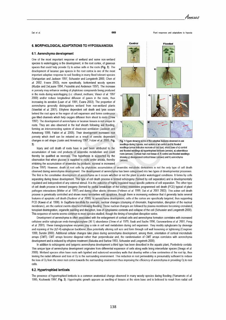
____________________________________________________________________________ Annexes
138

____________________________________________________________________________ Annexes
139

____________________________________________________________________________ Annexes
140

____________________________________________________________________________ Annexes
141

____________________________________________________________________________ Annexes
142

____________________________________________________________________________ Annexes
143

____________________________________________________________________________ Annexes
144
Formes réactives de l’oxygène, stress et mort cellulaire chez les plantes
Publication acceptée le 08 février 2008 dans le journal Comptes rendus - Biologies.
Les auteurs sont C. Parent, N. Capelli et J. Dat.
Résumé:
Les plantes sont constamment soumises à des variations environnementales. Ces changements
peuvent engendrer un stress qui modifie l’homéostasie cellulaire par la production de formes
réactives de l’oxygène. L’accumulation phytotoxique de ces différents radicaux oxygénés
peut entraîner la mort de la plante ; cependant, ils ont récemment été identifiés comme des
acteurs essentiels de la réponse au stress et leur rôle comme messager secondaire est
maintenant clairement établi. Leur implication dans la régulation de l’expression génique a
aussi permis de démontrer leur rôle d’inducteurs de la mort cellulaire programmée, mort
génétiquement contrôlée que l’on retrouve non seulement dans les processus
développementaux, mais également typiquement observée dans la réponse au stress. Cette
revue présente les récentes avancées dans la caractérisation du rôle des formes réactives de
l’oxygène chez les plantes.

____________________________________________________________________________ Annexes
145

____________________________________________________________________________ Annexes
146

____________________________________________________________________________ Annexes
147

____________________________________________________________________________ Annexes
148

____________________________________________________________________________ Annexes
149

____________________________________________________________________________ Annexes
150

____________________________________________________________________________ Annexes
151

Résumé de thèse

L'ennoyage est un phénomène courant qui se répercute sur les végétaux en diminuant
leur croissance, leur développement et leur régénération. Il entraîne une forte diminution du taux d’oxygène (hypoxie) dans le compartiment racinaire résultat de l’excès d’eau dans le sol. Face à ce stress, certaines plantes sont capables intensifier leur métabolisme anaérobie et développer des adaptations morphologiques (lenticelles hypertrophiées, aérenchymes, racines adventives). Le chêne sessile (Quercus petraea Matt L.) et le chêne pédonculé (Quercus robur L.) présentent une différence de tolérance à l’ennoyage du sol mais sont génétiquement proches. Cependant, il existe peu de connaissances sur les processus physiologiques mis en place par ces deux espèces durant ce stress. La croissance ainsi que certains changements morphologiques, anatomiques et physiologiques étudiés chez les deux espèces en réponse à un ennoyage, ont confirmé que le chêne sessile était davantage sensible à ce stress. Le chêne pédonculé, espèce plus tolérante, parvient à maintenir son statut hydrique de même que son activité photosynthétique plus longtemps et à des niveaux moins critiques que le sessile. L’hémoglobine non-symbiotique améliore la survie des plantes en conditions d’hypoxie en interagissant avec le monoxyde d’azote (NO). Le clonage d’une hémoglobine non-symbiotique chez le chêne sessile a permis d’isoler le gène QpHb1 codant pour une protéine de 161 acides aminés et présentant toutes les caractéristiques communes aux hémoglobines non-symbiotiques de classe 1. Chez les deux espèces, QpHb1 est davantage exprimé dans les racines que dans la tige ou les feuilles, suggérant un rôle particulier dans ce tissu. L’expression de QpHb1 analysée par Northern blotting a montré une chute d’expression dans les racines dès les premières heures de stress chez le chêne sessile qui se poursuit durant toute la durée de l’expérience (28 jours). Alors que chez le chêne pédonculé, l’expression de QpHb1 suit un schéma plus complexe : on observe un pic d’expression après 1h d’ennoyage suivi d’une forte inhibition après 3h. Cette régulation pourrait être synonyme d’une implication de l’hémoglobine non-symbiotique dans la signalisation rapide du stress et par conséquent dans la mise en place de la tolérance. D’ailleurs sa localisation par hybridation in situ au niveau racinaire a montré que QpHb1 se situait au niveau des cellules du protoderme, pouvant suggérer un rôle dans la détection des modifications de la rhizosphère mais aussi au niveau du protoxylème qui pourrait lui permettre de participer à la signalisation entre l’appareil racinaire et l’appareil aérien via la S-nitrosylation. Dans la réponse à un ennoyage de plusieurs semaines, le chêne pédonculé met en place des adaptations (aérenchymes, lenticelles hypertrophiées et racines adventives) et exprime davantage QpHb1 que le chêne sessile notamment dans les racines adventives. La régulation spatio-temporelle de QpHb1 pourrait être impliquée dans la capacité de tolérance ainsi que dans la cascade de signalisation menant au développement de ces adaptations.

Flooding is a common environmental stress inducing a decline in plant growth,
development and regeneration. Waterlogging leads to low oxygen levels (hypoxia) in the root environment due to an excess of water in the soil. In response to hypoxia, plants switch their metabolism from aerobic respiration to anaerobic fermentation and can develop morphological adaptations (aerenchyma, hypertrophied lenticels and adventitious roots). Sessile and pedunculate oak are two closely related species that display a strong differential tolerance to soil flooding. However, few reports exist about the physiological processes leading to their response to flooding. Growths, but also morphological, anatomical and physiological changes studied in both species in response to waterlogging, have confirmed that sessile oak is more sensitive to this stress. Pedunculate oak, the more tolerant species, succeeded in maintaining its water status and its photosynthetic activity at a higher level than sessile oak. Non-symbiotic hemoglobin enhances plant survival in hypoxic conditions by interacting with nitric oxide (NO). We cloned and characterized a novel non-symbiotic hemoglobin gene in sessile oak, QpHb1, coding for a 161 amino acid protein and showing all characteristics of class1 non symbiotic hemoglobins. In both oak species, QpHb1 is more highly expressed in roots than in stem or leaves, suggesting a particular role in this tissue. QpHb1 expression analyzed by northern blotting showed a strong decline from the first hours of stress in sessile oak which continued until the end of the experiment (28 days). In contrast, QpHb1 expression in pedunculate oak, exhibits a more complex scheme: an early rise in expression after 1h of waterlogging followed by a strong decline after 3h. This regulation could be related to the implication of non-symbiotic hemoglobin in rapid stress signaling and consequently in the induction of tolerance. Indeed, QpHb1 root localization performed by in situ hybridization showed a strong expression in protoderm cells, suggesting a sensing function of rhizospheric modifications. In addition, the protoxylem localization of QpHb1 could also indicate a role in root to shoot signalization by S-nitrosylation. In response to several weeks of flooding stress, pedunculate oak displayed morphological adaptations (aerenchyma, hypertrophied lenticels and adventitious roots) and exhibited a higher level of QpHb1 transcripts than sessile most notably in adventitious roots. The spatio-temporal regulation of QpHb1 could be involved in the tolerance capacity as well as in signaling cascade leading to developmental adaptations.