european journal of pharmacology - core.ac.uk · in human chondrocytes ( rufino et al., 2014a )....
TRANSCRIPT

Immunopharmacology and inflammation
Evaluation of the anti-inflammatory, anti-catabolic and pro-anaboliceffects of E-caryophyllene, myrcene and limonene in a cell modelof osteoarthritis
Ana Teresa Rufino a,b, Madalena Ribeiro a,b, Cátia Sousa a,b, Fernando Judas c,d,Lígia Salgueiro a,e, Carlos Cavaleiro a,e, Alexandrina Ferreira Mendes a,b,n
a Centre for Neuroscience and Cell Biology, University of Coimbra, Coimbra, Portugalb Faculty of Pharmacy, University of Coimbra, Coimbra, Portugalc Orthopedics Department and Bone Bank, University and Hospital Center of Coimbra, Coimbra, Portugald Faculty of Medicine, University of Coimbra, Coimbra, Portugale Centro de Estudos Farmacêuticos, Faculty of Pharmacy, University of Coimbra, Coimbra, Portugal
a r t i c l e i n f o
Article history:Received 23 October 2014Received in revised form14 January 2015Accepted 16 January 2015Available online 23 January 2015
Keywords:OsteoarthritisChronic inflammationNF-κBMAPKNatural products
a b s t r a c t
Osteoarthritis is a progressive joint disease and a major cause of disability for which no curativetherapies are yet available. To identify compounds with potential anti-osteoarthritic properties, in thisstudy, we screened one sesquiterpene, E-caryophyllene, and two monoterpenes, myrcene and limonene,hydrocarbon compounds for anti-inflammatory, anti-catabolic and pro-anabolic activities in humanchondrocytes. At non-cytotoxic concentrations, myrcene and limonene inhibited IL-1β-induced nitricoxide production (IC50¼37.3 μg/ml and 85.3 mg/ml, respectively), but E-caryophyllene was inactive.Myrcene, and limonene to a lesser extent, also decreased IL-1β-induced NF-κB, JNK and p38 activationand the expression of inflammatory (iNOS) and catabolic (MMP-1 and MMP-13) genes, while increasingthe expression of anti-catabolic genes (TIMP-1 and -3 by myrcene and TIMP-1 by limonene). Limoneneincreased ERK1/2 activation by 30%, while myrcene decreased it by 26%, relative to IL-1β-treated cells.None of the compounds tested was able to increase the expression of cartilage matrix-specific genes(collagen II and aggrecan), but both compounds prevented the increased expression of the non-cartilagespecific, collagen I, induced by IL-1β. These data show that myrcene has significant anti-inflammatoryand anti-catabolic effects in human chondrocytes and, thus, its ability to halt or, at least, slow downcartilage destruction and osteoarthritis progression warrants further investigation.
& 2015 Elsevier B.V. All rights reserved.
1. Introduction
Osteoarthritis (OA) is a multifactorial degenerative joint diseasecharacterized by inflammation and progressive loss of the articularcartilage, associated with changes in the subchondral bone and otherjoint tissues. It affects 10–15% of the world population and is a majorcause of disability, not only in the elderly, as well as in the workforcepopulation (Zhang and Jordan, 2010). Existing therapeutic approaches
are mainly symptomatic, thus novel drugs with disease-modifying andchondroprotective properties, the so-called disease-modifying osteoar-thritis drugs, are required to halt disease progression and decrease itshuge socio-economic impact (Goldring and Goldring, 2007; Kaplanet al., 2013).
Plant-derived compounds show important biological properties thatcan be explored in the context of OA for identification of compoundswith potential anti-osteoarthritic activity (Calixto et al., 2004; Khalifeand Zafarullah, 2011). Among compounds of plant origin, those foundin essential oils present favorable pharmacokinetic properties, namelylipophilicity and low molecular weight (Miguel, 2010). Our previousstudies have been focused in identifying essential oils with anti-inflammatory and anti-catabolic properties in human chondrocytes tobe used as sources of compounds with potential anti-osteoarthriticactivity (Neves et al., 2009; Rufino et al., 2014a). In this context, werecently identified the essential oils of Eryngium duriaei subsp. juresia-num and Lavandula luisieri as possessing anti-inflammatory properties
Contents lists available at ScienceDirect
journal homepage: www.elsevier.com/locate/ejphar
European Journal of Pharmacology
http://dx.doi.org/10.1016/j.ejphar.2015.01.0180014-2999/& 2015 Elsevier B.V. All rights reserved.
n Corresponding author at: Center for Neuroscience and Cell Biology, Universityof Coimbra, Edifício da Faculdade de Medicina, Polo I, 11 piso, Rua Larga, 3004-504Coimbra, Portugal. Tel.: þ351 239 480209; fax: þ351 239 488503.
E-mail addresses: [email protected] (A.T. Rufino),[email protected] (M. Ribeiro),[email protected] (C. Sousa), [email protected] (F. Judas),[email protected] (L. Salgueiro), [email protected] (C. Cavaleiro),[email protected] (A.F. Mendes).
European Journal of Pharmacology 750 (2015) 141–150

in human chondrocytes (Rufino et al., 2014a). The current studyaims at identifying active compounds of these essential oils andfurther characterizing their pharmacological properties in human chon-drocytes.
For this, mechanisms relevant as pharmacological targets forthe development of anti-osteoarthritic drugs need to be addressed.Although OA etiology is not yet completely understood, pro-inflammatory cytokines, like interleukin-1β (IL-1β), play a centralrole in disease development and progression by inducing theexpression of cartilage matrix-degrading enzymes and impairinganabolic and anti-catabolic responses in chondrocytes (Goldringet al., 2008). The consequent upregulated degradative process,together with impaired reparative responses, results in progressivecartilage loss, the hallmark of OA.
Matrix metalloproteases (MMPs) and aggrecanases are the mainenzymes responsible for hydrolyzing the major articular cartilage-specific matrix components, collagen II and aggrecan. This is accom-panied by impaired reparative responses involving downregulation ofthe natural MMP inhibitors, the tissue inhibitor of metalloproteases(TIMP) family, decreased synthesis of collagen II and aggrecan andincreased expression of non-articular cartilage matrix components, likecollagen I (Troeberg and Nagase, 2012). Moreover, increased productionof pro-inflammatory and pro-catabolic mediators, like nitric oxide(NO), amplifies and perpetuates cartilage destruction (Boileau et al.,2002; Rosa et al., 2008; Sasaki et al., 1998). The transcription factor,Nuclear Factor-κB (NF-κB), and the family of mitogen-activated proteinkinases (MAPK) play a central role in modulating the expression ofthose catabolic and inflammatorymediators (Goldring and Otero, 2011)and, thus, agents that suppress their activity have the potential toeffectively decrease cartilage destruction and, therefore, OA progression(Berenbaum, 2004). Furthermore, compounds that can also restoreanabolic and anti-catabolic gene expression have the potential to pro-mote cartilage repair.
Therefore, we used primary human chondrocyte culturesstimulated with IL-1β as an in vitro cartilage degradation modelthat emulates the damage seen in OA. Using this model, weevaluated the inhibition of IL-1β-induced NO production as asimple screening assay. The essential oils of E. duriaei subsp.juresianum and L. luisieri were separated into several fractionsthat were then screened using the assay mentioned above. Thechemical composition of the fractions tested was elucidated andcompounds present in the active fractions and commerciallyavailable were selected for screening using the same assay. Thecompounds selected were obtained with a high degree of purityfrom commercial sources and those found to be capable ofinhibiting IL-1β-induced NO production were selected for furtherassessment of anti-osteoarthritic potential. For this, we evaluatedtheir ability to modulate IL-1β-induced signaling pathways invol-ved in the expression of inflammatory and catabolic genes, namelyactivation of NF-κB and the MAPK family members, Jun terminalKinase (JNK), p38 and Extracellular signal-Regulated Kinase 1/2(ERK1/2). Then, we evaluated the ability of those compounds tocounteract the effects of IL-1β on the expression of inflammatory(iNOS), catabolic (MMP-1 and -13), anti-catabolic (TIMP-1 and -3)and extracellular matrix (collagen I, collagen II and aggrecan)genes in human articular chondrocytes.
2. Material and methods
2.1. Essential oil fractionation and chemical analysis
The essential oils of E. duriaei subsp. juresianum and L. luisieriwere fractionated by flash chromatography on silica gel 63–200 μm (Merck) using a 2 cm�40 cm Omnifit (Sigma-Aldrich)glass columns. Elution was made in step gradients from 100% of
n-pentane, n-pentane /ethyl ether mixtures, till a final concentra-tion of 100% ethyl ether. Collected fractions were analyzed bygas chromatography–mass spectroscopy (GC/MS) and combinedif having similar composition. Analysis was performed using aHewlett-Packard 6890 gas chromatograph fitted with a HP1 fusedsilica column (polydimethylsiloxane 30 m�0.25 mm i.d., filmthickness 0.25 μm), interfaced with an Hewlett-Packard massselective detector 5973 (Agilent Technologies) operated by HPEnhanced ChemStation software, version A.03.00. GC–MS para-meters: oven temperature program: 70–220 1C (3 1C min�1),220 1C (15 min); injector temperature: 250 1C; carrier gas: helium,adjusted to a linear velocity of 30 cm s�1; splitting ratio 1:40;interface temperature: 250 1C; MS source temperature: 230 1C; MSquadrupole temperature: 150 1C; ionization energy: 70 eV; ioniza-tion current: 60 μA; scan range: 35–350 units; scans s�1: 4.51.Compounds were identified through their GC retention and massspectra. Retention indices, calculated by linear interpolation rela-tive to retention times of C8–C23 of n-alkanes, were comparedwith those of authentic products included in laboratory data-base (CEF/Faculty of Pharmacy, University of Coimbra) and/or theliterature data. Acquired mass spectra were compared with refer-ence spectra from the laboratory database, Wiley/NIST library(McLafferty, 2009) and literature data (Adams, 1995; Cavaleiroet al., 2011; Joulain, 1998; Videira et al., 2013). Relative amountsof individual components were calculated based on the total ionchromatogram peak areas.
2.2. Cartilage samples and chondrocyte isolation
Human chondrocytes were isolated by enzymatic digestion(Rosa et al., 2008) of knee cartilage from the distal femoralcondyles of multi-organ donors (20–70 years old, mean¼52.9,n¼31) and patients (58–73 years old, mean¼65, n¼5) undergoingtotal knee arthroplasty at the Orthopaedic Department and BoneBank of the University and Hospital Center of Coimbra (CHUC).The cartilage samples presented variable degrees of degradation,ranging from intact to severely damaged. All procedures wereapproved by the Ethics Committee of CHUC (protocol approvalnumbers 8654/DC and HUC-13-05).
2.3. Cell cultures and treatments
Primary non-proliferating chondrocyte cultures were estab-lished from non-pooled cartilage samples. The human chondrocy-tic cell line, C28/I2, kindly provided by Prof. Mary Goldring(currently at the Hospital for Special Surgery, New York) andHarvard University, was used to evaluate NF-κB–DNA bindingactivity. The cell cultures were serum-starved for at least 8 h andmaintained thereafter in serum-free culture medium. The testcompounds, diluted in DMSO (Sigma-Aldrich, St Louis, MO, USA)to achieve the concentrations indicated in figures and theirlegends, were added to the chondrocyte cultures 30 min beforethe pro-inflammatory stimulus (IL-1β, 10 ng/ml) and maintainedfor the experimental period indicated in figure legends. The finalconcentration of DMSO did not exceed 0.1% (v/v). E-caryophyllene(purity 498.5%) and myrcene (purity Z95.0%) were from Sigma-Aldrich (St Louis, MO, USA) and limonene (purity �90%) wasfrom Fluka.
2.4. Nitric oxide production
Nitric oxide production was evaluated as the amount of nitriteaccumulated in primary chondrocyte culture supernatants after24 h treatment with IL-1β, following pre-treatment with non-cytotoxic concentrations of the fractions of the essential oils or thetest compounds. Nitrite concentration was measured using the
A.T. Rufino et al. / European Journal of Pharmacology 750 (2015) 141–150142

spectrophotometric method based on the Griess reaction (Greenet al., 1982).
2.5. Western blot analysis
Total and cytoplasmic extracts were prepared as describedbefore (Goldring and Otero, 2011). Proteins were separated bySDS-PAGE under reducing conditions and electrotransferred ontoPVDF membranes. These were probed overnight with the follow-ing primary antibodies: mouse monoclonal anti-human iNOS(R&D systems, Minneapolis, MN), mouse polyclonal anti-humanphospho-IκB-α or rabbit polyclonal anti-human IκB-α, anti-human phospho-JNK, anti-human phospho-p38 or anti-humanphospho-ERK1/2 (Cell Signaling Technology, Inc., Danvers, MA)and then with anti-rabbit or anti-mouse alkaline phosphatase-conjugated secondary antibodies (GE Healthcare, UK). Mouse anti-human β-Tubulin monoclonal antibody was used to detectβ-Tubulin as a loading control. Enhanced ChemiFluorescencereagent (GE Healthcare) was used to detect immune complexes.Image analysis was performed with ImageQuant TL software (GEHealthcare). The results presented are the normalized ratiobetween the intensities of the bands corresponding to the proteinof interest and to the protein used as loading control.
2.6. NF-κB transcription factor assay
A colorimetric ELISA-based assay (NoShift Transcription FactorAssay Kit, Novagen, La Jolla, CA) was used to evaluate the presenceof active NF-κB dimers, capable of binding to the cognate con-sensus oligonucleotide sequence. For this purpose, nuclear extractsfrom C-28/I2 cells were incubated with a biotinylated consensusNF-κB oligonucleotide (NoShift NF-κB Reagents; Novagen) and theassay performed according to the manufacturer's instructions. Theabsorbance intensity in each sample is directly proportional to theamount of NF-κB–DNA complexes formed and, thus, to theamount of active NF-κB dimers present in each sample. In parallel,the specificity of the reaction was confirmed in competition assayswhere addition of a 10-fold molar excess of non-biotinylated wild-type or mutant oligonucleotides (NoShift NF-κB Reagents; Nova-gen) to binding reactions containing nuclear extracts from IL-1βtreated cells abrogated or did not affect, respectively, the forma-tion of NF-κB–DNA complexes.
2.7. Total RNA extraction and quantitative real-time RT-PCR (qRT-PCR)
Total RNA was extracted from human condrocytes using TRI-zols Reagent (Invitrogen, Life Technologies, Co; Paisley, UK) andquantified using a NanoDrop ND-1000 spectrophotometer at260 nm. Purity and integrity of RNA were assessed as the 240/260 and 280/260 ratios. The cDNA was reverse-transcribed usingthe iScript Select cDNA Synthesis Kit (Bio-Rad), beginning with
1 mg of RNA. Specific sets of primers for iNOS, MMP-1, MMP-13,TIMP-1, TIMP-3, collagen II, collagen I, aggrecan and HPRT-1(Table 1) were designed using Beacon Designer software (PremierBiosoft International, Palo Alto, CA). PCR reactions were performedusing 25 μg/ml of transcribed cDNA in a final volume of 20 mL.
The efficiency of the amplification reaction for each gene wascalculated using a standard curve of a series of diluted cDNAsamples, and the specificity of the amplification products wasassessed by analyzing the melting curve generated in the process.
The results for each gene of interest were normalized againstHPRT-1, the housekeeping gene found to be the most stable underthe experimental conditions used. Gene expression changes wereanalyzed using the built-in iQ5 Optical system software v2, whichenables the analysis of the results with the Pfaffl method, avariation of the ΔΔCT method corrected for gene-specific effi-ciencies (Nolan et al., 2006)
2.8. Statistical analysis
Results are presented as mean7S. E. M. Each subject contrib-uted only once to the statistical analysis which was performedusing GraphPad Prism (version 5.00). The Kolmogorov–Smirnovtest was used to assess normality for the observations themselvesor for the observed differences. As this test showed that in allexperiments the results were normally distributed, the statisticalanalysis was performed using the paired t-test for comparison ofeach condition with its respective control and one-way ANOVA forcomparison of all conditions. Results were considered statisticallysignificant at Po0.05.
3. Results
3.1. Selection of inhibitors of IL-1-induced iNOS expression and NOproduction
Various fractions of the essential oils of E. duriaei subsp.juresianum and L. luisieri were separated in amounts sufficientfor pharmacological screening. The composition of these fractionswas fully elucidated and is reported in Table 2.
The fractions obtained were then tested at non-cytotoxicconcentrations ranging from 10 to 50 μg/ml (Supplementarydata). The results (Fig. 1 panel A) show that the hydrocarbon-containing fractions (F1 and FA) of both essential oils decreased IL-1β-induced NO production in a concentration-dependent manner,the highest concentrations achieving an inhibition of 55% and 75%,respectively, relative to cells treated with IL-1β alone. The otherthree fractions of the essential oil of L. luisieri also showed someinhibitory activity which, nonetheless, did not exceed 35% relativeto IL-1β-treated cells. Fractions F2.4 and F2.5 of the essential oil of E.duriaei subsp. juresianum were only tested at a concentration of10 mg/ml, as higher concentrations were found to be cytotoxic(Supplementary data). These fractions, as well as fraction F2.7 at
Table 1Oligonucleotide primer pairs used for qRT-PCR.
Gene name Genbank accession number Forward sequence Reverse sequence
iNOS NM_000625.4 50-AATCCAGATAA GTGACATAAG-30 50-CTCCACATTGT TGTTGAT-30
MMP-1 NM_001145938.1 50-GAGTCTCCCAT TCTACTG-30 50-TTATAGCATCA AAGGTTAGC-30
MMP-13 NM_002422.3 50-GTTTCCTATCTA CACCTACAC-30 50-CTCGGAGACTGG TAATGG-30
TIMP-1 NM_003254.2 50-TGTTGCTGTGGC TGATAG-30 50-CTGGTATAAGGT GGTCTGG-30
TIMP-3 NM_000362.4 50-CCATACACTATCCAC -30 50-TAACAGCATTGAACA -30
Collagen II NM_001844.4 50-GGCAGAGGTA TAATGATAAGG-30 50-ATTATGTCGTC GCAGAGG-30
Collagen I NM_000088.3 50GGAGGAGAGTCAGGA-30 50-GCAACACAGTTACAC-30
Aggrecan NM_001135 50-CCTGGTGTGGCT GCTGTC-30 50-CTGGCTCGGT GGTGAACTC-30
HPRT-1 NM_000194.2 50-TGACACTGGCA AAACAAT-30 50-GGCTTATATCC AACACTTCG-30
A.T. Rufino et al. / European Journal of Pharmacology 750 (2015) 141–150 143

a concentration of 50 mg/ml, also significantly inhibited IL-1β-ind-uced NO production, although to a lesser extent than found withF1. No significant effects were obtained with F2.3 at any of theconcentrations tested. Therefore, the hydrocarbon-containing frac-tions (F1 and FA) of both essential oils were considered the mostpromising for selection of compounds for further studies.
As shown in Table 2, these fractions (FA and F1) are composed ofmonoterpene and sesquiterpene hydrocarbons, mainly α-pineneand 3,5-dimethylene-1,4,4-trimethylcyclopentene, and E-caryo-phyllene, α-neocallitropsene, germacrene D, β-selinene and bicy-clogermacrene, respectively. Of these compounds, only α-pineneand E-caryophyllene are readily available from commercial sources
Table 2Composition of the fractions of the essential oils of E. duriaei subsp juresianum and Lavandula luisieri.
Eryngium duriaei subsp juresianum % Lavandula luisieri %
F1 E-caryophyllene 29.5 FA 3.5-dimethylene-1.4.4-trimethylcyclopentene 10.4α-neocallitropsene 50.2 α-pinene 26.9Germacrene D 2.7 β-pinene 3.5β-selinene 2.7 Δ-3-carene 5.2Bicyclogermacrene 6.3 Limonene 3.0Limonene 0.1 E-β-ocimene 1.6Myrcene t Cyclosativene 2.0
F2.3 Octanal 9.3 α-copaene 2.2Caryophyllene oxide 31.6 E-caryophyllene 3.9Isocaryophyllene-14-al 44.4 Alloaromadendrene 1.2
F2.4 Unknown 1 22.8 β-selinene 2.9Spathulenol 9.8 α-selinene 3.514-hydroxy-β-caryophyllene 38.0 δ-cadinene 6.7Unknown 2 6.6 Selina-3.7(11)-diene 4.5
F2.5 Unknown 1 8.8 FB trans-α-necrodyl acetate 30.5Spathulenol 21.4 Lavandulyl acetate 8.214-hydroxy-β-caryophyllene 45.2 cis-α-necrodyl acetate 3.8Unknown 2 2.9 1.8-cineole 32.4Unknown 3 3.7 Lyratyl acetate 2.4
F2.7 Caprylic acid 8.1 FC 1.10-di-epi-cubenol 4.8Buthyhidroxytoluene (solvent contaminant) 8.6 2.3.4.4-tetramethyl-5-methylene-cyclopent-2-enone 8.6Tetradecanoic acid 40.5 Linalool 11.9Hexadecanoic acid 8.5 epi-cubenol 1.3
trans-α-necrodol 20.1Lavandulol 2.2Viridiflorol 8.2T-cadinol 2.4T-muurolol 4.4cis-linalool oxide (THP) 3.2Unkown (C10H16O) 2.31.1.2.3-tetramethyl-4-hidroxymethyl-2-ciclopentene 7.214-norcadin-5-ene-4-one (Isomer) 4.3
FD trans-linalool oxide (THF) 12.6α-terpineol 7.1Verbenone 2.5α-muurolol 1.3trans-verbenol 3.1Globulol 2.5α-cadinol 48.7
Fig. 1. Effects of the fractions of the essential oils of Eryngium duriaei subsp juresianum and Lavandula luisieri (panel A) and of the test compounds, E-caryophyllene, myrceneand limonene, (panel B) on IL-1β-induced NO production. Human chondrocytes were left untreated (Control, Ctrl) or treated with IL-1β, 10 ng/ml, for 24 h, following pre-treatment for 30 min with the indicated concentrations of each fraction or pure compound. Each column represents the mean7S. E. M. of, at least, 4 independentexperiments. nPo0.05; nnPo0.01, nnnPo0.001 relative to IL-1β treated cells. §Po0.05; §§Po0.01, §§§Po0.001 relative to control cells and #Po0.05; ##Po0.01, ###Po0.001between the conditions indicated.
A.T. Rufino et al. / European Journal of Pharmacology 750 (2015) 141–150144
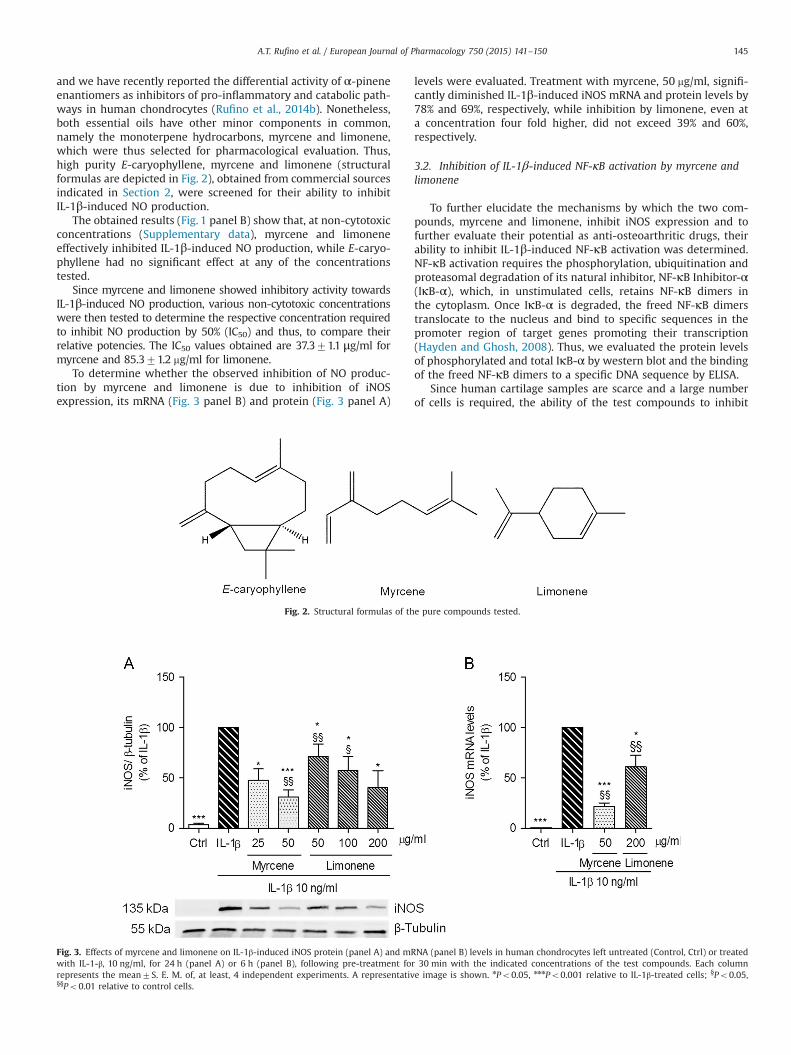
and we have recently reported the differential activity of α-pineneenantiomers as inhibitors of pro-inflammatory and catabolic path-ways in human chondrocytes (Rufino et al., 2014b). Nonetheless,both essential oils have other minor components in common,namely the monoterpene hydrocarbons, myrcene and limonene,which were thus selected for pharmacological evaluation. Thus,high purity E-caryophyllene, myrcene and limonene (structuralformulas are depicted in Fig. 2), obtained from commercial sourcesindicated in Section 2, were screened for their ability to inhibitIL-1β-induced NO production.
The obtained results (Fig. 1 panel B) show that, at non-cytotoxicconcentrations (Supplementary data), myrcene and limoneneeffectively inhibited IL-1β-induced NO production, while E-caryo-phyllene had no significant effect at any of the concentrationstested.
Since myrcene and limonene showed inhibitory activity towardsIL-1β-induced NO production, various non-cytotoxic concentrationswere then tested to determine the respective concentration requiredto inhibit NO production by 50% (IC50) and thus, to compare theirrelative potencies. The IC50 values obtained are 37.371.1 μg/ml formyrcene and 85.371.2 mg/ml for limonene.
To determine whether the observed inhibition of NO produc-tion by myrcene and limonene is due to inhibition of iNOSexpression, its mRNA (Fig. 3 panel B) and protein (Fig. 3 panel A)
levels were evaluated. Treatment with myrcene, 50 mg/ml, signifi-cantly diminished IL-1β-induced iNOS mRNA and protein levels by78% and 69%, respectively, while inhibition by limonene, even ata concentration four fold higher, did not exceed 39% and 60%,respectively.
3.2. Inhibition of IL-1β-induced NF-κB activation by myrcene andlimonene
To further elucidate the mechanisms by which the two com-pounds, myrcene and limonene, inhibit iNOS expression and tofurther evaluate their potential as anti-osteoarthritic drugs, theirability to inhibit IL-1β-induced NF-κB activation was determined.NF-κB activation requires the phosphorylation, ubiquitination andproteasomal degradation of its natural inhibitor, NF-κB Inhibitor-α(IκB-α), which, in unstimulated cells, retains NF-κB dimers inthe cytoplasm. Once IκB-α is degraded, the freed NF-κB dimerstranslocate to the nucleus and bind to specific sequences in thepromoter region of target genes promoting their transcription(Hayden and Ghosh, 2008). Thus, we evaluated the protein levelsof phosphorylated and total IκB-α by western blot and the bindingof the freed NF-κB dimers to a specific DNA sequence by ELISA.
Since human cartilage samples are scarce and a large numberof cells is required, the ability of the test compounds to inhibit
Fig. 2. Structural formulas of the pure compounds tested.
Fig. 3. Effects of myrcene and limonene on IL-1β-induced iNOS protein (panel A) and mRNA (panel B) levels in human chondrocytes left untreated (Control, Ctrl) or treatedwith IL-1-β, 10 ng/ml, for 24 h (panel A) or 6 h (panel B), following pre-treatment for 30 min with the indicated concentrations of the test compounds. Each columnrepresents the mean7S. E. M. of, at least, 4 independent experiments. A representative image is shown. nPo0.05, nnnPo0.001 relative to IL-1β-treated cells; §Po0.05,§§Po0.01 relative to control cells.
A.T. Rufino et al. / European Journal of Pharmacology 750 (2015) 141–150 145
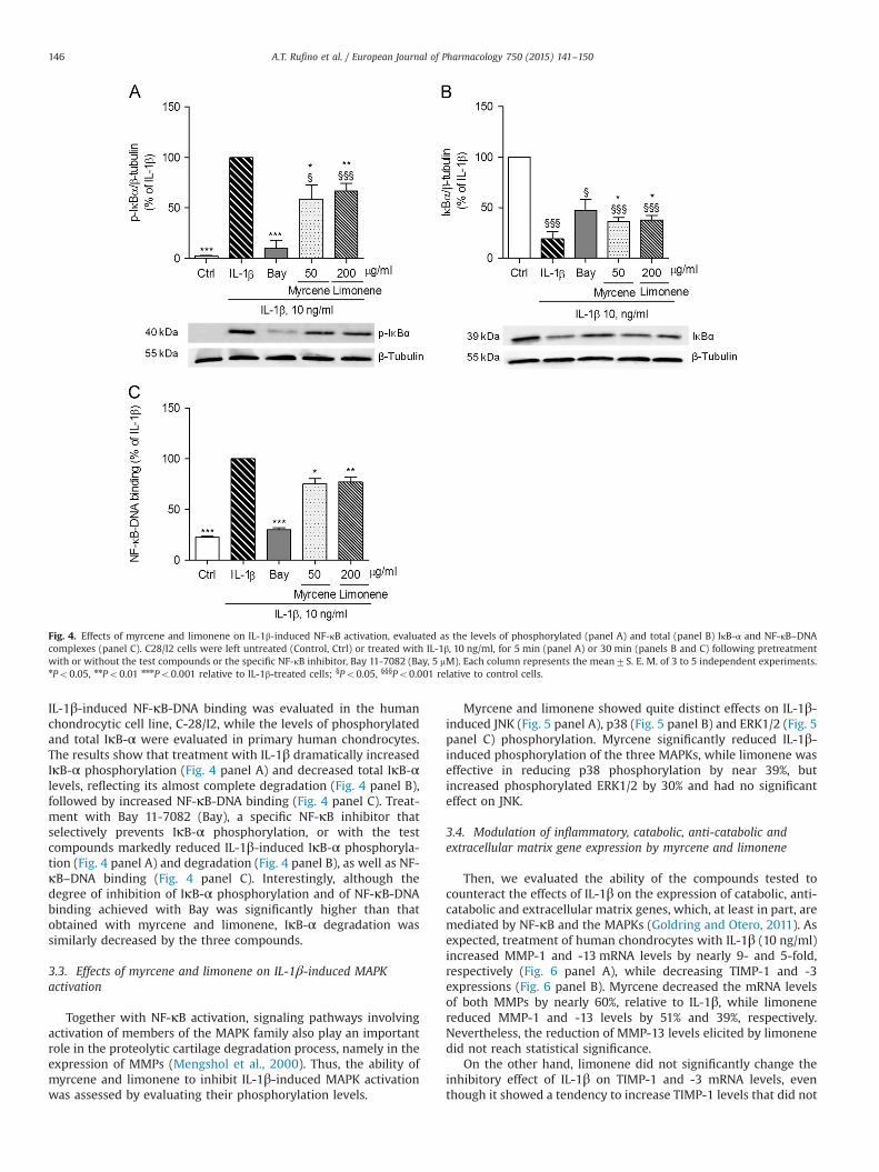
IL-1β-induced NF-κB-DNA binding was evaluated in the humanchondrocytic cell line, C-28/I2, while the levels of phosphorylatedand total IκB-α were evaluated in primary human chondrocytes.The results show that treatment with IL-1β dramatically increasedIκB-α phosphorylation (Fig. 4 panel A) and decreased total IκB-αlevels, reflecting its almost complete degradation (Fig. 4 panel B),followed by increased NF-κB-DNA binding (Fig. 4 panel C). Treat-ment with Bay 11-7082 (Bay), a specific NF-κB inhibitor thatselectively prevents IκB-α phosphorylation, or with the testcompounds markedly reduced IL-1β-induced IκB-α phosphoryla-tion (Fig. 4 panel A) and degradation (Fig. 4 panel B), as well as NF-κB–DNA binding (Fig. 4 panel C). Interestingly, although thedegree of inhibition of IκB-α phosphorylation and of NF-κB-DNAbinding achieved with Bay was significantly higher than thatobtained with myrcene and limonene, IκB-α degradation wassimilarly decreased by the three compounds.
3.3. Effects of myrcene and limonene on IL-1β-induced MAPKactivation
Together with NF-κB activation, signaling pathways involvingactivation of members of the MAPK family also play an importantrole in the proteolytic cartilage degradation process, namely in theexpression of MMPs (Mengshol et al., 2000). Thus, the ability ofmyrcene and limonene to inhibit IL-1β-induced MAPK activationwas assessed by evaluating their phosphorylation levels.
Myrcene and limonene showed quite distinct effects on IL-1β-induced JNK (Fig. 5 panel A), p38 (Fig. 5 panel B) and ERK1/2 (Fig. 5panel C) phosphorylation. Myrcene significantly reduced IL-1β-induced phosphorylation of the three MAPKs, while limonene waseffective in reducing p38 phosphorylation by near 39%, butincreased phosphorylated ERK1/2 by 30% and had no significanteffect on JNK.
3.4. Modulation of inflammatory, catabolic, anti-catabolic andextracellular matrix gene expression by myrcene and limonene
Then, we evaluated the ability of the compounds tested tocounteract the effects of IL-1β on the expression of catabolic, anti-catabolic and extracellular matrix genes, which, at least in part, aremediated by NF-κB and the MAPKs (Goldring and Otero, 2011). Asexpected, treatment of human chondrocytes with IL-1β (10 ng/ml)increased MMP-1 and -13 mRNA levels by nearly 9- and 5-fold,respectively (Fig. 6 panel A), while decreasing TIMP-1 and -3expressions (Fig. 6 panel B). Myrcene decreased the mRNA levelsof both MMPs by nearly 60%, relative to IL-1β, while limonenereduced MMP-1 and -13 levels by 51% and 39%, respectively.Nevertheless, the reduction of MMP-13 levels elicited by limonenedid not reach statistical significance.
On the other hand, limonene did not significantly change theinhibitory effect of IL-1β on TIMP-1 and -3 mRNA levels, eventhough it showed a tendency to increase TIMP-1 levels that did not
Fig. 4. Effects of myrcene and limonene on IL-1β-induced NF-κB activation, evaluated as the levels of phosphorylated (panel A) and total (panel B) IκB-α and NF-κB–DNAcomplexes (panel C). C28/I2 cells were left untreated (Control, Ctrl) or treated with IL-1β, 10 ng/ml, for 5 min (panel A) or 30 min (panels B and C) following pretreatmentwith or without the test compounds or the specific NF-κB inhibitor, Bay 11-7082 (Bay, 5 mM). Each column represents the mean7S. E. M. of 3 to 5 independent experiments.nPo0.05, nnPo0.01 nnnPo0.001 relative to IL-1β-treated cells; §Po0.05, §§§Po0.001 relative to control cells.
A.T. Rufino et al. / European Journal of Pharmacology 750 (2015) 141–150146

Fig. 5. Effects of myrcene and limonene on IL-1β- induced activation of JNK, p38 and ERK1/2 in human chondrocytes. Phosphorylated levels of JNK (panel A), p38 (panel B)and ERK (1/2) (panel C) were analyzed in total cell extracts of human chondrocytes left untreated (Control, Ctrl) or treated for 5 min with IL-1β, 10 ng/ml, following a pre-treatment for 30 min with the indicated concentrations of myrcene, limonene or a specific inhibitor of the activation of each MAPK. Each column represents the mean7S. E.M. of, at least, 4 independent experiments. Representative images are shown. nnPo0.01, nnnPo0.001 relative to IL-1β-treated cells and §§§Po0.001 relative to control cells.iJNK: JNK inhibitor, SP600125 (20 mM); ip38: p38 inhibitor, SB203580 (20 mM); iERK: ERK1/2 inhibitor, U0126 (10 mM).
Fig. 6. Effects of myrcene and limonene on IL-1β-induced changes in the expression of catabolic and anti-catabolic genes. mRNA levels of MMP-1 and MMP-13 (panel A) andTIMP-1 and TIMP-3 (panel B) were evaluated by qRT-PCR. Each bar represents the mean7S. E. M. of, at least, 4 independent experiments in which human chondrocytes wereleft untreated (Control, Ctrl) or treated for 12 h (panel A) or 24 h (panel B) with IL-1β, 10 ng/ml, in the presence or absence of the indicated compounds added to the cellcultures 30 min before IL-1β. nPo0.05, nnPo0.01, nnnPo0.001 relative to IL-1β-treated cells and §Po0.05, §§Po0.01, §§§Po0.001 relative to control cells.
A.T. Rufino et al. / European Journal of Pharmacology 750 (2015) 141–150 147

reach statistical significance. On the contrary, myrcene not onlycompletely reversed the inhibitory effect exerted by IL-1β, as iteffectively increased TIMP-1 and -3 levels approximately 2- and1.3-fold above those in untreated control cells, respectively, whichcorrespond to even larger increases if compared to TIMP-1 and-3 mRNA levels in cells treated with IL-1β alone.
To assess the potential ability of the test compounds to inhibit thedeleterious effects of IL-1β in anabolic responses that are essential forrepair of damaged articular cartilage, the expression of collagen II andaggrecan was evaluated. Furthermore, the ability of the test com-pounds to decrease the expression of the non-cartilage specific,collagen I gene, induced by IL-1β, was also evaluated. Chondrocytetreatment with 10 ng/ml IL-1β, for 24 h, significantly increasedcollagen I mRNA levels, while decreasing those of collagen II andaggrecan, relative to untreated control cells (Fig. 7). Treatment ofhuman chondrocytes with myrcene or limonene caused no significantchanges on collagen II (Fig. 7 panel A) and aggrecan (Fig. 7 panel B)mRNA levels compared to those in cells treated with IL-1β alone.Nonetheless, both treatments were able to completely abolish or evenreverse the increase in collagen I mRNA levels induced by IL-1β.
4. Discussion
The results obtained in this study identify two monoterpenehydrocarbons, myrcene and limonene, as capable of inhibiting IL-1β-induced NO production in human chondrocytes. The specific activities(activity/μg) relative to inhibition of IL-1-induced NO production, ofmyrcene and limonene are 1.33%/μg and 0.57%/μg, respectively, whilefor the active fractions (F1 and FA) of the essential oils of E. duriaeisubsp. juresianum and L. luisieri they are 1%/μg and 3.0%/μg, respec-tively. Since myrcene and limonene are only minor components ofthose fractions, it is likely that other constituents are also active andcontribute to the effects observed. Moreover, since both fractionscontain several distinct compounds, none of which is present insufficiently high amounts to justify the effects observed, either theactive compound in those fractions is significantly more potent thanmyrcene and limonene or various active compounds, including thesetwo, act synergistically, or at least, additively, to achieve a similar oreven higher degree of inhibition of IL-1-induced NO production.Unfortunately, as mentioned in Section 3.1, the major components ofthose fractions are either not readily available from commercialsources or have been previously studied, as is the case for (þ)-α-pinene that we showed to have anti-inflammatory and anti-catabolic
activities in human chondrocytes (Rufino et al., 2014b). Thus, identi-fication of the other active compounds in those fractions is, at present,impracticable.
On the other hand, the sesquiterpene hydrocarbon, E-caryo-phyllene, which is a major component of the active fraction of theessential oil of E. duriaei subsp. juresianum, is completely inactive.This finding is somewhat unexpected as E-caryophyllene has beenreported to exert anti-inflammatory effects by activating cannabi-noid CB2 receptors (Bento et al., 2011; Medeiros et al., 2007) andendogenous and synthetic cannabinoids have been reported todecrease inflammation in animal models of arthritis (Sumariwallaet al., 2004) and to inhibit IL-1-induced NO production in bovinechondrocytes (Mbvundula et al., 2005).
The two active compounds, myrcene and limonene, show clearqualitative and quantitative differences in terms of ability toinhibit IL-1β-induced responses. Myrcene was the most potentin inhibiting NO production, as indicated by an IC50 value less thanhalf of that found for limonene. Myrcene was also more effectivethan limonene in preventing other inflammatory and catabolicresponses in human chondrocytes, namely expression of iNOS,MMP-1 and MMP-13 induced by IL-1β, likely reflecting, at least inpart, the stronger inhibition of NF-κB and the ability to inhibit allthree MAPKs. These findings are in agreement with another studythat reported anti-inflammatory properties of myrcene in a mousemodel of pleurisy induced by zymosan and bacterial lipopolysac-charide where it inhibited the production of NO and inflammatorycytokines (Souza et al., 2003). Furthermore, myrcene, but notlimonene, caused a net increase in the expression of the anti-catabolic genes, TIMP-1 and -3, which in combination with thedecrease in MMP-1 and -13 expression can cause a significantreduction of the catabolic milieu characteristic of OA.
On the other hand, myrcene also completely prevented theincrease in collagen I induced by IL-1β. Collagen I is not normallyfound in articular cartilage and its expression increases in OA and inassociation with chondrocyte dedifferentiation, a process that involvesseveral alterations of chondrocyte gene expression and morphologyand leads to the formation of fibrocartilage (Martin et al., 2001).Therefore, even though it did not increase the specific anabolicresponses of human chondrocytes, myrcene may be effective inpreventing chondrocyte dedifferentiation associated with increasedcollagen I expression, while decreasing inflammatory and catabolicprocesses directly involved in cartilage destruction.
Reports on pharmacological properties of limonene are scarce, butit has been shown to have antimicrobial properties (Bevilacqua et al.,
Fig. 7. Effects of myrcene and limonene on IL-1β-induced changes in the expression of extracellular matrix genes. mRNA levels of collagens I and II (panel A) and aggrecan(panel B) were evaluated by qRT-PCR. Each bar represents the mean7S. E. M. of, at least, 4 independent experiments in which human chondrocytes were left untreated(Control, Ctrl) or treated for 24 h with IL-1β, 10 ng/ml, in the presence or absence of the indicated compounds added to the cell cultures 30 min before IL-1β. nPo0.05,nnnPo0.001 relative to IL-1β-treated cells and §§Po0.01, §§§Po0.001 relative to control cells.
A.T. Rufino et al. / European Journal of Pharmacology 750 (2015) 141–150148

2010) and anti-inflammatory effects in a mouse model of LPS-inducedacute lung injury by suppressing MAPK and NF-κB pathways (Chiet al., 2012). The results presented here only partially agree with thisstudy, since limonene inhibited NF-κB and p38 activation, but did notaffect IL-1β-induced JNK and actually potentiated ERK1/2 activation,suggesting that this compound has cell- and/or stimulus-specificeffects. On the other hand, ERK1/2 is required for a number of cellularprocesses, including activation of c-fos expression which, among otherfunctions, is involved in cell survival (Karin et al., 1997; Shaulian andKarin, 2002). Whether increased activation of ERK1/2 by limonenecontributes to enhance chondrocyte survival was not addressed in thisstudy, but is an interesting possibility to study further, as increasedchondrocyte death is a relevant feature of OA (Johnson et al., 2008).Nonetheless, since ERK1/2 has also been shown to inhibit proteoglycansynthesis and to promote inflammatory and catabolic responses inchondrocytes (Scherle et al., 1997), the net effect resulting from itsinduction by limonene is likely undesirable, compromising its poten-tial utility as a therapeutic agent in OA.
Form another point of view, it is intriguing that limoneneinduced ERK1/2 activation while decreasing p38 and not affectingJNK activation. This is even more puzzling as myrcene was able toinhibit the activation of all three MAPKs. Even though we cannotprovide an explanation for the differential effects of both com-pounds, it seems reasonable to admit that they may act on distinctmolecular targets. Although the exact signaling pathways that linkIL-1β binding to its receptor to downstream events are stillincompletely understood, the immediate upstream activators ofeach MAPK have been identified. While some of those, namely themitogen-activated protein kinase kinase 4 (MKK4 or MEK4), canactivate both JNK and p38, others specifically activate each of theseMAPK. MKK3 and MKK6 are p38 activators while MKK7 activatesJNK and MKK1 activates ERK1/2 (Weber et al., 2010). Thus,limonene may inhibit MKK3 or MKK6 without affecting MKK4 orMKK7 or any other upstream intermediate, while inducing MKK1activation, either direct or indirectly. On the other hand, myrcenemay act on another intermediate common to the three MAPK andalso to NF-κB or instead it may independently inhibit the MAPKand NF-κB signaling pathways. Clearly, further studies are requiredto identify the specific molecular targets of myrcene and limonene.Nonetheless, this may be a difficult task given the complexity ofeach of these pathways and the extensive cross-talk between them(Virtue et al., 2012; Weber et al., 2010).
In comparison with (þ)-α-pinene that we previously reported tohave anti-inflammatory and anti-catabolic properties in humanchondrocytes (Rufino et al., 2014b), myrcene shows potential advan-tages since, besides inhibiting iNOS expression and activity and NF-κBand JNK activation to a similar extent but at lower concentrations, itfurther decreased ERK1/2 and p38 activation and increased anti-catabolic responses, namely TIMP-1 and -3 expression. Moreover,myrcene can also promote the maintenance of the differentiatedchondrocyte phenotype, as it also decreased collagen I expression.Nonetheless, none of the compounds tested, nor (þ)-α-pinene, seemeffective in promoting the expression of articular cartilage matrix-specific genes and, thus, are unlike to promote the repair of areasalready damaged. Moreover, the potency of myrcene is relatively lowwhich may also hinder its therapeutic efficacy.
5. Conclusions
Myrcene has significant anti-inflammatory and anti-catabolicproperties in vitro suggesting that it may be useful to halt or, atleast, slow down cartilage destruction and, thus, OA progression.Future studies in in vivo models of OA are thus warranted toevaluate its potential as a disease-modifying osteoarthritis drug.
Acknowledgment
This work was co-funded by FEDER (QREN), through ProgramaMaisCentro under project CENTRO-07-ST24-FEDER-002006, and throughPrograma Operacional Factores de Competitividade – COMPETE andnational funds via FCT – Fundação Portuguesa para a Ciência e aTecnologia under projects PestC/SAU/LA0001/20132014, Pest-OE/SAU/UI0177/2011, PTDC/EME-TME/113039/2009, and the PhD fellowships,SFRH/BD/47470/2008, SFRH/BD/78188/2011 and SFRH/BD/79600/2011.
Appendix A. Supporting information
Supplementary data associated with this article can be found inthe online version at http://dx.doi.org/10.1016/j.ejphar.2015.01.018.
References
Adams, R.P., 1995. Identification of Essential Oil Components by Gas Chromato-graphy/Quadrupole Mass Spectroscopy. Allured Publishing Corporation, CarolStream, Illinois, USA.
Bento, A.F., Marcon, R., Dutra, R.C., Claudino, R.F., Cola, M., Leite, D.F., Calixto, J.B.,2011. β-Caryophyllene inhibits dextran sulfate sodium-induced colitis in micethrough CB2 receptor activation and PPARgamma pathway. Am. J. Pathol. 178,1153–1166.
Berenbaum, F., 2004. Signaling transduction: target in osteoarthritis. Curr. Opin.Rheumatol. 16, 616–622.
Bevilacqua, A., Corbo, M.R., Sinigaglia, M., 2010. In vitro evaluation of theantimicrobial activity of eugenol, limonene, and citrus extract against bacteriaand yeasts, representative of the spoiling microflora of fruit juices. J. Food Prot.73, 888–894.
Boileau, C., Martel-Pelletier, J., Moldovan, F., Jouzeau, J.Y., Netter, P., Manning, P.T.,Pelletier, J.P., 2002. The in situ up-regulation of chondrocyte interleukin-1-converting enzyme and interleukin-18 levels in experimental osteoarthritis ismediated by nitric oxide. Arthritis Rheumatol. 46, 2637–2647.
Calixto, J.B., Campos, M.M., Otuki, M.F., Santos, A.R., 2004. Anti-inflammatorycompounds of plant origin. Part II. Modulation of pro-inflammatory cytokines,chemokines and adhesion molecules. Planta Med. 70, 93–103.
Cavaleiro, C., Goncalves, M.J., Serra, D., Santoro, G., Tomi, F., Bighelli, A., Salgueiro, L.,Casanova, J., 2011. Composition of a volatile extract of Eryngium duriaei subsp.juresianum (M. Lainz) M. Lainz, signalised by the antifungal activity. J. Pharm.Biomed. Anal. 54, 619–622.
Chi, G., Wei, M., Xie, X., Soromou, L.W., Liu, F., Zhao, S., 2012. Suppression of MAPKand NF-kappaB pathways by limonene contributes to attenuation oflipopolysaccharide-induced inflammatory responses in acute lung injury.Inflammation 36, 501–511.
Goldring, M.B., Goldring, S.R., 2007. Osteoarthritis. J. Cell. Physiol. 213, 626–634.Goldring, M.B., Otero, M., 2011. Inflammation in osteoarthritis. Curr. Opin. Rheu-
matol. 23, 471–478.Goldring, M.B., Otero, M., Tsuchimochi, K., Ijiri, K., Li, Y., 2008. Defining the roles of
inflammatory and anabolic cytokines in cartilage metabolism. Ann. Rheum. Dis.67 (Suppl. 3), Siii75–Siii82.
Green, L.C., Wagner, D.A., Glogowski, J., Skipper, P.L., Wishnok, J.S., Tannenbaum, S.R.,1982. Analysis of nitrate, nitrite, and [15N]nitrate in biological fluids. Anal.Biochem. 126, 131–138.
Hayden, M.S., Ghosh, S., 2008. Shared principles in NF-kappaB signaling. Cell 132,344–362.
Johnson, E.O., Charchandi, A., Babis, G.C., Soucacos, P.N., 2008. Apoptosis inosteoarthritis: morphology, mechanisms, and potential means for therapeuticintervention. J. Surg. Orthop. Adv. 17, 147–152.
Joulain, D., 1998. The Atlas of Spectral Data of Sesquiterpene Hydrocarbons.Hamburg Verlag, Hamburg.
Kaplan, W., Wirtz, V.J., Mantel-Teeuwisse, A., Stolk, P., Duthey, B., Laing, R., 2013.Priority Medicines for Europe and the World—2013 Update. WHO LibraryCataloguing-in-Publication Data.
Karin, M., Liu, Z., Zandi, E., 1997. AP-1 function and regulation. Curr. Opin. Cell Biol.9, 240–246.
Khalife, S., Zafarullah, M., 2011. Molecular targets of natural health products inarthritis. Arthritis Res. Ther. 13, 102.
Martin, I., Jakob, M., Schafer, D., Dick, W., Spagnoli, G., Heberer, M., 2001.Quantitative analysis of gene expression in human articular cartilage fromnormal and osteoarthritic joints. Osteoarthr. Cartil. 9, 112–118.
Mbvundula, E.C., Bunning, R.A., Rainsford, K.D., 2005. Effects of cannabinoids onnitric oxide production by chondrocytes and proteoglycan degradation incartilage. Biochem. Pharmacol. 69, 635–640.
McLafferty, W., 2009. Wiley Registry of Mass Spectral Data 9th/NIST 08. MassSpectral Library.
Medeiros, R., Passos, G.F., Vitor, C.E., Koepp, J., Mazzuco, T.L., Pianowski, L.F.,Campos, M.M., Calixto, J.B., 2007. Effect of two active compounds obtained
A.T. Rufino et al. / European Journal of Pharmacology 750 (2015) 141–150 149

from the essential oil of Cordia verbenacea on the acute inflammatory responseselicited by LPS in the rat paw. Br. J. Pharmacol. 151, 618–627.
Mengshol, J.A., Vincenti, M.P., Coon, C.I., Barchowsky, A., Brinckerhoff, C.E., 2000.Interleukin-1 induction of collagenase 3 (matrix metalloproteinase 13) geneexpression in chondrocytes requires p38, c-Jun N-terminal kinase, and nuclearfactor kappaB: differential regulation of collagenase 1 and collagenase 3.Arthritis Rheumatol. 43, 801–811.
Miguel, M.G., 2010. Antioxidant and anti-inflammatory activities of essential oils: ashort review. Molecules 15, 9252–9287.
Neves, A., Rosa, S., Goncalves, J., Rufino, A., Judas, F., Salgueiro, L., Lopes, M.C.,Cavaleiro, C., Mendes, A.F., 2009. Screening of five essential oils for identifica-tion of potential inhibitors of IL-1-induced NF-kappaB activation and NOproduction in human chondrocytes: characterization of the inhibitory activityof alpha-pinene. Planta Med. 76, 303–308.
Nolan, T., Hands, R.E., Bustin, S.A., 2006. Quantification of mRNA using real-timeRT-PCR. Nat. Protoc. 1, 1559–1582.
Rosa, S.C., Judas, F., Lopes, M.C., Mendes, A.F., 2008. Nitric oxide synthase isoformsand NF-kappaB activity in normal and osteoarthritic human chondrocytes:regulation by inducible nitric oxide. Nitric Oxide 19, 276–283.
Rufino, A.T., Ferreira, I.V., Judas, F., Salgueiro, L., Lopes, M.C., Cavaleiro, C., Mendes, A.F.,2014a. Differential effects of Lavandula luisieri and Eryngium duriaei subsp.juresianum essential oils in cell models of two chronic inflammatory diseases.Pharm. Biol., http://dx.doi.org/10.3109/13880209.2014.970701.
Rufino, A.T., Ribeiro, M., Judas, F., Salgueiro, L., Lopes, M.C., Cavaleiro, C., Mendes, A.F.,2014b. Anti-inflammatory and chondroprotective activity of (þ)-alpha-pinene:structural and enantiomeric selectivity. J. Nat. Prod. 77, 264–269.
Sasaki, K., Hattori, T., Fujisawa, T., Takahashi, K., Inoue, H., Takigawa, M., 1998. Nitric oxidemediates interleukin-1-induced gene expression of matrix metalloproteinases and
basic fibroblast growth factor in cultured rabbit articular chondrocytes. J. Biochem.123, 431–439.
Scherle, P.A., Pratta, M.A., Feeser, W.S., Tancula, E.J., Arner, E.C., 1997. The effects ofIL-1 on mitogen-activated protein kinases in rabbit articular chondrocytes.Biochem. Biophys. Res. Commun. 230, 573–577.
Shaulian, E., Karin, M., 2002. AP‐1 as a regulator of cell life and death. Nat. Cell Biol.4, E131–E136.
Souza, M.C., Siani, A.C., Ramos, M.F., Menezes-de-Lima, O.J., Henriques, M.G., 2003.Evaluation of anti-inflammatory activity of essential oils from two Asteraceaespecies. Pharmazie 58, 582–586.
Sumariwalla, P.F., Gallily, R., Tchilibon, S., Fride, E., Mechoulam, R., Feldmann, M.,2004. A novel synthetic, nonpsychoactive cannabinoid acid (HU-320) withantiinflammatory properties in murine collagen-induced arthritis. ArthritisRheumatol. 50, 985–998.
Troeberg, L., Nagase, H., 2012. Proteases involved in cartilage matrix degradation inosteoarthritis. Biochim. Biophys. Acta 1824, 133–145.
Videira, R., Castanheira, P., Grãos, M., Salgueiro, L., Faro, C., Cavaleiro, C., 2013. Anecrodane monoterpenoid from Lavandula luisieri essential oil as a cell-permeable inhibitor of BACE-1, the β-secretase in Alzheimer's disease. FlavourFragr. J. 28, 380–388.
Virtue, A., Wang, H., Yang, X.F., 2012. MicroRNAs and toll-like receptor/interleukin-1receptor signaling. J. Hematol. Oncol. 5, 66.
Weber, A., Wasiliew, P., Kracht, M., 2010. Interleukin-1 (IL-1) pathway. Sci. Signal. 3,cm1.
Zhang, Y., Jordan, J.M., 2010. Epidemiology of osteoarthritis. Clin. Geriatr. Med. 26,355–369.
A.T. Rufino et al. / European Journal of Pharmacology 750 (2015) 141–150150