evaluating the role of pth in promotion of chondrosarcoma
TRANSCRIPT
Int. J. Mol. Sci. 2014, 15, 19816-19831; doi:10.3390/ijms151119816
International Journal of
Molecular Sciences ISSN 1422-0067
www.mdpi.com/journal/ijms
Article
Evaluating the Role of PTH in Promotion of Chondrosarcoma Cell Proliferation and Invasion by Inhibiting Primary Cilia Expression
Wei Xiang 1, Ting Jiang 2, Fengjing Guo 1, Tao Xu 2, Chen Gong 3, Peng Cheng 1, Libo Zhao 1,
Weiting Cheng 4 and Kai Xu 1,*
1 Department of Orthopedics, Tongji Hospital, Tongji Medical College,
Huazhong University of Science and Technology, Wuhan 430030, China;
E-Mails: [email protected] (W.X.); [email protected] (F.G.);
[email protected] (P.C.); [email protected] (L.Z.)
2 Department of Rehabilitation, Tongji Hospital, Tongji Medical College,
Huazhong University of Science and Technology, Wuhan 430030, China;
E-Mails: [email protected] (T.J.); [email protected] (T.X.)
3 Department of Oncology, Tongji Hospital, Tongji Medical College,
Huazhong University of Science and Technology, Wuhan 430030, China;
E-Mail: [email protected]
4 Department of Oncology, Wuhan Integrated Traditional Chinese Medicine and Western Medicine
Hospital, Wuhan No1. Hospital, Wuhan 430030, China; E-Mail: [email protected]
* Author to whom correspondence should be addressed; E-Mail: [email protected];
Tel.: +86-027-8366-5218; Fax: +86-027-8366-5299.
External Editor: William Cho
Received: 2 September 2014; in revised form: 15 October 2014 / Accepted: 23 October 2014 /
Published: 31 October 2014
Abstract: Chondrosarcoma is characterized by secretion of a cartilage-like matrix, with
high proliferation ability and metastatic potential. Previous studies have shown that
parathyroid hormone-related protein (PTHrP) has a close relationship with various tumor
types. The objectives of this study were to research the function played by PTHrP in
human chondrosarcoma, especially targeting cell proliferation and invasion, and to search
for the potential interaction between PTHrP and primary cilia in tumorigenesis. Surgical
resection tissues and the human chondrosarcoma cell line SW1353 were used in the
scientific research. Cells were stimulated with an optimum concentration of recombinant
OPEN ACCESS

Int. J. Mol. Sci. 2014, 15 19817
PTH (1-84), and siRNA was used to interfere with internal PTHrP. Cell proliferation and
invasion assays were applied, including MTS-8 cell proliferation assay, Western blot,
RT-PCR, Transwell invasion assay, and immunohistochemistry and immunofluorescence
assays. A high level of PTHrP expression was found in human chondrosarcoma tissues,
and recombinant PTH exhibited positive promotion in tumor cell proliferation and invasion.
In the meantime, PTHrP could inhibit the assembly of primary cilia and regulate
downstream gene expression. These findings indicate that PTHrP can regulate tumor cell
proliferation and invasion ability, possibly through suppression of primary cilia assembly.
Thus, restricting PTHrP over-expression is a feasible potential therapeutic method
for chondrosarcoma.
Keywords: chondrosarcoma; primary cilia expression; parathyroid hormone-related
protein; PTHrP
1. Introduction
Chondrosarcoma, as a type of tumor producing a cartilage-like matrix, is one of the most common
primary malignant bone tumors. This tumor generally occurs in patients aged more than 20 years [1–3],
and the 10-year survival rate ranges from 29% to 83%, based on the tumor’s degree of differentiation [4,5].
Because of its lack of vascularity and its high expression of a cartilage-like matrix, chondrosarcoma
has been confirmed to be less sensitive to routine chemotherapy and radiation therapy. Surgical
resection is still considered the best approach in treating chondrosarcoma [6]. Therefore, the study of
the pathogenesis mechanism of chondrosarcoma has great significance in guiding the selection of
treatment methods and developing novel therapies.
Parathyroid hormone-related protein (PTHrP) is a crucial factor in regulating calcium balance in the
internal environment, and it plays an indispensable role in the processes of bone and cartilage growth
and differentiation [7–11]. In normal cartilage, PTHrP can inhibit cartilage cell differentiation and
promote proliferation by a PTHrP/Ihh feedback loop [12]. PTHrP was found to be regulated by the
expression of Hedgehog (Hh) signaling pathway downstream transcription zinc finger protein GLI2
and to suppress the expression of effective Ihh protein in turn [7,10]. This feedback loop can control
cartilage cells’ maturation and postpone the process of endochondral ossification, thus helping to
maintain bone structure and function [12].
However, a high level of PTHrP may not only lead to teratogenesis, but it also may induce
malignancies, such as giant cell tumor of bone and various bone metastasis tumors. These tumors
were detected in high expression of PTHrP that leads to metastatic osteolytic destruction accompanied
with hypercalcemia [8–11]. In the current study, we confirmed that human chondrosarcoma tissues
expressed a high level of PTHrP, compared with adjacent tissues, and this protein contributes to
positive facilitation in promoting proliferation and invasion. What is more, this auxoaction realization
is through the regulation of Hh signaling pathway-associated primary cilia disassembly.
Primary cilia, as a type of extracellular organelle, have multifarious functions. For example, primary
cilia can serve as a control center for many signaling pathways, such as Hh and Wnt molecules [13–16].

Int. J. Mol. Sci. 2014, 15 19818
They also can detect extracellular mechanical stress stimulation and biochemical environment
changes [17–19]. In addition, primary cilia assembly is a sign of the end-point of the cell division
cycle [20]. Also, intraflagellar transport protein IFT88, activated mainly in primary cilia, represents the
function of internal material transport in cilia [21]. Previous studies have demonstrated that deficiency of
primary cilia has a close link with tumorigenesis, such as breast cancer, gastrointestinal tumor, renal
carcinoma, and others [16,22–24].
Therefore, through studying the interaction between PTHrP and primary cilia, we have been able to
initiate a new way to research the pathogenesis mechanism of chondrosarcoma and provide a foothold
for guiding clinical treatment of chondrosarcoma with great practical significance.
2. Results and Discussion
2.1. Human Chondrosarcoma Tissue Expressed an Elevated Level of PTHrP
PTHrP protein plays a crucial role in the process of normal bone tissue development [7–12].
Abnormal expression of PTHrP has been found in various tumor types. In this study, through
immunohistochemical staining, we found that human chondrosarcoma tissues apparently expressed
an elevated level of PTHrP, compared with adjacent tissues (Figure 1). Meanwhile, nearly half of the
total tumor cells (44.30% ± 4.52%) were stained positive with Ki67 primary antibody, which was
considered a biomarker for proliferation. Previous study confirmed that an Ihh/PTHrP negative loop
exists in normal cartilage growth and the differentiation process. PTHrP contributed to keeping
cartilage cells in the proliferation stage, rather than differentiation [12]. These results confirmed that
the unusual expression of PTHrP may be essential for promoting chondrosarcoma cell proliferation.
Figure 1. Human chondrosarcoma tissues expressed elevated level of parathyroid
hormone-related protein (PTHrP) accompanied by strong proliferation ability. The white
arrows refer to positive tumor cells. The left image (A) shows PTHrP expression in
chondrosarcoma tumor tissue and adjacent tissue; The right image (B) shows the
Ki67 expression.
The left image shows that chondrosarcoma tumor tissue expressed apparent PTHrP greater than
adjacent tissue. Nearly half of the tumor cells were stained positively, compared to paracarcinoma
tissue (right image). We collected five surgically resected specimens of chondrosarcoma in
immunohistochemical staining.

Int. J. Mol. Sci. 2014, 15 19819
2.2. PTH Promotes Chondrosarcoma Cell Proliferation by Suppressing Primary Cilia Assembly
PTH is believed to be a crucial factor in regulating tumor growth. It always leads to bone
destruction with consequent hypercalcaemia due to tumor cells’ proliferation and invasion [10]. In
order to study the mechanism of PTH effects on tumor cell proliferation, we treated chondrosarcoma
cells with different doses of recombinant PTH (1-84). The results indicated that the facilitation effect
increased serially in a range, with 100 nM the optimal concentration to promote proliferation (Figure 2A).
Subsequently, we added 100 nM PTH to both the serum-free culture medium and the chloral
hydrate culture medium. We observed that PTH could significantly reverse cell proliferation inhibition
through a serum-free medium, which could cause cells to go into a stationary phase and induce
primary cilia assembly. Further, PTH had little positive effect on the chloral hydrate-treated tumor
cells (Figure 2D). Chloral hydrate was thought to be a high-efficiency component to destroy the basal
body structure of primary cilia [16]. Therefore, we speculated that PTH may facilitate chondrosarcoma
cell proliferation by interacting with primary cilia. By destroying the primary structure of cilia, the
up-regulated proliferation ability caused by PTH was restrained significantly.
Figure 2. PTH promoted SW1353 cell proliferation by acting on primary cilia. Image A
shows the effect of different concentrations of PTH on SW1353 proliferation; Image B
shows the effect of PTH on tumor cell; Image C shows the effects that serum free medium
and PTH on tumor cell proliferation; Image D shows the influences that chloral hydrate
and PTH stimulation on proliferation ability. (* p < 0.05).
SW1353 cells were treated with different concentrations of recombinant PTH (1-84), and there is a
trend that cell proliferation increased and reached the peak at 100 nM (Figure 2A). A serum-free
medium was used to induce primary cilia assembly, and chloral hydrate (40 μM) to destroy them.

Int. J. Mol. Sci. 2014, 15 19820
Adding PTH (100 nM) could improve cilia assembly cell proliferation ability, rather than perturbing
cilia cells (Figure 2B–D). We used a microplate reader to assess the proliferation by the absorbance at
450 nm wavelength. Three independent experiments were conducted (* p < 0.05, n = 5).
2.3. PTH Enhances Chondrosarcoma Cells Invasion Capacity by Interacting with Primary Cilia
The destructive force of tumor cells in decomposing the extracellular matrix reflects the invasion
ability of tumor cells. Our Transwell invasion assays showed that more PTH-treated cells penetrated to
the lower surface than in the empty control group (Figure 3A,B, p < 0.05). After we used chloral
hydrate to destroy the cilia structure, these transmembrane cells decreased and could not make a
reversion by being treated with PTH (Figure 3A,C, p > 0.05). This phenomenon illustrated that
primary cilia could play a crucial role in PTH-mediated promotion of tumor cells penetrating the
extracellular matrix process.
Transwell assays were used to assess tumor cells’invasion ability. As these photos show,
PTH (100 nM) could significantly facilitate the cells’ penetration to the lower surface (p < 0.05), while
PTH could not improve invasion ability after destroying cilia with chloral hydrate (p < 0.05) (Figure 3A).
This histogram displays the average number of invading cells (B). Three independent experiments were
conducted. A p-value less than 0.05 was defined as a statistically significant criterion.
Figure 3. PTH enhances chondrosarcoma cells’capacity for invasion by interacting with
primary cilia. The white arrows refer to tumor cells. Image A is the control group; Image B
shows PTH stimulation on tumor cell invasion ability; Image C shows the effect that
chloral hydrate stimulation on tumor cell invasion; Image D shows the influence that PTH
caused after stimulating tumor cell on chloral hydrate; These histograms (E and F) display
the average number of invading cell on different conditions. (* p < 0.05).

Int. J. Mol. Sci. 2014, 15 19821
Western blot assays (Figure 4) also demonstrated that simple PTH could up-regulate expression of
tumor cell invasion-associated matrix metalloproteinases MMP2 and MMP9, especially post-treated
with serum-free starvation. But after destroying primary cilia with chloral hydrate, MMP2 and MMP9
were down-regulated and irrelevant in the presence of PTH. These results revealed that PTH could
facilitate chondrosarcoma cells’invasion ability through interaction with primary cilia to initiate relevant
function. Suffering from external damage to primary cilia had a negative regulatory effect for invasion.
Figure 4. PTH could regulate MMP2 and MMP9 proteins expression by primary cilia.
PTH (100 nM) (P) could increase MMP2 and MMP9 expression to some extent, and improve
serum-free (SF) medium-treated cells’ MMP expression. However, after using chloral hydrate (CH)
to destroy cilia, PTH could not affect the expression of both proteins. Three independent assays
were conducted.
2.4. PTH/PTHrP Can Regulate Primary Cilia Expression
PTH, as an important endocrine-regulation factor, is a downstream effect molecule of Hh pathway.
Primary cilia are a crucial part of the classical Hh pathway upstream structure in controlling cell cycle
course [20]. We conjectured theoretically that growth factor PTH may regulate the cells’ growth
progression by affecting primary cilia. In order to validate this hypothesis, we observed primary cilia
from a morphologic view (Figures 5 and 6). We first induced ciliogenesis with a serum-free culture
medium and then added PTH to stimulate chondrosarcoma cells. Newly appearing primary cilia could
be suppressed significantly by PTH (33.88% ± 3.12% to 17.35% ± 2.54%, p < 0.001).
Int. J. Mol. Sci. 2014, 15 19822
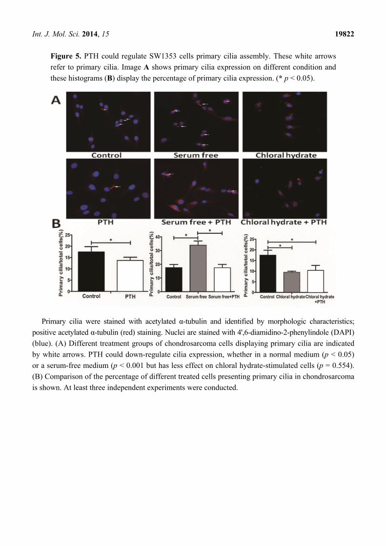
Figure 5. PTH could regulate SW1353 cells primary cilia assembly. These white arrows
refer to primary cilia. Image A shows primary cilia expression on different condition and
these histograms (B) display the percentage of primary cilia expression. (* p < 0.05).
Primary cilia were stained with acetylated α-tubulin and identified by morphologic characteristics;
positive acetylated α-tubulin (red) staining. Nuclei are stained with 4',6-diamidino-2-phenylindole (DAPI)
(blue). (A) Different treatment groups of chondrosarcoma cells displaying primary cilia are indicated
by white arrows. PTH could down-regulate cilia expression, whether in a normal medium (p < 0.05)
or a serum-free medium (p < 0.001 but has less effect on chloral hydrate-stimulated cells (p = 0.554).
(B) Comparison of the percentage of different treated cells presenting primary cilia in chondrosarcoma
is shown. At least three independent experiments were conducted.

Int. J. Mol. Sci. 2014, 15 19823
Figure 6. PTHrP silence could increase primary cilia expression. (A) Using siRNA to
silence PTHrP gene expression caused elevated ciliogenesis, compared with the empty
control (p = 0.003) and negative (p = 0.000) groups; (B) The relative percentage of
primary cilia in different treatment groups is shown. Three independent experiments were
conducted. The white arrows refer to primary cilia. (* p < 0.05).
At the same time, if we used chloral hydrate to destroy primary cilia and then treated them with
PTH, primary cilia showed no significant difference (9.38% ± 0.62% to 10.33% ± 2.67%, p > 0.05).
What is more, when we used chemical synthetic small interfering RNA to down-regulate endogenous
PTHrP expression, the percentage of primary cilia rose distinctly, compared with the empty control

Int. J. Mol. Sci. 2014, 15 19824
(31.50% ± 3.90% to 21.18% ± 3.97%, p < 0.05) and negative control (31.50% ± 3.90% to 17.88% ± 5.32%,
p < 0.001) groups (Figure 6).
Furthermore, knocking down endogenous PTHrP was accompanied by slightly elevated expression
of cilia relevant intraflagellar transport protein IFT88 (Figure 7). These outcomes reminded us that
PTH/PTHrP may have a reverse-regulation impact on primary cilia morphology expression and
eventually achieve function depending on complete cilia.
Figure 7. siRNA could decrease PTHrP expression in both protein and gene levels. The
left image A shows the effect of siRNA on PTHrP and IFT88 proteins expression; The
right one (B) display the interfere efficiency of siRNA on PTHrP in gene level. (* p < 0.05).
We used small interference RNA to reduce PTHrP RNA; three days later, PTHrP protein and gene
expression decreased distinctly, compared with the empty control (p < 0.05) and negative control
(p < 0.05) groups. At the same time, reducing PTHrP could slightly elevate cilia-related intraflagellar
transport protein IFT88 expression. Three independent experiments were conducted.
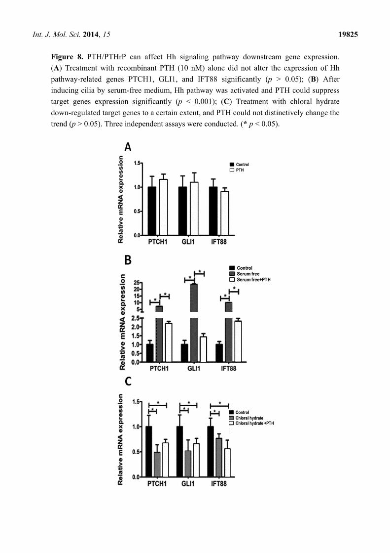
2.5. PTH/PTHrP Can Affect Hh Signaling Pathway Downstream Gene Expression
As a vital component of Hh signaling pathway, primary cilia play an important role in the
transferring signal [16]. Intraflagellar transport protein 88 (IFT88) is the most important transporting
signal molecule in primary cilia [21]. In addition, GLI1 and PTCH1 have been generally recognized as
the target genes of Hh signaling pathway [16]. We used RT-PCR to detect the influences that
PTH/PTHrP imposed on Hh signaling pathway. As Figure 8 shows, these results suggested that simple
treatment with PTH had no obvious effect on Hh pathways, compared with the control group
(Figure 8A, p > 0.05), while the pathways could be activated by serum-free stimulated and increased
IFT88, GLI1, and PTCH1 genes’expression, to some extent (Figure 8B, p < 0.05). In addition, PTH
could down-regulate Hh signaling pathway (Figure 8B, p < 0.001) by inhibiting ciliogenesis. After
destroying primary cilia by chloral hydrate, PTH had no significant effects on downstream gene
expression (Figure 8C, p > 0.05).
In addition, Western blot assay showed that in reducing endogenous PTHrP, intraflagellar
transport protein IFT88 expression was somewhat higher (Figure 7A). These findings illustrated that
PTH/PTHrP could affect Hh downstream gene expression through a negative regulation of primary cilia.
Int. J. Mol. Sci. 2014, 15 19825
Figure 8. PTH/PTHrP can affect Hh signaling pathway downstream gene expression.
(A) Treatment with recombinant PTH (10 nM) alone did not alter the expression of Hh
pathway-related genes PTCH1, GLI1, and IFT88 significantly (p > 0.05); (B) After
inducing cilia by serum-free medium, Hh pathway was activated and PTH could suppress
target genes expression significantly (p < 0.001); (C) Treatment with chloral hydrate
down-regulated target genes to a certain extent, and PTH could not distinctively change the
trend (p > 0.05). Three independent assays were conducted. (* p < 0.05).

Int. J. Mol. Sci. 2014, 15 19826
As a key regulatory factor, PTHrP plays an important part in the growth and development of bone
and cartilage tissue in vivo. In normal cartilage, PTHrP secretes through autocrine and paracrine matter.
Binding with PTHrP receptor-1, PTHrP can delay chondrocyte differentiation and promote cell
proliferation to regulate the developmental processes of cartilage [7–12]. However, abnormal
expression of PTHrP can cause a range of metabolic function disorders in vivo [8]. There have been a
variety of studies that found malignancy accompanied with high expression of PTHrP is also
associated with hypercalcemia [7–11]. PTHrP can activate oncogene Bcl-2 expression and is
considered to be an upstream regulator of Bcl-2 [25]. Otherwise, PTHrP is also regarded as a major
regulator of bone tumor-caused osteolytic destruction [8–11]. These studies suggested that the
abnormal expression of PTHrP has a close relationship with the occurrence of malignancy.
As a special type of tumor that can secrete cartilage matrix, chondrosarcoma occurs from abnormal
development of cartilage. The level of malignancy depends on the degree of tumor cell differentiation [16].
Although the specific pathogenesis is not clear, in this study we found that human chondrosarcoma
tissues had a distinct PTHrP expression, and the expression levels were positively correlated with the
tumor proliferation rate.
This phenomenon suggests that aberrant expression of PTHrP may cause cell proliferation to be
increased while differentiation is inhibited, leading to the occurrence of chondrosarcoma. Various
studies have shown that PTH is the decisive factor that causes osteolytic destruction in primary or
metastatic bone tumors [8–11]. In our in vitro experiments, an appropriate concentration of PTH (1-84)
could promote proliferation of SW1353 human chondrosarcoma cells and strengthen the ability to
secrete more matrix metalloproteinase MMP2 and MMP9, thereby enhancing degradation of
extracellular matrix and facilitating the ability to migrate. The Transwell invasion assay results also
confirmed this conclusion. Thus, PTHrP is likely to play a key role in regulating chondrosarcoma
dedifferentiation during development.
Primary cilia, as cell superficial structures, can detect the changes of extracellular mechanical
stimulation and biochemical environment [17–19]. Primary cilia are considered to be a critical point of
many signaling pathways, regulating interactions. Previous studies have found that normal cartilage
cells express a higher level of primary cilia than neoplastic chondrocytes [16], by which they can
feel external mechanical stress and internal osmotic pressure changes, thereby regulating cartilage
cells’ growth and differentiation [17,19,26]. Primary cilia historically have been regarded as a
biomarker for the end of division and out of cell cycle, which can objectively reflect the cell
proliferation stage, and the inner intraflagellar transport protein IFT88 represents the physiologic
function of cilia [20,21].
3. Experimental Section
The study was approved by the Ethics Committee of Tongji Medical College, Huazhong University
of Science and Technology, Wuhan, Hubei, China.
3.1. Cells and Reagents
The human chondrosarcoma cell line SW1353 was purchased from the Type Culture Collection of
the Chinese Academy of Sciences, Shanghai, China. Cells were cultured in Dulbecco’s modified Eagle

Int. J. Mol. Sci. 2014, 15 19827
medium/nutrient mixture F-12 (DMEM/F-12) with 10% fetal bovine serum, penicillin (100 U/mL) and
streptomycin (100 U/mL) and incubated at 37 °C with 5% CO2. Human recombinant PTH (1-84) was
purchased from ProSpec, Ness-Ziona, Israel.
3.2. Cell Proliferation Test
We used an assay of 3-(4,5-dimethylthiazol-2-yl)-5-(3-carboxymethoxyphenyl)-2-(4-sulfophenyl)-2H-
tetrazolium (MTS-8) to evaluate the cell proliferation rate. The steps were as follows: 2000 cells per
well were plated in 96-well plates. Different stimulation reagents were added to the cells based on the
experiment’s design. We added 100 μL DMEM/F12 with 10% fetal bovine serum and incubated for a
suitable time. Then 10 μL MST-8 (Boster, Wuhan, China) was added to each well. Two hours later,
the OD value (absor-bance at 450 nm wavelength) was measured using an enzyme micro-plate reader.
The cell viability was expressed by the OD value.
3.3. Transwell Invasion Assay
Transwell assay was used to assess chondrosarcoma cells’ invasion ability. The upper filter
membrane (pore diameter, 8 μm) of the Transwell plates was coated with 25 mg Matrigel (BD
Biosciences, San Jose, CA, USA) at 37 °C environment for 30 min. The lower chambers were filled
with cell culture medium DMEM/F-12 containing 10% fetal bovine serum. SW1353 cells were first
starved in serum-free DMEM for 4 h. After digestion 1 × 10⁵ cells were transferred onto the upper
surface. Based on experimental design, we added either 100 nM PTH, or 40 μM chloral hydrate and
also both. Chloral hydrate was used to disrupt the junction between primary cilium and the basal body.
Twenty-four hours later, we gently wiped the upper surface of the Matrigel. Paraformaldehyde at 4%
was used to fix the cells and allowed to penetrate to the lower surface for 15 min. After washing three
times, we stained the cells with crystal violet (Boster, Wuhan, China). Ten random visual fields were
selected and counted under the micro-scope (Olympus, Tokyo, Japan).
3.4. Immunohistochemical Studies
Human chondrosarcoma tissues were fixed in 4% paraformaldehyde. We then embedded tissues in
paraffin and sectioned it for immunohistochemical assays. All these experimental processes were
conducted using standard techniques based on previous studies [9,16]. Primary antibodies PTHrP
(1:100 dilution, Santa Cruz Biotechnology, Santa Cruz, CA, USA) and Ki67 (1:200 dilution, Cell
Signaling Technology, Danvers, MA, USA) were stained. All the sections were observed, and photos
were taken under microscopic magnification ×200.
3.5. Immunofluorescence Assay
Chondrosarcoma SW1353 cells of the proper density were inoculated on cover glasses and stimulated
with different reagents. Twenty-four hours later, they were fixed with 4% paraformaldehyde for 15 min,
blocked with 0.5% BSA at room temperature for 60 minutes, and then incubated with primary acetylated
α-tubulin antibody (1:300 dilution, Abcam, Cambridge, UK) overnight at 4 °C. Subsequently, cells were
incubated with CY3-conjugated goat anti-mouse IgG secondary antibody at room temperature, and the

Int. J. Mol. Sci. 2014, 15 19828
nuclei were stained with 1 µg/µL DAPI. PBS was used to wash the cells three times for 10 min during
each step in this process. Finally, images were visualized and recorded with a fluorescent microscope.
3.6. Western Blot Analysis
The steps for Western blot analysis were as previously described [10]. Total cell proteins (40 μg/lane)
were loaded and separated by use of sodium dodecyl sulfate (SDS)-polyacrylamide gels, and then the
proteins were trans-ferred onto PVDF membranes. We incubated these membranes overnight with
primary antibodies-MMP2 (1:1000 dilution, Cell Signaling Technology, Danvers, MA, USA); MMP9
(1:1000 dilution, Cell Signaling Technology, Danvers, MA, USA); IFT88 (1:500 dilution, ABGENT,
San Diego, CA, USA); PTHrP (1:100 dilution, Santa Cruz Biotechnology, Santa Cruz, CA, USA);
β-actin (1:400 dilution, Boster, Wuhan, China)-following up with secondary antibody horse-radish
peroxidase-labeled goat anti-rabbit and goat anti-mouse (1:5000 dilution, Boster, Wuhan, China) IgG for
one hour. The ECL Western blotting detection kit (Thermo Fisher Scientific, Geel, Antwerpen, Belgium)
was used to detect all the protein bands and visu-alized using an enhanced chemiluminescence system
(Bio-Rad, Philadelphia, PA, USA). All the values were expressed relative to β-actin.
3.7. Quantitative Real-Time PCR (qRT-PCR)
Ihh/PTHrP feedback loop-related genes (GLI1, PTCH1, IFT88, PTHrP) were measured by
real-time PCR (RT-PCR). After incubation for 24 h with different stimulation reagents in six-well
plates, RNA was extracted from the cells using RNeasy Mini Kit (Invitrogen Life Technologies,
New York, NY, USA), according to the manufacturer’s instructions. cDNA was synthesized from 2 to
5 μg of total RNA using the First-Strand Synthesis System for RT-PCR (Invitrogen Life Technologies,
New York, NY, USA), per the manufacturer’s instructions. The total PCR system contained cDNA,
SYBR Green, no RNA enzyme, water and primers. The primer sequences were as outlined in
the descriptions.
3.8. Small-Interfering RNA Transfection
SW1353 chondrosarcoma cells were transitorily transfected with 50 nM siRNA targeting PTHrP or
with a scrambled sequence (Negative Control siRNA) for 6 h using Lipofectamine 2000 (Invitrogen
Life Technologies, New York, NY, USA). Cells were maintained in growth media for 72 h. The
efficiency of the knockdown specific gene was evaluated by qRT-PCR and Western blot assays, as
previously described. Both small interfering RNA and negative control siRNA were synthesized by the
Guangzhou RiboBio Company, Guangzhou, China.
3.9. Statistical Analysis
Each experiment was conducted at least three times. These data were represented as mean ± SD.
We used the student’s t-test or one-way analysis of variance to analyze the differences among means.
A p-value less than 0.05 was defined as statistically significant. All statistical analyses were performed
using SPSS 20.0 (IBM, Armonk, NY, USA).

Int. J. Mol. Sci. 2014, 15 19829
4. Conclusions
In this study, we found that recombinant PTH (1-84) can increase human chondrosarcoma cell
proliferation activity, accompanied by inhibition of extracellular primary cilia generation and IFT88
expression. In addition, we used chloral hydrate to disrupt the junction of the cilium and the basal body
to inactivate cilia. After destroying the cilia by means of chloral hydrate, treatment with recombinant
PTH had no significant effect on Hh downstream genes GLI1 and PTCH1, together with intraflagellar
transport protein IFT88.
Interestingly, chemical destruction of the cilia may lead to the proliferation and invasion abilities
distinctly inhibited in tumor cells. Therefore, we first noticed that through dependence on primary cilia,
PTH can achieve its facilitation function and regulate ciliogenesis oppositely, eventually affecting the
proliferation and invasion activity of tumor cells. Details of the specific mechanism of PTH acting on
primary cilia are a possible focus of attention in future research.
The study of how PTHrP affects chondrosarcoma cell proliferation and invasion in regulating
primary cilia expression have provided us with a new way of viewing the pathogenesis of
chondrosarcoma. How to manipulate primary cilia assembly to restrain PTH-mediated tumorigenesis
could be a question deserving in-depth investigation. These conclusions may have great significance in
guiding future chemotherapy drug investigation and improving clinical treatment efficacy on
human chondrosarcoma.
Acknowledgments
This research was supported by the National Natural Science Foundation of China (Grant
No.81202121) and the Hubei Provincial Health Department of Young Scientists Fund (Grant
No. QJX2012-05).
Author Contributions
Wei Xiang was responsible for preparing the final illustrations and writing this manuscript.
Ting Jiang, Chen Gong, Peng Cheng and Libo Zhao equally took part in this study, they conducted
all experiments independently. Fengjing Guo and Tao Xu guided the statistical analysis of data.
Weiting Cheng mainly repeated some Western blots. Kai Xu was responsible for providing
experimental ideas, assembling a team, guiding experiments and funding this study.
Conflicts of Interest
The authors declare no conflict of interest.
References
1. Bjornsson, J.; McLeod, R.A.; Unni, K.K.; Ilstrup, D.M.; Pritchard, D.J. Primary chondrosarcoma
of long bones and limb girdles. Cancer 1998, 83, 2105–2119.
Int. J. Mol. Sci. 2014, 15 19830
2. Schrage, Y.M.; Briaire-de Bruijn, I.H.; de Miranda, N.F.; van Oosterwijk, J.; Taminiau, A.H.;
van Wezel, T.; Hogendoorn, P.C.; Bovee, J.V. Kinome profiling of chondrosarcoma reveals
src-pathway activity and dasatinib as option for treatment. Cancer Res. 2009, 69, 6216–6222.
3. Li, X.; Ye, H.; Cai, L.; Yu, F.; Chen, W.; Lin, R.; Zheng, C.; Xu, H.; Ye, J.; Wu, G.; et al.
Millimeter wave radiation induces apoptosis via affecting the ratio of bax/bcl-2 in sw1353 human
chondrosarcoma cells. Oncol. Rep. 2012, 27, 664–672.
4. Bovee, J.V.; Cleton-Jansen, A.M.; Taminiau, A.H.; Hogendoorn, P.C. Emerging pathways in the
development of chondrosarcoma of bone and implications for targeted treatment. Lancet Oncol.
2005, 6, 599–607.
5. Gelderblom, H.; Hogendoorn, P.C.; Dijkstra, S.D.; van Rijswijk, C.S.; Krol, A.D.; Taminiau, A.H.;
Bovee, J.V. The clinical approach towards chondrosarcoma. Oncologist 2008, 13, 320–329.
6. Dickey, I.D.; Rose, P.S.; Fuchs, B.; Wold, L.E.; Okuno, S.H.; Sim, F.H.; Scully, S.P.
Dedifferentiated chondrosarcoma: The role of chemotherapy with updated outcomes. J. Bone
Jt. Surg. 2004, 86, 2412–2418.
7. Johnson, R.W.; Merkel, A.R.; Danilin, S.; Nguyen, M.P.; Mundy, G.R.; Sterling, J.A.
6-thioguanine inhibition of parathyroid hormone-related protein expression is mediated by GLI2.
Anticancer Res. 2011, 31, 2705–2712.
8. Mak, I.W.; Turcotte, R.E.; Ghert, M. Parathyroid hormone-related protein (pthrp) modulates
adhesion, migration and invasion in bone tumor cells. Bone 2013, 55, 198–207.
9. Mak, I.W.; Cowan, R.W.; Turcotte, R.E.; Singh, G.; Ghert, M. Pthrp induces autocrine/paracrine
proliferation of bone tumor cells through inhibition of apoptosis. PLoS One. 2011, 6, e19975.
10. Johnson, R.W.; Nguyen, M.P.; Padalecki, S.S.; Grubbs, B.G.; Merkel, A.R.; Oyajobi, B.O.;
Matrisian, L.M.; Mundy, G.R.; Sterling, J.A. TGF-β promotion of Gli2-induced expression of
parathyroid hormone-related protein, an important osteolytic factor in bone metastasis, is
independent of canonical hedgehog signaling. Cancer Res. 2011, 71, 822–831.
11. Soki, F.N.; Park, S.I.; McCauley, L.K. The multifaceted actions of pthrp in skeletal metastasis.
Future Oncol. 2012, 8, 803–817.
12. Xu, K.; Guo, F.; Zhang, S.; Liu, C.; Wang, F.; Zhou, Z.; Chen, A. Blocking Ihh signaling pathway
inhibits the proliferation and promotes the apoptosis of PSCs. J. Huazhong Univ. Sci. Technol.
Med. Sci. 2009, 29, 39–44.
13. Steere, N.; Chae, V.; Burke, M.; Li, F.Q.; Takemaru, K.; Kuriyama, R. A Wnt/beta-catenin
pathway antagonist chibby binds cenexin at the distal end of mother centrioles and functions in
primary cilia formation. PLoS One 2012, 7, e41077.
14. May-Simera, H.L.; Kelley, M.W. Cilia, wnt signaling, and the cytoskeleton. Cilia 2012, 1,
doi:10.1186/2046-2530-1-7.
15. Lienkamp, S.; Ganner, A.; Walz, G. Inversin, wnt signaling and primary cilia. Differentiation
2012, 83, S49–S55.
16. Ho, L.; Ali, S.A.; Al-Jazrawe, M.; Kandel, R.; Wunder, J.S.; Alman, B.A. Primary cilia attenuate
hedgehog signalling in neoplastic chondrocytes. Oncogene 2013, 32, 5388–5396.
17. Hoey, D.A.; Tormey, S.; Ramcharan, S.; O’Brien, F.J.; Jacobs, C.R. Primary cilia-mediated
mechanotransduction in human mesenchymal stem cells. Stem Cells 2012, 30, 2561–2570.
Int. J. Mol. Sci. 2014, 15 19831
18. Proulx-Bonneau, S.; Annabi, B. The primary cilium as a biomarker in the hypoxic adaptation of
bone marrow-derived mesenchymal stromal cells: A role for the secreted frizzled-related proteins.
Biomark. Insights 2011, 6, 107–118.
19. Muhammad, H.; Rais, Y.; Miosge, N.; Ornan, E.M. The primary cilium as a dual sensor of
mechanochemical signals in chondrocytes. Cell Mol. Life Sci. 2012, 69, 2101–2107.
20. Goto, H.; Inoko, A.; Inagaki, M. Cell cycle progression by the repression of primary cilia
formation in proliferating cells. Cell Mol. Life Sci. 2013, 70, 3893–3905.
21. Irigoin, F.; Badano, J.L. Keeping the balance between proliferation and differentiation: The
primary cilium. Curr. Genomics 2011, 12, 285–297.
22. Yang, Y.; Roine, N.; Makela, T.P. Ccrk depletion inhibits glioblastoma cell proliferation in a
cilium-dependent manner. EMBO Rep. 2013, 14, 741–747.
23. Hassounah, N.B.; Bunch, T.A.; McDermott, K.M. Molecular pathways: The role of primary cilia
in cancer progression and therapeutics with a focus on hedgehog signaling. Clin. Cancer Res.
2012, 18, 2429–2435.
24. Basten, S.G.; Willekers, S.; Vermaat, J.S.; Slaats, G.G.; Voest, E.E.; van Diest, P.J.; Giles, R.H.
Reduced cilia frequencies in human renal cell carcinomas versus neighboring parenchymal tissue.
Cilia 2013, 2, doi:10.1186/2046-2530-2-2.
25. Bovee, J.V.; van den Broek, L.J.; Cleton-Jansen, A.M.; Hogendoorn, P.C. Up-regulation of
PTHrP and Bcl-2 expression characterizes the progression of osteochondroma towards
peripheral chondrosarcoma and is a late event in central chondrosarcoma. Lab. Invesigt. 2000, 80,
1925–1934.
26. Rich, D.R.; Clark, A.L. Chondrocyte primary cilia shorten in response to osmotic challenge and
are sites for endocytosis. Osteoarthr. Cartil. 2012, 20, 923–930.
© 2014 by the authors; licensee MDPI, Basel, Switzerland. This article is an open access article
distributed under the terms and conditions of the Creative Commons Attribution license
(http://creativecommons.org/licenses/by/4.0/).