evaluation of the biocompatibility of a new biomembrane 1. introduction 2. materials and methods
TRANSCRIPT

Vol. 7, No. 2, 2004 Evaluation of The Biocompatibility of a New Biomembrane 277Materials Research, Vol. 7, No. 2, 277-283, 2004. © 2004
*e-mail: [email protected]
Evaluation of The Biocompatibility of a New Biomembrane
Fatima Mruea*, Joaquim Coutinho Nettob, Reginaldo Cenevivaa, João José Lachatc,
José Antônio Thomazinic, Helder Tambelinid
aUniversity of São Paulo, Faculty of Medicine, Department of SurgerybUniversity of São Paulo, Faculty of Medicine, Department of BiochemistrycUniversity of São Paulo, Faculty of Medicine, Department of Morphology
dUniversity of São Paulo, General BioteriunFaculdade de Medicina de Ribeirão Preto - USP, Departamento de Bioquímica e
Imunologia, Av. Bandeirantes, 3900,14049-900 Ribeirão Preto - SP, Brazil
Received: August 18, 2003; Revised: January 18, 2004
Biocompatibility has been considered one of the most important items to validate a biomaterialfor its application in human organisms. The present work evaluates the biocompatibility of a newbiomembrane using in vivo assay in different animal species. The experiments to evaluate the cellularreaction were carried out through the implantation of the material into the subcutaneous tissue ofanimals and the results showed a good reaction of the host tissue without any signal of fibrosis orrejection. The cell adhesion experiments were done by means of the measure of the DNA content onthe material surface after its implantation into the subcutaneous tissue of animals and the resultsshowed a growing number of DNA that was proportional to the time of implantation. The healingprocess was evaluated using a dermal ulcer model and the results showed a good tissue repairresembling a physiologic process. The overall results presented here lead to the conclusion that thisnew biomembrane is a biocompatible material but more research must be done, as it is a new materialdesired for medical use.
Keywords: Biomaterial, biomembrane, biocompatibility
1. Introduction
To manage a variety of challenging clinical situationscaused by the loss, injury or disease of important or vitalorgans and tissues such as bone, soft tissue, heart, trachea,blood vessels, dermal ulcers, etc., researchers have long beenengaged in seeking an ideal material capable of meeting theessential criteria of biocompatibility, innocuity, easy handling,and affordability for the people who need it. Several materialshave been developed for these purposes, including silicon,polypropylene, hydroxyapatite and others. Becausebiocompatibility is one of the most important requirementsfor these materials, in-depth studies are required of their char-acteristics and of their interaction with the humans in whichthey are expected to work.
Biocompatibility can be evaluated by “in vivo” and “invitro” assays. Several evaluative methods have been de-scribed, such as cellular reaction1, cell adhesion2 and thehealing process3.
In this article, we present a new biomembrane designed
for medical applications in tissue repair, evaluating it fromthe standpoint of its biocompatibility.
2. Materials and Methods
The biomembrane
The new biomembrane presented here is made of natu-ral latex extracted from the rubber tree Hevea brasiliensisand manufactured by a process that differs from the con-ventional technique used in the rubber industry for manu-facturing gloves, drains, tires, urinary or tracheal tubes andother devices.
Manufacturing process
The process, which is very simple, is briefly describedbelow.
Natural latex is collected from the rubber tree, usingammonia as the only preservative. The latex thus collected
278 Mrué et al. Materials Research
is centrifuged to reduce its protein content, particularly theallergenic proteins. A sulfur composition is then added asthe only curing agent, after which the latex is polymerizedat low temperatures in a glass mold, a procedure that resultsin a biomembrane. Finally, the product is sterilized by theethylene oxide method.
Biocompatibility tests
Cellular reaction
The cellular reaction was studied through subcutaneous im-plants of 0.5 × 0.5 cm fragments of the biomembrane in the ven-tral wall of mongrel dogs. The implantation sites were at least 2.0cm apart. The experiment involved 6 dogs, each of which re-ceived 8 implanted fragments. Two fragments, including the sur-rounding tissue, were removed weekly from each dog, from the 1st
to the 4th week. The tissues thus removed were prepared for histo-logical examination using hematoxylin-eosin and Masson’s tri-chrome method. This study focused on the reactions to for-eign bodies and on the presence of fibrotic tissue.
Cell adhesion
This experiment involved the implantation of fragmentsof the biomembrane in the subcutaneous tissue of dogs, asdescribed above, and in the peritoneal cavity of rats. Thefragments were subsequently removed for quantitativeanalyses and morphological studies.
Quantitative analysis
The implanted fragments were removed according tothe following schedule: 0, 2, 6, 12 and 24 h after implanta-tion. They were then subjected to a DNA analysis, as de-scribed by Labarca and Paigen4.
Morphological analysis
The morphological features of the adhered cells wereexamined by scanning electron microscopy 12 and 24 hafter implantation.
Healing process
The healing process was studied using the dermal ulcermodel in rabbit ear5. Four 0.5 cm ulcers were made to thedepth of the cartilage in each rabbit ear, using a 0.5 cm circu-lar dermatologic punch device. Two groups were set up. Inone group, the ulcer was treated with a fragment of thebiomembrane, while in the other group, which served as thecontrol group, the ulcer was treated with ointment (Fibrase –Aché). The healing process was evaluated after periods of 1,3, 7 and 14 days following surgery, comprising 6 events ineach period. The treated ulcer was removed as described previ-ously and prepared for histological study using hematoxylin-eosin and Masson’s trichrome method. The study focused onthe presence of new vessels, epithelialization and fibrosis.
3. Results
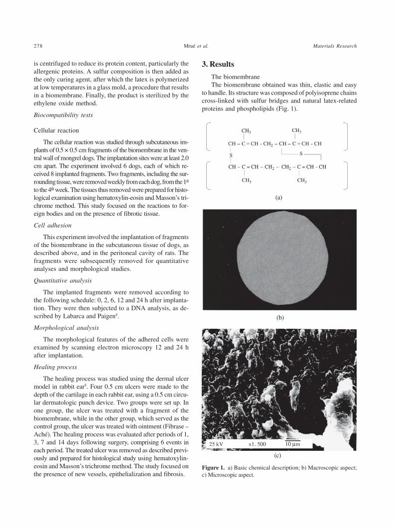
The biomembraneThe biomembrane obtained was thin, elastic and easy
to handle. Its structure was composed of polyisoprene chainscross-linked with sulfur bridges and natural latex-relatedproteins and phospholipids (Fig. 1).
Figure 1. a) Basic chemical description; b) Macroscopic aspect;c) Microscopic aspect.
(c)
(b)
(a)
Vol. 7, No. 2, 2004 Evaluation of The Biocompatibility of a New Biomembrane 279
Cellular reaction
Inflammatory cells
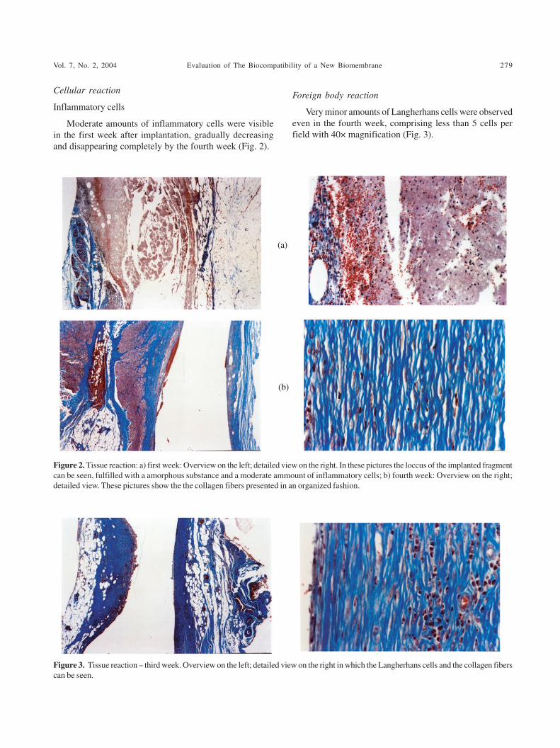
Moderate amounts of inflammatory cells were visiblein the first week after implantation, gradually decreasingand disappearing completely by the fourth week (Fig. 2).
Foreign body reaction
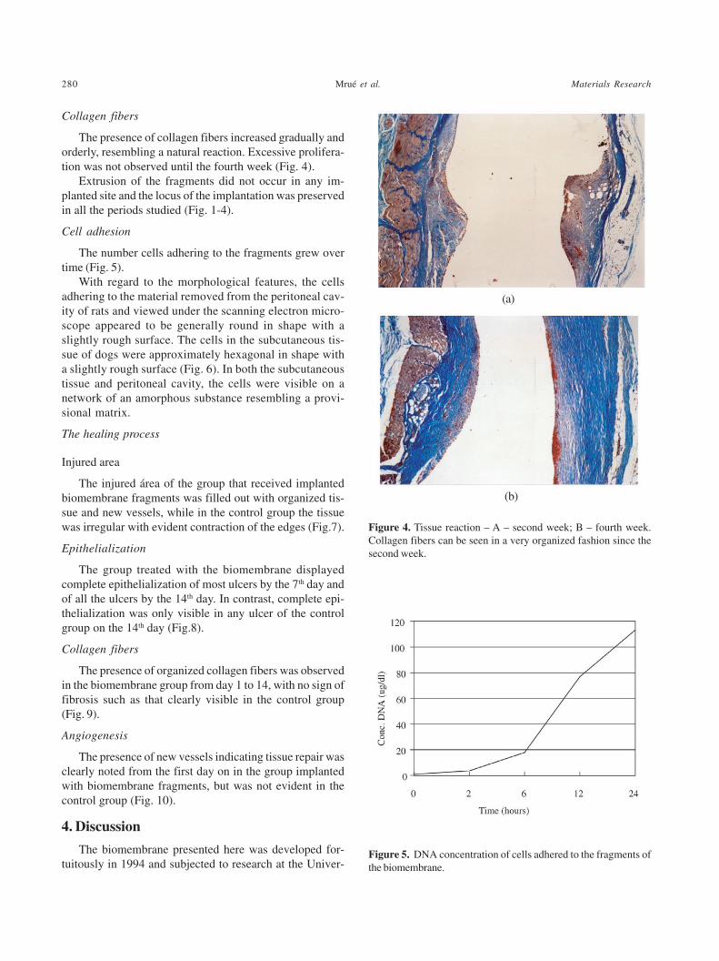
Very minor amounts of Langherhans cells were observedeven in the fourth week, comprising less than 5 cells perfield with 40× magnification (Fig. 3).
Figure 2. Tissue reaction: a) first week: Overview on the left; detailed view on the right. In these pictures the loccus of the implanted fragmentcan be seen, fulfilled with a amorphous substance and a moderate ammount of inflammatory cells; b) fourth week: Overview on the right;detailed view. These pictures show the the collagen fibers presented in an organized fashion.
(b)
(a)
Figure 3. Tissue reaction – third week. Overview on the left; detailed view on the right in which the Langherhans cells and the collagen fiberscan be seen.
280 Mrué et al. Materials Research
Collagen fibers
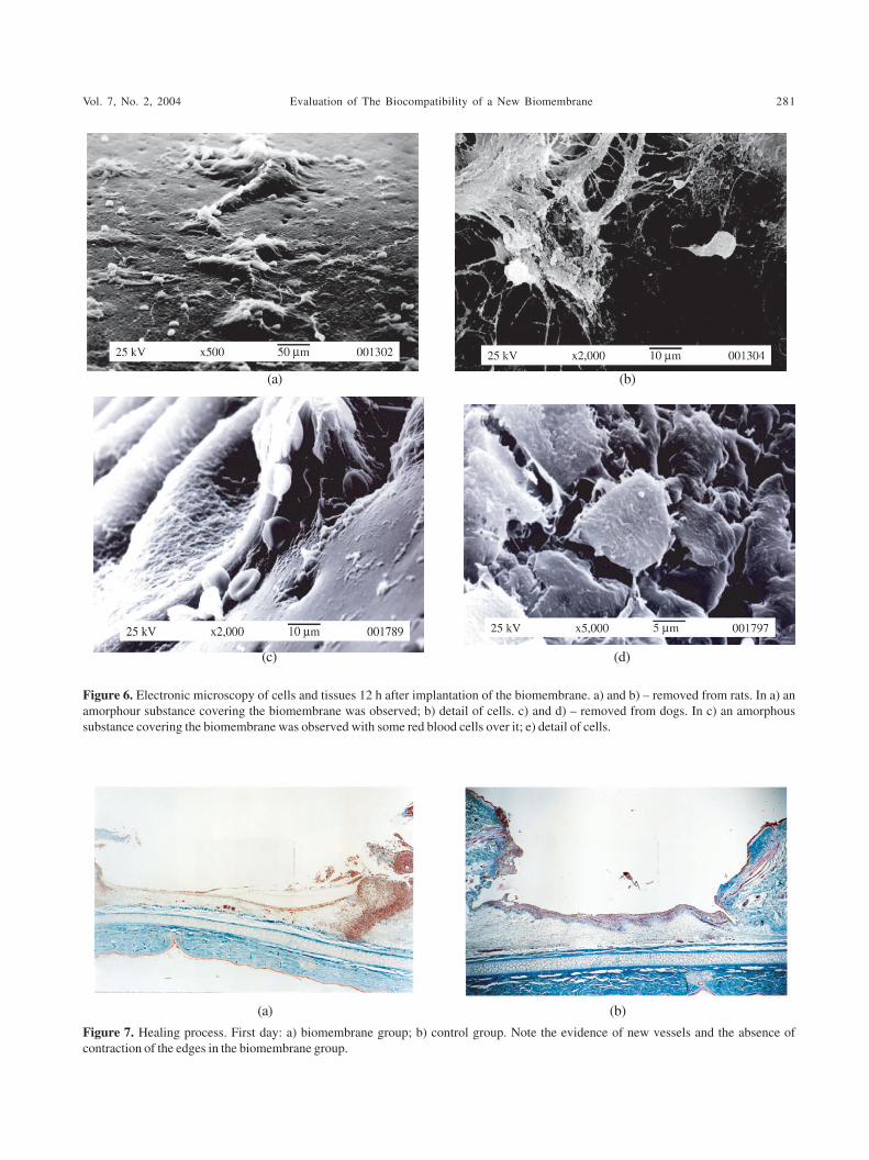
The presence of collagen fibers increased gradually andorderly, resembling a natural reaction. Excessive prolifera-tion was not observed until the fourth week (Fig. 4).
Extrusion of the fragments did not occur in any im-planted site and the locus of the implantation was preservedin all the periods studied (Fig. 1-4).
Cell adhesion
The number cells adhering to the fragments grew overtime (Fig. 5).
With regard to the morphological features, the cellsadhering to the material removed from the peritoneal cav-ity of rats and viewed under the scanning electron micro-scope appeared to be generally round in shape with aslightly rough surface. The cells in the subcutaneous tis-sue of dogs were approximately hexagonal in shape witha slightly rough surface (Fig. 6). In both the subcutaneoustissue and peritoneal cavity, the cells were visible on anetwork of an amorphous substance resembling a provi-sional matrix.
The healing process
Injured area
The injured área of the group that received implantedbiomembrane fragments was filled out with organized tis-sue and new vessels, while in the control group the tissuewas irregular with evident contraction of the edges (Fig.7).
Epithelialization
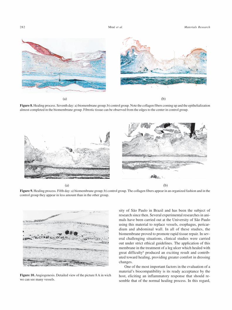
The group treated with the biomembrane displayedcomplete epithelialization of most ulcers by the 7th day andof all the ulcers by the 14th day. In contrast, complete epi-thelialization was only visible in any ulcer of the controlgroup on the 14th day (Fig.8).
Collagen fibers
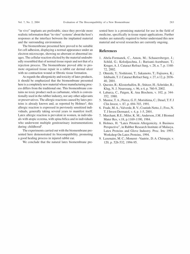
The presence of organized collagen fibers was observedin the biomembrane group from day 1 to 14, with no sign offibrosis such as that clearly visible in the control group(Fig. 9).
Angiogenesis
The presence of new vessels indicating tissue repair wasclearly noted from the first day on in the group implantedwith biomembrane fragments, but was not evident in thecontrol group (Fig. 10).
4. Discussion
The biomembrane presented here was developed for-tuitously in 1994 and subjected to research at the Univer-
(a)
(b)
Figure 4. Tissue reaction – A – second week; B – fourth week.Collagen fibers can be seen in a very organized fashion since thesecond week.
Figure 5. DNA concentration of cells adhered to the fragments ofthe biomembrane.
Vol. 7, No. 2, 2004 Evaluation of The Biocompatibility of a New Biomembrane 281
(a) (b)
Figure 6. Electronic microscopy of cells and tissues 12 h after implantation of the biomembrane. a) and b) – removed from rats. In a) anamorphour substance covering the biomembrane was observed; b) detail of cells. c) and d) – removed from dogs. In c) an amorphoussubstance covering the biomembrane was observed with some red blood cells over it; e) detail of cells.
Figure 7. Healing process. First day: a) biomembrane group; b) control group. Note the evidence of new vessels and the absence ofcontraction of the edges in the biomembrane group.
(a) (b)
(c) (d)
282 Mrué et al. Materials Research
Figure 10. Angiogenesis. Detailed view of the picture 8 A in wichwe can see many vessels.
Figure 8. Healing process. Seventh day: a) biomembrane group; b) control group. Note the collagen fibers coming up and the epithelializationalmost completed in the biomembrane group. Fibrotic tissue can be observed from the edges to the center in control group.
Figure 9. Healing process. Fifth day: a) biomembrane group; b) control group. The collagen fibers appear in an organized fashion and in thecontrol group they appear in less amount than in the other group.
(a) (b)
(a) (b)
sity of São Paulo in Brazil and has been the subject ofresearch since then. Several experimental researches in ani-mals have been carried out at the University of São Paulousing this material to replace vessels, esophagus, pericar-dium and abdominal wall. In all of these studies, thebiomembrane proved to promote rapid tissue repair. In sev-eral challenging situations, clinical studies were carriedout under strict ethical guidelines. The application of thismembrane in the treatment of a leg ulcer which healed withgreat difficulty6 produced an exciting result and contrib-uted toward healing, providing greater comfort in dressingchanges.
One of the most important factors in the evaluation of amaterial’s biocompatibility is its ready acceptance by thehost, eliciting an inflammatory response that should re-semble that of the normal healing process. In this regard,
Vol. 7, No. 2, 2004 Evaluation of The Biocompatibility of a New Biomembrane 283
“in vivo” implants are preferable, since they provide morerealistic information than “in vitro” systems7 about the host’sresponses at the interface between the implanted materialand the surrounding environment.
The biomembrane presented here proved to be suitablefor cell adhesion, displaying a normal appearance under anelectron microscope, showing no aberrant or abnormal im-ages. The cellular reaction elicited by the material undoubt-edly resembled that of normal tissue repair and not that of arejection process. The biomembrane proved able to pro-mote organized tissue repair in a rabbit ear dermal ulcerwith no contraction wound or fibrotic tissue formation.
As regards the allergenicity and toxicity of latex products,it should be emphasized that the biomembrane presentedhere is a completely new material whose manufacturing proc-ess differs from the traditional one. This biomembrane con-tains no toxic product such as carbamate, which is conven-tionally used in the rubber industry, nor any other adjuvantsor preservatives. The allergic reactions caused by latex pro-teins is already known and, as reported by Holmes8, thisallergic reaction is expressed in previously sensitized indi-viduals, generally taking several years to manifest itself.Latex allergic reaction is prevalent in women, in individu-als with atopic eczema, with spina bifica and in individualswho underwent multiple genitourinary instrumentationsduring childhood9.
The experiments carried out with the biomembrane pre-sented here demonstrated its biocompatibility, promotinga good healing process in injured rabbit ear.
We conclude that the natural latex biomembrane pre-
sented here is a promising material for use in the field ofmedicine, specifically in tissue repair applications. Furtherstudies are naturally required to better understand this newmaterial and several researches are currently ongoing.
References
1. Abela-Formanek, C., Amon, M.; Schauersberger, J.;Schild, G.; Kolodjaschna, J.; Barisani-Asenbauer, T.;Kruger, A. J. Cataract Refract Surg, v. 28, n. 7, p. 1160-72, 2002.
2. Ohnishi, Y.; Yoshitomi, T.; Sakamoto, T.; Fujisawa, K.;Ishibashi, T. J. Cataract Refract Surg, v. 27, n 12, p. 2036-40, 2001.
3. Quester, R.; Klosterhalfen, B.; Stützer, H.; Schröder, R.;Klug, N. J. Neurosurg, v. 96, n 4, p. 760-9, 2002.
4. Labarca, C., Paigen, K. Ana Biochem, v. 102, p. 344-352, 1980.
5. Mustoe, T. A.; Pierce, G. F.; Morishima, C.; Deuel, T. F. JClin Invest, v. 87, p. 694-703, 1991.
6. Frade, M. A.; Valverde, R. V.; Coutinh-Netto, J.; Foss, N.T. J Invest Dermatol, v. 4, p. 1-5, 2001.
7. Marchant, R.E.; Miler, K. M.; Anderson, J.M. J BiomedMater Res, v.18, p.1169-1190, 1984.
8. Holmes, H. “Latex Prtotein Allergenicity. A BusinessPerspective”, in Rubber Research Institute of Malasya.Latex Proteins and Glove Industry. Proc. Irtc 1993.Workshop On Latex Proteins, 1994.
9. Laxenaire, M. C.; Moneret - Vautrin , D. A. Chirurgie, v.120, p. 526-532, 1994-95.

284 Mrué et al. Materials Research