evolutionary origins of hepatitis a virus in small mammals
TRANSCRIPT

Evolutionary origins of hepatitis A virus insmall mammalsJan Felix Drexlera,b,1, Victor M. Cormana,b, Alexander N. Lukashevc, Judith M. A. van den Brandd, Anatoly P. Gmylc,e,Sebastian Brüninka, Andrea Raschea, Nicole Seggewiβf, Hui Fengg, Lonneke M. Leijtend, Peter Valloh,Thijs Kuikend, Andreas Dotzauerf, Rainer G. Ulrichi, Stanley M. Lemong, Christian Drostena,b,and the Hepatovirus Ecology Consortium2
aInstitute of Virology, University of Bonn Medical Centre, 53127 Bonn, Germany; bGerman Centre for Infection Research, Bonn-Cologne, Germany;cChumakov Institute of Poliomyelitis and Viral Encephalitides, Moscow 142782, Russia; dDepartment of Viroscience, Erasmus MC, 3000 CA, Rotterdam, TheNetherlands; eLomonosov Moscow State University, Moscow 119991, Russia; fLaboratory of Virus Research, University of Bremen, 28359 Bremen, Germany;gLineberger Comprehensive Cancer Center, The University of North Carolina at Chapel Hill, Chapel Hill, NC 27599-7292; hInstitute of Evolutionary Ecology andConservation Genomics, University of Ulm, 89069 Ulm, Germany; and iFriedrich-Loeffler-Institut, Institute for Novel and Emerging Infectious Diseases, 17493Greifswald–Insel Riems, Germany
Edited by Francis V. Chisari, The Scripps Research Institute, La Jolla, CA, and approved October 2, 2015 (received for review August 26, 2015)
Hepatitis A virus (HAV) is an ancient and ubiquitous human path-ogen recovered previously only from primates. The sole species ofthe genus Hepatovirus, existing in both enveloped and nonenvel-oped forms, and with a capsid structure intermediate between thatof insect viruses andmammalian picornaviruses, HAV is enigmatic inits origins. We conducted a targeted search for hepatoviruses in15,987 specimens collected from 209 small mammal species globallyand discovered highly diversified viruses in bats, rodents, hedge-hogs, and shrews, which by pairwise sequence distance comprise13 novel Hepatovirus species. Near-complete genomes from nine ofthese species show conservation of unique hepatovirus features,including predicted internal ribosome entry site structure, a trun-cated VP4 capsid protein lacking N-terminal myristoylation, a car-boxyl-terminal pX extension of VP1, VP2 late domains involved inmembrane envelopment, and a cis-acting replication element withinthe 3Dpol sequence. Antibodies in some bat sera immunoprecipi-tated and neutralized human HAV, suggesting conservation of crit-ical antigenic determinants. Limited phylogenetic cosegregationamong hepatoviruses and their hosts and recombination patternsare indicative of major hepatovirus host shifts in the past. Ancestralstate reconstructions suggest a Hepatovirus origin in small insectiv-orous mammals and a rodent origin of human HAV. Patterns ofinfection in small mammals mimicked those of human HAV in hep-atotropism, fecal shedding, acute nature, and extinction of the virusin a closed host population. The evolutionary conservation of hep-atovirus structure and pathogenesis provide novel insight into theorigins of HAV and highlight the utility of analyzing animal reser-voirs for risk assessment of emerging viruses.
hepatitis A virus | viral evolution | pathogenesis | zoonosis | small mammals
Small mammals such as bats and rodents have been implicatedfrequently in the evolution and spread of emerging viruses
(1). It is uncertain whether this reflects unique aspects of theirphysiology, immune response to infectious agents, or ecologicaltraits facilitating virus maintenance such as rapid population turn-over or tendencies to form large and gregarious social groups (2, 3).The emergence of Ebola virus from bats (4) and hantaviruses fromrodents (5) exemplifies the prominent contributions of these taxa toemerging zoonotic threats to human health, but the extent to whichsuch species have contributed to the evolution of well-establishedhuman pathogens such as hepatitis A virus (HAV) is less clear.HAV is unique among the Picornaviridae, a large and diverse
family of positive-strand RNA viruses (6), not only in its tropismfor the liver but also in its structure and life cycle. It infects via thefecal–oral route and is shed in feces as a naked, nonenvelopedparticle, but circulates in the blood cloaked in an envelope derivedfrom host cell membranes (7). Recent X-ray studies have revealedthat HAV possesses a primitive capsid structure related to that ofpicorna-like viruses infecting insects, hinting at both an ancient
evolutionary relationship and a novel mechanism of cell entry (8).The origins of HAV, however, remain shrouded in mystery. De-spite evidence of limited replication in guinea pigs (9), only higherprimates have been shown to be fully permissive for infection.HAV strains show little variation in nucleotide sequence over timeor geography, forming six closely related genotypes comprisingonly a single serotype (10). Unlike other human hepatitis viruses,HAV infections never persist and uniformly engender lifelong,likely antibody-mediated immunity against reinfection (11).HAV has thus disappeared previously from small, isolated humanpopulations (12, 13), raising questions as to how it could haveevolved in early human hunter–gatherer societies.
ResultsIdentification of Nonprimate Hepatoviruses. To elucidate the evolu-tionary origins of HAV, we sought the presence of HAV-relatedviruses in 15,987 specimens (tissue, blood, and feces) collectedglobally from 209 species of small mammals in five different mam-malian orders: Rodentia (rodents), Scandentia (treeshrews), Chi-roptera (bats), Eulipotyphla (hedgehogs, shrews), and Afrosoricida
Significance
The origins of human hepatitis A virus (HAV) are unknown. Weconducted a targeted search for HAV-related viruses in smallmammals sampled globally and discovered highly diversifiedviruses in bats, rodents, hedgehogs, and shrews. We demon-strate that these viruses share unique biological features withHAV, including structural, genomic, antigenic, and pathogenicproperties. We found evidence of major shifts of HAV-relatedviruses between mammalian hosts in the past, suggesting bothan origin of this viral genus in small mammals and a zoonoticorigin of human HAV. Our data show that risk assessments foremerging viruses can benefit greatly from the analysis of viralinfection patterns that evolved within animal reservoirs.
Author contributions: J.F.D. designed research; J.M.A.v.d.B., A.P.G., S.B., A.R., N.S., H.F., L.M.L.,and P.V. performed research; H.E.C. performed and designed field work; R.G.U. contributednew reagents/analytic tools; J.F.D., V.M.C., A.N.L., T.K., A.D., R.G.U., S.M.L., and C.D. ana-lyzed data; and J.F.D., S.M.L., and C.D. wrote the paper.
The authors declare no conflict of interest.
This article is a PNAS Direct Submission.
Data deposition: The sequences reported in this paper have been deposited in the Gen-Bank database, www.ncbi.nlm.nih.gov/genbank (accession nos. KT452631–KT452765).
See Commentary on page 15010.1To whom correspondence should be addressed. Email: [email protected] complete list of the Hepatovirus Ecology Consortium can be found in the SupportingInformation.
This article contains supporting information online at www.pnas.org/lookup/suppl/doi:10.1073/pnas.1516992112/-/DCSupplemental.
15190–15195 | PNAS | December 8, 2015 | vol. 112 | no. 49 www.pnas.org/cgi/doi/10.1073/pnas.1516992112
Dow
nloa
ded
by g
uest
on
Oct
ober
18,
202
1

(tenrecs; Fig. 1A, Fig. S1A, and Table S1). Hepatoviruses wereidentified by a broadly reactive nested RT-PCR assay targeting theVP2 domain that is among the most highly conserved segments ofthe polyprotein-coding RNA. A total of 117 specimens (0.7%)were positive, originating from five continents and a total of 28different nonprimate hosts, including 13 bat species, 13 rodentspecies, 1 shrew, and 1 hedgehog species (GenBank accession nos.KT452631–KT452747). Bayesian phylogenetic reconstructiondemonstrated considerably more diversity among these novelviruses than primate HAVs, with seven deeply branching cladesforming an extended Hepatovirus tree (Fig. 1B and Fig. S1B).Phylogenetically basal clades contained shrew and bat viruses fromCentral Europe (clade I), African rodent viruses (clade II),Malagasy and European bat viruses (clade III), American, European,and African bat viruses (clade IV), and African and European batand hedgehog viruses (clade V). Rodent viruses from North America(clade VI), Asia, and Central Europe (clade VII) clustered phylo-genetically in an immediate sister relationship to primate hepatovirusesthat form an apical monophyletic group. Pairwise sequence distancesindicate these novel viruses represent 13 new Hepatovirusspecies (Fig. S1C).Only limited phylogenetic cosegregation was evident for these
novel nonprimate hepatoviruses and their mammalian host spe-cies. When categorized according to host order (Fig. 1C, nodesa–c), viruses from cricetid rodents in Central Europe and Asia(node a1), Northern America (node a2), and murid rodentsfrom Africa (node a3) were not monophyletic. Similarly, virusesfrom different bat hosts sampled globally occupied three differentphylogenetic positions (nodes b1–b3), whereas viruses fromEulipotyphla (hedgehogs and shrews) occupied two differentphylogenetic positions (nodes c1–c2). Even at the level of hostsuperorder, viruses from Laurasiatheria (bats, hedgehogs, andshrews), and Euarchontoglires (rodents) were intermixed. In twoinstances, however, pairs of genetically closely related viruses wereidentified in the same bat genus in biogeographically distinct re-gions. One virus pair was obtained from Rhinolophus bats fromSouthern Europe and Africa and a second pair from Miniopterusbats in Eastern Europe and Madagascar (Fig. 1C). These two viruspairs may suggest some limited degree of ancient host–virus re-lationships. Finally, closely related viruses were detected in co-occurring rodent hosts belonging to three highly divergent familiesin Central European and Northern American sampling sites (Fig.1C). Taken together, the phylogenetic relationships evident amongnonprimate hepatoviruses strongly suggest multiple host shifts in thepast and spillover infections in co-occurring species.Consistent with the long branches in the hepatovirus phyloge-
netic tree segregating small mammal viruses, the patristic distanceof hepatoviruses was about fourfold greater in small mammals thanin primates (Fig. 1D). Parsimony-based ancestral state reconstruc-tions (ASRs) (3) consistently projected the primate HAV ancestorto a rodent host (Fig. 1E). In contrast, the origin of all mammalianhepatoviruses was consistently projected to a Laurasiatherian hostand an insectivorous diet. Bats were most relevant hepatovirusdonors for projected host switches, followed by rodents and theEulipotyphla (Fig. 1F). No host switch was projected to involve aprimate donor. These results collectively suggest a long and com-plex evolutionary history of hepatoviruses in small mammals.
Nonprimate Hepatovirus Genome Structure. We determined 14 near-complete genome sequences of nonprimate hepatoviruses repre-senting 9 predicted species (Fig. 1C). Each is similar in organization
Fig. 1. Hepatovirus evolutionary relationships. (A) Number of sampled hostgenera, specimens, and dates of collection. Country abbreviations, see TableS1. (B) Hepatovirus VP2 phylogeny (MrBayes, GTR+G+I nucleotide substitutionmodel). (C) Cladogram of hepatovirus hosts (Left) and Hepatovirus phylogeny(Right) as in B. Circled numbers, predicted viral species. Diamonds, viral fullgenome characterizations. Circles at nodes, posterior probabilities >0.9.(D) Hepatovirus patristic distance per host order; aa, amino acid. (E) Parsimony-
based ancestral state reconstructions (ASR) as described previously (3) using10,000 tree replicates of a VP2 phylogeny (MrBayes, WAG aa substitutionmodel) according to host order (Left) or predominantly insectivorous diet(Right). (F) Averaged number of host switches from ASR shown in E origi-nating from and received by each hepatovirus host order.
Drexler et al. PNAS | December 8, 2015 | vol. 112 | no. 49 | 15191
MICRO
BIOLO
GY
SEECO
MMEN
TARY
Dow
nloa
ded
by g
uest
on
Oct
ober
18,
202
1
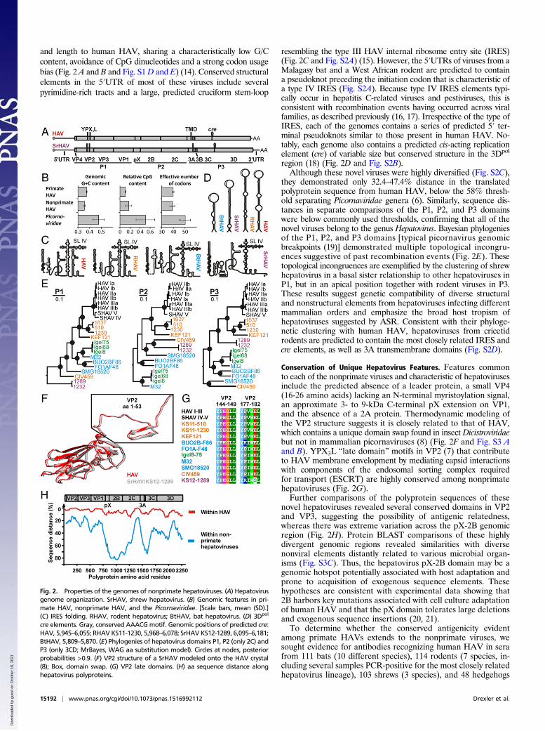
and length to human HAV, sharing a characteristically low G/Ccontent, avoidance of CpG dinucleotides and a strong codon usagebias (Fig. 2 A and B and Fig. S1D and E) (14). Conserved structuralelements in the 5′UTR of most of these viruses include severalpyrimidine-rich tracts and a large, predicted cruciform stem-loop
resembling the type III HAV internal ribosome entry site (IRES)(Fig. 2C and Fig. S2A) (15). However, the 5′UTRs of viruses from aMalagasy bat and a West African rodent are predicted to containa pseudoknot preceding the initiation codon that is characteristic ofa type IV IRES (Fig. S2A). Because type IV IRES elements typi-cally occur in hepatitis C-related viruses and pestiviruses, this isconsistent with recombination events having occurred across viralfamilies, as described previously (16, 17). Irrespective of the type ofIRES, each of the genomes contains a series of predicted 5′ ter-minal pseudoknots similar to those present in human HAV. No-tably, each genome also contains a predicted cis-acting replicationelement (cre) of variable size but conserved structure in the 3Dpol
region (18) (Fig. 2D and Fig. S2B).Although these novel viruses were highly diversified (Fig. S2C),
they demonstrated only 32.4–47.4% distance in the translatedpolyprotein sequence from human HAV, below the 58% thresh-old separating Picornaviridae genera (6). Similarly, sequence dis-tances in separate comparisons of the P1, P2, and P3 domainswere below commonly used thresholds, confirming that all of thenovel viruses belong to the genus Hepatovirus. Bayesian phylogeniesof the P1, P2, and P3 domains [typical picornavirus genomicbreakpoints (19)] demonstrated multiple topological incongru-ences suggestive of past recombination events (Fig. 2E). Thesetopological incongruences are exemplified by the clustering of shrewhepatovirus in a basal sister relationship to other hepatoviruses inP1, but in an apical position together with rodent viruses in P3.These results suggest genetic compatibility of diverse structuraland nonstructural elements from hepatoviruses infecting differentmammalian orders and emphasize the broad host tropism ofhepatoviruses suggested by ASR. Consistent with their phyloge-netic clustering with human HAV, hepatoviruses from cricetidrodents are predicted to contain the most closely related IRES andcre elements, as well as 3A transmembrane domains (Fig. S2D).
Conservation of Unique Hepatovirus Features. Features commonto each of the nonprimate viruses and characteristic of hepatovirusesinclude the predicted absence of a leader protein, a small VP4(16-26 amino acids) lacking an N-terminal myristoylation signal,an approximate 3- to 9-kDa C-terminal pX extension on VP1,and the absence of a 2A protein. Thermodynamic modeling ofthe VP2 structure suggests it is closely related to that of HAV,which contains a unique domain swap found in insect Dicistroviridaebut not in mammalian picornaviruses (8) (Fig. 2F and Fig. S3 Aand B). YPX3L “late domain” motifs in VP2 (7) that contributeto HAV membrane envelopment by mediating capsid interactionswith components of the endosomal sorting complex requiredfor transport (ESCRT) are highly conserved among nonprimatehepatoviruses (Fig. 2G).Further comparisons of the polyprotein sequences of these
novel hepatoviruses revealed several conserved domains in VP2and VP3, suggesting the possibility of antigenic relatedness,whereas there was extreme variation across the pX-2B genomicregion (Fig. 2H). Protein BLAST comparisons of these highlydivergent genomic regions revealed similarities with diversenonviral elements distantly related to various microbial organ-isms (Fig. S3C). Thus, the hepatovirus pX-2B domain may be agenomic hotspot potentially associated with host adaptation andprone to acquisition of exogenous sequence elements. Thesehypotheses are consistent with experimental data showing that2B harbors key mutations associated with cell culture adaptationof human HAV and that the pX domain tolerates large deletionsand exogenous sequence insertions (20, 21).To determine whether the conserved antigenicity evident
among primate HAVs extends to the nonprimate viruses, wesought evidence for antibodies recognizing human HAV in serafrom 111 bats (10 different species), 114 rodents (7 species, in-cluding several samples PCR-positive for the most closely relatedhepatovirus lineage), 103 shrews (3 species), and 48 hedgehogs
Fig. 2. Properties of the genomes of nonprimate hepatoviruses. (A) Hepatovirusgenome organization. SrHAV, shrew hepatovirus. (B) Genomic features in pri-mate HAV, nonprimate HAV, and the Picornaviridae. [Scale bars, mean (SD).](C) IRES folding. RHAV, rodent hepatovirus; BtHAV, bat hepatovirus. (D) 3Dpol
cre elements. Gray, conserved AAACGmotif. Genomic positions of predicted cre:HAV, 5,945–6,055; RHAV KS11-1230, 5,968–6,078; SrHAV KS12-1289, 6,095–6,181;BtHAV, 5,809–5,870. (E) Phylogenies of hepatovirus domains P1, P2 (only 2C) andP3 (only 3CD; MrBayes, WAG aa substitution model). Circles at nodes, posteriorprobabilities >0.9. (F) VP2 structure of a SrHAV modeled onto the HAV crystal(8); Box, domain swap. (G) VP2 late domains. (H) aa sequence distance alonghepatovirus polyproteins.
15192 | www.pnas.org/cgi/doi/10.1073/pnas.1516992112 Drexler et al.
Dow
nloa
ded
by g
uest
on
Oct
ober
18,
202
1

(Table S2). Sera from bats, but no other animals, were reactive inimmunofluorescence assays (IFAs) of HAV-infected cells at1:40–1:400 endpoint dilutions, with fluorescence patterns thatclosely resembled those of a monoclonal antibody control serum(Fig. 3A). Of eight positive sera (7.3%), six were from West Af-rican Eidolon helvum, and one each from Central African Rou-settus aegyptiacus and Micropteropus pusillus. To confirm theseresults, we assessed the ability of Eidolon sera to immunoprecip-itate (IP) human HAV. Four of the six IFA-positive sera that wereavailable in sufficient volumes were strongly reactive in thisassay, some exceeding the precipitating activity of anti-HAVreference sera (Fig. 3B). These four sera also effectively neutral-ized human HAV infectivity (Table S3). These results hint atsignificant conservation of capsid antigenicity between the Eidolonhepatovirus lineage and human HAV and are consistent withconservation of the sequences of several neutralization epitopeslocated in the capsid proteins VP3, VP2, and VP1 (Fig. 3C andFig. S4 A and B). An alternative explanation for the presence ofsuch antibodies in African bats could be exposure to humanHAV, perhaps by ingestion of contaminated surface water inhighly endemic areas (22). The unique ecologic traits of bats in-cluding their longevity and spatial mobility compared with othersmall mammals may facilitate repeated encounters with human
HAV in endemic areas. However, it seems unlikely that such anexposure would be of sufficient magnitude to evoke antibodyresponses without replication of the virus.Because of the genetic relatedness between rodent hepatoviruses
and human HAV, the absence of cross-reactive antibodies inrodent sera was surprising. It may be possible that rodents differfrom other small mammal hosts in the magnitude and onsetof their antibody response after hepatovirus infection, whichwould be consistent with lack of antibodies in guinea pigs up to60 d after experimental infection with HAV (9). Alternative ex-planations may include subtle differences in complex epitopes locatedin the hepatovirus antigenic sites. Resurrection or isolation ofrodent viruses for infection studies and development of hepatovirus-specific serologic assays will aid comparative investigations infurther studies.
Pathogenesis and Ecology of Nonprimate Hepatoviruses.Hepatovirusinfection patterns in bats, rodents, hedgehogs, and shrews weresimilar to those of HAV in primates. Viral RNA abundancewas highest in liver vs. other solid organs and blood in rodents,shrews, and hedgehogs. In bats, liver, and several extrahepatictissues, including predominantly spleen, lung, and intestine,showed high viral RNA concentrations (Fig. 4A). Viral RNAwas identified by in situ hybridization in bat hepatocytes and inlarge mononuclear cells within the germinal center of spleniclymphatic nodules (Fig. 4B). Comparable data are not availablefor primate hepatoviruses, although HAV antigen has beenidentified in splenic macrophages (23). Minus-strand RNA (areplicative intermediate) was detected in the liver and spleen ofbats, but only in the liver of hedgehogs (Fig. S4C). We concludethat these novel viruses are hepatotropic, but may also replicatewithin the spleen in bats. Liver tissues from 23 PCR-positive and49 PCR-negative bats, rodents, shrews, and hedgehogs were ex-amined histologically. In some PCR-positive animals we observeda mild increase in periportal inflammatory mononuclear cell in-filtrates, but there was no consistent difference with control, PCR-negative animals, and no severe liver pathology.We next assessed infection outcome in 24 hedgehogs that were
serially monitored over a 220-d period in a German animalshelter. Nine of these animals tested positive for hepatoviruses inup to three separate specimens. The viruses identified in theseanimals were 100% identical in their respective VP2 sequences,consistent with the very low nucleotide substitution rate of hu-man HAV (10) (Fig. S4D). Continuous fecal shedding of virus(∼106 RNA copies per gram feces) was observed over 79–142 din four animals, resembling HAV shedding in humans (24)(Fig. 4C). Importantly, in consecutive specimens taken less than15 d apart, the second specimen consistently showed a significantdecrease in virus concentration, suggesting resolving acute in-fections (Fig. 4C). Four animals completely cleared the infectionduring the study period. When released, all nine infected hedgehogswere in good physical condition, corroborating low hepatoviruspathogenicity in these small mammals.Finally, to gain insight into the epizootic nature of nonprimate
hepatoviruses, we analyzed specimens obtained over a 3-y periodin a German bat maternity roost within which females annuallyform a colony lasting for approximately 3 mo, giving birth tooffspring in a synchronized fashion (25). Bat hepatovirus wasdetected in fecal droppings immediately on colony formation inearly May 2008 (Fig. 4D and clade IV in Fig. 1B). The detectionrate decreased significantly 1 mo later, only to rise again afterparturition in mid-July. Shortly before bats left the colony towardthe end of that month, the detection rate fell for the second time.These data suggest an absence of persistent infections in themajority of animals, followed by a new wave of acute infectionsin offspring, and resemble HAV outbreaks in children’s camps inthe prevaccine era (26). Resampling of the same maternity roost(containing largely the same individuals) 2 y later failed to identify
Fig. 3. Antigenic relatedness of human and nonprimate hepatoviruses.(A) (Upper) immunofluorescence assay (IFA) showing a bat serum reactingwith human HAV-infected FRhK-4 cells [red (Cy2); mixed with 50% non-infected cells as internal negative controls]. (Lower) Same cells stained with amonoclonal antibody control (mAb 7E7, 100% infected cells). Blue (DAPI),nuclei. (B) Immunoprecipitation (IP) of HAV by bat sera [IF, IP, neutralizationtest (NT); red, positive in all assays; gray, discordant assay results; empty,negative in all assays], human sera, and controls. See SI Materials andMethods for details on control sera. Dotted line, threshold precipitationseparating positive and negative control sera. (C) aa sequence distance be-tween HAV (genotype Ia, GenBank accession no. AB020564), the EidolonBtHAV M32, a RHAV (RMU10-1637), and a SrHAV (KS12-1232).
Drexler et al. PNAS | December 8, 2015 | vol. 112 | no. 49 | 15193
MICRO
BIOLO
GY
SEECO
MMEN
TARY
Dow
nloa
ded
by g
uest
on
Oct
ober
18,
202
1

the same hepatovirus lineage, suggesting its extinction in this pop-ulation. Instead, a highly divergent hepatovirus which was geneti-cally related to viruses from shrews (clade I in Fig. 1B) wasidentified in six specimens (Fig. S4E), illustrating that diverse hep-atoviruses can be carried by a single host species. The level of aminoacid sequence divergence between the two hepatovirus lineagesinfecting the roost in 2008 and 2010 exceeded 30% in the partialVP2 domain (Fig. S4E), which is beyond the distance separatingenterovirus serotypes in this genomic region (27), thus hinting at theexistence of divergent serotypes in the genus Hepatovirus.
DiscussionWe identified multiple, highly diversified hepatoviruses in nu-merous small mammal taxa, extending the host associations ofthe genus Hepatovirus well beyond primates. Our findings renderthis picornavirus genus exceptionally speciose, comparable onlyto the genus Enterovirus of the family Picornaviridae after de-cades of investigation (6).The unique properties of human HAV that are shared by these
novel nonprimate hepatoviruses and that distinguish it from othermammalian picornaviruses likely reflect those of ancestral virusesinfecting small mammals before formation of the primate hep-atovirus lineage. Whether the putative hepatovirus introduction tookplace in the primate stem lineage preceding the split of Hominoideaand Cercopithecoidea about 25 Mya (28) remains unknown because
of the scarcity of HAV strains recovered from nonhuman primates.The survival of hepatoviruses before their introduction into primateswas likely mediated by large population sizes and/or high populationturnover of small mammal hosts (1, 3, 5). On the virus side, anunusually broad host range and genetic plasticity is likely to havecontributed further to hepatovirus maintenance and evolution.The existence of evolutionarily ancestral hepatoviruses in bats
and shrews compared with the presence of more closely relatedviruses in rodents and primates is reminiscent of hantavirus hostassociations, in which pathogenic human viruses originate fromrodents, whereas ancestral viruses occur in bats and Eulipotyphla(29). The relevance of these Laurasiatherian hosts for the evo-lutionary origins of human hepatitis viruses is demonstrated bythe recent detections of ancestral hepatitis B, C, and E viruses inbats (30–32). It remains to be determined whether Laurasiatheriagenerally harbor a wider genetic diversity of viruses than Euarch-ontoglires and whether ecological traits such as insectivorous dietsinfluence viral diversity. However, reconstructions that point to aLaurasiatherian host and an insectivorous diet for ancestral hep-atoviruses provide a novel link to the structural phylogeny of theHAV capsid and its close relationship to picorna-like viruses ofinsects (8) and together suggest more distant ancestry in a pri-mordial insect-borne virus. Such a scenario is paralleled by recentsuggestions of similar ancestry for other mammalian viruses (33–35) and provides a new perspective on the origins of this ancienthuman pathogen.A major barrier to viral host switches is receptor use (36). Human
HAV uses the T-cell Ig and mucin domain 1 (TIM-1) for cell entry(37). It will be important to investigate whether receptor use isconserved among the nonprimate hepatoviruses we identified and todetermine whether these viruses are capable of infecting primatehosts. Of note, human TIM-1 is used by other zoonotic viruses forcell entry, including the bat-borne Ebola virus, the arthropod-bornedengue virus and several rodent- and bat-borne New World arena-viruses (38–40), illustrating that dependency on this molecule for cellentry may not represent an insuperable barrier to a viral host switch.Zoonotic infections with emerging viruses have become in-
creasingly relevant for human health due to the invasion of pristinehabitats by humans and their livestock, advancing global mobility,and the rapid spread of pathogens within dense human populations(41–43). The antigenic conservation we observed between HAVand nonprimate hepatoviruses from Eidolon bats suggests theseviruses belong to a common serotype and that the introduction ofsuch viruses into human populations would be limited by herdimmunity and vaccine-induced immune responses. However, thismay not apply to all of the novel hepatovirus species we identified,some of which on the basis of diversity within the P1 domain likelycomprise distinct serotypes. Our study exemplifies the utility oflooking beyond phylogenetic criteria alone when conducting riskassessment for emerging RNA viruses and the need to includefunctional, ecologic, and pathogenic analyses of animal reservoirs.
Materials and MethodsSampling was done as described previously (3). Hepatovirus detection,quantification, and genomic characterizations were done by PCR-basedtechniques (see oligonucleotide sequences in Tables S4 and S5). Serologicand histopathologic analyses were done as described previously (17). Detailsof these and evolutionary analyses are given in SI Materials and Methods.
ACKNOWLEDGMENTS. We thank Monika Eschbach-Bludau, Tobias Bleicker,Andres Moreira Soto, Breno Dominguez Souza, Leonardo Calderón Obaldía,Debby van Riel, Marcel A. Müller, Jens P. Teifke and Kevin L. McKnight forassistance and advice; Tabea Binger, Heather J. Baldwin, Augustina Annan,Gael Darren Maganga, Peter H.C. Lina, Markus Dietz, Jacques B. Pir, HeikePhilipps, Gertraude Göpner, Manfred Göpner, Lena Grosche, Frauke Meier,MyriamGötz, Ioan Coroiu, Regina Klüppel-Hellmann, Anda Culisier, Mircea-DanHarlav, Sabrina Stölting, Carsten Dense, Dieudonné Nkoghe, MathieuBourgarel, Stoian Yordanov, Tasnim Suleiman, Ndapewa Ithete, ThomasJäkel, Thomas Kruppa, Stephan Günther, Koussay Dellagi, Hervé Pascalis,Julien Mélade, Pablo Tortosa, Peter Witkowski, Georg Herrler, Jörg Thiel,
Fig. 4. Hepatovirus infection patterns in small mammals. (A) Hepatovirus RNAabundance in solid organs and blood per host taxon determined by qPCR. Li,liver; Sp, spleen; Ki, kidney; Lu, lung; In, intestine; Br, brain; He, heart; Bl, blood.Boxes, median, interquartile range; outliers (circles); extreme values (asterisks).Next to host pictograms, numbers of analyzed animals; Below each organ, ra-tios of specimens testing positive per specimens available for testing. (B) In situhybridization of BtHAV RNA in E. helvum liver (Upper Left, 400×magnification,animal M32), spleen (Upper Right, 400×, animal GH297), lung (Lower Left,200×, animal M32), and intestine (Lower Right, 200×, animal M32). (C) Hep-atovirus detections and RNA concentrations determined by qPCR in ninehedgehogs sampled longitudinally. (D) Hepatovirus amplification in a bat ma-ternity roost. Triangles, viral RNA concentrations in pooled fecal specimens overfive sampling points. Secondary y axis and orange line, detection rate.
15194 | www.pnas.org/cgi/doi/10.1073/pnas.1516992112 Drexler et al.
Dow
nloa
ded
by g
uest
on
Oct
ober
18,
202
1

Margrit Bemmann, Katarina Kühn, Engelbert Kampling, Mechthild Budde,Dagmar Funck, Jona Freise, Brita Oltmann, Sebastian Guenther, Brigitte Bannert,Matthias Wenk, Henrike Gregersen, Johannes Lang, Anita Plenge-Bönig, NicoleSchauerte, SusanneModrow, GerhardMaluck, Richard Kruczewski, Julie Lichière,Maria Nemeth, Anne Balkema-Buschmann, Susanne Jäckel, Horst Schirrmeier,Hanan Sheikh Ali, Christian Kretzschmar, Konrad Wanka, Ute Wessels, TheresWollny, Kathrin Baumann, Grit Möwert, Franziska Thomas, Bärbel Hammerschmidt,Daniel Windolph, Josephine Schlosser, Marc Mertens, Paul Dremsek, KatjaPlifke, Kerstin Tauscher, Angele Breithaupt, Nadja Lorenz, Christian Korthase,André Schütte, Julie Elkins, Sylvia Ferguson, Christiane Denys, Emilie Lecompte,René Ryll, Christin Trapp, AnkeMandelkow, Nicole Schmidt, Annemarie Steiner,Maxi Zumpe, Samuel Bernstein, Stefan Fischer, Sandra Blome, Denny Maaz,Christoph Gertler, Michael Faulde, Jens Lewitzki, Mathias Schlegel, Julia Zeitler,Wolfgang Fiedler, Elisabeth Glatthaar, and Sabine Sauer for assistance withfield work and necropsy; and Mediagnost for the mAb 7E7. Samples from small
mammal monitoring in Germany were kindly provided by Sabrina Schmidt,Ulrike M. Rosenfeld, Daniela Reil, Christian Imholt, Nastasja Kratzmann, StephanDrewes, Stefan Fischer, and Jens Jacob from UFOPLAN Projects 370941401and 371348401. Several samples for this study were taken during field workfor projects granted to C.D. by the German Federal Ministry of Educationand Research (BMBF) (Projects 01KI1005A and 01KI1016D), the German Re-search Foundation (DFG) (Projects DR 772/3-1, DR 772/12-1, and DR 772/10-1),and to R.G.U. by the German Armed forces (M/SABX/005 and E/U2AD/CF512/DF557) and the BMBF (Projects 01KI1018 and 01KI1303). Additionalfunding was obtained by the European Commission through the COMPAREproject (to C.D.) (643476), Russian Scientific Foundation Grant 14-15-00619(to A.N.L.), German Tönjes-Vagt-Stiftung Project XXIX (to A.D.), NationalInstitutes of Health Grants R01-AI103083 and U19-AI109965 (to S.M.L.), andthe US Department of Defense (HR0011-13-2-0020) and the DFG (DR810/1-1) (to J.F.D.).
1. Luis AD, et al. (2013) A comparison of bats and rodents as reservoirs of zoonotic vi-ruses: Are bats special? Proc R Soc Biol Sci 280(1756):20122753.
2. Brook CE, Dobson AP (2015) Bats as ‘special’ reservoirs for emerging zoonotic path-ogens. Trends Microbiol 23(3):172–180.
3. Drexler JF, et al. (2012) Bats host major mammalian paramyxoviruses. Nat Commun 3:796.4. Leroy EM, et al. (2005) Fruit bats as reservoirs of Ebola virus. Nature 438(7068):
575–576.5. Han BA, Schmidt JP, Bowden SE, Drake JM (2015) Rodent reservoirs of future zoonotic
diseases. Proc Natl Acad Sci USA 112(22):7039–7044.6. Knowles NJ, et al. (2012) Family Picornaviridae. Virus taxonomy. Ninth Report of the
International Committee on Taxonomy of Viruses, eds King AMQ, Adams MJ,Carstens EB, Lefkowitz EJ (Elsevier/Academic Press, Amsterdam), pp 855–880.
7. Feng Z, et al. (2013) A pathogenic picornavirus acquires an envelope by hijackingcellular membranes. Nature 496(7445):367–371.
8. Wang X, et al. (2015) Hepatitis A virus and the origins of picornaviruses. Nature517(7532):85–88.
9. Hornei B, et al. (2001) Experimental hepatitis A virus infection in guinea pigs. J MedVirol 64(4):402–409.
10. Cristina J, Costa-Mattioli M (2007) Genetic variability and molecular evolution ofhepatitis A virus. Virus Res 127(2):151–157.
11. Walker CM, Feng Z, Lemon SM (2015) Reassessing immune control of hepatitis A virus.Curr Opin Virol 11:7–13.
12. Wong DC, Purcell RH, Rosen L (1979) Prevalence of antibody to hepatitis A andhepatitis B viruses in selected populations of the South Pacific. Am J Epidemiol 110(3):227–236.
13. Skinhoj P, Mikkelsen F, Hollinger FB (1977) Hepatitis Ain Greenland: Importance of specificantibody testing in epidemiologic surveillance. Am J Epidemiol 105(2):140–147.
14. Aragonès L, Guix S, Ribes E, Bosch A, Pintó RM (2010) Fine-tuning translation kineticsselection as the driving force of codon usage bias in the hepatitis A virus capsid. PLoSPathog 6(3):e1000797.
15. Brown EA, Day SP, Jansen RW, Lemon SM (1991) The 5′ nontranslated region ofhepatitis A virus RNA: Secondary structure and elements required for translationin vitro. J Virol 65(11):5828–5838.
16. Hellen CU, de Breyne S (2007) A distinct group of hepacivirus/pestivirus-like internalribosomal entry sites in members of diverse picornavirus genera: Evidence for mod-ular exchange of functional noncoding RNA elements by recombination. J Virol81(11):5850–5863.
17. Drexler JF, et al. (2013) Evidence for novel hepaciviruses in rodents. PLoS Pathog 9(6):e1003438.
18. Yang Y, Yi M, Evans DJ, Simmonds P, Lemon SM (2008) Identification of a conservedRNA replication element (cre) within the 3Dpol-coding sequence of hepatoviruses.J Virol 82(20):10118–10128.
19. Lukashev AN (2010) Recombination among picornaviruses. Rev Med Virol 20(5):327–337.
20. Beard MR, Cohen L, Lemon SM, Martin A (2001) Characterization of recombinanthepatitis A virus genomes containing exogenous sequences at the 2A/2B junction.J Virol 75(3):1414–1426.
21. Emerson SU, Huang YK, McRill C, Lewis M, Purcell RH (1992) Mutations in both the 2Band 2C genes of hepatitis A virus are involved in adaptation to growth in cell culture.J Virol 66(2):650–654.
22. Esona MD, et al. (2010) Reassortant group A rotavirus from straw-colored fruit bat(Eidolon helvum). Emerg Infect Dis 16(12):1844–1852.
23. Mathiesen LR, et al. (1978) Localization of hepatitis A antigen in marmoset organsduring acute infection with hepatitis A virus. J Infect Dis 138(3):369–377.
24. Yotsuyanagi H, et al. (1996) Prolonged fecal excretion of hepatitis A virus in adultpatients with hepatitis A as determined by polymerase chain reaction. Hepatology24(1):10–13.
25. Drexler JF, et al. (2011) Amplification of emerging viruses in a bat colony. EmergInfect Dis 17(3):449–456.
26. Werzberger A, et al. (1992) A controlled trial of a formalin-inactivated hepatitis Avaccine in healthy children. N Engl J Med 327(7):453–457.
27. Nasri D, et al. (2007) Typing of human enterovirus by partial sequencing of VP2. J ClinMicrobiol 45(8):2370–2379.
28. Springer MS, et al. (2012) Macroevolutionary dynamics and historical biogeographyof primate diversification inferred from a species supermatrix. PLoS One 7(11):e49521.
29. Guo WP, et al. (2013) Phylogeny and origins of hantaviruses harbored by bats, in-sectivores, and rodents. PLoS Pathog 9(2):e1003159.
30. Drexler JF, et al. (2013) Bats carry pathogenic hepadnaviruses antigenically related tohepatitis B virus and capable of infecting human hepatocytes. Proc Natl Acad Sci USA110(40):16151–16156.
31. Drexler JF, et al. (2012) Bats worldwide carry hepatitis E virus-related viruses that forma putative novel genus within the family Hepeviridae. J Virol 86(17):9134–9147.
32. Quan PL, et al. (2013) Bats are a major natural reservoir for hepaciviruses and pegi-viruses. Proc Natl Acad Sci USA 110(20):8194–8199.
33. Zirkel F, et al. (2011) An insect nidovirus emerging from a primary tropical rainforest.MBio 2(3):e00077–e11.
34. Marklewitz M, Zirkel F, Kurth A, Drosten C, Junglen S (2015) Evolutionary and phe-notypic analysis of live virus isolates suggests arthropod origin of a pathogenic RNAvirus family. Proc Natl Acad Sci USA 112(24):7536–7541.
35. Li CX, et al. (2015) Unprecedented genomic diversity of RNA viruses in arthropodsreveals the ancestry of negative-sense RNA viruses. eLife 4:4.
36. Drosten C (2013) Virus ecology: A gap between detection and prediction. EmergMicrobes Infect 2(5):e31.
37. Feigelstock D, Thompson P, Mattoo P, Zhang Y, Kaplan GG (1998) The human homologof HAVcr-1 codes for a hepatitis A virus cellular receptor. J Virol 72(8):6621–6628.
38. Kondratowicz AS, et al. (2011) T-cell immunoglobulin and mucin domain 1 (TIM-1) is areceptor for Zaire Ebolavirus and Lake Victoria Marburgvirus. Proc Natl Acad Sci USA108(20):8426–8431.
39. Meertens L, et al. (2012) The TIM and TAM families of phosphatidylserine receptorsmediate dengue virus entry. Cell Host Microbe 12(4):544–557.
40. Jemielity S, et al. (2013) TIM-family proteins promote infection of multiple envelopedviruses through virion-associated phosphatidylserine. PLoS Pathog 9(3):e1003232.
41. Brockmann D, Helbing D (2013) The hidden geometry of complex, network-drivencontagion phenomena. Science 342(6164):1337–1342.
42. Plowright RK, et al. (2015) Ecological dynamics of emerging bat virus spillover. Proc RSoc Biol Sci 282(1798):20142124.
43. Pigott DM, et al. (2014) Mapping the zoonotic niche of Ebola virus disease in Africa.eLife 3:e04395.
44. Corman VM, et al. (2014) Characterization of a novel betacoronavirus related tomiddle East respiratory syndrome coronavirus in European hedgehogs. J Virol 88(1):717–724.
45. Alcaide M, et al. (2009) Disentangling vector-borne transmission networks: A uni-versal DNA barcoding method to identify vertebrate hosts from arthropod blood-meals. PLoS One 4(9):e7092.
46. Ronquist F, Huelsenbeck JP (2003) MrBayes 3: Bayesian phylogenetic inference undermixed models. Bioinformatics 19(12):1572–1574.
47. Maurer-Stroh S, Eisenhaber B, Eisenhaber F (2002) N-terminal N-myristoylation ofproteins: Prediction of substrate proteins from amino acid sequence. J Mol Biol 317(4):541–557.
48. Robert X, Gouet P (2014) Deciphering key features in protein structures with the newENDscript server. Nucleic Acids Res 42(Web Server issue):W320–W324.
49. Zuker M (2003) Mfold web server for nucleic acid folding and hybridization pre-diction. Nucleic Acids Res 31(13):3406–3415.
50. Simmonds P (2012) SSE: A nucleotide and amino acid sequence analysis platform. BMCRes Notes 5:50.
51. Tamura K, et al. (2011) MEGA5: Molecular evolutionary genetics analysis usingmaximum likelihood, evolutionary distance, and maximum parsimony methods. MolBiol Evol 28(10):2731–2739.
52. Hofmann K, Stoffel W (1993) TMbase: A database of membrane spanning proteinsegments. Biol Chem Hoppe Seyler, 10.1515/bchm3.1993.374.1-6.143.
53. Müller MA, et al. (2007) Coronavirus antibodies in African bat species. Emerg InfectDis 13(9):1367–1370.
54. Houde A, et al. (2007) Comparative evaluation of new TaqMan real-time assays forthe detection of hepatitis A virus. J Virol Methods 140(1-2):80–89.
55. Dotzauer A, et al. (2000) Hepatitis A virus-specific immunoglobulin A mediates in-fection of hepatocytes with hepatitis A virus via the asialoglycoprotein receptor.J Virol 74(23):10950–10957.
56. Bininda-Emonds OR, et al. (2007) The delayed rise of present-day mammals. Nature446(7135):507–512.
Drexler et al. PNAS | December 8, 2015 | vol. 112 | no. 49 | 15195
MICRO
BIOLO
GY
SEECO
MMEN
TARY
Dow
nloa
ded
by g
uest
on
Oct
ober
18,
202
1