examining pathogenesis and preventatives in spontaneous
TRANSCRIPT

University of Arkansas, Fayetteville University of Arkansas, Fayetteville
ScholarWorks@UARK ScholarWorks@UARK
Theses and Dissertations
12-2020
Examining Pathogenesis and Preventatives in Spontaneous and Examining Pathogenesis and Preventatives in Spontaneous and
Staphylococcus-Induced Bacterial Chondronecrosis with Staphylococcus-Induced Bacterial Chondronecrosis with
Osteomyelitis in Broilers Osteomyelitis in Broilers
Nnamdi Simon Ekesi University of Arkansas, Fayetteville
Follow this and additional works at: https://scholarworks.uark.edu/etd
Part of the Animal Diseases Commons, Animal Experimentation and Research Commons, Animal
Studies Commons, Bacteriology Commons, Pathogenic Microbiology Commons, and the Poultry or Avian
Science Commons
Citation Citation Ekesi, N. S. (2020). Examining Pathogenesis and Preventatives in Spontaneous and Staphylococcus-Induced Bacterial Chondronecrosis with Osteomyelitis in Broilers. Theses and Dissertations Retrieved from https://scholarworks.uark.edu/etd/3838
This Dissertation is brought to you for free and open access by ScholarWorks@UARK. It has been accepted for inclusion in Theses and Dissertations by an authorized administrator of ScholarWorks@UARK. For more information, please contact [email protected].
Examining Pathogenesis and Preventatives in Spontaneous and Staphylococcus-Induced
Bacterial Chondronecrosis with Osteomyelitis in Broilers
A dissertation submitted in partial fulfilment
of the requirements for the degree of
Doctor of Philosophy in Cell and Molecular Biology
by
Nnamdi Simon Ekesi
Ashford University
Bachelor of Sciences in Biology, 2013
December 2020
University of Arkansas
This dissertation is approved for recommendation to Graduate Council.
__________________________________
Douglas D Rhoads Ph.D.
Dissertation Director
__________________________________ ___________________________________
Jeannine M Durdik Ph.D. Ines Pinto Ph.D.
Committee Member Committee Member
___________________________________
Charles Rosenkrans, Jr. Ph.D.
Committee Member

ABSTRACT Bacterial chondronecrosis with osteomyelitis is the most important cause of lameness in
broilers. This is important to poultry production, as it poses animal welfare issues, and causes a
significant loss in revenue. The remediation of this disease requires the study of its etiology with
fitting models and evaluating preventatives. The research reported herein covers genomic
virulence analysis of BCO isolates, mainly Staphylococcus aureus, and Escherichia coli
retrieved from lame birds. We found that S. aureus isolates were closest to chicken strains in
Europe but may have been in the Arkansas area for a decade. Phylogenomics suggest our S.
aureus is restricted to poultry, while the E. coli spans various hosts. This dissertation includes the
analyses of mainly BCO isolates to determine virulence using the embryo lethality assay. Human
S. aureus was the most lethal to layer and broiler embryos. Staphylococcus agnetis 908 that may
induce lameness to >50% did not show virulence. This inconsistency among others compelled us
to offer that embryo lethality assay may not be an effective tool for estimating the pathogenicity
of BCO isolates. Furthermore, research reported herein covers the investigation of the feed
additive Availa-ZMC for lameness reduction potential in broilers using litter and wire-flooring
models for inducing lameness. Availa-ZMC (a mixture of organic trace minerals) resulted in a
reduction of lameness by 20% in the wire-flooring model, and 25% in the challenge on litter
flooring model. Finally, this dissertation reports on a Typhoid-Mary experiment in broilers raised
on the wire flooring to determine whether broilers challenged with S. agnetis can transmit the
bacterium to birds in the same pen thereby spreading BCO and lameness. The Typhoid Mary
experiment shows that young broilers exposed to S. agnetis at an early age harbor the bacterium
and if mixed with unexposed birds 10 days later can transmit the bacterium to their pen mates.

©2020 by Nnamdi Simon Ekesi
All Rights Reserved
ACKNOWLEDGEMENTS
This dissertation is a cumulative product of efforts from a great research team.
My sincere gratitude goes to Dr. Doug Rhoads for teaching me and nurturing my goals
with constant patience, and directness. You taught me structure, enhanced my critical thinking
skills, and encouraged my creativity. You have given me invaluable skills and made me more
confident to face my future endeavors. I will always be grateful to you. Thank you!
I am also very grateful to Dr. Adnan Alrubaye for taking me under your wing in the farm
projects. You superbly taught me a whole lot about animal husbandry. I appreciate you.
To my committee—Dr. Jeannine Durdik, Dr. Ines Pinto, and Dr. Charles Rosenkrans Jr.,
I sincerely thank you for devoting your time and resources to my goals. Thank you very much
for all the insightful advice and feedback you have offered through the candidacy, coursework,
research, and dissertation processes.
To my former and present lab mates—Dr. Ojha, Dr. Alzahrani, Dr. Estill, Dr. Dey, Dr.
Parveen, Sura Zaki, Tim Lipnack, Abdulkarim Shwani, Amer Hasan, Layla Al-Mitib, Duaa
Almansaf, Katie Lee, and Sonali Lenaduwe thank you all for your contributions to my success
and making the lab a wonderful place to work. I am happy to have met all of you.
Thank you, Dr. Nick Anthony, and the University of Arkansas farm crew thank you for
your hard work, and your commitment to sources various supplies I used for my research. I am
very grateful.
Thank you, mom, for teaching me the value of education, and hard work. Thank you to
my brothers, sisters, and friends. Your constant support got me this far.

DEDICATION
I dedicate this dissertation to the loving memories of my brother, Roy Emenike “Chicago” Ekesi.

TABLE OF CONTENTS
Introduction ..................................................................................................................................... 1
Chicken Domestication ............................................................................................................... 2
Broiler Production in the USA .................................................................................................... 3
Growth of the broiler Industry ................................................................................................. 3
Modern Broilers Selection and Breeding Techniques ............................................................. 6
Impact of Genetic Selection in Broiler Production: Pros and Cons ........................................ 9
Lameness in Broiler Chickens ................................................................................................... 13
The Issue ................................................................................................................................ 13
Prevalence of Bacterial Chondronecrosis with Osteomyelitis Lameness ............................. 14
Economics of BCO Lameness ............................................................................................... 15
Pathogenesis BCO-Lameness ................................................................................................... 16
Blood supply to proximal heads of rapidly growing broiler important to lameness. ............ 18
Vertebral anatomy and blood supply in broilers important to BCO lameness ...................... 21
Bacteria and Lameness .............................................................................................................. 24
Microbiota and Lameness...................................................................................................... 25
Stress, Immune responses, and BCO ..................................................................................... 29
Microbiology of Common Pathogenic BCO isolates ............................................................ 31
Staphylococcus spp. ........................................................................................................... 31
Staphylococcus agnetis .................................................................................................. 31
Staphylococcus aureus ................................................................................................... 32
Escherichia coli .............................................................................................................. 45
Enterococcus cecorum ................................................................................................... 52
Clinical Diagnosis of Terminal BCO ........................................................................................ 53
The S. agnetis induced BCO Lameness model ......................................................................... 57

Human Osteomyelitis ................................................................................................................ 69
References ................................................................................................................................. 70
CHAPTER 2 ................................................................................................................................. 96
Analysis of Genomes of Bacterial Isolates from Lameness Outbreaks in Broilers ...................... 96
Analysis of Genomes of Bacterial Isolates from Lameness Outbreaks in Broilers ...................... 97
Abstract: .................................................................................................................................... 97
Introduction ............................................................................................................................... 98
Methods ..................................................................................................................................... 98
Results and Discussion ............................................................................................................ 100
BCO Genome Assemblies ................................................................................................... 104
Phylogenetic Comparison .................................................................................................... 106
Conclusions ............................................................................................................................. 111
Acknowledgements ................................................................................................................. 111
References ............................................................................................................................... 112
Appendix ................................................................................................................................. 115
CHAPTER 3 ............................................................................................................................... 127
Investigation of Embryo Lethality Assay for Assessing Virulence of Isolates from Bacterial
Chondronecrosis with Osteomyelitis in Lame Broilers. ................................................. 127
Investigation of Embryo Lethality Assay for Assessing Virulence of Isolates from Bacterial
Chondronecrosis with Osteomyelitis in Lame Broilers. ................................................. 128
Abstract ................................................................................................................................... 128
Introduction ............................................................................................................................. 129
Methods ................................................................................................................................... 130

Microbiology ....................................................................................................................... 130
ELA 130
Electro-transformation ......................................................................................................... 130
Results 131
Embryo Lethality Assay with BCO isolates ........................................................................ 131
Is ELA Virulence Readily Transferable by Electroporation? ............................................. 133
Discussion ............................................................................................................................... 134
References ............................................................................................................................... 135
Appendix ................................................................................................................................. 139
CHAPTER 4 ............................................................................................................................... 143
Chondronecrosis with Osteomyelitis in Broilers: Further Defining Lameness-Inducing Models
with Wire or Litter Flooring, to Evaluate Protection with Organic Trace Minerals ....... 143
Disclaimer ............................................................................................................................... 144
Chondronecrosis with Osteomyelitis in Broilers: Further Defining Lameness-Inducing Models
with Wire or Litter Flooring, to Evaluate Protection with Organic Trace Minerals ....... 145
Abstract ................................................................................................................................... 145
Introduction ............................................................................................................................. 146
Materials and Methods ............................................................................................................ 147
Lameness Trials ................................................................................................................... 147
Histological Evaluation of Intestinal Villi ........................................................................... 148
Assay of Intestinal Gene Expression ................................................................................... 149
Phagocytosis Assay ............................................................................................................. 150
Statistical Analyses .............................................................................................................. 150

Results 151
Discussion ............................................................................................................................... 154
References ............................................................................................................................... 157
Appendix ................................................................................................................................. 160
CHAPTER 5 ............................................................................................................................... 165
Typhoid Mary Experiment: BCO incidence variation due to bird-to-bird intra-pen transmission
when administering Staphylococcus agnetis in the water ............................................... 165
Disclaimer ............................................................................................................................... 166
Typhoid Mary Experiment: BCO incidence variation due to bird-to-bird intra-pen transmission
when administering Staphylococcus agnetis in the water ............................................... 167
Summary ................................................................................................................................. 167
Materials and methods ............................................................................................................ 167
Animal Housing, Care, and Treatment ................................................................................ 167
Clinical Diagnosis of Lameness .......................................................................................... 169
Results 170
Discussion ............................................................................................................................... 171
References ............................................................................................................................... 176
CHAPTER 6 ............................................................................................................................... 177
Conclusions ................................................................................................................................. 177
Conclusions ................................................................................................................................. 178
Chapter 7 ..................................................................................................................................... 180
Appendix ..................................................................................................................................... 180

LIST OF TABLES
Table 1. Timeline for critical technologies employed for poultry breeding in the 20th century. .... 8
Table 2. The number of idiopathic disorders of broiler chickens ................................................. 11
Table 3: Comparison of microbial communities in different groups ............................................ 29
Table 4. Virulence Traits in Pathotypes of Extraintestinal Pathogenic E. coli ............................. 48
Table 5. Virulence factors in ExPEC ............................................................................................ 49
Table 6: BCO isolates from lame birds based on-site sampled .................................................... 62
Table 7: BCO isolates from lame birds based on the bone lesion……………………………… 62
Table 8. BCO Microbiological sampling from the blood of healthy or lame birds. ..................... 64
Table 9. Microbiological sampling of bone and blood samples from two commercial broiler
farms experiencing BCO outbreaks.. .......................................................................................... 102
Table 10. Bacterial genome assemblies used or produced in these analyses are listed .............. 106
Table 11. Genomes used for phylogenomic analyses. ................................................................ 118
Table 12. Proteome differences in four genomes of S. aureus infecting chickens. .................... 123
Table 13. Sources of bacterial isolates utilized for ELA ........................................................... 139
Table 14. Feed supplementation for the three treatments ........................................................... 160
Table 15. Lame, and Mortality by pen, and ending BW for three treatments ............................ 160
Table 16. Bacterial survival in adherent peripheral blood monocytes........................................ 161
Table 17. Protocol for Typhoid Mary Experiment ..................................................................... 175
Table 18. Total and percent lameness for the four treatment groups. ......................................... 176

LIST OF FIGURES
Figure 1. USDA Report: Broilers produced by the pound in the USA since 1968 ........................ 5
Figure 2. US broiler production in pounds with the estimated monetary value from 2008 to 2018.
......................................................................................................................................................... 5
Figure 3: Example of generation and multiplication scheme in modern broiler production .......... 8
Figure 4: Age-related changes in the size of the University of Alberta Meat Control strains
unselected since 1957 and 1978, compared to the Ross 308 broilers ........................................... 10
Figure 5: Classification of Broiler chicken Breast meat myopathies ........................................... 12
Figure 6: Factors that contribute to the incidence of lameness in broilers. .................................. 14
Figure 7. Routes of bacterial infections in rapidly growing birds ................................................ 18
Figure 8. The diagram on the left pane and photograph on the right panel shows the blood supply
and the anatomical structures of the long bone in growing broilers ............................................. 20
Figure 9. Diagram of the femoral proximal head illustrating the formation of osteochondrotic
clefts/crypts at the boundary between the growth plate and the epiphysis ................................... 20
Figure 10. Arterial blood supply to the leg bone of a growing bird ............................................. 21
Figure 11. The skeleton of a bird .................................................................................................. 22
Figure 12. Broiler bone histopathology image for formalin-fixed 5 μM sections from 5-week
stained with hematoxylin and eosin .............................................................................................. 23
Figure 13. BCO progression on femoral (A-D) and tibial (E-H) proximal heads that result in
lameness in broiler chickens ......................................................................................................... 55
Figure 14. Common clinical presentations of proximal femoral and tibial BCO lesions.. ........... 56
Figure 15. Tibial dyschondroplasia is characterized by abnormal masses. .................................. 56
Figure 16. Vertebral BCO in broiler that exhibited paraplegic hock-resting posture. ................. 57
Figure 17. The minimum effective dosage of S. agnetis in drinking water for the induction of
lameness. ....................................................................................................................................... 59
Figure 18: Most effective days for the administration of S. agnetis in drinking water for induction
of lameness for broilers reared on wire flooring. .......................................................................... 59
Figure 19. Cumulative percent lameness for birds raised on litter for 56 days (L56; 4 pens; 200
birds) ............................................................................................................................................. 61
Figure 20. Incidence of lameness with S. agnetis, S. saprophyticus, and S. epidermidis. S. agnetis
in drinking water. .......................................................................................................................... 65
Figure 21. Dendrogram based on genomic BLAST for 17,824 E. coli genomes from NCBI. .. 115
Figure 22. Phylogenetic tree for 61 E. coli genomes based on Average Nucleotide Identity .... 116
Figure 23. Phylogenetic tree for49 S. aureus genomes based on Average Nucleotide Identity . 117
Figure 24. Embryo lethality assay in layer chicken line embryos to estimate the lethal dosage of
bacterial isolates.. ........................................................................................................................ 140
Figure 25. Layer chicken line embryo lethality for injection of 105 or 106 CFU ....................... 141
Figure 26. Broiler chicken line embryo lethality for injections of 105 or 106 CFU of bacterial. 142
Figure 27. Embryo lethality is transferable from E. coli 1413 to 1409. ..................................... 142
Figure 28. Cumulative lameness for broilers raised on wire-flooring on control feed or feed
supplemented with Zinpro Availa-ZMC ..................................................................................... 161
Figure 29. Tibial (upper) and femoral (lower) lesion diagnoses for all lame birds raised on wire-
flooring in experiment 1.............................................................................................................. 162
Figure 30. Cumulative lameness for broilers with a bacterial challenged, raised on litter-flooring,
on control feed or feed supplemented with Zinpro Availa-ZMC in experiment 2 ..................... 163
Figure 31. Tibial (upper panel) and femoral (lower panel) lesion diagnoses for all lame birds
raised on litter-flooring in experiment 2. .................................................................................... 163
Figure 32. Villus length at d57 for apparently healthy birds from three treatment groups. ....... 164
Figure 33: Expression of intestinal barrier integrity-related genes from three treatment groups in
experiment 2................................................................................................................................ 164
Figure 34. Pen Setup in A364 for the Typhoid Mary experiment. ............................................. 173
Figure 35. Cumulative % lameness per treatment from day 34 through day 56. ....................... 174
Figure 36. Tibial and Femoral BCO lesion diagnoses for the four treatment groups ................ 175

LIST OF PUBLISHED ARTICLES
Chapter 4: Alrubaye A.A.K., Ekesi N.S., Hasan A., Elkins E., Ojha S., Zaki S., Dridi S.,
Wideman R.F., Rebollo M.A., Rhoads D.D. (2020). Chondronecrosis with Osteomyelitis in
Broilers: Further Defining Lameness-Inducing Models with Wire or Litter Flooring, to Evaluate
Protection with Organic Trace Minerals. Poultry
Science….……………………………………………………………………………In Press

1
Introduction
Literature Review
2
Introduction/ Literature Review
Chicken Domestication
Domestic Chicken—Gallus gallus domesticus, is the most widely distributed poultry species
(Miao., et al, 2013; Zhang., et al, 2017). Chickens have been important to human societies for
thousands of years. They are food; the meat and eggs serve as a reliable source of protein.
Chicken also served anthropomorphic purposes in entertainment (cockfights), religious practices,
and ornamentation. Domestic chickens make good biological and medical models. From an
archaeological perspective, domestic chickens are closely associated with humans; they have
been dispersed primarily by human activity. This makes chickens an important biological marker
of agricultural, cultural contacts, and trade between societies and civilizations (Mwacharo.,
2013A & B; Peters., 2016). There is evidence that multiple domestications of Red Jungle Fowl,
the primary parents of the most recent domestic chickens, began between the ending of the
Pleistocene and the beginning of the Holocene era in southern China, South Asia, and Southeast
Asia (Tixier-Boichard., et al, 2011; Miao., et al, 2013; Peters., et al, 2016; and Bosse, 2019).
Journals and archaeological evidence suggest that chickens got to Europe via southern (through
Greece and Persia) and northern (through china and Russia) trading routes (Crawford., 1990; and
Tixier-Boichard., 2011). Archaeozoological evidence suggests that domestic chickens were
raised in Africa, particularly ancient Egypt around 1307–1196 BC (Houlihan., 1986; Mwacharo.,
2013). Chickens showed up in Sudan 1650 BC, and Kenya around 800AD (Houlihan., 1986;
Marshall., 2000; Mwacharo., 2013). The sequence of events concerning the spread of chickens
across the rest of Africa is not fully understood (Blenck., 2000; Tixier-Boichard., 2011; and
Mwacharo., 2013). Mitochondrial DNA from 3000-year old chickens at the Teouma site
(Vanuatu) reveals that chicken spread from southeast Asia to Oceania between 1400–900 BC

3
(Storey., 2010; Miao., 2013). In the Americas, lineage and propagation of chickens are debatable
(Maio., 2013). Some studies reported that DNA and carbon dating evidence suggest that
Polynesian chickens were introduced in the Americas (Chile) in the pre-Columbian AD 1304–
1424 (Storey., 2007; Maio., 2013). Storey, et al. (2007) suggested, based on dating and DNA
evidence, that chickens were introduced to the Americas before the arrival of the Spanish or
Portuguese, but they were of Polynesian origin. Gongora et al. (2008) also countered the
Polynesian-Chilean American contact view citing that pre-Columbian chickens sequences lie
among European/Indian subcontinental/Chinese haplotype rather than Polynesia.
Broiler Production in the USA
Growth of the broiler Industry
Broilers and layers are the two parts of commercial chicken production. Broilers yield
meat and layers produce eggs. My dissertation focuses on broiler chickens. Our team worked to
better understand the mechanisms of pathogenesis behind the incidence of lameness in the
production of rapidly growing birds. We induced the disease using models. We tested
formulations postulated to reduce bacterial chondronecrosis with osteomyelitis (BCO) leading to
lameness in commercial broiler production farms. We also surveyed multiple commercial farms
and sampled lame broilers. Our goal was to improve models for studying BCO, enhance animal
health and welfare, improve meat quality, incorporate sustainable practices, and improve
productivity.
The poultry industry in the United States of America is one of the most successful sectors
in US agricultural production. Around the early 1900s, poultry production in the USA, like in
most parts of the world, was mainly practiced in small non-specialized units using diverse breeds
of chickens that already existed on the continent (Sainsbury, 2000; Muir & Aggrey, 2003). By

4
the late 1930s to mid-1940s, there was an explosion in poultry production in the US and across
Europe (Sansbury, 2000). Various genetic improvement programs were introduced. Poultry
breeders utilized line- and cross-breeding techniques adapted from plant breeders (Sansbury.,
2000). After successful crossbreeds were introduced, the numbers of poultry breeding programs
dwindled. The poultry breeders became streamlined and specialized to service large-scale poultry
production (Sainsbury, 2000).
Over 50 years after poultry intensification, poultry production in the US and around the
world shifted from a small scale non-specialized side activity to a global-scale specialized and
integrated industry. This level of global integration drives international trade by ensuring
standardization of poultry practices and products, particularly through shared policies and
transfer of technologies (Sainsbury, 2000; Bessei, 2018). Integrated production generally
involves contracting production to local farmers. In 2000, 57 million tons of chicken meat was
produced around the world (Executive guide to world poultry trends, 2000). In 2019, world
poultry meat production output was 128 million tons (Food and Agricultural Organization;
Executive guide to world poultry trends., 2020). The US poultry industry produced about 9
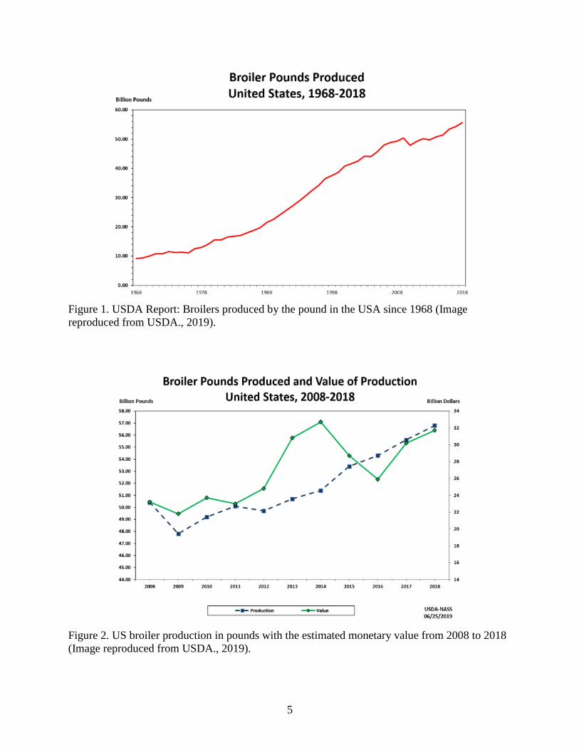
billion pounds (4.5 million tons) of broiler meat in 1968, and about 56 billion pounds (28 million
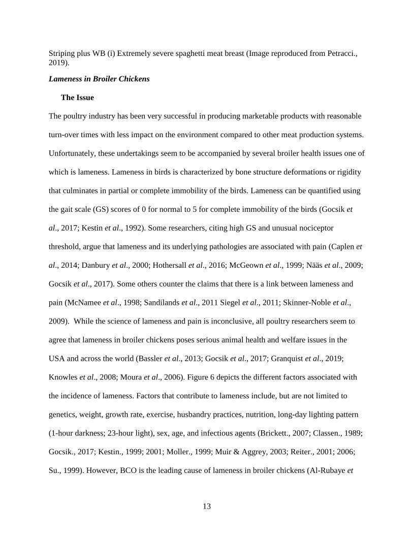
tons) in 2018—a 600% growth in productivity (Figure 1: USDA., 2019). In terms of monetary
value, US poultry production was priced at 32 billion dollars and it is still growing (Figure 2).
5
Figure 1. USDA Report: Broilers produced by the pound in the USA since 1968 (Image
reproduced from USDA., 2019).
Figure 2. US broiler production in pounds with the estimated monetary value from 2008 to 2018
(Image reproduced from USDA., 2019).

6
Modern Broilers Selection and Breeding Techniques
Until the early 1900s, the only way to select and breed chickens was to identify the best breeder
for the phenotype of interest and mate them for the next generation (Muir & Aggrey, 2003).
However, technologies applied in poultry breeding have since advanced. The technologies were
geared for (i) management of poultry reproduction, (ii) tracking of pedigrees, (iii) mating, and
(iv) accurate utilization of true breeding values of potential candidates (Muir & Aggrey, 2003).
Before the 1940s, breeding technologies were strictly aimed at producing purebred stock from
pure-breeding lines. But shortly after breeding programmes were implemented, broiler producers
began merging specialized lines and crossing them to make commercial production animals with
distinct breeding goals (Sainsbury, 2000; Muir & Aggrey, 2003). Today, broiler products are
usually three-way or four-way crosses between pure breeding lines over four generations (Muir
& Aggrey, 2003; Paxton, 2010 Pollock, 1999; Sainsbury, 2000). A common generation and
multiplication from pure breeding line to broiler products are described below:
(i)Pure-breeding line: Chickens are owned by primary breeder companies and kept on biosecure
farms for selection programmes. Breeding companies maintain up to ten pure-breeding lines for
their numerous broiler lines (Figure 3; Muir & Aggrey, 2003; Pollock., 1999).
(ii) Great-grandparent stock: These come from pure-breeding lines; they are used primarily to
multiply the line and produce tens of thousands which are needed for generating the grandparent
lines. They are subject to mass selection for selected traits. They are heavily controlled by the
primary breeding companies. In figure 3, they have designated flocks A males and females, B
males and females, C males and females, and D males and females (Muir & Aggrey, 2003;
Pollock., 1999).

7
(iii) Grandparent stock: are the first-generation in the four-way ABCD cross. They are A males
x B females and C males x D females from Great-grandparent stocks that are used to produce
hybrid AB or CD hybrids parents. Hundreds of thousands of Grandparent stocks are distributed
to the local distributor of parent stocks or integrated production companies (Muir & Aggrey,
2003; Pollock., 1999).
(iv) Parent Stock: are AB-hybrid males and CD-hybrid females. They are mainly owned and
maintained by broiler production companies (Muir & Aggrey, 2003; Pollock., 1999).
(v) Broilers: are the commercial products of crossing parent stocks. They are meat-type chickens
that are raised, slaughtered, processed for large scale meat consumption (Muir & Aggrey, 2003;
Pollock., 1999).
Expectations for future meat demands often drive poultry breeding goals. Hence the
intensification of artificial selection fuelling modern broiler production (Muir & Aggrey, 2003,
Paxton, 2010; Pollock.,1999). In the 20th century, Poultry breeders kept the pace of production
by adapting numerous vital selections and breeding technological innovations (Table 1, Muir &
Aggrey, 2003).

8
Figure 3: Example of generation and multiplication scheme in modern broiler production from
elite/ pedigree to commercial broilers products. (Reproduced from Paxton., et al, 2010).
Table 1. Timeline for critical technologies employed for poultry breeding in the 20th century
(Reproduced from Muir & Aggrey, 2003).

9
Impact of Genetic Selection in Broiler Production: Pros and Cons
The poultry industry has seen great successes largely due to its ability to economically produce
acceptable products (Anthony, 1998; Muir & Aggrey, 2003; Paxton., 2010; Tallentire, 2018).
This success is the cumulative effect of selection intensities, shorter generation times, and
lessened environmental impacts, among many other benefits (Anthony, 1998; Muir & Aggrey,
2003; Tallentire., 2018). It is important to explore the advantages and disadvantages that have
surfaced from intensive selective broiler breeding.
Compared to the early 20th century, the modern market-weight broiler’s time of production has
substantially dropped. In the 1920s, it took an average of 112 days to raise 2.5 lb live-weight
birds. In 2019, 6 lb live-weight birds can be raised in 47 days (Muir, 2013; NCC, 2020). The
“Feed to Meat Gain” or the pound-amount of feed used to produce a one-pound live-weight
broiler in 2019 was about 4.7 lb feed per 1 lb broiler (NCC, 2020). In 2019, the feed to meat gain
was 1.80 lb per 1 lb of broiler produce with acceptable meat yield (Anthony, 1998; Muir, 2013;
NCC, 2020). Modern broilers have been heavily selected for growth rate since the 1950s. By
2015, the growth rate has increased by 400% (Figure 4; Muir & Aggrey, 2003; Renema, 2007;
NCC, 2020). In addition to meat quality and quantity, the monetary value and exports of broilers
have been considerable (Anthony, 1998; Muir & Aggrey, 2003; USDA, 2019). The mortality
rate of chickens has dropped from 18% to 5% since 1925 (Muir & Aggrey, 2003; NCC, 2020).
Interestingly, broiler meat production has had a relatively lower impact on the environment
compared to beef and pork production (Anthony, 1998; Tallentire., 2018). This is associated with
a drop in the fossil fuel emission of greenhouse gases during feed production, combined with the
reduced nutrient loss from poultry manure. These benefits increase even more in feed-efficient
birds (Muir & Aggrey, 2003; Tallentire, 2016; 2018).

10
Although the poultry industry has recorded immense feats particularly as it concerns the artificial
selection of desirable broiler traits. There are growing concerns over the negative effects of
genetic selection in broiler production (Anthony, 1998; Hock., 2014; Muir & Aggrey, 2003).
Hock (2014) counted 23 classes of organ system metabolic disorders important to broiler
chickens and turkeys (Table 2; Hock, 2014). Some reports suggest that in rapidly growing birds,
one of the main issues observed is increased carcass fat deposition (Anthony, 1998; Tumová.,
2010). Then there are issues of broiler liveability, immune function, and reproductive
complications at the breeder level (Anthony, 1998; Hock, 2014).
Figure 4: Age-related changes in the size of the University of Alberta Meat Control strains
unselected since 1957 and 1978, compared to the Ross 308 broilers (2005) for Day 0, 28, and 56.
(Image reproduced from Zuidhof., 2014).

11
Table 2. The number of idiopathic disorders of broiler chickens and turkeys reported in the
literature (Reproduced from Hock, 2014)
Organ system Number
Skeletal disorders 11
Muscle disorders 4
Integument 3
Cardiovascular disease 2
Reproduction 3
Around the globe, some broilers develop ascites, a condition caused by increased pulmonary
pressure and resultant hypoxia that culminates in the accumulation of fluids in the peritoneal
cavity resulting in abdominal swelling (Al-Zahrani, et al., 2019; Anthony, 1998; Parveen, et al.,
2019; Wideman, 2000). More issues emanating and associated with the rapid growth rate of
broilers include muscle abnormalities resulting from the production of high-yield birds heavy
with meat that surpasses several metabolic and/or anatomical limits (Anthony, 1998).
Concerning animal behavior, Bokkers et al. (2003) write that no difference was found in resting
demeanor between fast- and slow-growing broilers raised to 13 weeks, as the birds seemed
motivated to perform all kinds of behavior in a feasible environment. They noted, however, that
for fast- and slow-growing broilers the ability to carry out certain behaviors became tasked with
age, probably due to their weight (Bokkers., 2003). In the past 30 years, there has been an
increased incidence of breast meat abnormalities like wooden-breast (WB), white-striping, and
spaghetti-meat (SM) (SM) in broilers (Abash et al., 2016; Petracci et al., 2015; 2019; Sihvo et
al., 2014; 2017). White stripes are recognizable by the accumulation of lipids and proliferation of
connective tissue line up in the same direction as the striations of the muscles (Figure 5B-D;
Petracci, et al., 2015; 2019). WB was first described in 2014 by Silvo, et al (Sihvo, et al., 2014).

12
WB mainly affects the pectoralis major and sometimes pectoralis minor. It presents a confined
lesion at 2 weeks of age that develops as a fibrotic injury with a hardened and pale appearance in
the pectoral muscles (Figure 5E-H; Abasht, et al., 2016; Petracci, et al., 2015; 2019; Sihvo, et
al., 2014; 2017). SM affects broiler chicken pectoralis major muscles impairing its integrity. It is
characterized by a soft consistency in the ventro-cranial segment due to poor adhesion of
Musculo-fibers (Figure 5I; Petracci et al., 2015; 2019; Tasoniero, et al., 2020).
Selection for rapid growth is also associated with numerous skeletal defects that clinically are
important to various degrees of locomotion. The group of locomotion difficulties resulting from
skeletal diseases is called Lameness. Skeletal defects include tibial dyschondroplasia, epiphyseal
ischaemic necrosis, epiphyseal separation, skeletal fracture, valgus-varsus deformity, angular
bone deformity, twisted leg, spondylolisthesis (kinky-back), gastrocnemius tendon rupture,
among others (Havenstein et al., 1994; 2003; Julian, 1998; Muir & Aggrey, 2003).
Figure 5: Classification of Broiler chicken Breast meat myopathies. (a) Normal breast (b)
Moderate White Striping breast (c) Severe White Striping breast (d) Represents moderate White
Striping thigh (e) Woody-breast (WB) with focal, hardened, and pale areas, without hemorrhages
(f) Extremely severe WB case (g) Is the same case as figure f with hemorrhages (h) White
13
Striping plus WB (i) Extremely severe spaghetti meat breast (Image reproduced from Petracci.,
2019).
Lameness in Broiler Chickens
The Issue
The poultry industry has been very successful in producing marketable products with reasonable
turn-over times with less impact on the environment compared to other meat production systems.
Unfortunately, these undertakings seem to be accompanied by several broiler health issues one of
which is lameness. Lameness in birds is characterized by bone structure deformations or rigidity
that culminates in partial or complete immobility of the birds. Lameness can be quantified using
the gait scale (GS) scores of 0 for normal to 5 for complete immobility of the birds (Gocsik et
al., 2017; Kestin et al., 1992). Some researchers, citing high GS and unusual nociceptor
threshold, argue that lameness and its underlying pathologies are associated with pain (Caplen et
al., 2014; Danbury et al., 2000; Hothersall et al., 2016; McGeown et al., 1999; Nääs et al., 2009;
Gocsik et al., 2017). Some others counter the claims that there is a link between lameness and
pain (McNamee et al., 1998; Sandilands et al., 2011 Siegel et al., 2011; Skinner-Noble et al.,
2009). While the science of lameness and pain is inconclusive, all poultry researchers seem to
agree that lameness in broiler chickens poses serious animal health and welfare issues in the
USA and across the world (Bassler et al., 2013; Gocsik et al., 2017; Granquist et al., 2019;
Knowles et al., 2008; Moura et al., 2006). Figure 6 depicts the different factors associated with
the incidence of lameness. Factors that contribute to lameness include, but are not limited to
genetics, weight, growth rate, exercise, husbandry practices, nutrition, long-day lighting pattern
(1-hour darkness; 23-hour light), sex, age, and infectious agents (Brickett., 2007; Classen., 1989;
Gocsik., 2017; Kestin., 1999; 2001; Moller., 1999; Muir & Aggrey, 2003; Reiter., 2001; 2006;
Su., 1999). However, BCO is the leading cause of lameness in broiler chickens (Al-Rubaye et

14
al., 2015; 2017; Bradshaw et al., 2002; Dinev., 2009; Jiang et al., 2015; Thorp et al., 1993; 1994;
1997; Wideman., 2016; Wideman et al., 2012; 2013; 2015; Wideman and Prisby 2013).
Figure 6: Factors that contribute to the incidence of lameness in broilers (Image reproduced from
Kierończyk et al., 2017).
Prevalence of Bacterial Chondronecrosis with Osteomyelitis Lameness
BCO-lameness is important to the poultry industry for economic and animal welfare reasons.
Over 1.5 % meat-type chickens raised to processing weights at 5-8 weeks within the past 20
years in the USA may be affected with spontaneous BCO and lameness (Dinev, 2009; Stalker et

15
al., 2010; Wideman, 2016; Wideman et al., 2012; Wideman and Prisby, 2013). This number may
be even higher. According to Zinpro, a spontaneous outbreak of lameness can affect over 15% of
commercial broiler flocks (Rebello., 2019). A cross-sectional study of broiler flocks across
Britain, France, Italy, and the Netherlands indicated that there was a 16% prevalence of
lameness, with GS of at least 3 or more (Bassler et al., 2013; Gocsik et al., 2017). A similar
study in Sweden suggested a 14-26% prevalence with GS≥3 (Sanotra et al., 2003). A
longitudinal survey of 20 broiler flocks in Victoria, Australia revealed that BCO occurs
throughout the lifespan of broiler at a very high rate, with different lesions diagnosed in about
28% of the birds (Wijesurendra et al., 2017).
Economics of BCO Lameness
Over the past 70 years, the market of broiler production has seen a dramatic change from
smallholder chicken farms to a more intensive and integrated multibillion-dollar set-up operated
by a few corporations (Lowder et al., 2009). Lameness causes financial loss in poultry revenue.
This is due to increased mortality, culling of lame birds at different stages of production, and
condemning birds during processing. According to the Farm Model, the economic impact of
lameness is a function of the frequency of the incidence of lameness in birds with GS≥3 and its
impact on poultry productivity. Poultry production is expressed in terms of production costs,
gross margin (revenues - variable costs), and the net profit per kilogram of delivered broiler
(Gocsik et al; 2017). The Farm model considers increased mortality, higher feed conversion,
increased condemnation rate at slaughter, and lower weight gain in estimating the economic
burden of lameness (Gocsik et al; 2017). The damages due to the mortality of lame birds can be
estimated with Equation 1. And the damages incurred from condemning lame market-age birds
at processing are estimated with Equation 1 and Equation 2 (Gocsik et al., 2017; Nääs et al.,
16
2009). Considering a lot of factors, the poultry industry in the USA loses over $100 million per
year which amounts to $.016 per broiler (Al-Rubaye et al., 2015; Aydin, 2018; Cook, 2000;
Weaver, 1998). This affects production costs and thus the shelf-price of poultry products (Cook.,
2000; Weaver., 1998).
(Equation 1 is reproduced from Gocsik et al.,2017).
(Equation 2 is reproduced from Gocsik et al., 2017).
Pathogenesis BCO-Lameness
BCO was formerly referred to as femoral head necrosis (FHN), proximal femoral degeneration,
or bacterial chondronecrosis but the name was changed as researchers learned that proximal
tibiotarsus and the fourth thoracic (T4) vertebra (with spondylitis) are also affected (Jiang et al.,
2015; McNamee & Smyth., 2000). Broilers can grow to about 8 Lbs in 8 weeks (Wideman.,
2016). This weight gain cannot be sustained without an equivalent increase in the size and
strength of the skeletal frame of the bird. The mechanism of rapid bone growth is important to
BCO and lameness. Growth of long bones in young broilers involves elongation of growth plates
at both ends of the bone shaft/diaphysis, as well as an increase in the diameter as a result of the
dynamic remodelling of the cortical bone (Wideman., 2016; Wideman & Prisby., 2013).
Growing broiler birds see about four-times growth in length of femur and tibia, with a mid-shaft
diameter increase that is three to five times the original width within the same time frame
(Applegate & Lilburn., 2002; Bond et al., 1991; Wideman, 2016; Yair et al., 2012). Wideman
(2016), notes that broilers are more susceptible to lameness than layers as the former has a

17
disproportionate weight gain ratio to skeletal structure maturation than it does cranial-caudal
redistribution of muscles mass (Wideman, 2016). Rapidly growing birds had a higher incidence
of lameness and efforts that reduce early growth lessens the disease in broilers (Wideman.,
2016). Dr. Wideman developed a wire-model flooring for inducing lameness in growing birds
(Wideman et al., 2012). This model creates shear stress in rapidly growing young birds, inducing
lameness with or without bacteria administration in water (Al-Rubaye et al., 2015; 2017;
Wideman et al., 2012, 2013, 2014; Wideman and Prisby, 2013; Wideman, 2016). Trials on the
wire-flooring system utilizing different broiler product lines revealed that they were all
susceptible to the incidence of BCO-lameness with some lines showing sire-effects (Al-Rubaye
et al., 2017; Wideman et al., 2013, 2014). The incidence of BCO lameness appears to begin with
mechanical micro-fracturing of poorly mineralized columns of cartilage cells (chondrocytes) in
the proximal growth plates of the femora and tibiae of early rapid-growing young broilers (Petry
et al., 2018; Wideman., 2016; Wideman & Prisby., 2013). The micro-fractures generate
osteochondrotic crypts that get colonized by hematogenously distributed opportunistic bacteria
(Al-Rubaye et al., 2015; Jiang et al., 2015; Mandal et al., 2016; Petry et al., 2018; Wideman.,
2016; Wideman & Prisby, 2013; Weimer et al., 2020). These bacteria come vertically from
broiler parent breeders to their chicks, or horizontally from a contaminated hatchery, and
eggshells (Stalker et al., 2010; Wideman., 2016). Bacteria may get translocated into the chick’s
blood supply through the respiratory system, gastrointestinal tract, or integumentary system
(Figure 7; Al-Rubaye et al., 2015; 2017; Wideman et al., 2012, 2013, 2014; Wideman and
Prisby, 2013; Wideman, 2016). Translocated bacteria get hematogenously distributed to both
ends of the growth plate by the numerous terminal epiphyseal and physeal vascular plexuses

18
(Figure 8; Wideman, 2016; Wideman & Prisby, 2013). Since the blood supply of broilers is
important to the incidence of lameness, it is crucial to study its anatomical composition.
Figure 7. Routes of bacterial infections in rapidly growing birds. Bacteria transmitted to chicks
from parent breeders, contaminated hatchery sources, eggshells, or bacteria that is translocated
into bird’s circulatory system via the integument, respiratory system or gastrointestinal tract gets
distributed hematogenously and colonize the osteochondrotic crypts from microfractures
resulting from mechanical stress (Image reproduced from Wideman, 2016; Wideman & Prisby,
2013).
Blood supply to proximal heads of rapidly growing broiler important to lameness.
There are three main structures in the blood supply of broiler long bones. These include (1)
cartilaginous epiphysis (e), (2) the physis (p) also known as the growth plate (GP), and (3) the
metaphysis (m). Cartilaginous epiphysis (e) is composed of articular cartilage (a) and hyaline
cartilage (hy). The physis (p) or the growth plate (GP) comprises a cartilaginous matrix and long
maturation columns of chondrocytes in consecutive layers with unique characteristics. The
physis/gp spans the germinal chondrocytes (stem cells) of the resting zone (rz), to the highly
mitotic proliferating zone (pz), the prehypertrophic zone (phz), and then the hypertrophic zone
(hz). The metaphysis (m) is composed of the degenerative calcifying chondrocytes as well as the
newly formed osteoid in the calcifying zone (cz). In the metaphysis, the spicules of trabecular

19
bone support the growth plate’s scaffolding and the resorption zone (rez) wherein the trabecular
bone thins out to form the medullary cavity (mc) of the diaphysis (d) (Figure 8 through
9; Wideman & Prisby, 2013; Wideman, 2012). In Figures 8 through 10, blood flows from the
epiphyseal vascular supply (ev), travels either through epiphyseal vascular canals (ec) within the
hy of the e or through the junctional canals (jc) moving down the growth plate. Branches of the
ev can also terminate as epiphyseal vascular capillary complexes (evc) within the hz or they can
become penetrating epiphyseal vessels (pev) that terminate as a penetrating vascular capillary
plexus (pvp) and supplies blood to the rz, pz, and phz collectively called the maturing zone of the
growth plate. The proximally traveling nutrient artery (ana) coming from mc divides severally
inside the diaphysis (d) to form metaphyseal vessels (mv) within the m. The mv terminates as
metaphyseal vascular capillary plexuses (mvp) and supplies the czi. The pvp or mvp does not
usually cross the hz like the transphyseal vessels (tp) does. The pvp and mvp loop back around to
form fenestrated capillaries that return as venules coursing through the same canal (Figures 8 –
10; Wideman., 2016; Wideman & Prisby., 2013).

20
Figure 8. The diagram on the left pane and photograph on the right panel shows the blood supply
and the anatomical structures of the long bone in growing broilers (Reproduced from Wideman
& Prisby., 2013; Wideman., 2016).
Figure 9. Diagram of the femoral proximal head illustrating the formation of osteochondrotic
clefts/crypts at the boundary between the growth plate and the epiphysis (Wideman &Prisby.,
2013).

21
Figure 10. Arterial blood supply to the leg bone of a growing bird (Reproduced from Wideman,
2016).
Vertebral anatomy and blood supply in broilers important to BCO lameness
Of the five thoracic vertebrae in broilers, the fourth thoracic vertebra (T4) moves freely and
separates the notarium and synsacrum (Figure 11; Baumel et al., 1993; Wideman, 2016).
Wideman (2016), describes T4 be fused to the caudal surface of notarium and the cranial surface
of synsacrum (Wideman, 2016). He mentioned that the fusion of these bones is only partial until
the birds reach sexual maturity, perhaps to allow room for the continuing longitudinal growth of
the vertebral body in young birds (Wideman, 2016; Wideman and Prisby, 2013). The structure
and position than T4 with respect to the more rigid/inflexible cranial T3 and caudal T5

22
encourages the erosion of epiphysis and physis of T4 which is important to the incidence of
vertebral BCO (Wideman., 2016).
Figure 11. The skeleton of a bird. The image highlights the exposure of the very flexible thoracic
vertebrae 4 or T4 (Image reproduced from Wideman, 2016; Wideman and Prisby, 2013).
The T4 vertebral body in rapidly growing broiler chickens is prone to various deformities and
non-inflammatory mechanical collapse. They are also susceptible to downward rotation

23
(subluxation), and scoliosis/lateral displacement (Figure 12; Wideman, 2016; Wideman and
Prisby, 2013). The clinical presentation of T4 subluxation is called spondylolisthesis or spine
slippage or “kinky back,” a condition in which spinal cord compression leads to paraplegia, a
hock- or rumps sitting position, and permanent immobility (Figure 12; Wideman, 2016;
Wideman and Prisby, 2013).
Figure 12. Broiler bone histopathology image for formalin-fixed 5 μM sections from 5-week
stained with hematoxylin and eosin. (A) Normal T4 (B) The boundary between the epiphysis (e)
and physis (p) normally should be seamless. (C) In broilers that appear to be clinically healthy
narrow osteochondrotic clefts or voids (arrows) containing cellular debris can be detected at the
boundary between the epiphysis (e) and the physis (p). Osteochondrotic clefts may interrupt the
local vasculature, cause distortions in the epiphyseal-physeal cartilage (*) and constitute wound
sites that are favourably colonized by opportunistic bacteria (modified from Figure 6 in
McCaskey et al., 1982).
In Figure 12 A-C, T4 from apparently healthy broiler birds present uninfected minor
microfractures, osteochondrotic clefts, and subclinical deformations in their epiphyseal and
growth plate layers. At the time when these findings were reported, only a few healthy birds
were diagnosed as clinical spondylolisthesis or kinky back (McCaskey et al., 1982; Wideman.,
2016; Wideman & Prisby, 2013; Wise., 1970). Kinky back (KB) may have genetic backgrounds
as studies conducted on broiler lines deliberately selected for KB showed the incidence of
spondylolisthesis. In another study where birds with KB were nursed back to health and bred for

24
two generations, the offspring presented with KB (Khan., 1977; Wideman., 2016; Wideman &
Prisby., 2013). In summary, the thoracic vertebra of rapidly growing meat-type chicken
undergoes wear and tear that results in skewed vertebral bodies. This condition combines with
numerous microfractures in the epiphysis and the physis of the vertebra and may stay non-
clinical or may even progress to the non-infectious and heritable spondylolisthesis (Wideman.,
2016; Wideman & Prisby., 2013). Non-infectious non-inflammatory osteochondrotic lesions are
by themselves not considered the main initiator of lameness in broilers. The collection of crypts
or clefts resulting from microfractures, packed with exposed collagen structures and fed by good
vascularities, maybe good infection sites for opportunistic bacteria microbes that play a role in
the onset of vertebral BCO (McNamee et al., 1998; Wideman, 2016; Wideman and Prisby,
2013).
Bacteria and Lameness
The etiology of BCO lameness is not fully understood, but bacteria are highly involved in the
incidence of the disease. As aforementioned, the unsupported mass of rapidly growing birds
causes microfractures and hence osteochondritic crevices. The crevices contain exposed collagen
matrices that may favor inhabitation and colonization by hematogenously distributed
opportunistic bacteria from various sources (Wideman & Prisby, 2013; Wideman, 2012; 2015;
2016). As discussed above, the vessels for blood supply to tibia, femur, and vertebra, narrows
into capillaries. These capillaries are networks of fenestrated endothelium large enough to allow
the translocation of some blood components, including bacteria, into the cartilaginous matrices
(Wideman & Prisby, 2013, Wideman et al., 2012; 2013; 2015; 2016). Translocated bacteria
adhere to exposed collagen complexes obstruct the epiphyseal and metaphyseal blood vessels
(Wideman, 2016; Wideman & Prisby, 2013). Such obstruction permits bacterial foci formation

25
and occludes pathogens from the broiler’s responses or antibiotics (Wideman, 2016; Wideman &
Prisby, 2013). Multiple opportunistic microbes, including Staphylococcus spp., Escherichia coli,
Enterococcus cecorum, Salmonella spp., have been isolated from BCO lesions (Al-Rubaye et al.,
2012; 2015; 2017; Dinev, 2009; Jiang et al., 2015; Joiner et al., 2005; Mandal et al., 2016;
Martin et al., 2011; Stalker et al., 2010; Thorp et al., 1993; Wideman., 2016, Wideman and
Pevzner, 2012, Wideman and Prisby., 2013; Wideman et al., 2012; 2013; 2015; Wijesurendra et
al; 2017). While we need to characterize BCO isolates to specify their role(s) in the incidence of
lameness, we also must ascertain where they are coming from and how they are getting into the
blood. In Figure 7, we proposed that bacteria important to be BCO may be translocated from the
respiratory tract, the integument, or the gut microbiome (Wideman, 2016). It is therefore
incumbent to analyze the microbial populations of the broiler chicken and their significance to
the infection process of BCO.
Microbiota and Lameness
Chicken’s natural microflora is associated with enrichment of intestinal villus and crypt
morphology (Jiang et al., 2016; Mandal, et al., 2016; Yeoman, et al., 2012). Analysis of villus
length and macroscopic pathology in lameness revealed that villus length improved with
probiotic treatments than the control group (Al-Rubaye, et al., 2020). The gut microbiome
promotes broiler growth by boosting energy-filled short-chain fatty acids. It is also involved in
nutrient absorption, detoxification, polysaccharides metabolism, immune system regulation, and
the general well-being of birds (Clavijo et al., 2018; Yeoman, et al., 2012). Microbiomes in
organs other than gut are also important to animal health. Studies of organ microflora dysbiosis
in humans, for example, are implicated in the pathogenesis of a host of diseases including
colorectal cancers, inflammatory bowel diseases, and so on (Mandel et al., 2016). Microbial

26
communities of the gut or other tissues important to lameness and other disorders are not fully
characterized or understood (Jiang et al., 2015; Yeoman, et al., 2012). However, most chicken
microbiome analysis seems to lean towards the gut compared to the blood, trachea, or feces, and
least of them the bones (Jiang et al., 2015; Lim et al., 2015; Mandal et al., 2016; Sohail et al.,
2015). Jiang et al. (2015) suspect that multiple bacterial species shuttle mainly from the gut
communities into the bloodstream forming niches across tissues. They emphasized the
importance of the gut microbes despite awareness about microbial communities present in yolk
remnants, and respiratory tracts in apparently healthy birds (Jiang et al., 2015). There are still
important questions to be answered: 1) Are all translocated bacteria commensals in BCO? 2)
How do commensals with benefits become pathogenic? 3) Do some of these microbes come in
as commensals evade immune effectors and then develop virulence? 4) Is translocation an
acquired virulence factor? 5) Is translocation a synergistic property that drives further
translocation? and 6) How and where can the immune system be boosted to better handle these
invasions? In attempts to characterize tissue (including gut) microbiomes, Mandal et al. (2016)
sampled the blood performing deep sequencing and analysing bacterial 16S rRNA sequences for
bacterial communities in 240 healthy birds and 12 lame birds. They discovered that 97% of the
phyla level communities in chicken blood was Proteobacteria (60%), Bacteroidetes (14%),
Firmicutes (11%), Actinobacteria (10%), and Cyanobacteria (2%) (Mandal et al., 2016). These
characterizations were determined from about 40 operational taxonomic units (OTUs) regardless
of age, host physiology, or environmental conditions. Linear discriminant analysis effect size
(LEfSe) showed significant population of Staphylococcus, Microbacterium, and Granulicatella
in lame vs healthy bird’s blood (Mandal et al., 2016). Wei et al. (2013) analyzed the intestinal
microbiome of broilers using all available published and unpublished data. They identified 915
27
OTUs equivalent to species that delineated with a 3% phylogenetic distance. The species were
grouped into 13 phyla comprising 70% Firmicutes, 12% are Bacteroidetes, and 9%
proteobacteria. These data made up 90% of all phyla. They identified 117 genera, a majority of
which include Clostridium, Ruminococcus, Lactobacillus, and Bacteroides. The main
representative at the genus level for Firmicutes was the ethanol metabolizing Ethanoligenes
bacteria. While Desulfohalobium was the most represented Proteobacteria. Actinobacteria (with
1% of sequences revealing Bifidobacterium) was represented in minute quantities. Other phyla
with small representations include Cyanobacteria, Spirochaetes, Synergisteles, Fusobacteria,
Tenericutes, and Verrucomicrobia (Wei et al., 2013; Clavijo et al., 2018). Understanding the
distribution of microbes may explain the routes and conditions necessary for bacterial
colonization important to BCO and other dysbiosis associated diseases. Clavijo et al. (2018)
analyzed the redistribution of microbial communities across the tissues of the gastrointestinal
tract (GIT) and found that the taxonomic profiles described for different parts of this system
vary. Factors include but are not confined to diet, sex, genetics, use of antimicrobials, and
sampling techniques. The chicken crop and gizzard are mainly populated by the genus
Lactobacillus and Clostridiaceae family. The crop environment promotes bacteria metabolization
of starch and fermentation of lactate. In the gizzard, gastric juices, pepsin, and hydrochloric acid
acidify this environment lowering the fermentation and general bacterial activity. The small
intestine contains the highest bacterial cell count of mainly Lactobacillus (70%), Enterococcus,
and Clostridiaceae (Clavijo et al., 2018). They also noted that the ceca are considered the richest
in species diversity to some extent for its capacity to hold food for 12 to 20 hours as well as for
its major water reabsorption role, the concentration of urea, and fermentation of undigested
carbohydrates from the intestines. The ceca are rich in the phyla Firmicutes, Bacteroides, and

28
Proteobacteria, and Clostridiaceae. Further, the abundant microorganisms of unknown
phylotypes belong to Firmicutes making this phylum to be of special interest. The gut
microbiome of chickens contains taxa with Campylobacter jejuni, Campylobacter coli,
Salmonella enterica, Escherichia coli, and Clostridium perfringens that may be harmful to birds
and human (Clavijo et al., 2018; Oakley et al., 2014). Campylobacter spp is considered harmful
to humans but not birds. Salmonella enterica may be deadly to birds and humans depending on
the age of the bird, the serotype of the Salmonella spp, and the health condition of the bird.
Salmonella spp is of lower prevalence. Escherichia coli, in chicken intestines, also has a low
abundance thorough out the lifespan of apparently healthy birds. Avian pathogenic Escherichia
coli (APEC) has virulence factors important to various diseases in birds (Clavijo et al., 2018;
Oakley et al., 2014). Jiang et al. (2015) surveyed the microbial communities of 97 femoral or
tibial heads from normal and lame broilers representing various ages, lines, lesion types, floor
types, to understand the long bone’s microbial importance to BCO. This study revealed a 91%
prevalence of Proteobacteria, 6% Firmicutes, and ~2% Actinobacteria phyla. Several other phyla
represented at lesser amounts, include Tenericutes, Bacteroidetes, Acidobacteria,
Verrucomicrobia, Nitrospirae, and Cyanobacteria that accounted for less than 0.4% of the total
phyla count. The overrepresented species were of Staphylococcus spp (Jiang et al., 2015). Table
3 accounts for differences between treatments recorded from these analyses (Jiang et al., 2015).
We have isolated multiple Staph species from birds that develop BCO on our facilities as well as
sick broilers from commercial farms (Al-Rubaye et al., 2012; 2015; 2017; Ekesi, 2020; Shwani
et al., 2020). Jiang et al. concluded that diminished species diversity is associated with a higher
degree of BCO lesions and lameness (Jiang et al., 2015). Further analysis of the BCO microbial
communities is needed for understanding the etiology of lameness and potential remediation.

29
Table 3: Comparison of microbial communities in different groups (Table reproduced from Jiang
et al., 2015)
Stress, Immune responses, and BCO
In lameness literature, the role of stress and immune responses revolves around microbial
proliferation important to the incidence of BCO that causes lameness in affected birds. The
incidence levels of lameness, the weight-induced microfractures, and subsequent bacterial
infection of physis and epiphysis of birds may initiate BCO (McNamee & Smyth, 2000;
Wideman & Prisby, 2012; Wideman et al., 2012; Wijesurendra et al., 2017). Environmental
stressors and septicemic pathogens, such as chicken anaemia virus (CAV) or infectious bursal
disease virus (IBDV) can cause immunosuppression that furthers the proliferation of microbes
that leads to the formation of BCO lameness (Wideman, 2016; Wideman & Prisby, 2013). The
wire-flooring model for inducing lameness in broilers causes chronic stress that results in
immunosuppression of broilers as shown by their elevated blood corticosterone levels (Wideman
& Prisby, 2012; Wideman et al., 2012). Injection of glucocorticoids, specifically dexamethasone,
resulted in femoral head necrosis (FHN) lesions, and the intravenous administration of
prednisolone also causes epiphyseolysis; separation of the epiphysis from the physis. (Cui et al.,

30
1997; Durairaj et al., 2012; Wideman and Prisby., 2013). In many of the dexamethasone
lameness trials, the responses were not typical of those recorded in spontaneous BCO cases.
Particularly, the administration of dexamethasone shrunk growth rates in broiler birds even at the
lowest dose that induced lameness in the birds (Wideman and Pevzner., 2012, Wideman and
Prisby., 2013). In turkey osteomyelitis complex (TOC), environmental stressors were associated
with the eruption of opportunistic pathogens harbored sub-clinically in the proximal tibia of
turkeys (Huff et al., 1998-2000; 2006; Wideman and Pevzner., 2012, Wideman and Prisby,
2013). Rodgers JD et al. (2006) developed an ELISA with nuclease protein as an antigen to
capture S. aureus-specific antibodies produced in response to bacteria administered to 500
broiler birds by aerosol. Bacteria were administered on Day 1 post-hatch with or without co-
infectors to induce BCO lameness. Co-infectors were CAV and IBDV. They found 71% of
serum samples from aerosolized S. aureus-treated birds had antibodies for nuclease protein. Only
35% of serum samples had antibodies for nuclease when there was no co-infection (Rodgers et
al., 2006). The co-infection was reported to have resulted in profound effects until day 42
(Rodgers et al., 2006). These findings are important as they highlight adaptive immune/ humoral
responses are activated in the birds infected by S. aureus, and co-infection with CAV and IBDV
drives the development of nuclease-specific antibodies for up to 42 days in broilers. Further,
because in our Staphylococcus BCO lameness model, we see an increasing amount of lameness
around this period, this needs to be examined further. We are working on characterizing the
pattern of innate immunity important to be BCO and lameness in our lab using phagocytosis
assays for BCO bacteria and chicken macrophage in directed genome evolution trials (Zaki, 2020
Dissertation). Lowder et al. (2009) wrote that S. aureus isolates gained mobile genetic elements
coincident with the jump from humans to birds to adapt to the avian ecosystem. Consequently,

31
they showed resistance to heterophil phagocytic killing in in vitro assays. A better understanding
of this mechanism is important, as it may help explain how BCO isolates bypass the immune
factors in the disease process of birds that get infected.
Microbiology of Common Pathogenic BCO isolates
Even though some of the findings described herein are not particular to chickens, the
mechanisms of pathogenesis of each microbe described may be important in the incident of BCO
and lameness.
Staphylococcus spp.
About 60 Staphylococcus species have been identified (Szafraniec et al., 2020). All
Staphylococcus spp can cause diseases (Crossley et al., 2009). The staph genome is dynamic;
the assortment of virulence factors varies by species and strains.
Staphylococcus agnetis
S. agnetis is a common cause of BCO lameness (Al-Rubaye et al., 2015; 2017). The species was
named after Europe’s first female veterinary surgeon, Agnes Sjöberg (1888–1964), who
struggled her way into the profession despite resistance from her male colleagues. S. agnetis is a
Gram-positive-staining, and a coagulase-variable bacterium (Adkins et al., 2017; Szafraniec et
al., 2020; Taponen et al., 2012). It is generally coagulase-negative after 4 hours, but over 25 %
of the isolates show coagulase-positivity after 24 hours. S. agnetis cells are facultatively
anaerobic, non-spore-forming, non-motile cocci which grow either singly, or in pairs or small
clusters. The bacteria colonies may grow to 3mm after 24 hours of incubation at 37 °C. The
bacterium is catalase-positive and oxidase-negative. It is round, opaque, smooth, non-hemolytic,
and light grey on bovine blood agar. S. agnetis is resistant to polymyxins, deferoxamine, and
lysozyme. But it is susceptible to lysostaphin and novobiocin. This bacterium is negative for

32
clumping factors and hydrolyses DNA at 37 °C giving off a degradation halo hue. S. agnetis
metabolizes and produces acids aerobically with D-glucose, D-fructose, D-mannose, lactose,
sucrose, and D-ribose. Phylogenies based on 16S rRNA sequence analysis, two housekeeping
genes (rpoB and tuf), or DNA fingerprinting with amplified fragment length polymorphism,
show S. agnetis forms a separate branch within the Staphylococcus genus (Al-Rubaye et al.,
2015; Adkins et al., 2017; Taponen et al., 2012). The closest species that have also been
recovered in BCO lameness include S. hyicus and S. chromogenes. S. agnetis are commonly
isolated in milk samples in the incidence of bovine intramammary infections that results in
subclinical or mild clinical mastitis in dairy cattle, and more recently BCO lameness lesions
(Adkins et al., 2017; Al-Rubaye et al., 2015; 2017; Taponen et al., 2012). S. agnetis is the main
BCO isolate on our farm and is induces lameness to statistically significant degrees using wire or
litter- flooring. The role of S. agnetis in BCO and lameness in infected birds will be discussed
further below.
Staphylococcus aureus
S. aureus is a coagulase-positive, Gram-positive bacterium. Although the production of
coagulase differentiates S. aureus from other Staphylococcal species, the coagulase gene (coa) is
not associated with virulence (Crossley et al., 2009). S. aureus is non-motile, non-spore-forming,
catalase-positive, and oxidase-negative. S. aureus is a facultatively anaerobic bacterium. The
bacterium has cell-bound clumping factors. It is slightly tolerant of sodium chloride. It ferments
mannitol and produces hyaluronidase. The physical appearance may differ by media. The reason
S. aureus causes more incidence of BCO and lameness in birds that develop the disease is not
known than any know BCO isolate. This implies, however, that S. aureus does have an inherent
capability to cause damage (McNamee et al., 2000). S. aureus infects several various hosts. Like

33
S. agnetis, S. aureus is also implicated in cattle mastitis. S. aureus also causes childhood
osteomyelitis, and hospital or community-acquired infections (Adkins et al., 2017; Al-Rubaye et
al., 2015; McNamee et al., 2000). Almost all recovered S. aureus recovered from sick animals or
humans have virulence capsules that inhibit them from being phagocytized (McNamee et al.,
2000). It has been suggested that these capsules facilitate adherence to chicken cartilage, but the
actual role in bone and joint infection is not understood (McNamee et al., 2000). S. aureus is the
most predominant of all the disease-causing Staphylococci (Crossley et al. 2009). Compared to
S. epidermidis (another common BCO isolate), S. aureus contains 18 distinct genomic islands
that house virulence genes that disrupt host defenses (Foster, 2005; Gill et al., 2005). According
to Crossley et al. (2009), much of the work done to characterize S. aureus has been on a limited
number of strains derived from a primary strain NCTC 8325. This isolate was retrieved
originally in 1960 from a sepsis patient in the UK and is the reference genome for NCBI. There
are 11,870 S. aureus genome assembly entries (Crossley et al., 2009). Different strains of S.
aureus have been derived from NCTC 8325 for various purposes, where the derivative strains
still preserve the ancestral lineage (Crossley et al., 2009). Comparing derived isolates with
clinical S. aureus, Φ13 is integrated into the att site on hlb gene that normally expresses β-toxin
interrupts its function (Crossley et al., 2009). This may be observed in strain NCTC 8325-4
where the temperate Φ13 is excised, hlb gene production of β-toxin is restored. This is important
not only because of the β-toxin negative effect of Φ13 integration but also because phage that
utilizes the att hlb site alters functions for a combination of genes (sea, sak, scn, chp) with
virulence factors for disruption host immune functions (Crossley et al., 2009). More
discrepancies and connotations exist between derived and clinical S. aureus strains, but these
notions are not without controversy. Studies suggest that mutations exist in at least tcaR and

34
rsbU genes that regulate the expression of specific virulence factors in all NCTC 8325
derivatives making them references and models for studying the regulation of pathogenicity
(Crossley et al., 2009; Sassi et al., 2014). Crossley et al. (2009) utilized genome-level
transcriptional profile analysis of NCTC 8325-4 strain with clinical isolate UAMS-1 and argues
that there are no such mutations in either tcaR or rsbU. This study revealed that over 300 genes
were expressed in a strain-dependent manner comparing NCTC 8325-4 and UAMS-1 strain
(without tcaR or rsbU) (Crossley et al., 2009). About half of these genes are under SigB regulon
control (Cassat et al., 2005; Crossley et al., 2009). A correlation study was performed in over
400 S. aureus isolated from different infections and geolocations for variable genes and virulence
with pulse-field gel electrophoresis (Booth et al., 2001; Crossley et al., 2009). Five of the 90
identified lineages were important to 65% of infections (Booth, et al., 2001; Crossley et al.,
2009). Collagen-binding adhesin (cna) gene was in three of the five lineages. Booth et al. (2009)
indicated, however, that no cna-containing lineages encoded the fibronectin-binding protein gene
fnbB. Lack of fnbB does not eliminate the relevance of S. aureus fibronectin binding capability
as they still possess a highly conserved fnbA. This does not make fnbB irrelevant either, as this
gene is positioned in a location termed region of difference (RD5). A region that houses the
surface-associated protein SasG and the regulatory elements sarT and sarU (Crossley et al.,
2009). In a similar study of 334 S. aureus strains, 33 genes were determined important for
virulence; cna was one of seven genes prevalent in invasive species (Crossley et al., 2009).
Otsuka, et al. studied15 clonal clinical community-acquired methicillin-resistant S. aureus (CA-
MRSA) particularly to osteomyelitis, and necrotizing pneumonia. They found cna in nine of
them (Otsuka et al., 2006). The 15 CA-MRSA strains were either categorized as pandemic
(Sequence type ST30) or continent-specific (ST1, ST8, OR ST80). Five of the cna-specific
35
strains were classified as pandemic CA-MRSA strains; they encoded gene bbp for adhesin that
binds sialoprotein in bones and lacked fnbB. This absence, Otsuka et al. (2006) noted, implies
the lack of RD5-linked genes (sasG and sarT) in the pandemic strain. The combination of bbp
and cna has been considered for the potential to galvanize pathogenicity to the point of
pandemicity (Crossley et al., 2009). Genome-level analysis suggests that certain S. aureus strains
may exhibit higher virulence and that predominantly clonal populations possess the ability to
cause disease. However, this is not always the case. Feil et al. (2003) performed a genome-scale
microarray study that analysed core variable (CV) genes scattered throughout the chromosome
and identified10 dominant lineages of S. aureus that lacked genes for diseases and were not
linked to infectious processes. Crossley et al. (2009) added that multilocus sequence typing
(MLST) failed to separate commensal versus invasive S. aureus based on clonal lineages. Feng
et al. (2008) did a comparative genomic analysis of S. aureus pathogenic and non-pathogenic
strains as well and argued that since no genotypic difference was recorded between the two
groups, that observed differences between pathogenic and non-pathogenic S. aureus are a
function of its state. One explanation for the divergent findings is that S. aureus is an
opportunist; isolates recovered from a sick host may say more about the state of the host than it
does the strain isolated. Conversely, S. aureus strains retrieved from healthy hosts may be
important to invasive diseases (Crossley et al., 2009). Study and analysis of clinical isolates and
NCTC 8325 derived strains in many in vivo and in vitro models show many structures, their
interaction, and disease processes of S. aureus. The main classes of virulence factors in S. aureus
are described below.
1) Surface Virulence Factors: In S. aureus, cell surface-linked pathogenicity involves
exposed cell wall structures like teichoic acid (WTA), lipoteichoic acid (LTA), lipoprotein, and

36
peptidoglycan (PG). The host immune systems may recognize such structures and express
cytokines and chemokines in response. These structures collectively play important roles in
helping S. aureus evade host defenses, and enhance colonization (Crossley et al., 2009). Studies
suggest that WTA is important to nasal colonization, as this precedes many S. aureus infections
(Baur et al., 2004; Crossley et al., 2009; Weidenmaier et al., 2004; van Dalen et al., 2020;
Winstel et al., 2015). S. aureus strains without these structural factors had poor adherence to
endothelial frame and were not virulent in rabbit endocarditis models (Weidenmaier, et al., 2004;
2005; Wertheim, et al., 2005). LTA and PG produce inflammatory responses that recruits
neutrophils that trigger the onset of septic shock (Crossley et al., 2009; Fournier& Philpott.,
2005; Ginsburg, 2002; Kengatharan et al., 1998). The involvement of TLR 2 in the host response
to LTA and PG are inconclusive (Crossley et al., 2009). In S. aureus LTA (ltaS gene) controls
autolysin activity, surface hydrophobicity, and therefore biofilm formation in a strain-dependent
manner (Crossley et al., 2009; Fedtke et al., 2007).
2) Surface Adhesins: S. aureus cell surface adhesins include a variety of proteins. Bone
sialoprotein-binding protein (bbp) that binds sialoproteins in bones and may result in
Staphylococcus-related osteomyelitis and arthritis (Campoccia et al., 2009; Crossley et al., 2009;
Persson et al., 2009; Tung et al., 2000). S. aureus expresses serine–aspartate repeat proteins
sdrC, sdrD, sdrE that contiguous and have adhesive properties (Crossley et al., 2009). SdrC
facilitates interactions with NRXN1 in the host extracellular matrix to promote bacterial
adhesion and consequently pathogenesis (Crossley et al., 2009; Askarian et al., 2016; 2017).
sdrD promotes the adhesion to host DSG1 decreasing bacterial clearance by posing resistance
against neutrophil killing in the blood (Crossley et al., 2009; Askarian et al., 2016; 2017). SdrE
interacts with host complement factor H/CFAH and improves the resistance to bacterial killing

37
by innate immune components in blood and therefore decreasing bacterial clearance (Crossley et
al., 2009; Sharp et al., 2012). SdrE protein interacts with host complement regulator C4BPA
inhibiting bacterial opsonization and killing by interrupting the activation of the host classical
pathway (Crossley et al., 2009; Hair, et al., 2013). Protein A (spa) plays a role in the inhibition
of the host innate and adaptive immune defenses, as they bind and capture the Fab and Fc arms
of immunoglobulins (Ig) with their five Ig-binding domains. As a result, S. aureus is therefore
protected from opsonophagocytosis (phagocyte killing of bacteria). Down the line, host B-cell
response is averted as there is a decrease of Ig-secreting cell proliferation in the bone marrow
and a decreased long-term antibody production (Crossley et al., 2009; Forsgren & Sjöquist.,
1966; Graille et al., 2000; Moks et al., 1986). Protein A hinders osteogenesis by (i) preventing
osteoblast proliferation, (ii) expression of alkaline phosphatase, (iii) expression of type I
collagen, as well as (iv) expression of osteopontin and osteocalcin. Protein A is a pro-
inflammatory factor in the lung. It binds and activates tumor necrosis factor-alpha receptor
1TNFRSF1A (Crossley et al., 2009; Forsgren & Sjöquist., 1966; Graille et al., 2000; Moks et al.,
1986; Widaa et al., 2012). S. aureus produces surface-associated molecules and secreted proteins
that bind fibronectin (Crossley et al., 2009). The two main classes of these proteins are
fibronectin-binding proteins A (fnbA) and B (fnbB). In the chromosome of S. aureus, the genes
for fnbA and fnbB are contiguous and either of them are is sufficient for proper binding of
fibronectin; however, the only fnbA can mediate platelet clumping (Crossley et al., 2009;
Heilmann, et al 2004; Peacook et al., 2000). The fnbA binds fibrinogen and elastin while fnbB
binds elastin but they both are associated with invasive diseases (Peacook, et al., 2000). Collagen
adhesin (cna) introduced earlier facilitates S. aureus adhesion to collagenous tissues such as
cartilages that may be important to bone disorders (Crossley et al., 2009). The cna protein is an
38
important virulence agent in many different animal models of Staphylococcal infections
including arthritis, endocarditis, and keratitis. It stops the activation of the classical complement
pathway by interacting and interfering with host C1q by forming a link between C1r with C1q
(Kang et al., 2013; Patti et al., 1993; 2002; Rhem et al., 2000; Xu et al., 2004). Fibrinogen
binding factors or clumping factors A (clfA) and B (clfB) are expressed on the cell surface of S.
aureus and are important to infections. Clumping factors A (clfA) enables the specific bacterial
binding to the gamma-chain of human fibrinogen. This stimulates aggregation of bacteria which
inhibits phospholipase A2 from bacterial phospholipid hydrolysis and hence inhibited the killing
and clearance of bacteria (Crossley et al., 2009). ClfA plays a role in the pathogenesis of sepsis
and septic arthritis (Crossley et al., 2009; Dominiecki & Weiss, 1999; O'Brien et al., 2002;
Palmqvist et al., 2004; Siboo et al., 2001). Fibrinogen-binding protein B clfB facilitates bacterial
attachment to the alpha- and beta-chains of human fibrinogen, thereby causing aggregation of
bacterial in clumps. It interacts with cytokeratin k10 and is partly implicated in the binding of
highly keratinized squamous epithelial cells from the nasal cavity. clfB binds the mice model
cytokeratin. And it causes platelet aggregation in humans (Crossley et al., 2009; Dominiecki &
Weiss, 1999; O'Brien, et al., 2002; Palmqvist, et al., 2004; Siboo, et al., 2001). S. aureus also
expresses sas genes; sasA is involved in platelet binding, and SasG is important for biofilm
formation (Crossley et al., 2009).
3) Secreted Adhesins: S. aureus possesses a group of secretable adhesive proteins
collectively termed SERAM (secretable expanded repertoire adhesive molecules). Extracellular
fibrinogen-binding protein (Efb) is a SERAM that is important to virulence (Crossley et al.,
2009; Posner et al., 2016; Pickering et al., 2019). Efb protein interacts with the alpha chain of
fibrinogen and fibrin product promoting interrupting normal platelets-fibrinogen functions that

39
result in the inhibition of platelet aggregation (Crossley et al., 2009; Posner et al., 2016). Efb also
forms a fibrinogen protective shield around the bacteria and inhibits phagocytic clearance by the
host. It does this by binding the C3b complement placed on its surface for opsonization with the
C-terminal end and then attaches to fibrinogen via its N-terminal (Bodén & Flock,1994; Crossley
et al., 2009). S. aureus also secretes Atl and Aaa proteins. Atl protein is the major autolysin of S.
aureus, which has been implicated in cell separation of newly divided S. aureus (Crossley et al.,
2009; Singh, 2014; Sugai, et al., 1995). Even though Atl participates in cell wall turnover, this
does not contribute to virulence (Takahashi et al., 2002). Aaa (Alias Sle1), binds host proteins
like fibrinogen, fibronectin, and vitronectin resulting in pathogenicity in mouse models
(Kajimura, et al., 2005; Crossley et al., 2009).
4) Surface Polysaccharides: S. aureus expresses capsular polysaccharides (cap genes), and
polysaccharide intercellular adhesin (icaADBC) (Crossley et al., 2009; Visansirikul et al., 2020).
Polysaccharides capsules coat C3b decreasing opsonization and phagocytosis of S. aureus by
phagocytic cells (Crossley et al., 2009; Visansirikul et al., 2020). Polysaccharide intercellular
adhesin is important to S. aureus biofilm formation in vitro (Crossley et al., 2009).
5) Extracellular Virulence Factors/ Exotoxins: S. aureus makes several extracellular
toxins important for diseases. These toxins are group as exfoliative toxins, enterotoxins, and
TSST-1 (Bukowski et al., 2010; Crossley et al., 2009). S. aureus encodes toxins in a strain-
dependent manner. A number of the predominant S. aureus diseases produce specific toxins.
Exfoliative toxins are common to Staphylococcal scalded skin syndrome (SSSS), bullous
impetigo is due to enterotoxins in staph food poisoning, and TSST-1 is associated with toxic
shock syndrome (Bukowski et al.,2010; Crossley et al., 2009). Many exotoxins cause
superantigenicity (a strong activation of the immune system) that causes disease (Thomas, et al;

40
2007). All S. aureus isolates encode α- and β-toxin (hla and hlb respectively) which are linked to
increased virulence in animal models of Staphylococcal diseases (Crossley et al., 2009; Walker,
et al., 1992; Wilke, et al., 2010). Alpha-toxin (hla) in staph binds the membrane of various
eukaryotic cells (particularly erythrocytes) drilling pores in them that results in ion imbalance
and consequently lysis (Crossley et al., 2009; Walker, et al., 1992; Wilke, et al., 2010). α-Toxin
is dermonecrotic and neurotoxic. Animal studies reveal that β-Toxin (Beta-hemolysins) encoded
on hlb causes cell lysis. These exotoxins also interact with blood cell membranes and cause them
to lyse (Crossley et al., 2009). While it is suggested that α-toxin is a factor in biofilm formation,
strains UAMS-1 and Sanger-252 that have a missense mutation in hla, and insertion of lysogenic
prophage in hlb do not produce alpha and beta-toxins (Crossley et al., 2009). The implication of
this is that α- and β-toxin are not necessary for virulence, but when made they cause infections.
The δ-toxin (a small, helical, amphipathic peptide) lysis mammalian cells, especially RBCs. γ-
Toxin and PVL (lukS-PV, lukF-PV) are bicomponent toxins made by interaction with hlgA and
hlgC (Crossley et al., 2009). A signature of several S. aureus diseases, including toxic shock
syndrome, is secretions of exfoliative toxins. Staph diseases with exfoliative toxins are
associated with strains that produce one of etA, etB, etC, and etD genes. Exfoliative toxin A
possesses serine protease-like properties; it binds profilaggrin on the skin and cleaves substrates
after acidic residues (Bailey, et al., 1990; Crossley et al., 2009; Dancer, et al., 1990).
Staphylococcus aureus produces many enterotoxins and exotoxins one of which is TSST-1 that
has superantigenic properties and has heat-stability crucial to food poisoning (Hamad, et al.,
1997; Shupp et al., 2002). TSST-1 can cross epithelial surfaces. This is an ability lacking in most
enterotoxins. Enterotoxins can penetrate the intestinal lining and initiate a local immune response
that includes activation of mast cells to produce histamine with frequent triggers of
41
vomiting/emesis (Hamad, et al., 1997; Shupp, et al., 2002). Toxic shock syndrome toxin-
1(TSST-1) is associated with toxic shock syndrome disease presented by high fever, rash,
hypotension, peeling of the skin, and multi-organ failure that often results in death (Crossley et
al., 2009). Though TSST-1 is mostly linked with toxic shock syndrome, enterotoxins like SEA,
SEB, and SEC, have superantigenicity and cause the disease if they get introduced via other
means than food (Crossley et al., 2009). TSST-1 is unique in its ability to cross the vaginal
mucosa though, hence the link with menstruation-associated toxic shock syndrome (Crossley et
al., 2009).
6) Enzymes: S. aureus produces and employs exoenzymes for metabolism and tissue
invasion important in pathogenesis. Enzyme classes include proteases, lipases, hyaluronidases,
and nucleases (Crossley et al., 2009; Drapeau et al., 1972; Prokesová, et al., 1992). Enzyme sspA
breaks the carboxyl-terminal of aspartate and glutamate of the peptide bond and along with
extracellular proteases triggers pathogenesis in human tissues. SspA engages in proteolytic
maturation of thiol protease sspB and inactivation of SspC (an inhibitor of SspB). SspA is
important to the degradation of the fibronectin-binding protein (FnBP) and surface protein A
crucial for the adherence to host cells. It also shields bacteria against host defenses by cleaving
IgG, IgA, and IgM (Crossley et al., 2009; Drapeau, et al., 1972; Prokesová, et al., 1992).
Cysteine protease SspB is involved in the inhibition of host innate mechanisms. It breaks down
host elastin, fibrinogen, fibronectin, and kininogen (Massimi, et al., 2001; Rice et al., 1992;
Shaw et al., 2004; 2005). SspB stops the phagocytosis of opsonized S. aureus and decreases the
surface expression of CD31 on neutrophils (Massimi, et al., 2001; Rice, et al., 1992; Shaw, et
al., 2004; 2005). It degrades host galectin-3/LGALS3, thereby preventing the neutrophil
activation by lectin (Massimi, et al., 2001; Rice, et al., 1992; Shaw, et al., 2004; 2005). Some

42
protease enzymes in S. aureus play regulatory roles in virulence (Crossley et al., 2009). The Clp
proteolytic complexes (clpX and clpP) are ATP-dependent and they direct protease to specific
substrates. A mutation in clpX reduced S. aureus biofilm formation, while a mutation in ClpP
increased biofilm formation. In the absence of clpP, clpX can serve as chaperone (Crossley et al.,
2009; Frees et al., 2004; Gersch et al., 2015).
7) Iron acquisition: S. aureus requires iron, which is limited in the host, for its infection
process (Crossley et al., 2009). S. aureus meets its iron needs through (1) siderophore which
consists of heme and high-affinity chelators, and (2) direct reuptake with surface proteins. Four
well-studied siderophores include staphyloferrin A, staphyloferrin B, aureochelin, and
staphylobactin. Stapyloferin A and B are encoded respectively on SfaA and SfaB genes. The
enzymes required for making staphylobactin are encoded on a nine-gene sbnABCDEFGHI
operon (Conroy et al., 2019; Crossley et al., 2009). Mutation of sbnE resulted in stunted growth
in iron-restricted media and lowered pathogenicity in the murine kidney model (Crossley et al.,
2009). Siderophores attach to host iron and passage them into the bacterial cell via iron-regulated
ABC transporter, such as the ferric hydroxamate uptake Fhu system. Fhu system has an iron-
binding lipoprotein ligand, integral membrane proteins, and ATPase (Crossley et al., 2009;
Hannauer et al., 2015). S. aureus has two other systems for the extraction of iron from Heme.
The first system designated isd includes five transcriptional units isdA, isdB, isdCDEFsrtBisdG,
isdH, and isdI (Crossley et al., 2009). The second is designated HtsABC for the heme transport
system, so named as they were identified from the search for proteins like ABC transporters
(Crossley et al., 2009). These systems are important to S. aureus pathogenesis, as 95% of the
iron in the human host is bound to heme proteins (Crossley et al., 2009). IsdB protein pulls heme
from oxidized methemoglobin (metHb) and transports it to IsdA or IsdC proteins, and on to

43
membrane transporter/IsdEF for internalization. The isdB protein induces resistance to hydrogen
peroxide and neutrophils killing (Bowden et al., 2014; 2018; Crossley et al., 2009; Gaudin et al.,
2011).
8) Regulation of S. aureus virulence: like every other process involved in bacterial
physiological processes, S. aureus produces virulence molecules in a regulated manner to boost
pathogenicity. It produces the accessory gene regulator, agr that is involved in the regulation of
several virulence S. aureus important to disease processes (Bibalan et al., 2014; Crossley et al.,
2009). The agr system is a two-part system composed of RNAII and RNAIII transcriptional units
which are respectively controlled by P2 and P3 promoters (Bibalan et al., 2014; Crossley et al.,
2009; Novick et al., 1995). The RNAII transcript traverses operon agrACDB that houses agrA
(regulator) and agrC (membrane sensor). AgrD encodes the extracellular-bound autoinducing
peptide (AIP) that is important to quorum sensing and is processed by agrB molecule (Abdelnour
et al., 1993; Booth et al., 1995; Cheung et al., 1994). AgrA plays a role in post-exponential phase
expression of a string of secreted proteins. (Bibalan et al., 2014; Crossley et al., 2009)
(Abdelnour et al., 1993; Booth et al., 1995; Cheung et al., 1994). Sar genes in S. aureus
regulates Agr gene functions (Crossley et al., 2009). SarA controls the production of virulence
factors and drives the biofilm formation process in a cell density-dependent manner
(Balamurugan et al., 2017; Bibalan et al., 2014; Crossley et al., 2009). SarA is also important to
multi-drug resistance mechanisms (Valle, et al., 2003). SarA is on a locus with three overlapping
transcripts sarA, sarC, and sarB with a similar terminus and promoters P1, P2, and P3 (Crossley
et al., 2009). SarA is regulates RNAII and RNAIII transcripts on agr locus and serves as a
transcriptional activator for fnbA, fnbB, hemolysins (hla, hld, hlgB, and hlgC), serine proteases
(splA, splB, splD and splF), bap that are strain-dependently important to biofilm formation in

44
diseases (Crossley et al., 2009; Valle et al., 2003). SarA down-regulates protein A (spa), lipase
(lip), thermonuclease (nuc), immunodominant staphylococcal antigen B (isaB), staphylococcal
serine and cysteine proteases (sspA and sspB), staphostatin B (sspC), metalloprotease aureolysin
(aur) and collagen adhesin (cna) (Crossley et al., 2009). Homologs of SarA include SarR, SarV,
SarX, SarZ, Rot, and MgrA whose interactions are not fully understood, but sarA and SarR
regulates agr RNAII expression. SarA activates and SarR represses the P2 in transcription
(Crossley et al., 2009; Reyes et al., 2011). In one study, sarV (rat) and mgrA(norR) are looped in
a feedback mechanism; mutation of mgrA results in increased autolysis (Crossley et al., 2009;
Ingavale et al., 2003). MgrA is responds to reactive oxidative changes that result in resistance to
certain antibiotics (Williams et al., 2006; 2015). There are more factors of regulation employed
in S. aureus virulence. The S. aureus exoprotein expression gene (saeRS) regulates exotoxins
(Crossley et al., 2009; Feng et al., 2008). Staphylococcal respiratory response gene (srrAB)
responds to changes in oxygen levels (Crossley et al., 2009; Ulrich et al., 2007). The two-
component response regulator gene (vraSR) that regulates agr and responds to cell wall-directed
antibiotics and increases resistance to β-lactams and vancomycin (Crossley et al., 2009;
Taglialegna et al., 2019). The two-component response regulator gene (graRS) that promotes
resistance to vancomycin and antimicrobial peptides (Crossley et al., 2009; Meehl et al., 2007;
Hu et al., 2016). The two-component response regulator gene (apsRS) that promotes resistance to
antimicrobial peptides (Crossley et al., 2009). The two-component response regulator gene
(alsSD) for Cell wall integrity and biofilm formation (Crossley et al., 2009). The metabolic
pathway regulation gene (Spx) for Stress response, as well as biofilm formation (Crossley et al.,
2009). The modulator of the sarA gene (Msa) which increases the expression of sarA, and the
virulence factors under its control (Crossley et al., 2009). The peptidoglycan synthesis gene
45
(murF) which is involved in the expression of cell-associated or extracellular virulence factors
(Crossley et al., 2009; Sobral et al., 2006).The concepts discussed in this unit encapsulates broad
association of structures, , patterns, , and regulatory mechanisms important to the pathogenesis
of S. aureus in many different diseases in different organisms. We find that S. aureus disease
mechanism per disease and organism seem to be resultant of the combination of factors utilized.
In chapter two we will discuss further how some of these processes may be involved in the
pathogenesis of a clonal population of S. aureus isolated from BCO lame birds.
Escherichia coli
E. coli is a rod-shaped, Gram-negative, facultatively anaerobic bacterium that possesses both
respiratory and fermentative metabolism (coliform). It is a member of the Enterobacteriaceae
family of the phylum γ-Proteobacteria. E. coli occurs in straight rod stacks which are 1.0–1.5-μm
wide, and about 2–6-μm-long. It does not produce oxidase. E. coli can be motile (with lateral
flagella) or non-motile (with polar flagella). E. coli is methyl red-positive, citrate-negative, and
Voges–Proskauer-negative. This bacterium is phylogenetically closest to Shigella spp
(Desmarchelier & Fegan., 2016; Percival et al., 2014). E. coli is a physiologically and
metabolically versatile organism classified as pathogenic or non-pathogenic. The non-pathogenic
or commensal strains are part of the normal gut microflora of endotherms (Mellata, 2013; Kaper
2005; Köhler& Dobrindt., 2011). Pathogenic strains have also been identified and associated
with various types of intestinal (IPEC- intestinal pathogenic E. coli) and extraintestinal (ExPEC)
ailments. IPEC are obligate pathogens and ExPEC are facultative. IPEC is phylogenetically and
epidemiological distant from ExPEC and commensal E. coli. This is because ExPECs and non-
pathogenic E. coli share large genomic segments (Köhler & Dobrindt., 2011). Commensal E. coli
strains are not prevalent in disease cases expect in immunocompromised hosts, or in situations
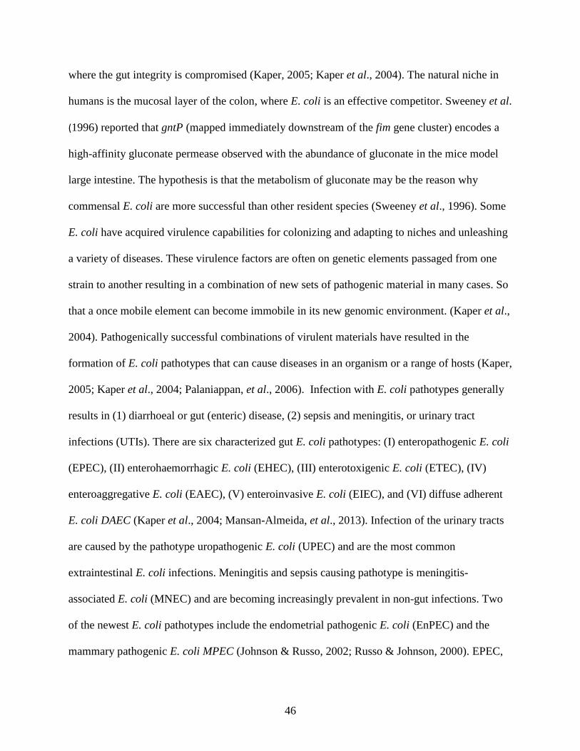
46
where the gut integrity is compromised (Kaper, 2005; Kaper et al., 2004). The natural niche in
humans is the mucosal layer of the colon, where E. coli is an effective competitor. Sweeney et al.
(1996) reported that gntP (mapped immediately downstream of the fim gene cluster) encodes a
high-affinity gluconate permease observed with the abundance of gluconate in the mice model
large intestine. The hypothesis is that the metabolism of gluconate may be the reason why
commensal E. coli are more successful than other resident species (Sweeney et al., 1996). Some
E. coli have acquired virulence capabilities for colonizing and adapting to niches and unleashing
a variety of diseases. These virulence factors are often on genetic elements passaged from one
strain to another resulting in a combination of new sets of pathogenic material in many cases. So
that a once mobile element can become immobile in its new genomic environment. (Kaper et al.,
2004). Pathogenically successful combinations of virulent materials have resulted in the
formation of E. coli pathotypes that can cause diseases in an organism or a range of hosts (Kaper,
2005; Kaper et al., 2004; Palaniappan, et al., 2006). Infection with E. coli pathotypes generally
results in (1) diarrhoeal or gut (enteric) disease, (2) sepsis and meningitis, or urinary tract
infections (UTIs). There are six characterized gut E. coli pathotypes: (I) enteropathogenic E. coli
(EPEC), (II) enterohaemorrhagic E. coli (EHEC), (III) enterotoxigenic E. coli (ETEC), (IV)
enteroaggregative E. coli (EAEC), (V) enteroinvasive E. coli (EIEC), and (VI) diffuse adherent
E. coli DAEC (Kaper et al., 2004; Mansan-Almeida, et al., 2013). Infection of the urinary tracts
are caused by the pathotype uropathogenic E. coli (UPEC) and are the most common
extraintestinal E. coli infections. Meningitis and sepsis causing pathotype is meningitis-
associated E. coli (MNEC) and are becoming increasingly prevalent in non-gut infections. Two
of the newest E. coli pathotypes include the endometrial pathogenic E. coli (EnPEC) and the
mammary pathogenic E. coli MPEC (Johnson & Russo, 2002; Russo & Johnson, 2000). EPEC,

47
ETEC, and EHEC have been observed in animals to employ similar pathogenesis molecules and
patterns seen in human diseases (Kaper et al., 2004). About BCO lameness, ExPEC also causes
significant economic losses in animal production, especially the poultry industry. Avian
pathogenic E. coli (APEC) behave like human pathogenic ExPEC strains. A recent examination
of virulence genes in human and avian ExPEC suggest that poultry products are important to
ExPEC that causes sepsis infection in human. A conclusion was drawn since poultry
contaminated with higher levels of E. coli has more multidrug resistance (MDR) compared to
other meat sources (Manges et al., 2007). Such finding implies that increased consumption of
poultry worldwide could be a contributing factor to antibiotic resistance in human ExPEC, and
hence the emergence of ExPEC diseases in humans (Pitout et al., 2012). A globalized market
coupled with ease of travel plays a role in the spread of the infection as well (van der Bij &
Pitout, 2012). Preventing and eradicating E. coli diseases in both humans and animals requires
full classification of virulence factors per pathotype and understanding their individual and
combined characteristics. There are four main phylogenetic groups of Escherichia coli A, B1,
B2, and D, of which B2 is the most abundant and pathogenic strains in ExPEC infections (Table
4; Mellata, 2013). E. coli is classified into 150 to 200 serotypes or serogroups based on somatic
(O), capsular (K), fimbrial (F) and flagellar (H) antigens. At least 53 H antigens, 188 O
liposaccharides antigens, and numerous different capsular K polysaccharide antigens.

48
Table 4. Virulence Traits in Pathotypes of Extraintestinal Pathogenic E. coli determined to be
associated with pathogenicity (Reproduced from Köhler & Dobrindt, 2011).
Although many ExPEC and their roles in the disease mechanism have been mostly characterized,
there are many unique factors between pathogenic and non-pathogenic E. coli (Kaper et al.,
2004; Köhler& Dobrindt., 2011). With ExPEC, a model has been postulated for the site of
isolation combined with two detected associated virulence factors per pathotype and is described
(Köhler& Dobrindt., 2011). ExPEC is diverse with only a few shared pathogenic factors, but
there is still no methodology to clearly define commensals apart from ExPECs. The classical
serotyping method is useful for IPEC classification because a good number of virulent strains in
this category are established well within the O: H serotypes (Köhler& Dobrindt., 2011). To some
degree, highly pathogenic ExPEC strains seem to be confined within a few O serogroups and
pathogenic factor combinations (See Table 5; Köhler& Dobrindt., 2011).

49
Table 5. Virulence factors in ExPEC (Reproduced from Köhler & Dobrindt, 2011)
Multilocus enzyme electrophoresis MLEE analysis reported that only a few E. coli genotypes
exist despite its diversity and forms the A-E phylogroups (Köhler& Dobrindt., 2011; Mellata,
2013). This was valuable as it helped distinguish many ExPEC from IPEC or non-pathogenic.
Many ExPECs belonged to B2 and to some degree D phylogroups. IPEC or non-pathogenic
strains are mainly A1 and B1 isolates. Multilocus VNTR analysis (MLVA) a relatively fast DNA
Sequence typing technique has been applied in the characterization of IPEC particularly EHEC

50
and is used for epidemiological inspection (Köhler& Dobrindt., 2011). Multilocus sequence
typing (MLST) employed for its worldwide comparability and usability, reveal E. coli strains to
be of comparable delineation (Köhler& Dobrindt., 2011; Martin, et al., 1998). Because
sequencing and analysis of various chromosomal regions do not characterize the phylogenetic
composition of E. coli strains, MLST is useful still for phylotype determination (Köhler&
Dobrindt., 2011). MLST-based molecular epidemiology reveals that phylogroup B2 (already
described to contain most ExPEC) is the evolutionarily oldest strains within the group (Köhler&
Dobrindt., 2011; Tenaillon, et al., 2010). Next-generation and large-scale sequencing give a
better in-depth phylogenomic analysis, as well as strain classification (Köhler& Dobrindt.,
2011). ExPECs and APECs were initially treated separately as initial studies did not connect
both to human and animal diseases in terms of virulence and hosts (Mellata, 2013). The
importance of APEC pathogenicity was initially minimized, as they were designated
opportunistic pathogens effectively reliant on external stress conditions (Mellata, 2013). This
notion has since been reversed, by studies that reveal significant variances in the distribution of
virulence elements in E. coli retrieved from chickens with colibacillosis and feces of healthy
ones (Johnson, et al., 2012; Mellata, 2013; Schouler, et al., 2012). Comparison of human
pathogenic E. coli, strains from chicken colibacillosis, with strains isolated from feces identified
virulence factors that may categorize APECs. Examination of these factors has shown them
important to APEC infection processes (Mellata, 2013). Also, APEC that was once thought of as
a distinct population, is now divided into several subtypes due to pathogenicity factors that are
important to diverse chicken diseases. Although the genetic traits that define APECs are not fully
categorized, the subsets do contain a combination of unique virulence stet of genes. These factors
include (I) iss, tsh, iucC, cvi, iutA, hylA, iss, iroN, and ompT genes carried on colicin V plasmids,

51
(II) toxin genes (astA, vat), (III) iron acquisition system genes (irp2 and iucD), (IV) adhesin
genes (papC and tsh), and (IV) the ColV genes cva-cvi (Mellata, 2013). Schouler et al. suggested
that the following four pathogenic factors can be used for strain determination as they can detect
over 70% APECs: (I) iutA and sitA for iron acquisition, (II) fimbriae P (F11), (III) frzorf4 for
sugar breakdown, and (IV) O-antigen O78, and T6SS aec26, aec4 (Schouler, et al., 2012). APEC
poultry diseases are generally regarded as colibacillosis. The presentation of colibacillosis
depends on the strain involved, the route of entry, and external factors. Like many microbes,
APEC harnesses host weaknesses to advance its pathogenesis. APEC causes local infections
such as cellulitis, salpingitis, synovitis, and omphalitis. They also cause septicemia, fibrinous
lesions of internal organs, and death of infected birds (Mellata, 2013). APEC is said to pose a
zoonotic risk as they share pathogenic factors and phylogenic features with human ExPEC.
Various epidemiological studies suggest poultry as a conduit for Human ExPEC, as avian
ExPEC (that was comparable genetically) has been isolated from the gut of healthy birds and
poultry meat (Manges & Johnson, 2012). Experimental evidence suggests that human ExPECs
can cause disease in chickens models, and Avian ExPECs alike to human animal-models
(Mellata, 2013). Other reports suggest that some ExPECs from ST95 and ST23 clonal groups
may be endemic to a range of hosts with the capability to cause many infections (Mellata, 2013).
This was a major concern because poultry colibacillosis was initially treated with antibiotics that
were important for AMR resistance in human ExPECs. Many countries have curbed the use of
antibiotics for livestock rearing, perhaps it might help mitigate the acquisition of antimicrobial
resistance in human and poultry pathogens. How do we now control colibacillosis outbreaks in
poultry? Not Long Ago, after a 3-year consecutive microbiologically sampling of 650,000
chickens from 38 broiler flocks in two large farms, Dinev reported the isolation of E. coli in over

52
90% of FHN associated with osteomyelitis. Urging that E. coli had a primary role in the etiology
of BCO in commercial birds particularly FHN (Dinev, 2009; Wijesurendra et al., 2017) Dinev,
noted that in contrast to E. coli, S. aureus seemed endemic to the parent stock lines (Dinev.,
2009). Wijesurendra et al. also examined chickens from 20 broiler farms in Australia and
reported that E. coli plays a crucial role in the bacteremia and hematological spread crucial to
BCO formation (Wijesurendra et al., 2017). Based on the amount of APEC isolated,
Wijesurendra et al. proposed that a remedy for BCO should be targeted at the species
(Wijesurendra et al., 2017). In Chapter 2, we report on E. coli isolated from BCO birds in three
different farms in the Arkansas area.
Enterococcus cecorum
Enterococcus cecorum, in the past 15 years, has become an important emerging pathogen to the
incidence of BCO lameness, even though it was initially considered a commensal in the
mammalian gut microflora when it was first described in 1983 (Dolka et al., 2017; Jung et al.,
2018; Kierończyk et al., 2017). E. cecorum is Gram-positive cocci, that often occur in pairs
(diplococci) or short chains. E. cecorum is a non-spore-forming, facultatively anaerobic bacteria
of the phylum Firmicutes. They are tolerant of diverse environments; they thrive in under
varying oxygen contents, temperature conditions, high sodium chloride, and pH. There was an
initial outbreak of E. cecorum in 2001 in Scotland, and multiple outbreaks have since followed
across Europe, United States, Iran, and Southern Africa (Jung et al., 2018). Pathogenic E.
cecorum has a signature inflammatory mass that occurs in the spinal cavities of the flexible
thoracic vertebrae. The infections of pathogenic E. cecorum goes by Vertebral BCO, vertebral
osteomyelitis, vertebral enterococcal osteomyelitis and arthritis, enterococcal spondylitis, and
‘kinky-back.’ E. cecorum has also been isolated from tibial and femoral head lesions (Jung et al.,
53
2018; Personal observation). Kinky back (KB) incidentally is the also colloquial name for the
developmental spinal anomaly, spondylolisthesis (Jung et al., 2018; Muir & Aggrey, 2003).
Broilers with KB experience higher morbidity and mortality as a result of a combination of
sepsis at the early growing days, starvation, and dehydration due to paralysis of infected birds in
the late growth stage (Jung et al., 2018). Embryo lethality assay has been used to determine the
pathogenicity and lethal dosage of pathogenic E. cecorum (Borst et al., 2015; Jung et al., 2018).
Some E. cecorum are inherently resistant to β-lactam-based antibiotics (like carbapenems,
cephalosporins, and penicillin), various aminoglycosides, and more recently vancomycin (Fisher
&Philips., 2009; Ryan & Ray., 2004). In chapter 3, we discuss our attempt to characterize the
pathogenicity of E. cecorum recovered from a commercial farm.
Clinical Diagnosis of Terminal BCO
Significant progress has been made in the study of BCO lameness since the first report of the
condition. Bacterial chondronecrosis with osteomyelitis, formerly known as femoral head
necrosis, is a complex disease with etiology that is not fully characterized. There is some
consistency, however, in the manifestation of terminal BCO in spontaneous cases or
Staphylococcal-induced models. Terminal BCO presents varying necrotic degradation and
bacterial infection of (1) the proximal ends and growth plate of long bones (tibiae and femora),
and (2) the flexible thoracic vertebrae T4 (Figures 13-15; Applegate et al., 2002; 2017; Al-
Rubaye et al., 2012; 2015; 2017; Dinev, 2009; Jiang et al., 2015; Joiner et al., 2005; Mandal et
al., 2016; Martin et al., 2011; McNamee & Smyth., 2000; Stalker et al., 2010; Thorp et al., 1993;
Wideman., 2016, Wideman and Pevzner., 2012, Wideman and Prisby., 2013; Wideman et al.,
2012; 2013; 2015; Wijesurendra et al; 2017). Common clinical presentations of BCO on long

54
bones include (I) femoral head transition, FHT (Figure 13B), (II) femoral head necrosis, FHN
(Figures 13 C-D, and 14-3), (IV) tibial head necrosis with caseous, THNC (Figures 13-F-H; 14-
6), (V) femoral head separation, FHS (Figure 14-2), (VI) tibial head necrosis, THN (Figure 14-
5), (VII) tibial head necrosis severe, THNS (Figure 14-6), (VIII) Tibial Dyschondroplasia, TD
(Figure 15), (IX) twisted leg, and (X) BCO diagnosis of unknown origins. The distal portions of
the long bones are less affected that the proximal heads. The presentations can be one-sided,
contralateral, or bilateral (Applegate et al., 2002; 2017; Al-Rubaye et al., 2012; 2015; 2017; Al-
Rubaye, personal observation; Dinev, 2009; Ekesi, personal observation, Jiang et al., 2015;
Joiner et al., 2005; Mandal et al., 2016; Martin et al., 2011; McNamee & Smyth., 2000; Rhoads,
personal observation; Stalker et al., 2010; Thorp et al., 1993; Wideman., 2016, Wideman and
Pevzner., 2012, Wideman and Prisby., 2013; Wideman et al., 2012; 2013; 2015; Wideman,
personal observation, Wijesurendra et al; 2017). Lameness associated with the T4 thoracic
vertebrae is generally termed spondylopathy or “Kinky back” for non-bacterial deformities, and
vertebral or enterococcal spondylitis if an infection is involved (Figure 16; Dinev et al., 2012).
The levels of inflammation in the flexible vertebra vary between cases (Dinev et al., 2012).

55
Figure 13. BCO progression on femoral (A-D) and tibial (E-H) proximal heads that result in
lameness in broiler chickens. (A) Normal femoral head with a white cap of epiphyseal cartilage
(e). (B) Epiphyseolysis or femoral head separation (FHS) epiphysis. Early necrosis (n) has begun
on the exposed underlying surface of the growth plate or physis (p). (C) Fractured growth plate
revealing necrotic void (nv) within the metaphysis (m). (D) The necrosis weakened diaphysis is
fractured during the disarticulation of the femoral head from acetabulum. The femoral epiphysis,
physis, and a great part of the metaphysis remain in the acetabulum. Abundant fibrinonecrotic
exudate (fe) oozes out in the terminal or most severe femoral head necrosis (FHN). (E) Normal
tibia comprising e, p, m with a center of ossification (designated *); the growth plate of the
healthy tibia is structurally in place with struts of trabecular bone in the metaphysis. (F-H) Levels
of bacterial permeation and sequestrate(s), arrows pointing to microfractures, along with necrotic
voids (nv) below the physis (Image Reproduced from Applegate et al., 2017).

56
Figure 14. Common clinical presentations of proximal femoral and tibial BCO lesions. (1)
Normal proximal femoral head; (2) FHS: epiphyseolysis; (3) FHN; (4)Normal proximal tibial
head with struts of trabecular bone in the metaphyseal zone fully supporting the growth plate; (5)
Tibial head necrosis (THN); (6) THNs with caseous (Reproduced from Mandal et al., 2016).
Figure 15. Tibial dyschondroplasia is characterized by abnormal masses of proliferated,
avascular, prehypertrophic cartilage in the proximal metaphyseal region of the proximal
tibiotarsus. This is attributable to the formation and retention of a large cartilage mass filling in
the entire metaphysis (Image reproduced from Dinev et al., 2012).
57
Figure 16. Vertebral BCO in broiler that exhibited paraplegic hock-resting posture, and spinal
compression due to build-up of abscess in vertebral bodies (follow white arrows). (A) Externally
visible nodal swelling of the flexible thoracic vertebral body with yellow discoloration of the
translucent remnants of the vertebral body. (B) The caudal portion of the vertebral body filled
with caseous abscess. (C) The epiphysis and growth plate have been replaced with necrotic
abscess. Osteochondrosis (follow black arrows) in the cranial articular cartilage (Image
reproduced from Applegate et al., 2017).
The S. agnetis induced BCO Lameness model
Our S. agnetis models for studying BCO lameness use different flooring systems: litter, wire, or
a combination of both. Ever since Dr. Wideman invented the elevated wire-flooring structure for
inducing lameness in broilers, we have induced lameness at high incidence (Al-Rubaye., 2015;
2017; Wideman et al, 2012, 2013, 2014; Wideman and Prisby, 2013; Wideman, 2016). This
model is primarily directed towards tibial and femoral BCO and has proven effective (Al-
Rubaye., 2015; 2017; Wideman et al., 2012, 2013, 2014; Wideman and Prisby, 2013; Wideman,
2016). Utilizing wire-floor, we found that S. agnetis is the prevalent isolate on our research farm
recovered from BCO lesions irrespective of site sampled or lesion type (Tables 4-5; Al-Rubaye.,
2015; 2017). In this model, we found administering S. agnetis in drinking water increases the
incidence of BCO lameness (Al-Rubaye et al., 2015; 2017). The finished genome sequence for a
58
S. agnetis BCO isolate has been published (Al-Rubaye et al., 2015). Our team also reported that
BCO is sometimes connected with significant bacteraemia (Al-Rubaye et al., 2015; 2017). The
broiler lines tested for susceptibility to lameness showed that all lines are predisposed, but line
differences and sire-effects may exist between birds (Wideman et al., 2013; 2014). In either the
wire or litter floor model, rapidly growing broilers that seem healthy (or have normal gait) can
over 24 hours, exhibit early symptoms of lameness. Depending on the project goals, we
generally place 60-day-old broilers per pen and then cull on day 15 to 50 of the clinically
healthiest birds (Al-Rubaye et al., 2015; 2017; Wideman et al., 2012, 2014). Pen configuration,
bird densities, food, and water (ad libitum), and environmental conditions (as light, and
temperature) are as described (Al-Rubaye et al., 2015; 2017; Wideman et al., 2012, 2013, 2014).
The bacterial administration in the broiler’s drinking water supply is also described (Al-Rubaye
et al., 2015; 2017; 2020 A & B). From a series of experiments, the minimum effective dose of S.
agnetis isolate 908 in drinking water starting on day 20 for two days is 105 CFU/mL on wire-
flooring (Figure 17).

59
Figure 17. The minimum effective dosage of S. agnetis in drinking water for the induction of
lameness. The cumulative percentage lameness is plotted for broilers reared on wire flooring and
administered the indicated concentrations of S. agnetis in the drinking water. The birds were
provided with tap water throughout (0 CFU/mL) or with S. agnetis in the drinking water on days
5 and 6 and again on days 15 and 16. Each treatment was for one pen of 50 birds. Values are
calculated as the percentage of the total number of birds in the treatment group. Final lameness
counts for challenges ≥105 CFU/mL were significantly different (P ≤ 10−7) from the challenges
with ≤104 CFU/mL (Image reproduced from Al-Rubaye et al., 2017).
Figure 18: Most effective days for the administration of S. agnetis in drinking water for induction
of lameness for broilers reared on wire flooring. Cumulative percent lameness (y-axis) is plotted
for broilers reared on wire flooring with tap water throughout (No Staph; 1 pen; 50 birds), or
administered S. agnetis at 105 CFU/mL (per treatment 2 pens, 50 birds/pen) for 2 days beginning
at 10, 20, or 30 days of age. Values are calculated as the cumulative lameness percentage for
days 40 through 56 based on the number of birds in the treatment group (Image reproduced from
Al-Rubaye et al., 2017).

60
Early symptoms of lameness will include, a poor gait (Score of 3), hesitancy to stand, eagerness
to sit, and slight wing-tip dipping (Al-Rubaye et al., 2015). The birds are encouraged to walk by
gentle prodding or herding with a common kitchen broom from Day 15 through Day 56. This is
done to minimize birds’ distress and diagnose the ailment in a timely fashion (Al-Rubaye et al.,
2015; Wideman, 2016; Wideman et al., 2012; Wideman and Prisby, 2013). Once birds present
the earliest sign of lameness, they are humanely euthanized and necropsied to grade or score
BCO, if any, on the femora, tibiae, and sometimes the vertebrae. In this model, we only
categorize birds presenting with specific lesions as lame. The percent (%) cumulative lameness
per pen/treatment is calculated with the formulas below:
% 𝑐𝑢𝑚𝑢𝑙𝑎𝑡𝑖𝑣𝑒 𝑙𝑎𝑚𝑒𝑛𝑒𝑠𝑠 𝑝𝑒𝑟 𝑝𝑒𝑛 =# 𝑜𝑓 𝑙𝑎𝑚𝑒 𝑖𝑛 𝑝𝑒𝑛
# 𝑜𝑓 𝑏𝑖𝑟𝑑𝑠 𝑖𝑛 𝑝𝑒𝑛 𝑜𝑛 𝐷𝑎𝑦 15 𝑋 100
% 𝑐𝑢𝑚𝑢𝑙𝑎𝑡𝑖𝑣𝑒 𝑙𝑎𝑚𝑒𝑛𝑒𝑠𝑠 𝑝𝑒𝑟 𝑡𝑟𝑒𝑎𝑡𝑚𝑒𝑛𝑡 =# 𝑜𝑓 𝑙𝑎𝑚𝑒 𝑓𝑜𝑟 𝑇𝑟𝑒𝑎𝑡𝑚𝑒𝑛𝑡
# 𝑜𝑓 𝑏𝑖𝑟𝑑𝑠 𝑖𝑛 𝑇𝑟𝑒𝑎𝑡𝑚𝑒𝑛𝑡 𝑜𝑛 𝐷𝑎𝑦 15 𝑋 100
In this model, treatment effects are evaluated for significant differences (P < 0.05) mainly by
logistic regression (binomial distribution) using the GLM procedure implemented in the R
Foundation for Statistical Computing. Blood and BCO lesions are usually assessed for
bacteremia and bacterial species utilizing protocols described (Al-Rubaye et al., 2015; 2017).
We find that for broilers raised on (a) litter, (b) litter for 35 days and then transferred to wire, or
(c) wire, the incidence of lameness was highest in birds raised on the wire-floor with the
administration of S. agnetis in water for two days on Day 20 (Figure 19). The incidence of
lameness on the wire using the S. agnetis model is ≥71%. Birds initially raised on litter and
transferred to wire have a lameness incidence of ~52%, compared to the birds raised on only

61
litter floor with ~3%. The more recent incidences of lameness on the wire-floor are higher, over
80%, and those of broilers raised on wood shaving litter floor has risen to 50% (Unpublished).
We believe that the repeated experiments in the same building on our research facility may have
facilitated the increase in amount, as well as, mutation of S. agnetis and made it hypervirulent
(Alrubaye et al., 2020a, 2020b; Shwani et al., 2020).
Figure 19. Cumulative percent lameness for birds raised on litter for 56 days (L56; 4 pens; 200
birds), litter for 35 days then transferred to wire till 56 (L35W; 6 pens; 300 birds), or wire for 56
days (W56; 4 pens; 200 birds). Lameness % (y-axis) was calculated based on the number of
birds in the treatment group. The figure includes an average % lameness and ±S.D. The %
Lameness per pen was 2.5 ± 2.5 for L56, 52.5 ± 15.1 for L35W, and 71 ±10.4 for W56. The P-
values for L56 v L35W, L56 v W56, and L35W v W56 are all significant (P<0.05) Image
reproduced from Al-Rubaye et al., 2017.

62
Table 6: BCO isolates from lame birds based on-site sampled (Reproduced from Al-Rubaye et
al., 2015).
Table 7: BCO isolates from lame birds based on the bone lesion (Reproduced from Al-Rubaye et
al., 2015).
The S. agnetis model has been used to study bacterial translocation into the blood, and
colonization of the growth plates of the proximal femora and tibiae in rapidly growing birds.
This was done by tracing and quantifying administered S. agnetis in the blood at different times
for birds raised for 56 days on litter, wire, or initially of litter and then moved to the wire. Table
6 shows that only a few bacteria were picked up from the blood of apparently healthy birds
through Day 41 in birds raised on litter and wire (Al-Rubaye et al., 2017). The colony count in
broiler blood increased by Day 49. The colony count in blood of birds raised on the wire-floor,
and those initially raised on litter and transferred to wire had approximately 10 times more than
those in the blood of those raised on litter. Of the bacteria recovered on Day 49 from healthy
birds raised on litter, 70% were not S. agnetis. Approximately 50% of apparently healthy birds
63
raised on wire-floor, or initially on litter and transferred to wire-floor after 35 days were S.
agnetis. Birds raised on wire-floor that were positive for S. agnetis had a higher colony count
(Unpublished). Some of the sampled lame birds raised on litter that were positive for S. agnetis
had about 120 CFUs/mL. Lame birds raised on wire had a higher average colony count per mL
of blood. In trials including other BCO isolates, S. agnetis induced the highest amount of
lameness (Figure 20). Because non-agnetis isolates (S. saprophyticus and S. epidermidis) had a
lower incidence of lameness in birds that developed lameness, we studied them to see if they can
confer probiotic protection against BCO lameness. The one-dose treatment of non-agnetis
isolates in the drinking water of broilers was not sufficient to protect lame growing birds from
BCO lameness. We did not follow up on the continual dosage experiment, but studies like these
are promising for BCO remedies.

64
Table 8. BCO Microbiological sampling from the blood of healthy or lame birds raised on litter
(L56), litter for 35 days then transferred to wire (L35W), or wire (W56) at various times. For
lame vs healthy birds tested, the table includes average colony count per 0.1 mL of blood plated
on CHROMagar Orientation, the percentage of birds positive for S. agnetis or non-agnetis
samples Diagnoses as S. agnetis or non-S. agnetis was based on colony color on CHROMagar
Orientation and qPCR-HRM of 16S V2 region (Reproduced from Al-Rubaye et al., 2017).

65
Figure 20. Incidence of lameness with S. agnetis, S. saprophyticus, and S. epidermidis. S. agnetis
in drinking water. Cumulative percent lameness (y-axis) is plotted for broilers reared on wire-
flooring with three different Staphylococcus spp administered at 105 CFU/mL in drinking water
on days 20 and 21 (3 pens per treatment; 60 birds/pen). Values are calculated as the cumulative
lameness percentage for days 35 through 56 based on the number of birds in that treatment
group. Only the S. agnetis treatment was statistically different from None (P = 0.021). Average
(±S.D.) % Lameness per pen was None 71.7 (±10); S. agnetis 80.5 (±1.8); S. saprophyticus 57.4
(±5.2); S. epidermidis 52.3 (±3.5). (Reproduced from Al-Rubaye et al., 2017).
An important question in the incidence of lameness whether in the lab or on a commercial farm
for birds that develop the illness is the mode of transfer of transmission if it is contagious. We
observed that some pens in our BCO trials showed lameness faster than others, so we performed
a typhoid Mary experiment to study the potential intra-pen transmission of BCO. This work is
discussed further in chapter five. Also, owing to a publication that S. agnetis has been identified
in the guts of sheep scab mites (Hogg et al., 1999), we requested an entomologist look for mites
on our poultry research facility. They did not find any (unpublished). In a recent project, we had
an infestation of domestic flies (Musca domestica) in our facility, we retrieved some flies and
preserved in the -80C freezer. In the future, we will be evaluating their microbiota to determine if
houseflies are potential propagators of S. agnetis or other non-agnetis BCO agents that are

66
important to the incidence of BCO lameness. We also used the S. agnetis induced lameness
model to evaluate the effectiveness of various probiotics and prebiotics for the ability to reduce
lameness results are published (Alrubaye et al., 2020). In some of the trials to evaluate probiotic
effects on the incidence of BCO, we utilized transepithelial electrical resistance (TEER) and
Local Short Circuit (LSC) to evaluate the integrity of fresh gut (illeal) samples using the Ussing
chamber. The results are published (Alrubaye et al., 2020). We evaluated the adherent illeal
microbiome for control treatment vs two probiotic treatment groups with or with S. agnetis at
Day14 or 59. We found that the age of birds was important for shaping the gut bacterial
population associated with ileal mucosa of broiler chickens. Significant differences were
observed in the abundance of phyla and genera taxonomic groups. Alpha diversity and beta
diversity between the two different age groups were significant. Some commercial products may
confer protections against BCO. This is important because our model is effective for evaluating
the formulations proposed for remediation of BCO lameness (Unpublished). Using the S. agnetis
model, we performed bone histopathology analysis that has been published, and some of which
will be discussed in chapter four (Alrubaye et al., 2020a; 2020b). We graded the villi integrity
and measured the length of villi per treatment of CHR Hansen probiotics. We found that illeal
villus lengths correlated with reduced lameness for probiotic treatment (Al-Rubaye et al., 2020a,
2020b). Zinpro OTM micronutrients given to birds in feed at different levels significantly
reduced the incidence of lameness (Alrubaye et al., 2020a). The evaluation of Zinpro
micronutrients as a potential treatment of the BCO lameness manuscript is published and is
discussed herein in chapter four. Paired ileal and jejunal samples from broilers utilized for TEER
and LSC were stored in RNA later and used for expression analysis for multiple gut genes. We
investigated the expression levels of three chicken mucin genes, Muc4, Muc13, and Muc16,

67
predicted to contain transmembrane domains (Lang et al., 2006; Forder et al., 2012). Our
expression analyses of lung tissues as a control revealed that Muc4 is not significantly expressed,
but Muc13 and Muc16 appear to be expressed in the lung. Using the Reverse Transcriptase
quantitative PCR (RT-qPCR), we found that only Muc13 (not Muc4 or Muc16) is expressed at
detectable levels in either small intestinal tissue with respect to the TATA Binding Protein (TBP)
as a reference (Livak et al., 2001; Radonic et al., 2004). The differential expression for Occludin
1 (OCLN1), a gap junction protein alpha 1 (GJA1), Claudin 1 (CLDN1), and Catenin beta 1
(CTNNB1) with administrations of different levels of Zinpro micronutrients is described further
in Chapter four (Alrubaye et al., 2020 A). Recently, we utilized the S. agnetis lameness model to
perform whole-genome resequencing to identify genetic determinants of resistance to BCO that
cause lameness. We looked for SNP regions that distinguish birds that develop BCO lameness
early in the infection cycle versus those that survive to the end of the trial (56 days). Even though
this work is on-going, we believe this technique is sufficiently cost-effective for locating genetic
markers for many different quantitative traits in the incidence of lameness in broiler flocks. The
S. agnetis model for inducing BCO that causes lameness has been utilized to quantify the effects
of dietary mycotoxins in the incidence of BCO lameness. Dietary mycotoxins have been linked
to the onset of tibial dyschondroplasia (Wijesurendra et al., 2017). Mycotoxins can exacerbate
the incidence of lameness by contributing to the compromise of gut integrity and increased
translocation of pathogens into the bloodstream (BCO Lameness, 2019; Wijesurendra et al.,
2017). Even though S. agnetis was not administered in water in our recent trial, we induced
lameness by exposing birds to mycotoxins in feed and raising them on wire flooring. We found
that mycotoxins in broiler feed (simulating “spoiled” feed) did not significantly increase the
incidence of BCO lameness in broilers raised on litter- or wire-flooring systems (Unpublished).
68
S. agnetis BCO lameness model is efficient in testing various factors important to the disease. In
other aspects of studying BCO lameness, colleagues of mine have been working to perfect
techniques for typing Staphylococcus species important to the disorder (Zaki, Unpublished
Dissertation). This technique is important because it is less culture-dependent as it involves
targeting and amplifying conserved sequence regions of the bacterial 16S rRNA gene using
specific primer pairs for at least 5 of the 9 hypervariable regions (V1-V9) region (Chakravorty et
al., 2007). This technique includes the high-resolution melting curve (HRM) in the real-time
PCR amplification of different hypervariable regions. This technique has the potential to be cost-
effective as a primer-based fluorescent-marked DNA fragment analsysis for reliable species
typing. This would be crucial to BCO lameness and other microbial infections (Steer et al.,
2009; Tong et al., 2012). In addition to perfecting the PCR-HRM species determination
techniques, my colleague is working on chicken macrophage functions in the clearance of BCO
bacteria during the infection process in vitro (Zaki, Unpublished Dissertation). The team has
been uncovering the ability of pathogenic S. agnetis (isolated from chicken and/or cattle milk) to
evade the killing mechanism performed by macrophages (Zaki, Unpublished Dissertation). Since
Lowder et al. (2009) found that S. aureus has virulence properties that ensure reduced
phagocytic killing of the bacteria, we performed heterophil/neutrophil assay in-vitro with S.
agnetis and other BCO isolates using isolated WBC from leghorns and Arkansas random-bred
(ARB) chicken blood. Even though our results were inconclusive, we believe the protocol could
be perfected, and it has the potential to reveal the innate chicken immune responses to BCO
species that will be important for solving lameness. Overall, the multiple methodologies relevant
in the S. agnetis model for inducing BCO lameness are very efficient and cost-effective for

69
studying the mechanisms of pathogenesis of the disease and may be applied to other infectious
disorders.
Human Osteomyelitis
One in 10, 000 children (more in boys than girls) are prone to acute osteomyelitis in western
countries, and even higher in other countries (Heikki & Pääkkönen, 2014). Adults can also
develop osteomyelitis, especially smokers, or people suffering from kidney disease, and diabetes.
Bacteria may travel from injuries, spread from neighboring tissues with septic arthritis or
cellulitis or hematogenous seeding during bacteremia. Most cases of childhood osteomyelitis are
from seeding through the blood supply. And like many diseases, if osteomyelitis is not caught
early and treated it can be devastating and have a long-term effect especially in children with
lower access to resources. S. aureus, among other bacteria, is the leading of childhood
osteomyelitis (Heikki & Pääkkönen, 2014; Maleb et al., 2017). Clinical symptoms in childhood
osteomyelitis include limping, limited mobility, pain, fatigue, fever, focal tenderness, visible
redness, and swelling around long bones more in the leg than the arm (Heikki & Pääkkönen,
2014; Bhowmik et al., 2018). In the pathogenesis of osteomyelitis when the bone is infected
WBCs enter the area. As WBCs attempt to phagocytize bacteria, chemicals are secreted that
degrade bone structures (Bhowmik et al., 2018). Vaso-occlusion occurs as pus clogs blood
vessels in bones impairing blood flow and creating a bacterial foci. The pathogenicity of human
osteomyelitis rivals that of BCO in broilers, there have not been any chicken models utilized to
study human osteomyelitis. Rabbit, rat, Ovine/sheep, canine, goat, porcine, guinea pig, and
hamster models have been utilized to study human cases (Reizner et al., 2014). Rabbit models
involved either drilling holes or directly injecting various concentrations of S. aureus inoculum
into the site of implantation, as most of these studies are of orthopaedic background. This action

70
resulted in a range of effects from chronic staphylococcal osteomyelitis in 88 % rabbits that got 2
x 108 CFUs after tibial fracture & rod insertion to no significant difference in infection rate
between treatment and control groups (Reizner et al., 2014). Injection of S. aureus into femoral
arteries of New Zealand white rabbits at 108 CFUs resulted in septic shock and death with 72
hours (Poultsides et al., 2008; Reizner et al., 2014). The same experiment with 108 CFUs
resulted in 80% osteomyelitis. Although there are successes recorded with some models more
than others, Reizner, et al. writes that there is a need for some new clinically relevant models to
reference prosthetic joint infection and hence the staphylococcal osteomyelitis (Reizner et al.,
2014). Judging from similarities in pathogenesis between human and chicken osteomyelitis, we
propose that broiler chickens might be suitable model to study staphylococcal osteomyelitis.
References
Aarestrup, F. M., Larsen, H. D., Eriksen, N. H., Elsberg, C. S., & Jensen, N. E. (1999).Frequency
of alpha- and beta-haemolysin in staphylococcus aureus of bovine and human origin. A
comparison between pheno- and genotype and variation in phenotypic expression. APMIS: Acta
Pathologica, Microbiologica Et Immunologica Scandinavica, 107(4), 425.
Abasht, B., Mutryn, M. F., Michalek, R. D., & Lee, W. R. (2016). Oxidative stress and metabolic
perturbations in wooden breast disorder in chickens. PloS One, 11(4), e0153750.
doi:10.1371/journal.pone.0153750.
Abdelnour, A., Arvidson, S., Bremell, T., Rydén, C., & Tarkowski, A. (1993). The accessory
gene regulator (agr) controls staphylococcus aureus virulence in a murine arthritis model.
Infection and Immunity, 61(9), 3879-3885. doi:10.1128/iai.61.9.3879-3885.1993
Adair, B.M. 2000. Immunopathogenesis of chicken anaemia virus infection. Developmental and
Comparative Immunology, 24: 247–255.
Adams, D. S., McDonald, J. S., Hancock, D., & McGuire, T. C. (1988). Staphylococcus aureus
antigens reactive with milk immunoglobulin G of naturally infected dairy cows. Journal of
Clinical Microbiology, 26(6), 1175-1180. doi:10.1128/JCM.26.6.1175-1180.1988
Adkins, P. R. F., Middleton, J. R., Calcutt, M. J., Stewart, G. C., & Fox, L. K. (2017). Species
identification and strain typing of staphylococcus agnetis and staphylococcus hyicus isolates

71
from bovine milk by use of a novel multiplex PCR assay and pulsed-field gel electrophoresis.
Journal of Clinical Microbiology, 55(6), 1778-1788. doi:10.1128/JCM.02239-16
Almeida Paz, I., Mendes, A., Takita, T., Vulcano, L., Guerra, P., Wechsler, F., . . . Quinteiro, R.
(2005). Comparison of techniques for tibial dyschondroplasia assessment in broiler chickens.
Revista Brasileira De Ciência Avícola, 7(1), 27-31. doi:10.1590/S1516-635X2005000100005
Al-Rubaye, A. A. K., Couger, M. B., Ojha, S., Pummill, J. F., Koon, 2., Joseph A, Wideman, J.,
Robert F, & Rhoads, D. D. (2015). Genome analysis of staphylococcus agnetis, an agent of
lameness in broiler chickens. PloS One, 10(11), e0143336. doi:10.1371/journal.pone.0143336
Al-Rubaye, A. A. K., Couger, M. B., Ojha, S., Pummill, J. F., Koon, 2., Joseph A, Wideman, J.,
Robert F, & Rhoads, D. D. (2015). Genome analysis of staphylococcus agnetis, an agent of
lameness in broiler chickens. PloS One, 10(11), e0143336. doi:10.1371/journal.pone.0143336
Al-Rubaye, A. A. K., Ekesi, N. S., Zaki, S., Emami, N. K., Wideman, J., Robert F, & Rhoads, D.
D. (2017). Chondronecrosis with osteomyelitis in broilers: Further defining a bacterial challenge
model using the wire flooring model. Poultry Science, 96(2), 332-340. doi:10.3382/ps/pew299
Alrubaye, A., N. S. Ekesi., A. Hasan., D. A. Koltes., R. Wideman Jr., and D. Rhoads. 2020.
Chondronecrosis with osteomyelitis in broilers: Further defining a bacterial challenge model
using standard litter flooring and protection with probiotics. Poult. Sci. In Review.
Al-Zahrani, K., Licknack, T., Watson, D. L., Anthony, N. B., & Rhoads, D. D. (2019). Further
investigation of mitochondrial biogenesis and gene expression of key regulators in ascites-
susceptible and ascites- resistant broiler research lines. Plos One, 14(3), e0205480.
doi:10.1371/journal.pone.0205480
Anthony, N. B. (1998). a review of genetic practices in poultry: Efforts to improve meat quality.
Journal of Muscle Foods, 9(1), 25-33. doi:10.1111/j.1745-4573.1998.tb00641.x
Applegate, T. J., & Lilburn, M. S. (2002). Growth of the femur and tibia of a commercial broiler
line. Poultry Science, 81(9), 1289-1294. doi:10.1093/ps/81.9.1289
Applegate, T., Jordan, D. B., Hargis, P. B. M., Tellez, G., Bielke, L. R., Nair, P. V., . . . Schneitz,
D. C. (2017). Achieving sustainable production of poultry meat volume 3: Health and welfare.
Cambridge: Burleigh Dodds Science Publishing. doi:10.4324/9781351114325
Askarian, F., Ajayi, C., Hanssen, A., van Sorge, N. M., Pettersen, I., Diep, D. B., . . .
Johannessen, M. (2016). The interaction between staphylococcus aureus SdrD and desmoglein 1
is important for adhesion to host cells. Scientific Reports, 6(1), 22134. doi:10.1038/srep22134
Askarian, F., Uchiyama, S., Valderrama, J. A., Ajayi, C., Sollid, J. U. E., van Sorge, N. M., . . .
Johannessen, M. (2016; 2017). Serine aspartate repeat protein D increases staphylococcus aureus
virulence and survival in blood. Infection and Immunity, 85(1) doi:10.1128/IAI.00559-16
Aydin, A. (2018) Leg Weaknesses and Lameness Assessment Methods in Broiler Chickens.
Arch Animal Husb & Dairy Sci. 1(2). AAHDS.MS.ID.000506.

72
Bailey, C. J., & Smith, T. P. (1990). The reactive serine residue of epidermolytic toxin A.
Biochemical Journal, 269(2), 535-537. doi:10.1042/bj2690535
Balamurugan, P., Praveen Krishna, V., Bharath, D., Lavanya, R., Vairaprakash, P., & Adline
Princy, S. (2017). Staphylococcus aureus quorum regulator SarA targeted compound, 2-
[(methylamino) methyl]phenol inhibits biofilm and down-regulates virulence genes. Frontiers in
Microbiology, 8, 1290. doi:10.3389/fmicb.2017.01290
Balda, M. S., Flores‐Maldonado, C., Cereijido, M., & Matter, K. (2000). Multiple domains of
occludin are involved in the regulation of paracellular permeability. Journal of Cellular
Biochemistry, 78(1), 85-96. doi:10.1002/(SICI)1097-4644(20000701)78:1<85::AID-
JCB8>3.0.CO;2-F
Bassler, A., Arnould, C., Butterworth, A., Colin, L., Jong, d., I.C., Ferrante, V., Ferrari, P.,
Haslam, S. A., Wemelsfelder, F., & Blokhuis, H. J. (2013). Potential risk factors associated with
contact dermatitis, lameness, negative emotional state, and fear of humans in broiler chicken
flocks. Poultry Science, 92(11), 2811-2826. https://doi.org/10.3382/ps.2013-03208
Baumel., J. J; Whitmer., L.M, Nuttall Ornithological Club, & World Association of Veterinary
Anatomists. International Committee on Avian Anatomical Nomenclature. (1993); pp. 45-132.
Handbook of avian anatomy: Nomina anatomica avium (Second ed.). Cambridge, Mass: Nuttall
Ornithological Club.
Baur, S., Rautenberg, M., Faulstich, M., Grau, T., Severin, Y., Unger, C., . . . Weidenmaier, C.
(2014). A nasal epithelial receptor for staphylococcus aureus WTA governs adhesion to
epithelial cells and modulates nasal colonization. PLoS Pathogens, 10(5), e1004089.
doi:10.1371/journal.ppat.1004089
BCO Lameness. (2019, December 09). Retrieved July 17, 2020, from
https://www2.biomin.net/us/species/poultry/bco-lameness/
Bessei, W. (2018). Impact of animal welfare on worldwide poultry production. World's Poultry
Science Journal, 74(2), 211-224. doi:10.1017/S0043933918000028
Bhowmik, D., Bhanot, R., Gautam, D., Rai, P., & Kumar, K. P. S. (2018). Osteomyelitis-
symptoms, causes and treatment. Research Journal of Science and Technology, 10(2), 165-177.
doi:http://dx.doi.org/10.5958/2349-2988.2018.00024.4
Bibalan, M. H., Shakeri, F., Javid, N., Ghaemi, A., & Ghaemi, E. A. (2014). Accessory Gene
Regulator Types of Staphylococcus aureus Isolated in Gorgan, North of Iran. Journal of clinical
and diagnostic research: JCDR, 8(4), DC07–DC9. https://doi.org/10.7860/JCDR/2014/6971.4219
Bischoff, M., Dunman, P., Kormanec, J., Macapagal, D., Murphy, E., Mounts, W., . . . Projan, S.
(2004). Microarray-based analysis of the staphylococcus aureus sigmaB regulon. Journal of
Bacteriology, 186(13), 4085.

73
Bischoff, M., Entenza, J. M., & Giachino, P. (2001). Influence of a functional sigB operon on the
global regulators sar and agr in staphylococcus aureus. Journal of Bacteriology, 183(17), 5171-
5179. doi:10.1128/JB.183.17.5171-5179.2001
Bodén, M. K., & Flock, J. I. (1994). Cloning and characterization of a gene for a 19 kDa
fibrinogen-binding protein from staphylococcus aureus. Molecular Microbiology, 12(4), 599
Bokkers, E. A. M., & Koene, P. (2003). Behaviour of fast- and slow growing broilers to 12
weeks of age and the physical consequences. Applied Animal Behaviour Science, 81(1), 59-72.
doi:10.1016/S0168-1591(02)00251-4
Bond, P. L., Sullivan, T. W., Douglas, J. H., & Robeson, L. G. (1991). Influence of age, sex, and
method of rearing on tibia length and mineral deposition in broilers. Poultry Science, 70(9),
1936.
Booth, M. C., Pence, L. M., Mahasreshti, P., Callegan, M. C., & Gilmore, M. S. (2001). Clonal
associations among Staphylococcus aureus isolates from various sites of infection. Infection and
immunity, 69(1), 345–352. https://doi.org/10.1128/IAI.69.1.345-352.2001
Booth, M., Atkuri, R., Nanda, S., Iandolo, J., & Gilmore, M. (1995). Accessory gene regulator
controls staphylococcus aureus virulence in endophthalmitis. Investigative Ophthalmology &
Visual Science, 36(9), 1828.
Bosse, M. (2019). No "doom" in chicken domestication? PLoS Genetics, 15(5), e1008089.
doi:10.1371/journal.pgen.1008089
Bowden, C. F. M., Chan, A. C. K., Li, E. J. W., Arrieta, A. L., Eltis, L. D., & Murphy, M. E. P.
(2018). Structure–function analyses reveal key features in staphylococcus aureus IsdB-associated
unfolding of the heme-binding pocket of human hemoglobin. The Journal of Biological
Chemistry, 293(1), 177-190. doi:10.1074/jbc.m117.806562
Bowden, C. F. M., Verstraete, M. M., Eltis, L. D., & Murphy, M. E. P. (2014). Hemoglobin
binding and catalytic heme extraction by IsdB near iron transporter domains. Biochemistry,
53(14), 2286-2294. doi:10.1021/bi500230f
Bradshaw RH, Kirkden RD, Broom DM (2002) A review of the aetiology and pathology of leg
weakness in broilers in relation to welfare. Avian Poult Biol Rev 13: 45–103.
Bradshaw, R. H., Kirkden, R. D., & Broom, D. M. (2002). A review of the aetiology and
pathology of leg weakness in broilers in relation to welfare. Avian and Poultry Biology Reviews,
13(2), 45-103. doi:10.3184/147020602783698421
Brickett KE, Dahiya JP, Classen HL, Annett CB, Gomis S (2007) The impact of nutrient density,
feed form, and photoperiod on the walking ability and skeletal quality of broiler chickens. Poult
Sci 86: 2117–2125.
Bukowski, M., Wladyka, B., & Dubin, G. (2010). Exfoliative toxins of staphylococcus aureus.
Toxins, 2(5), 1148-1165. doi:10.3390/toxins2051148
74
Butterworth A (1999) Infectious components of broiler lameness: A review. World's poultry
Science Journal 56: 327–352.
Campoccia, D., Speziale, P., Ravaioli, S., Cangini, I., Rindi, S., Pirini, V., . . . Arciola, C. R.
(2009). The presence of both bone sialoprotein-binding protein gene and collagen adhesin gene
as a typical virulence trait of the major epidemic cluster in isolates from orthopedic implant
infections. Biomaterials, 30(34), 6621-6628. doi:10.1016/j.biomaterials.2009.08.032
Caplen, G., Hothersall, B., Nicol, C.J., Parker, R.M.A., Waterman-Pearson, A.E., Weeks, C.A. &
Murrell, J.C. (2014) Lameness is consistently better at predicting broiler chicken performance in
mobility tests than other broiler characteristics. Animal Welfare, 23: 179–187.
doi:10.7120/09627286.23.2.179
Carnaghan, R. B. (1966). Spinal cord compression in fowls due to spondylitis caused by
staphylococcus pyogenes. Journal of Comparative Pathology, 76(1), 9.
Cassat, J. E., Dunman, P. M., McAleese, F., Murphy, E., Projan, S. J., & Smeltzer, M. S. (2005).
Comparative genomics of staphylococcus aureus musculoskeletal isolates. Journal of
Bacteriology, 187(2), 576-592. doi:10.1128/jb.187.2.576-592.2005
Chakravorty, S., Helb, D., Burday, M., Connell, N., & Alland, D. (2007). A detailed analysis of
16S ribosomal RNA gene segments for the diagnosis of pathogenic bacteria. Journal of
microbiological methods, 69(2), 330–339. https://doi.org/10.1016/j.mimet.2007.02.005
Chart, H., Baskerville, A., Humphrey, T. J., & Rowe, B. (1992). Serological responses of
chickens experimentally infected with salmonella enteritidis PT4 by different routes.
Epidemiology and Infection, 109(2), 297-302. doi:10.1017/S0950268800050251
Cheung, A. L., Eberhardt, K. J., Chung, E., Yeaman, M. R., Sullam, P. M., Ramos, M., & Bayer,
A. S. (1994). Diminished virulence of a sar-/agr- mutant of staphylococcus aureus in the rabbit
model of endocarditis. The Journal of Clinical Investigation, 94(5), 1815-1822.
doi:10.1172/JCI117530
Classen HL, Riddell C (1989) Photoperiodic effects on performance and leg abnormalities in
broiler chickens. Poult Sci 68: 873–879.
Clavijo, V., & Flórez, M. J. V. (2018). The gastrointestinal microbiome and its association with
the control of pathogens in broiler chicken production: A review. Poultry Science, 97(3), 1006-
1021. doi:10.3382/ps/pex359
Conroy, B. S., Grigg, J. C., Kolesnikov, M., Morales, L. D., & Murphy, M. E. P. (2019).
Staphylococcus aureus heme and siderophore-iron acquisition pathways. Biometals, 32(3), 409-
424. doi:10.1007/s10534-019-00188-2
Cook, M. E. (2000). Skeletal deformities and their causes: Introduction. Poultry Science, 79(7),
982-984. doi:10.1093/ps/79.7.982

75
Crawford, R.D. (1990). Origin and history of poultry species. Poultry Breeding and Genetics,
Elsevier (1990), pp. 1-42
Crossley K. B & Archer G. L (2009) The staphylococci in human disease. Smeltzer M.S, Chia
L.Y & Nada. H, & Hart. M.E (Eds.), Molecular Basis of Pathogenicity (pp. 65-108). Hoboken,
N. J: Blackwell Publishing.
Cui, Q., Wang, G.-J., Su, C.-C., and Balian, G. (1997). Lovastatin prevents steroid induced
adipogenesis and osteonecrosis. Clin. Orthop. Relat. Res. 344, 8–19.
Danbury, T. C., Weeks, C. A., Waterman-Pearson, A. E., Kestin, S. C., & Chambers, J. P.
(2000). Self-selection of the analgesic drug carprofen by lame broiler chickens. Veterinary
Record, 146(11), 307-311. https://doi.org/10.1136/vr.146.11.307
Danbury, T. C., Weeks, C. A., Waterman-Pearson, A. E., Kestin, S. C., & Chambers, J. P.
(2000). Self-selection of the analgesic drug carprofen by lame broiler chickens. Veterinary
Record, 146(11), 307-311. https://doi.org/10.1136/vr.146.11.307
Dancer, S. J., Garratt, R., Saldanha, J., Jhoti, H., & Evans, R. (1990). The epidermolytic toxins
are serine proteases. FEBS Letters, 268(1), 129-132. doi:10.1016/0014-5793(90)80990-Z
Dawes, M. E., Griggs, L. M., Collisson, E. W., Briles, W. E., & Drechsler, Y. (2014). Dramatic
differences in the response of macrophages from B2 and B19 MHC-defined haplotypes to
interferon gamma and polyinosinic: Polycytidylic acid stimulation. Poultry Science, 93(4), 830-
838. doi:10.3382/ps.2013-03511
Desmarchelier. P & Fegan. N (2011) Pathogens in Milk: Escherichia coli. Encyclopedia of Dairy
Sciences. Pages 60-66
Dhamad, A. E., Greene, E., Sales, M., Nguyen, P., Beer, L., Liyanage, R., & Dridi, S. (2020).
75-kDa glucose-regulated protein (GRP75) is a novel molecular signature for heat stress
response in avian species. American Journal of Physiology: Cell Physiology, 318(2), C289-
C303. doi:10.1152/ajpcell.00334.2019
Dinev, I (2009) Clinical and morphological investigations on the prevalence of lameness
associated with femoral head necrosis in broilers. Br Poult Sci 50: 284–290. pmid:19637027
Dinev, I., Denev, S. A., & Edens, F. W. (2012). Comparative clinical and morphological studies
on the incidence of tibial dyschondroplasia as a cause of lameness in three commercial lines of
broiler chickens. Journal of Applied Poultry Research, 21(3), 637-644. doi:10.3382/japr.2010-
00303
Dinges, M. M., Orwin, P. M., & Schlievert, P. M. (2000). Exotoxins of staphylococcus aureus.
Clinical Microbiology Reviews, 13(1), 16-34. doi:10.1128/CMR.13.1.16-34.2000
Dolka, B., Chrobak-Chmiel, D., Czopowicz, M., & Szeleszczuk, P. (2017). Characterization of
pathogenic enterococcus cecorum from different poultry groups: Broiler chickens, layers,
turkeys, and waterfowl. PloS One, 12(9), e0185199. doi:10.1371/journal.pone.0185199

76
Dominiecki, M. E., & Weiss, J. (1999). Antibacterial action of extracellular mammalian group
IIA phospholipase A2 against grossly clumped staphylococcus aureus. Infection and Immunity,
67(5), 2299-2305.
Drapeau, G. R., Boily, Y., & Houmard, J. (1972). Purification and properties of an extracellular
protease of staphylococcus aureus. Journal of Biological Chemistry, 247(20), 6720.
Drechsler, Y., Tkalcic, S., Saggese, M. D., Shivaprasad, H. L., Ajithdoss, D. K., & Collisson, E.
W. (2013). A DNA vaccine expressing env and gag offers partial protection against
reticuloendotheliosis virus in the prairie chicken (tympanicus cupido). Journal of Zoo and
Wildlife Medicine, 44(2), 251-261. doi:10.1638/2011-0229R1.1
Drevets, D. A., Canono, B. P., & Campbell, P. A. (2015). Measurement of bacterial ingestion
and killing by macrophages. Current Protocols in Immunology, 109, 14.6.1.
Durairaj, V., Clark, F. D., Coon, C. C., Huff, W. E., Okimoto, R., Huff, G. R., et al. (2012).
Effects of high fat diets or prednisolone treatment on femoral head separation in chickens. Br.
Poult. Sci. 53, 198–203.
Fedtke, I., Mader, D., Kohler, T., Moll, H., Nicholson, G., Biswas, R., . . . Peschel, A. (2007). A
staphylococcus aureus ypfP mutant with strongly reduced lipoteichoic acid (LTA) content: LTA
governs bacterial surface properties and autolysin activity. Molecular Microbiology, 65(4), 1078-
1091. doi:10.1111/j.1365-2958.2007.05854.x
Feil, E. J., Cooper, J. E., Grundmann, H., Robinson, D. A., Enright, M. C., Berendt, T., . . . Day,
N. P. J. (2003). How clonal is staphylococcus aureus? Journal of Bacteriology, 185(11), 3307-
3316. doi:10.1128/jb.185.11.3307-3316.2003
Feng, Y., Chen, C., Su, L., Hu, S., Yu, J., & Chiu, C. (2008). Evolution and pathogenesis of
staphylococcus aureus : Lessons learned from genotyping and comparative genomics. FEMS
Microbiology Reviews, 32(1), 23-37. doi:10.1111/j.1574-6976.2007.00086.x
Feßler, A. T., Li, J., Kadlec, K., Wang, Y., & Schwarz, S. (2018). Chapter 4 - antimicrobial
resistance properties of staphylococcus aureus. (pp. 57-85) Elsevier Inc. doi:10.1016/B978-0-12-
809671-0.00004-8
Fisher K, Phillips C (June 2009). "The ecology, epidemiology and virulence of Enterococcus".
Microbiology. 155 (Pt 6): 1749–57. doi:10.1099/mic.0.026385-0. PMID 19383684
Forder, R. E. A., Nattrass, G. S., Geier, M. S., Hughes, R. J., & Hynd, P. I. (2012). Quantitative
analyses of genes associated with mucin synthesis of broiler chickens with induced necrotic
enteritis. Poultry Science, 91(6), 1335-1341. doi:10.3382/ps.2011-02062
Forsgren, A., & Sjöquist, J. (1966). "protein A" from S. aureus. I. pseudo-immune reaction with
human gamma-globulin. The Journal of Immunology (1950), 97(6), 822
Foster, T. J. (2005). Immune evasion by staphylococci. Nature Reviews. Microbiology, 3(12),
948-958. doi:10.1038/nrmicro1289

77
Fournier, B., & Philpott, D. J. (2005). Recognition of staphylococcus aureus by the innate
immune system. Clinical Microbiology Reviews, 18(3), 521-540. doi:10.1128/cmr.18.3.521-
540.2005
Frees, D., Chastanet, A., Qazi, S., Sørensen, K., Hill, P., Msadek, T., & Ingmer, H. (2004). Clp
ATPases are required for stress tolerance, intracellular replication and biofilm formation in
staphylococcus aureus. Molecular Microbiology, 54(5), 1445-1462. doi:10.1111/j.1365-
2958.2004.04368.x
Gaudin, C. F. M., Grigg, J. C., Arrieta, A. L., & Murphy, M. E. P. (2011). Unique heme-iron
coordination by the hemoglobin receptor IsdB of staphylococcus aureus. Biochemistry, 50(24),
5443-5452. doi:10.1021/bi200369p
Gersch, M., Famulla, K., Dahmen, M., Göbl, C., Malik, I., Richter, K., . . . Sieber, S. A. (2015).
AAA+ chaperones and acyldepsipeptides activate the ClpP protease via conformational control.
Nature Communications, 6(1), 6320. doi:10.1038/ncomms7320
Gill, S. R., Fouts, D. E., Archer, G. L., Mongodin, E. F., DeBoy, R. T., Ravel, J., . . . Fraser, C.
M. (2005). Insights on evolution of virulence and resistance from the complete genome analysis
of an early methicillin-resistant staphylococcus aureus strain and a biofilm-producing
methicillin-resistant staphylococcus epidermidis strain. Journal of Bacteriology, 187(7), 2426-
2438. doi:10.1128/jb.187.7.2426-2438.2005
Ginsburg, I. (2002). Role of lipoteichoic acid in infection and inflammation. The Lancet
Infectious Diseases, 2(3), 171-179. doi:10.1016/s1473-3099(02)00226-8
Gocsik, É, Silvera, A. M., Hansson, H., Saatkamp, H. W., Blokhuis, H. J., & Blokhuis, H. J.
(2017). Exploring the economic potential of reducing broiler lameness. British Poultry Science,
58(4), 337-347. https://doi.org/10.1080/00071668.2017.1304530
Gongora, J., Rawlence, N. J., Mobegi, V. A., Jianlin, H., Alcalde, J. A., Matus, J. T., . . . Uppsala
universitet. (2008). Indo-european and asian origins for chilean and pacific chickens revealed by
mtDNA. Proceedings of the National Academy of Sciences of the United States of America,
105(30), 10308-10313. doi:10.1073/pnas.0801991105
Graille, M., Stura, E. A., Corper, A. L., Sutton, B. J., Taussig, M. J., Charbonnier, J., &
Silverman, G. J. (2000). Crystal structure of a staphylococcus aureus protein A domain
complexed with the fab fragment of a human IgM antibody: Structural basis for recognition of
B-cell receptors and superantigen activity. Proceedings of the National Academy of Sciences -
PNAS, 97(10), 5399-5404. doi:10.1073/pnas.97.10.5399
Granquist, E. G., Vasdal, G., Jong, D., I.C., & Moe, R. O. (2019). Lameness and its relationship
with health and production measures in broiler chickens. Animal, 13(10), 2365-2372.
https://doi.org/10.1017/S1751731119000466
Greene, E., Flees, J., Dhamad, A., Alrubaye, A., Hennigan, S., Pleimann, J., . . . Dridi, S. (2019).
Double-stranded RNA is a novel molecular target in osteomyelitis pathogenesis: A translational

78
avian model for human bacterial chondronecrosis with osteomyelitis. The American Journal of
Pathology, 189(10), 2077.
Hair, P. S., Foley, C. K., Krishna, N. K., Nyalwidhe, J. O., Geoghegan, J. A., Foster, T. J., &
Cunnion, K. M. (2013). Complement regulator C4BP binds to staphylococcus aureus surface
proteins SdrE and bbp inhibiting bacterial opsonization and killing. Results in Immunology, 3,
114-121. doi:10.1016/j.rinim.2013.10.004
Hamad, A. R., Marrack, P., & Kappler, J. W. (1997). Transcytosis of staphylococcal
superantigen toxins. The Journal of Experimental Medicine, 185(8), 1447-1454.
doi:10.1084/jem.185.8.1447
Hannauer, M., Sheldon, J. R., & Heinrichs, D. E. (2015). Involvement of major facilitator
superfamily proteins SfaA and SbnD in staphyloferrin secretion in staphylococcus aureus. FEBS
Letters, 589(6), 730-737. doi:10.1016/j.febslet.2015.02.002
Havenstein, G. B., Ferket, P. R., & Qureshi, M. A. (2003). d representative 1957 and 2001
broiler diets1. Poultry Science, 82(10), 1500.
Havenstein, G. B., Ferket, P. R., Scheideler, S. E., & Larson, B. T. (1994). Growth, livability,
and feed conversion of 1957 vs 1991 broilers when fed "typical" 1957 and 1991 broiler diets.
Poultry Science, 73(12), 1785.
Heikki, P., & Pääkkönen, M. (2014). Acute osteomyelitis in children. The New England Journal
of Medicine, 370(4), 352-360. doi:http://dx.doi.org/10.1056/NEJMra1213956
Heilmann, C., Niemann, S., Sinha, B., Herrmann, M., Kehrel, B. E., & Peters, G. (2004).
Staphylococcus aureus fibronectin-binding protein (FnBP)-mediated adherence to platelets, and
aggregation of platelets induced by FnBPA but not by FnBPB. The Journal of infectious
diseases, 190(2), 321–329. https://doi.org/10.1086/421914
Hocking, P. M. (2014). Unexpected consequences of genetic selection in broilers and turkeys:
Problems and solutions. British Poultry Science, 55(1), 1-12.
doi:10.1080/00071668.2014.877692
Hogg, J. C., & Lehane, M. J. (1999). Identification of bacterial species associated with the sheep
scab mite (psoroptes ovis) by using amplified genes coding for 16S rRNA. Applied and
Environmental Microbiology, 65(9), 4227-4229. doi:10.1128/aem.65.9.4227-4229.1999
Hothersall, B., Caplen, G., Parker, R.M.A., Nicol, C.J., Waterman-Pearson, A.E., Weeks, C.A. &
Murrell, J.C. (2016) Effects of carprofen, meloxicam and butorphanol on broiler chickens’
performance in mobility tests. Animal Welfare, 25: 55–67. doi:10.7120/09627286.25.1.055
Houlihan, P. F., & Goodman, S. M. (1986). The birds of ancient Egypt. Warminster, England:
Aris & Philips.

79
Hu, Q., Peng, H., & Rao, X. (2016). Molecular events for promotion of vancomycin resistance in
vancomycin intermediate staphylococcus aureus. Frontiers in Microbiology, 7, 1601.
doi:10.3389/fmicb.2016.01601
Hudson, B. P., Dozier, W. A., Wilson, J. L., Sander, J. E., & Ward, T. L. (2004). Reproductive
performance and immune status of caged broiler breeder hens provided diets supplemented with
either inorganic or organic sources of zinc from hatching to 65 wk of age. Journal of Applied
Poultry Research, 13(2), 349-359. doi:10.1093/japr/13.2.349
Huff, G. R., Huff, W. E., Balog, J. M., and Rath, N. C. (1998). Effects of dexamethasone
immunosuppression on turkey osteomyelitis complex in an experimental Escherichia coli
respiratory infection. Poult. Sci. 77, 654–661.
Huff, G. R., Huff, W. E., Balog, J. M., and Rath, N. C. (1999). Sex differences in the resistance
of turkeys to Escherichia coli challenge after immunosuppression with dexamethasone. Poult.
Sci. 78, 38–44.
Huff, G. R., Huff, W. E., Rath, N. C., and Balog, J. M. (2000). Turkey osteomyelitis complex.
Poult. Sci. 79, 1050–1056.
Huff, G., Huff, W., Rath, N., Balog, J., Anthony, N. B., and Nestor, K. (2006). Stress-induced
colibacillosis and turkey osteomyelitis complex in turkeys selected for increased body weight.
Poult. Sci. 85, 266–272.
Ingavale, S. S., Van Wamel, W., & Cheung, A. L. (2003). Characterization of RAT, an autolysis
regulator in staphylococcus aureus. Molecular Microbiology, 48(6), 1451-1466.
doi:10.1046/j.1365-2958.2003.03503.x
Jiang, T., Mandal, R. K., Wideman, J., Robert F, Khatiwara, A., Pevzner, I., & Min Kwon, Y.
(2015). Molecular survey of bacterial communities associated with bacterial chondronecrosis
with osteomyelitis (BCO) in broilers. PloS One, 10(4), e0124403. doi:
10.1371/journal.pone.0124403
Johnson, J. R., & Russo, T. A. (2002). Extraintestinal pathogenic escherichia coli: “The other
bad E coli ”. The Journal of Laboratory and Clinical Medicine, 139(3), 155-162.
doi:10.1067/mlc.2002.121550
Johnson, T. J., Logue, C. M., Johnson, J. R., Kuskowski, M. A., Sherwood, J. S., Barnes, H. J., . .
. Nolan, L. K. (2012). Associations between multidrug resistance, plasmid content, and virulence
potential among extraintestinal pathogenic and commensal escherichia coli from humans and
poultry. Foodborne Pathogens and Disease, 9(1), 37.
Joiner, K. S., Hoerr, F. J., van Santen, E., & Ewald, S. J. (2005). The avian major
histocompatibility complex influences bacterial skeletal disease in broiler breeder chickens. Los
Angeles, CA: SAGE Publications. doi:10.1354/vp.42-3-275
Julian, R. J. (1998). Rapid growth problems: Ascites and skeletal deformities in broilers. Poultry
Science, 77(12), 1773-1780. doi:10.1093/ps/77.12.1773
80
Jung, A., Chen, L. R., Suyemoto, M. M., Barnes, H. J., & Borst, L. B. (2018). A review of
enterococcus cecorum infection in poultry. Avian Diseases, 62(3), 261.
Kajimura, J., Fujiwara, T., Yamada, S., Suzawa, Y., Nishida, T., Oyamada, Y., . . . Sugai, M.
(2005). Identification and molecular characterization of an N‐acetylmuramyl‐l‐alanine amidase
Sle1 involved in cell separation of staphylococcus aureus. Molecular Microbiology, 58(4), 1087-
1101. doi:10.1111/j.1365-2958.2005.04881.x
Kang, M., Ko, Y., Liang, X., Ross, C. L., Liu, Q., Murray, B. E., & Höök, M. (2013). Collagen-
binding microbial surface components recognizing adhesive matrix molecule (MSCRAMM) of
gram-positive bacteria inhibit complement activation via the classical pathway. The Journal of
Biological Chemistry, 288(28), 20520-20531. doi:10.1074/jbc.m113.454462
Kaper, J. B. (2005). Pathogenic Escherichia coli. International Journal of Medical Microbiology,
295(6-7), 355-356. doi:10.1016/j.ijmm.2005.06.008
Kaper, J. B., Nataro, J. P., & Mobley, H. L. T. (2004). Pathogenic escherichia coli. Nature
Reviews. Microbiology, 2(2), 123-140. doi:10.1038/nrmicro818
Kengatharan, K. M., De Kimpe, S., Robson, C., Foster, S. J., & Thiemermann, C. (1998).
Mechanism of gram-positive shock: Identification of peptidoglycan and lipoteichoic acid
moieties essential in the induction of nitric oxide synthase, shock, and multiple organ failure.
The Journal of Experimental Medicine, 188(2), 305-315. doi:10.1084/jem.188.2.305
Kestin SC, Gorden S, Su G, Sorensen P (2001) Relationships in broiler chickens between
lameness, live weight, growth rate and age. Vet Rec 148: 195–197.
Kestin SC, Knowles TG, Tinch AE, Gregory NE (1992) The prevalence of leg weakness in
broiler chickens assessed by gait scoring and its relationship to genotype. Vet Rec 131: 190–194.
Kestin SC, Su G, Sorensen P (1999) Different commercial broiler crosses have different
susceptibilities to leg weakness. Poult Sci 78: 1085–1090.
Kestin, S. C., Knowles, T. G., Tinch, A. E., & Gregory, N. G. (1992). Prevalence of leg
weakness in broiler chickens and its relationship with genotype. The Veterinary Record, 131(9),
190.
Khan, M. A., Olson, N. O., & Overman, D. O. (1977). Spontaneous spondylolisthesis in
embryonic and adult chick. Poultry Science, 56(2), 689.
Kibenge, F. S., Wilcox, G. E., & Pass, D. A. (1983). Pathogenicity of four strains of
staphylococci isolated from chickens with clinical tenosynovitis. Avian pathology: journal of the
W.V.P.A, 12(2), 213–220. https://doi.org/10.1080/03079458308436164
Knowles, T. G., Kestin, S. C., Haslam, S. M., Brown, S. N., Green, L. E., Butterworth, A., . . .
Nicol, C. J. (2008). Leg disorders in broiler chickens: Prevalence, risk factors and prevention.
PloS One, 3(2), e1545. doi:10.1371/journal.pone.0001545

81
Köhler, C., & Dobrindt, U. (2011). What defines extraintestinal pathogenic Escherichia coli?
International Journal of Medical Microbiology, 301(8), 642.
Lam, K. M. (1998). Alteration of chicken heterophil and macrophage functions by the infectious
bursal disease virus. Microbial Pathogenesis, 25(3), 147-155. doi:10.1006/mpat.1998.0224 Lang,
T., Hansson, G. C., Samuelsson, T., Sahlgrenska akademin, Göteborgs universitet, Gothenburg
University, . . . Sahlgrenska Academy. (2006). An inventory of mucin genes in the chicken
genome shows that the mucin domain of Muc13 is encoded by multiple exons and that ovomucin
is part of a locus of related gel-forming mucins. BMC Genomics, 7(1), 197-197.
doi:10.1186/1471-2164-7-197
Lassiter, K., Greene, E., Piekarski, A., Faulkner, O. B., Hargis, B. M., Bottje, W., & Dridi, S.
(2015). Orexin system is expressed in avian muscle cells and regulates mitochondrial dynamics.
American Journal of Physiology. Regulatory, Integrative and Comparative Physiology, 308(3),
R173-R187. doi:10.1152/ajpregu.00394.2014
Lee, G. M., Jackson, G. D., & Cooper, G. N. (1983). Infection and immune responses in
chickens exposed to salmonella typhimurium. Avian Diseases, 27(3), 577.
Lim, S., Cho, S., Caetano-Anolles, K., Jeong, S. G., Oh, M. H., Park, B. Y., . . . Ham, J. S.
(2015). Developmental dynamic analysis of the excreted microbiome of chickens using next-
generation sequencing. Journal of Molecular Microbiology and Biotechnology, 25(4), 262-268.
doi:10.1159/000430865
Lindsay, J. A., & Foster, S. J. (1999). Interactive regulatory pathways control virulence
determinant production and stability in response to environmental conditions in staphylococcus
aureus. Molecular & General Genetics, 262(2), 323.
Livak, K. J., & Schmittgen, T. D. (2001). Analysis of relative gene expression data using real-
time quantitative PCR and the 2(-delta delta C(T)) method. Methods (San Diego, Calif.), 25(4),
402.
Lowder, B. V., Guinane, C. M., Nouri L. Ben Zakour, Weinert, L. A., Conway-Morris, A.,
Cartwright, R. A., . . . Fitzgerald, J. R. (2009). Recent human-to-poultry host jump, adaptation,
and pandemic spread of staphylococcus aureus. Proceedings of the National Academy of
Sciences - PNAS, 106(46), 19545-19550. doi:10.1073/pnas.0909285106
Maleb, A., Frikh, M., Lahlou, Y. B., Chagar, B., Lemnouer, A., & Elouennass, M. (2017).
Bacteriological aspects of chronic osteoarticular infections in adults: The influence of the
osteosynthesis material. BMC Research Notes, 10(1), 635-5. doi:10.1186/s13104-017-2976-z
Mandal, R. K., Jiang, T., Al-Rubaye, A. A., Rhoads, D. D., Wideman, R. F., Zhao, J., . . . Kwon,
Y. M. (2016). An investigation into blood microbiota and its potential association with bacterial
chondronecrosis with osteomyelitis (BCO) in broilers. Scientific Reports, 6(1), 25882.
doi:10.1038/srep25882
Manges, A. R., & Johnson, J. R. (2012). Food-borne origins of escherichia coli causing
extraintestinal infections. Clinical Infectious Diseases, 55(5), 712.

82
Manges, A. R., Smith, S. P., Lau, B. J., Nuval, C. J., Eisenberg, J. N. S., Dietrich, P. S., & Riley,
L. W. (2007). Retail meat consumption and the acquisition of antimicrobial resistant escherichia
coli causing urinary tract infections: A case-control study. Foodborne Pathogens and Disease,
4(4), 419.
Mansan-Almeida, R., Pereira, A. L., & Giugliano, L. G. (2013). Diffusely adherent escherichia
colistrains isolated from children and adults constitute two different populations. BMC
Microbiology, 13(1) doi:10.1186/1471-2180-13-22
Marshall F (2000) The origins and spread of domestic animals in East Africa. In: The Origins
and Development of African Livestock: Archaeology, genetics, Linguistics and Ethnography
(eds Blench RM, MacDonald KC), pp. 191–221. UCL press, London, UK.
Martin C. J. Maiden, Bygraves, J. A., Feil, E., Morelli, G., Russell, J. E., Urwin, R., . . . Spratt,
B. G. (1998). Multilocus sequence typing: A portable approach to the identification of clones
within populations of pathogenic microorganisms. Proceedings of the National Academy of
Sciences - PNAS, 95(6), 3140-3145. doi:10.1073/pnas.95.6.3140
Martin, L. T., Martin, M. P., & Barnes, H. J. (2011). Experimental reproduction of enterococcal
spondylitis in male broiler breeder chickens. United States: American Association of Avian
Pathologists 953 College Station Road, Athens, GA 30602-4875. doi:10.1637/9614-121410-
Reg.1
Massimi, I., Park, E., Rice, K., Muller-Esterl, W., Sauder, D., & McGavin, M. J. (2002).
Identification of a novel maturation mechanism and restricted substrate specificity for the SspB
cysteine protease of staphylococcus aureus. The Journal of Biological Chemistry, 277(44),
41770.
McCallum, N., Bischoff, M., Maki, H., Wada, A., & Berger-Bächi, B. (2004). TcaR, a putative
MarR-like regulator of sarS expression. Journal of bacteriology, 186(10), 2966–2972.
https://doi.org/10.1128/jb.186.10.2966-2972.2004
McCaskey, P. C., Rowland, G. N., Page, R. K., & Minear, L. R. (1982). Focal failures of
endochondral ossification in the broiler. Avian Diseases, 26(4), 701.
McGeown, D., Danbury, T. C., Waterman-Pearson, A. E., & Kestin, S. C. (1999). Effect of
carprofen on lameness in broiler chickens. Veterinary Record, 144(24), 668-671.
https://doi.org/10.1136/vr.144.24.668
McKnight, L., G. Page, and Y. Han. 2020. Effect of replacing in-feed antibiotics with synergistic
organic acids, with or without trace minerals and/or water acidification, on growth performance
and health of broiler chickens under a Clostridium perfringens type A challenge. Avian Dis.
online:null. doi 10.1637/aviandiseases-D-19-00115

83
McNamee PT, McCullagh JJ, Thorp BH, Ball HJ, Graham D, et al. (1998) Study of leg
weakness in two commercial broiler flocks. Vet Rec 143: 131–135. pmid:9725184
McNamee PT, Smyth JA (2000) Bacterial chondronecrosis with osteomyelitis ('femoral head
necrosis') of broiler chickens: A review. Avian Pathol 29: 253–270. pmid:19184815
McNamee, P. T., & Smyth, J. A. (2000). Bacterial chondronecrosis with osteomyelitis ('femoral
head necrosis') of broiler chickens: A review. Avian Pathology: Journal of the W.V.P.A, 29(5),
477.
McNamee, P. T., McCullagh, J. J., Rodgers, J. D., Thorp, B. H., Ball, H. J., Connor, T. J., . . .
Smyth, J. A. (1999). Development of an experimental model of bacterial chondronecrosis with
osteomyelitis in broilers following exposure to staphylococcus aureus by aerosol, and inoculation
with chicken anemia and infectious bursal disease viruses. Avian Pathology, 28(1), 26-35.
doi:10.1080/03079459995019
McNamee, P.T., Mccullagh, J.J., Thorp, B.H., Ball, H.J., Graham, D., Mccullough, S.J.,
Mcconaghy, D. & Smyth, J.A. (1998) Study of leg weakness in two commercial broiler flocks.
Veterinary Record, 143: 131–135. doi:10.1136/vr.143.5.131
Meehl, M., Herbert, S., Gotz, F., & Cheung, A. (2007). Interaction of the GraRS two-component
system with the VraFG ABC transporter to support vancomycin-intermediate resistance in
staphylococcus aureus. Antimicrobial Agents and Chemotherapy, 51(8), 2679-2689.
doi:10.1128/aac.00209-07
Mellata, M. (2013). Human and avian extraintestinal pathogenic escherichia coli: Infections,
zoonotic risks, and antibiotic resistance trends. Foodborne Pathogens and Disease, 10(11), 916.
Miao, Y., Peng, M., Wu, G., Ouyang, Y., Yang, Z., Yu, N., . . . Zhang, Y. (2013). Chicken
domestication: An updated perspective based on mitochondrial genomes. Heredity, 110(3), 277-
282. doi:10.1038/hdy.2012.83
MOKS, T., ABRAHMSEN, L., NILSSON, B., HELLMAN, U., SJOQUIST, J., & UHLEN, M.
(1986). Staphylococcal protein A consists of five IgG-binding domains. European Journal of
Biochemistry, 156(3), 637-643. doi:10.1111/j.1432-1033.1986.tb09625.x
Moller AP, Sanotra GS, Vestergaard KS (1999) Developmental instability and light regime in
chickens. Appl Anim Behav Sci 62: 57–71.
Moura, D. J., Nääs, I. A., Pereira, D. F., Silva, R., & Camargo, G. A. (2006). Animal welfare
concepts and strategy for poultry production: A review. Revista Brasileira De Ciência Avícola,
8(3), 137-147. https://doi.org/10.1590/S1516-635X2006000300001
Muir, W. M. (William M.), & Aggrey, S. E. (Samuel E.). (2003). Poultry genetics, breeding and
biotechnology. Oxon [England];Cambridge, MA;: CABI Pub. doi:10.1079/9780851996608.0000

84
Mwacharo, J. M., Bjørnstad, G., Han, J. L., & Hanotte, O. (2013). The history of african village
chickens: An archaeological and molecular perspective. The African Archaeological Review,
30(1), 97-114. doi:10.1007/s10437-013-9128-1
Mwacharo, J. M., Nomura, K., Hanada, H., Han, J. L., Amano, T., & Hanotte, O. (2013).
Reconstructing the origin and dispersal patterns of village chickens across east africa: Insights
from autosomal markers. Molecular Ecology, 22(10), 2683-2697. doi:10.1111/mec.12294
Nääs, I. A., Paz, I. C. L. A., Baracho, M. S., Menezes, A. G., Bueno, L. G. F., Almeida, I. C. L.,
& Moura, D. J. (2009). Impact of lameness on broiler well-being. Journal of Applied Poultry
Research, 18(3), 432-439. https://doi.org/10.3382/japr.2008-00061
Novick, R. P., Projan, S. J., Kornblum, J., Ross, H. F., Ji, G., Kreiswirth, B., Vandenesch, F., &
Moghazeh, S. (1995). The agr P2 operon: an autocatalytic sensory transduction system in
Staphylococcus aureus. Molecular & general genetics : MGG, 248(4), 446–458.
https://doi.org/10.1007/BF02191645
Oakley, B. B., Lillehoj, H. S., Kogut, M. H., Kim, W. K., Maurer, J. J., Pedroso, A., . . . Cox, N.
A. (2014). The chicken gastrointestinal microbiome. FEMS Microbiology Letters, 360(2), 100.
O'Brien, L., Kerrigan, S. W., Kaw, G., Hogan, M., Penadés, J., Litt, D., Fitzgerald, D. J., Foster,
T. J., & Cox, D. (2002). Multiple mechanisms for the activation of human platelet aggregation
by Staphylococcus aureus: roles for the clumping factors ClfA and ClfB, the serine-aspartate
repeat protein SdrE and protein A. Molecular microbiology, 44(4), 1033–1044.
https://doi.org/10.1046/j.1365-2958.2002.02935.x
Oliveira, A. M., MacKellar, A., Hume, L., Huntley, J. F., Thoday, K. L., & van den Broek, A. H.
M. (2006). Immune responses to staphylococcus aureus and psoroptes ovis in sheep infected
with P. ovis — the sheep scab mite. Veterinary Immunology and Immunopathology, 113(1), 64-
72. doi:10.1016/j.vetimm.2006.04.005
Otsuka, T., Saito, K., Dohmae, S., Takano, T., Higuchi, W., Takizawa, Y., . . . Yamamoto, T.
(2006). Key adhesin gene in community-acquired methicillin-resistant staphylococcus aureus.
Biochemical and Biophysical Research Communications, 346(4), 1234-1244.
doi:10.1016/j.bbrc.2006.06.038
Palaniappan, R. U., Zhang, Y., Chiu, D., Torres, A., Debroy, C., Whittam, T. S., & Chang, Y. F.
(2006). Differentiation of Escherichia coli pathotypes by oligonucleotide spotted array. Journal
of clinical microbiology, 44(4), 1495–1501. https://doi.org/10.1128/JCM.44.4.1495-1501.2006
Palmqvist, N., Patti, J. M., Tarkowski, A., Josefsson, E., Sahlgrenska akademin, Göteborgs
universitet, . . . Sahlgrenska Academy. (2004). Expression of staphylococcal clumping factor A
impedes macrophage phagocytosis. Microbes and Infection, 6(2), 188-195.
doi:10.1016/j.micinf.2003.11.005
Parveen, A. (2019). Chasing the genetics of ascites in broilers using whole genome resequencing
85
Patti, J. M., Boles, J. O., & Hook, M. (1993;2002;). Identification and biochemical
characterization of the ligand binding domain of the collagen adhesin from staphylococcus
aureus. Biochemistry, 32(42), 11428-11435. doi:10.1021/bi00093a021
PATTISON M (1992) Impacts of bone problems on the poultry meat industry. In: Whitehead C.
C., editor. Bone Biology and Skeletal Disorders in Poultry.: Abingdon: Carfax Publishing
Company. pp. 329–338.
Paxton, H., Anthony, N. B., Corr, S. A., & Hutchinson, J. R. (2010). The effects of selective
breeding on the architectural properties of the pelvic limb in broiler chickens: A comparative
study across modern and ancestral populations. Journal of Anatomy, 217(2), 153-166.
doi:10.1111/j.1469-7580.2010.01251.x
Peacock, S. J., Day, N. P. J., Thomas, M. G., Berendt, A. R., & Foster, T. J. (2000). Clinical
isolates of staphylococcus aureus exhibit diversity in fnb genes and adhesion to human
fibronectin. The Journal of Infection, 41(1), 23-31. doi:10.1053/jinf.2000.0657
Percival, L.S & Williams, W.D (2014) Chapter Six - Escherichia coli. Microbiology of
Waterborne Diseases. Pages 89-117
Persson, L., Johansson, C., & Ryden, C. (2009). Antibodies to staphylococcus aureus bone
sialoprotein-binding protein indicate infectious osteomyelitis. Clinical and Vaccine Immunology,
16(6), 949-952. doi:10.1128/cvi.00442-08
Peters, J., Lebrasseur, O., Deng, H., & Larson, G. (2016). Holocene cultural history of red jungle
fowl (gallus gallus) and its domestic descendant in east asia. Quaternary Science Reviews, 142,
102-119. doi:10.1016/j.quascirev.2016.04.004
Petracci, M., Mudalal, S., Soglia, F., & Cavani, C. (2015). Meat quality in fast-growing broiler
chickens. World's Poultry Science Journal, 71(2), 363-374. doi:10.1017/S0043933915000367
Petracci, M., Soglia, F., Madruga, M., Carvalho, L., Ida, E., & Estévez, M. (2019). Wooden‐
Breast, white striping, and spaghetti meat: Causes, consequences and consumer perception of
emerging broiler meat abnormalities. Comprehensive Reviews in Food Science and Food Safety,
18(2), 565-583. doi:10.1111/1541-4337.12431
Petry, B., Savoldi, I. R., Ibelli, A. M. G., Paludo, E., de Oliveira Peixoto, J., Jaenisch, F. R. F., . .
. Ledur, M. C. (2018). New genes involved in the bacterial chondronecrosis with osteomyelitis in
commercial broilers. Livestock Science, 208, 33-39. doi:10.1016/j.livsci.2017.12.003
Pickering, A. C., Vitry, P., Prystopiuk, V., Garcia, B., Höök, M., Schoenebeck, J., . . . Fitzgerald,
J. R. (2019). Host-specialized fibrinogen-binding by a bacterial surface protein promotes biofilm
formation and innate immune evasion. PLoS Pathogens, 15(6), e1007816.
doi:10.1371/journal.ppat.1007816
Piekarski-Welsher, A., E. Greene, K. Lassiter, B. C. Kong, S. Dridi, and W. Bottje. 2018.
Enrichment of Autophagy and Proteosome Pathways in Breast Muscle of Feed Efficient Pedigree
Male Broilers. Front. Physiol. 9. doi 10.3389/fphys.2018.01342

86
Pitout, J. D. D. (2012). Extraintestinal pathogenic escherichia coli: A combination of virulence
with antibiotic resistance. Frontiers in Microbiology, 3, 9. doi:10.3389/fmicb.2012.00009
Pollock, D. L. (1999). A geneticist's perspective from within a broiler primary breeder company.
Poultry Science, 78(3), 414-418. doi:10.1093/ps/78.3.414
Posner, M. G., Upadhyay, A., Abubaker, A. A., Fortunato, T. M., Vara, D., Canobbio, I., Bagby,
S., & Pula, G. (2016). Extracellular Fibrinogen-binding Protein (Efb) from Staphylococcus
aureus Inhibits the Formation of Platelet-Leukocyte Complexes. The Journal of biological
chemistry, 291(6), 2764–2776. https://doi.org/10.1074/jbc.M115.678359
Poultsides, L. A., Papatheodorou, L. K., Karachalios, T. S., Khaldi, L., Maniatis, A., Petinaki, E.,
& Malizos, K. N. (2008). Novel model for studying hematogenous infection in an experimental
setting of implant-related infection by a community-acquired methicillin-resistant S. aureus
strain. Journal of Orthopaedic Research, 26(10), 1355.
Prokesová, L., Potuzníková, B., Potempa, J., Zikán, J., Radl, J., Hachová, L., . . . John, C. (1992).
Cleavage of human immunoglobulins by serine proteinase from staphylococcus aureus.
Immunology Letters, 31(3), 259.
Qureshi, M. A., Hussain, I., & Heggen, C. L. (1998). Understanding immunology in disease
development and control. Poultry Science, 77(8), 1126-1129. doi:10.1093/ps/77.8.1126 R.M.
Blenck, R.D. MacDonald (Eds.), The Origins and Development of African Livestock, UCL
Press, London, New York (2000), pp. 368-448
Rebollo, M. (2019). BCO Lameness in Chickens: The Tip of an Iceberg. Retrieved May 2, 2020,
from https://essentialfeed.zinpro.com/2019/11/bco-lameness-in-chickens-the-tip-of-an-iceberg.
Reiter K (2006) Behaviour and welfare of broiler chicken. Archiv. fur Geflugelkunde 70: 208–
215.
Reiter K, Kutritz B (2001) Behaviour and leg weakness in different broiler breeds. Archiv fur
Geflugelkunde 65: 137–141
Reizner, W., Hunter, J., O’Malley, N., Southgate, R., Schwarz, E., & Kates, S. (2014). A
systematic review of animal models for staphylococcus aureus osteomyelitis. European Cells &
Materials, 27, 196-212.
RENEMA, R. A., RUSTAD, M. E., & ROBINSON, F. E. (2007). Implications of changes to
commercial broiler and broiler breeder body weight targets over the past 30 years. World's
Poultry Science Journal, 63(3), 457-472. doi:10.1017/S0043933907001572
Reyes, D., Andrey, D. O., Monod, A., Kelley, W. L., Zhang, G., & Cheung, A. L. (2011).
Coordinated regulation by AgrA, SarA, and SarR to control agr expression in staphylococcus
aureus. Journal of Bacteriology, 193(21), 6020-6031. doi:10.1128/jb.05436-11

87
Rhem, M. N., Lech, E. M., Patti, J. M., McDevitt, D., Hook, M., Jones, D. B., & Wilhelmus, K.
R. (2000). The collagen-binding adhesin is a virulence factor in staphylococcus aureus keratitis.
Infection and Immunity, 68(6), 3776-3779. doi:10.1128/iai.68.6.3776-3779.2000
Rice, K., Peralta, R., Bast, D., Azavedo, J. d., & McGavin, M. J. (2001). Description of
staphylococcus serine protease (ssp) operon in staphylococcus aureus and nonpolar inactivation
of sspA-encoded serine protease. Infection and Immunity, 69(1), 159-169.
doi:10.1128/IAI.69.1.159-169.2001
Rodgers, J. D., McCullagh, J. J., McNamee, P. T., Smyth, J. A., & Ball, H. J. (2006). The anti-
nuclease humoral immune response of broiler chickens exposed to staphylococcus aureus,
infectious bursal disease virus and chicken anaemia virus in an experimental model for bacterial
chondronecrosis and osteomyelitis. Avian Pathology, 35(4), 302-308.
doi:10.1080/03079450600821174
Rodgers, J.D., McCullagh, J.J., McNamee, P.T., Smyth, J.A. & Ball, H.J. (1999). Comparison of
Staphylococcus aureus recovered from personnel in a poultry hatchery and in broiler parent
farms with those isolated from skeletal disease in broilers. Veterinary Microbiology, 69, 189–
198.
Russo, T., & Johnson, J. (2000). Proposal for a new inclusive designation for extraintestinal
pathogenic isolates ofEscherichia coli: ExPEC. The Journal of Infectious Diseases, 181(5), 1753-
1754. doi:10.1086/315418
Ryan KJ, Ray CG, eds. (2004). Sherris Medical Microbiology (4th ed.). McGraw Hill. pp. 294–
5. ISBN 0-8385-8529-9.
Sainsbury, D. (2000). Poultry health and management: Chicken, turkey, ducks, geese, and quail
(Fourth ed.). Oxford;Malden, Mass;: Blackwell Science.
Sandilands, V., Brocklehurst, S., Sparks, N., Baker, L., Mcgovern, R., Thorp, B. & Pearson, D.
(2011) Assessing leg health in chickens using a force plate and gait scoring: how many birds is
enough? Veterinary Record, 168: 77. doi:10.1136/vr.c5978
Sanotra, G.S., Berg, C. & Lund, J.D. (2003) A comparison between leg problems in Danish and
Swedish broiler production. Animal Welfare, 12: 677–683.
Sassi, M., Felden, B., & Augagneur, Y. (2014). Draft genome sequence of staphylococcus aureus
subsp. aureus strain HG003, an NCTC 8325 derivative. Genome Announcements (Washington,
DC), 2(4) doi:10.1128/genomeA.00855-14
Schmittgen, T. D., & Livak, K. J. (2008). Analyzing real-time PCR data by the comparative CT
method. Nature Protocols, 3(6), 1101-1108. doi:10.1038/nprot.2008.73 Schouler, C., Schaeffer,
B., Brée, A., Mora, A., Dahbi, G., Biet, F., . . . Moulin-Schouleur, M. (2012). Diagnostic strategy
for identifying avian pathogenic escherichia coli based on four patterns of virulence genes.
Journal of Clinical Microbiology, 50(5), 1673-1678. doi:10.1128/JCM.05057-11
88
Schulzke, J. D., Gitter, A. H., Mankertz, J., Spiegel, S., Seidler, U., Amasheh, S., . . . Fromm, M.
(2005). Epithelial transport and barrier function in occludin-deficient mice. Biochimica Et
Biophysica Acta, 1669(1), 34.
Sharma, H. (2016). Staphylococcus aureus and toxic shock syndrome (Order No. 13832841).
Available from ProQuest Dissertations & Theses Global. (2164561210). Retrieved from
https://search.proquest.com/docview/2164561210?accountid=8361
Sharp, J. A., Echague, C. G., Hair, P. S., Ward, M. D., Nyalwidhe, J. O., Geoghegan, J. A., . . .
Cunnion, K. M. (2012). Staphylococcus aureus surface protein SdrE binds complement regulator
factor H as an immune evasion tactic. PloS One, 7(5), e38407.
doi:10.1371/journal.pone.0038407
Shaw, L. N., Golonka, E., Szmyd, G., Foster, S. J., Travis, J., & Potempa, J. (2005). Cytoplasmic
control of premature activation of a secreted protease zymogen: Deletion of staphostatin B
(SspC) in staphylococcus aureus NCTC 8325-4 yields a profound pleiotropic phenotype. Journal
of Bacteriology, 187(5), 1751-1762. doi:10.1128/JB.187.5.1751-1762.2005
Shaw, L., Golonka, E., Potempa, J., & Foster, S. J. (2004). The role and regulation of the
extracellular proteases of staphylococcus aureus. Microbiology, 150(1), 217-228.
doi:10.1099/mic.0.26634-0
Shupp, J. W., Jett, M., & Pontzer, C. H. (2002). Identification of a transcytosis epitope on
staphylococcal enterotoxins. Infection and Immunity, 70(4), 2178-2186.
doi:10.1128/IAI.70.4.2178-2186.2002
Siboo, I. R., Cheung, A. L., Bayer, A. S., & Sullam, P. M. (2001). Clumping factor A mediates
binding of staphylococcus aureus to human platelets. Infection and Immunity, 69(5), 3120-3127.
doi:10.1128/iai.69.5.3120-3127.2001
Siegel, P. B., Barger, K., & Siewerdt, F. (2019). Limb health in broiler breeding: History using
genetics to improve welfare. Journal of Applied Poultry Research, 28(4), 785-790.
doi:10.3382/japr/pfz052
Siegel, P.B., Gustin, S.J. & Katanbaf, M.N. (2011) Motor ability and self-selection of an
analgesic drug by fast-growing chickens. Journal of Applied Poultry Research, 20: 249–252.
doi:10.3382/japr.2009-00118
Sihvo, H. -., Immonen, K., & Puolanne, E. (2014). Myodegeneration with fibrosis and
regeneration in the pectoralis major muscle of broilers. Veterinary Pathology, 51(3), 619-623.
doi:10.1177/0300985813497488
Sihvo, H. -., Lindén, J., Airas, N., Immonen, K., Valaja, J., & Puolanne, E. (2017). Wooden
breast myodegeneration of pectoralis major muscle over the growth period in broilers. Veterinary
Pathology, 54(1), 119-128. doi:10.1177/0300985816658099

89
Singh, V. K. (2014). High-level expression and purification of atl, the major autolytic protein of
staphylococcus aureus. International Journal of Microbiology, 2014(2014), 615965.
doi:10.1155/2014/615965
Sirri, F., Maiorano, G., Tavaniello, S., Chen, J., Petracci, M., & Meluzzi, A. (2016). Effect of
different levels of dietary zinc, manganese, and copper from organic or inorganic sources on
performance, bacterial chondronecrosis, intramuscular collagen characteristics, and occurrence
of meat quality defects of broiler chickens. Poultry Science, 95(8), 1813-1824.
doi:10.3382/ps/pew064
Skinner-Noble, D.O. & Teeter, R.G. (2009) An examination of anatomic, physiologic, and
metabolic factors associated with the well-being of broilers differing in field gait score. Poultry
Science, 88: 2–9. doi:10.3382/ps.2006-00450
SMITH, H. W. (1954). Experimental staphylococcal infection in chickens. Journal of Pathology
and Bacteriology, 67(1), 81. Wise, D. R. (1970). Spondylolisthesis ('kinky back') in broiler
chickens. Research in Veterinary Science, 11(5), 447.
Sobral, R. G., Ludovice, A. M., Lencastre, H. d., & Tomasz, A. (2006). Role of murF in cell wall
biosynthesis: Isolation and characterization of a murF conditional mutant of staphylococcus
aureus. Journal of Bacteriology, 188(7), 2543-2553. doi:10.1128/JB.188.7.2543-2553.2006
Sohail, M. U., Hume, M. E., Byrd, J. A., Nisbet, D. J., Shabbir, M. Z., Ijaz, A., & Rehman, H.
(2015). Molecular analysis of the caecal and tracheal microbiome of heat-stressed broilers
supplemented with prebiotic and probiotic. Avian Pathology, 44(2), 67-74.
doi:10.1080/03079457.2015.1004622
Stalker, M. J., Brash, M. L., Weisz, A., Ouckama, R. M., & Slavic, D. (2010). Arthritis and
osteomyelitis associated with enterococcus cecorum infection in broiler and broiler breeder
chickens in ontario, canada. United States
Star, L., van der Klis, J D, Rapp, C., & Ward, T. L. (2012). Bioavailability of organic and
inorganic zinc sources in male broilers. Poultry Science, 91(12), 3115-3120.
doi:10.3382/ps.2012-02314
Steer, P. A., Kirkpatrick, N. C., O'Rourke, D., & Noormohammadi, A. H. (2009). Classification
of fowl adenovirus serotypes by use of high-resolution melting-curve analysis of the hexon gene
region. Journal of clinical microbiology, 47(2), 311–321. https://doi.org/10.1128/JCM.01567-08
Storey, A. A., Ramírez, J. M., Quiroz, D., Burley, D. V., Addison, D. J., Walter, R., . . . Matisoo-
Smith, E. A. (2007). Radiocarbon and DNA evidence for a pre-columbian introduction of
polynesian chickens to chile. Proceedings of the National Academy of Sciences of the United
States of America, 104(25), 10335-10339. doi:10.1073/pnas.0703993104
Storey, A. A., Spriggs, M., Bedford, S., Hawkins, S. C., Robins, J. H., Huynen, L., & Matisoo-
Smith, E. (2010). Mitochondrial DNA from 3000-year old chickens at the teouma site, vanuatu.
Journal of Archaeological Science, 37(10), 2459-2468. doi:10.1016/j.jas.2010.05.006

90
Su G, Sorensen P, Kestin SC (1999) Meal feeding is more effective than early feed restriction at
reducing the prevalence of leg weakness in broiler chickens. Poult Sci 78: 949–955.
Sugai, M., Komatsuzawa, H., Akiyama, T., Hong, Y. M., Oshida, T., Miyake, Y., . . . Suginaka,
H. (1995). Identification of endo-beta-N-acetylglucosaminidase and N-acetylmuramyl-L-alanine
amidase as cluster-dispersing enzymes in staphylococcus aureus. Journal of Bacteriology,
177(6), 1491-1496. doi:10.1128/JB.177.6.1491-1496.1995
Sweeney, N. J., Klemm, P., McCormick, B. A., Moller-Nielsen, E., Utley, M., Schembri, M. A.,
Laux, D. C., & Cohen, P. S. (1996). The Escherichia coli K-12 gntP gene allows E. coli F-18 to
occupy a distinct nutritional niche in the streptomycin-treated mouse large intestine. Infection
and immunity, 64(9), 3497–3503. https://doi.org/10.1128/IAI.64.9.3497-3503.1996
Szafraniec, G. M., Szeleszczuk, P., & Dolka, B. (2020). A review of current knowledge on
staphylococcus agnetis in poultry. Animals (Basel), 10(8), 1421. doi:10.3390/ani10081421
Taglialegna, A., Varela, M. C., Rosato, R. R., & Rosato, A. E. (2019). VraSR and Virulence
Trait Modulation during Daptomycin Resistance in Methicillin-Resistant Staphylococcus aureus
Infection. mSphere, 4(1), e00557-18. https://doi.org/10.1128/mSphere.00557-18
Takahashi, J., Komatsuzawa, H., Yamada, S., Nishida, T., Labischinski, H., Fujiwara, T., . . .
Sugai, M. (2002). Molecular characterization of an atl null mutant of staphylococcus aureus.
Microbiology and Immunology, 46(9), 601-612. doi:10.1111/j.1348-0421.2002.tb02741.x
Tallentire, C. W., Leinonen, I., & Kyriazakis, I. (2016). Breeding for efficiency in the broiler
chicken: A review. Agronomy for Sustainable Development, 36(4), 1-16. doi:10.1007/s13593-
016-0398-2
Taponen, S., Supre, K., Piessens, V., Van Coillie, E., De Vliegher, S., & Koort, J. M. K. (2011;
2012;). Staphylococcus agnetis sp. nov., a coagulase-variable species from bovine subclinical
and mild clinical mastitis. International Journal of Systematic and Evolutionary Microbiology,
62(1), 61-65. doi:10.1099/ijs.0.028365-0
Tasoniero, G., Zhuang, H., Gamble, G. R., & Bowker, B. C. (2020). Effect of spaghetti meat
abnormality on broiler chicken breast meat composition and technological quality. Poultry
Science, 99(3), 1724-1733. doi:10.1016/j.psj.2019.10.069
Tenaillon, O., Skurnik, D., Picard, B., & Denamur, E. (2010). The population genetics of
commensal escherichia coli. Nature Reviews. Microbiology, 8(3), 207-217.
doi:10.1038/nrmicro2298
Thomas, D., Chou, S., Dauwalder, O., & Lina, G. (2007). Diversity in staphylococcus aureus
enterotoxins. Chemical Immunology and Allergy, 93, 24.
Thorp, B. H. (1994). Skeletal disorders in the fowl: A review. Avian Pathology, 23(2), 203-236.
doi:10.1080/03079459408418991 Thorp, B. H., & Waddington, D. (1997). Relationships
between the bone pathologies, ash and mineral content of long bones in 35-day-old broiler
chickens. Research in Veterinary Science, 62(1), 67-73. doi:10.1016/S0034-5288(97)90183-1
91
Thorp, B. H., Whitehead, C. C., Dick, L., Bradbury, J. M., Jones, R. C., & Wood, A. (1993).
Proximal femoral degeneration in growing broiler fowl. Avian Pathology, 22(2), 325-342.
doi:10.1080/03079459308418924.
Tixier-Boichard, M., Bed'hom, B., & Rognon, X. (2011). Chicken domestication: From
archeology to genomics. Comptes Rendus Biologies, 334(3), 197.
Tong, S. Y., & Giffard, P. M. (2012). Microbiological applications of high-resolution melting
analysis. Journal of clinical microbiology, 50(11), 3418–3421.
https://doi.org/10.1128/JCM.01709-12
Tůmová, E., & Teimouri, A. (2010). Fat deposition in the broiler chicken: a review. Scientia
Agriculturae Bohemica, 41, 121-128.
TUNG, H., GUSS, B., HELLMAN, U., PERSSON, L., RUBIN, K., & RYDÉN, C. (2000). A
bone sialoprotein-binding protein from staphylococcus aureus: A member of the staphylococcal
sdr family. Biochemical Journal, 345(3), 611-619. doi:10.1042/bj3450611
U.S. Broiler Performance.” The National Chicken Council, Mar. 2020,
www.nationalchickencouncil.org/about-the-industry/statistics/u-s-broiler-performance.
Ulrich, M., Bastian, M., Cramton, S. E., Ziegler, K., Pragman, A. A., Bragonzi, A., . . . Döring,
G. (2007). The staphylococcal respiratory response regulator SrrAB induces ica gene
transcription and polysaccharide intercellular adhesin expression, protecting staphylococcus
aureus from neutrophil killing under anaerobic growth conditions. Molecular Microbiology,
65(5), 1276-1287. doi:10.1111/j.1365-2958.2007.05863.x
United States Department of Agriculture. (2019). Retrieved April 15, 2020, from
https://www.nass.usda.gov/Charts_and_Maps/Poultry/index.php
Valle, J., Toledo-Arana, A., Berasain, C., Ghigo, J., Amorena, B., Penadés, J. R., & Lasa, I.
(2003). SarA and not sigmaB is essential for biofilm development by staphylococcus aureus.
Molecular Microbiology, 48(4), 1075.
van Dalen, R., Peschel, A., & van Sorge, N. M. (2020). Wall teichoic acid in staphylococcus
aureus host interaction. Trends in Microbiology (Regular Ed.), doi:10.1016/j.tim.2020.05.017
van der Bij, A. K, & Pitout, J. D. D. (2012). The role of international travel in the worldwide
spread of multiresistant enterobacteriaceae. Journal of Antimicrobial Chemotherapy, 67(9),
2090-2100. doi:10.1093/jac/dks214
van Wamel, W. J., Rooijakkers, S. H., Ruyken, M., van Kessel, K. P., & van Strijp, J. A. (2006).
The innate immune modulators staphylococcal complement inhibitor and chemotaxis inhibitory
protein of Staphylococcus aureus are located on beta-hemolysin-converting bacteriophages.
Journal of bacteriology, 188(4), 1310–1315. https://doi.org/10.1128/JB.188.4.1310-1315.2006

92
Visansirikul, S., Kolodziej, S. A., & Demchenko, A. V. (2020). Staphylococcus aureus capsular
polysaccharides: A structural and synthetic perspective. Organic & Biomolecular Chemistry,
18(5), 783-798. doi:10.1039/c9ob02546d
Walker, B., Krishnasastry, M., Zorn, L., Kasianowicz, J., & Bayley, H. (1992). Functional
expression of the alpha-hemolysin of staphylococcus aureus in intact escherichia coli and in cell
lysates. deletion of five C-terminal amino acids selectively impairs hemolytic activity. Journal of
Biological Chemistry, 267(15), 10902.
Weaver, T. (1998). Giving broilers a firmer leg to stand on. Agricultural Research, 46(5), 6.
Wei, S., Morrison, M., & Yu, Z. (2013). Bacterial census of poultry intestinal microbiome.
Poultry Science, 92(3), 671-683. doi:10.3382/ps.2012-02822
Weidenmaier, C., Kokai-Kun, J. F., Kristian, S. A., Chanturiya, T., Kalbacher, H., Gross, M., . . .
Peschel, A. (2004). Role of teichoic acids in staphylococcus aureus nasal colonization, a major
risk factor in nosocomial infections. Nature Medicine, 10(3), 243-245. doi:10.1038/nm991
Weidenmaier, C., Kokai-Kun, J., Kristian, S. A., Chanturiya, T., Kalbacher, H., Gross, M., . . .
Peschel, A. (2004). Role of teichoic acids in staphylococcus aureus nasal colonization, a major
risk factor in nosocomial infections. Nature Medicine, 10(3), 243-5.
doi:http://dx.doi.org/10.1038/nm991
Weidenmaier, C., Peschel, A., Xiong, Y., Kristian, S., Dietz, K., Yeaman, M., & Bayer, A.
(2005). Lack of wall teichoic acids in staphylococcus aureus leads to reduced interactions with
endothelial cells and to attenuated virulence in a rabbit model of endocarditis. The Journal of
Infectious Diseases, 191(10), 1771-1777. doi:10.1086/429692
Weimer, S. L., Wideman, R. F., Scanes, C. G., Mauromoustakos, A., Christensen, K. D., &
Vizzier-Thaxton, Y. (2020). Broiler stress responses to light intensity, flooring type, and leg
weakness as assessed by heterophil to lymphocyte ratios, serum corticosterone, infrared
thermography, and latency to lie. Poultry Science, doi:10.1016/j.psj.2020.03.028
Wertheim, H. F., Melles, D. C., Vos, M. C., van Leeuwen, W., van Belkum, A., Verbrugh, H. A.,
& Nouwen, J. L. (2005). The role of nasal carriage in staphylococcus aureus infections. The
Lancet Infectious Diseases, 5(12), 751-762. doi:10.1016/s1473-3099(05)70295-4
Widaa, A., Claro, T., Foster, T. J., O’Brien, F. J., & Kerrigan, S. W. (2012). Staphylococcus
aureus protein A plays a critical role in mediating bone destruction and bone loss in
osteomyelitis. PloS One, 7(7), e40586. doi:10.1371/journal.pone.0040586
Wideman RF (2000). Cardio-pulmonary hemodynamics and ascites in broiler chickens. Avian
Biol Res. 2000;11(1):21–44.
Wideman RF Jr, Hamal KR, Stark JM, Blankenship J, Lester H, et al. (2012) A wire-flooring
model for inducing lameness in broilers: Evaluation of probiotics as a prophylactic treatment.
Poult Sci 91: 870–883. pmid:22399726

93
Wideman, J., R F, Al-Rubaye, A., Gilley, A., Reynolds, D., Lester, H., Yoho, D., . . . Pevzner, I.
(2013). Susceptibility of 4 commercial broiler crosses to lameness attributable to bacterial
chondronecrosis with osteomyelitis. Poultry Science, 92(9), 2311-2325. doi:10.3382/ps.2013-
03150
Wideman, J., R F, Al-Rubaye, A., Gilley, A., Reynolds, D., Lester, H., Yoho, D., . . . Pevzner, I.
(2013). Susceptibility of 4 commercial broiler crosses to lameness attributable to bacterial
chondronecrosis with osteomyelitis. Poultry Science, 92(9), 2311-2325. doi:10.3382/ps.2013-
03150
Wideman, J., R F, Al-Rubaye, A., Kwon, Y. M., Blankenship, J., Lester, H., Mitchell, K. N., . . .
Schleifer, J. (2015). Prophylactic administration of a combined prebiotic and probiotic, or
therapeutic administration of enrofloxacin, to reduce the incidence of bacterial chondronecrosis
with osteomyelitis in broilers. Poultry Science, 94(1), 25-36. doi:10.3382/ps/peu025
Wideman, J., R F, Hamal, K. R., Stark, J. M., Blankenship, J., Lester, H., Mitchell, K. N., . . .
Pevzner, I. (2012). A wire-flooring model for inducing lameness in broilers: Evaluation of
probiotics as a prophylactic treatment. Poultry Science, 91(4), 870-883. doi:10.3382/ps.2011-
01907
Wideman, R. F. (2016). Bacterial chondronecrosis with osteomyelitis and lameness in broilers: A
review. Poultry Science, 95(2), 325-344. doi:10.3382/ps/pev320Wideman, R. F., & Pevzner, I.
(2012). Dexamethasone triggers lameness associated with necrosis of the proximal tibial head
and proximal femoral head in broilers. Poultry Science, 91(10), 2464-2474. doi:10.3382/ps.2012-
02386
Wideman, R. F., Al-Rubaye, A., Reynolds, D., Yoho, D., Lester, H., Spencer, C., . . . Pevzner, I.
Y. (2014). Bacterial chondronecrosis with osteomyelitis in broilers: Influence of sires and
straight-run versus sex-separate rearing. Poultry Science, 93(7), 1675-1687.
doi:10.3382/ps.2014-03912
Wideman, R. F., Blankenship, J., Pevzner, I. Y., & Turner, B. J. (2015). Efficacy of 25-OH
vitamin D3 prophylactic administration for reducing lameness in broilers grown on wire
flooring. Poultry Science, 94(8), 1821-1827. doi:10.3382/ps/pev160
Wideman. (2013). Bone circulatory disturbances in the development of spontaneous bacterial
chondronecrosis with osteomyelitis: A translational model for the pathogenesis of femoral head
necrosis. Frontiers in Endocrinology (Lausanne), 3 doi:10.3389/fendo.2012.00183
Wijesurendra, D. S., Chamings, A. N., Chamings, A. N., Bushell, R. N., Rourke, D. O.,
Stevenson, M., . . . Stent, A. (2017). Pathological and microbiological investigations into cases
of bacterial chondronecrosis and osteomyelitis in broiler poultry. Avian Pathology, 46(6), 683-
694. doi:10.1080/03079457.2017.1349872
Wilke, G. A., Wardenburg, J. B., & Novick, R. P. (2010). Role of a disintegrin and
metalloprotease 10 in staphylococcus aureus α-hemolysin–mediated cellular injury. Proceedings
of the National Academy of Sciences - PNAS, 107(30), 13473-13478.
doi:10.1073/pnas.1001815107
94
Williams, W. A., Duguid, E. M., Bae, T., Schneewind, O., He, C., Chen, P. R., . . . Argonne
National Lab. (ANL), Argonne, IL (United States). Advanced Photon Source (APS).
(2006;2015;). An oxidation-sensing mechanism is used by the global regulator MgrA in
staphylococcus aureus. Nature Chemical Biology, 2(11), 591-595. doi:10.1038/nchembio820
Winstel, V., Kühner, P., Salomon, F., Larsen, J., Skov, R., Hoffmann, W., . . . Weidenmaier, C.
(2015). Wall teichoic acid glycosylation governs staphylococcus aureus nasal colonization.
Mbio, 6(4), e00632. doi:10.1128/mBio.00632-15
Wyers, M., Cherel, Y., & Plassiart, G. (1991). Late clinical expression of lameness related to
associated osteomyelitis and tibial dyschondroplasia in male breeding turkeys. Avian Diseases,
35(2), 408. McGuckin, M.A., et al., Mucin dynamics and enteric pathogens. 2011. 9: p. 265.
Xu, Y., Rivas, J., Brown, E., Liang, X., & Höök, M. (2004). Virulence potential of the
staphylococcal adhesin CNA in experimental arthritis is determined by its affinity for collagen.
The Journal of Infectious Diseases, 189(12), 2323-2333. doi:10.1086/420851
Yair, R., Uni, Z., & Shahar, R. (2012). Bone characteristics of late-term embryonic and hatchling
broilers: Bone development under extreme growth rate. Poultry Science, 91(10), 2614-2620.
doi:10.3382/ps.2012-02244
Yanyan Sun, Dr Yanming Han, Jilan Chen, Aixin Ni, Ying Jiang, Yunlei Li, Ziyan Huang, Lei
Shi, Hong Xu, Chao Chen, and Dongli Li (2020) Effect of replacing in-feed antibiotics with
synergistic organic acids on growth performance, health, carcass, and immune and oxidative
status of broiler chickens under Clostridium perfringens type A challenge. Avian Diseases In-
Press.
Yeoman, C. J., Chia, N., Jeraldo, P., Sipos, M., Goldenfeld, N. D., & White, B. A. (2012). The
microbiome of the chicken gastrointestinal tract. Animal Health Research Reviews, 13(1), 89-99.
doi:10.1017/S1466252312000138
Zakaria, H., Jalal, M., AL-Titi, H., & Souad, A. (2017). Effect of sources and levels of dietary
zinc on the performance, carcass traits and blood parameters of broilers. Revista Brasileira De
Ciência Avícola, 19(3), 519-526. doi:10.1590/1806-9061-2016-0415
Zeissig, S., Bürgel, N., Günzel, D., Richter, J., Mankertz, J., Wahnschaffe, U., . . . Schulzke, J.
(2007). Changes in expression and distribution of claudin 2, 5 and 8 lead to discontinuous tight
junctions and barrier dysfunction in active Crohn’s disease. Gut, 56(1), 61-72.
doi:10.1136/gut.2006.094375
Zhang, L., Zhang, P., Li, Q., Gaur, U., Liu, Y., Zhu, Q., . . . Li, D. (2017). Genetic evidence
from mitochondrial DNA corroborates the origin of tibetan chickens. PLoS One, 12(2)
doi:http://dx.doi.org/10.1371/journal.pone.0172945
Zhu., X.Y. and Hester, P.Y. (2000). The etiology of staphylococcosis. Avian and Poultry
Biology Reviews, 11: 97–112.

95
Zuidhof, M. J., Schneider, B. L., Carney, V. L., Korver, D. R., & Robinson, F. E. (2014).
Growth, efficiency, and yield of commercial broilers from 1957, 1978, and 2005. Poultry
Science, 93(12), 2970-2982. doi:10.3382/ps.2014-04291

96
CHAPTER 2
Analysis of Genomes of Bacterial Isolates from Lameness Outbreaks in Broilers
97
Analysis of Genomes of Bacterial Isolates from Lameness Outbreaks in Broilers
Abstract:
We investigated lameness outbreaks at commercial broiler farms in Arkansas. From Bacterial
Chondronecrosis with Osteomyelitis (BCO) lesions, we isolated distinct bacterial species.
Genomes assembled from Escherichia coli isolates were quite different between farms, and more
similar to genomes from very different geographical locations. Genomes for Staphylococcus
aureus were highly related to chicken isolates from Europe, but present in the Arkansas area for
at least a decade. Phylogenomics suggest that this S. aureus is restricted to poultry, while the E.
coli phylogenomics suggests broader host transfers. The results show that BCO-lameness
pathogens on particular farms can differ significantly. Isolate-specific genome characterizations
will help further our understanding of the disease mechanisms of BCO-lameness, a significant
animal welfare issue.
Keywords: E. coli; S. aureus; lameness; broiler; genome
Running Title: Broiler Lameness Bacterial Genomes

98
Introduction
Lameness poses animal health welfare issues which results in significant losses in poultry
production. Modern broilers selectively bred for rapid growth are particularly prone to leg
problems (1). Bacterial chondronecrosis with osteomyelitis (BCO) is the leading cause of lameness
in broiler and broiler breeder flocks (1-3). In birds that develop lameness, bacteria translocate into
the bloodstream via the integument, respiratory system or gastrointestinal tract (1, 4, 5). Bacteria
may have come from the immediate environment, or vertical transfer though the egg (6). Bacteria
that survive in the blood may colonize the proximal growth plate of the rapidly growing leg bones
inducing BCO (1, 5, 7). Stressors, or other factors contributing to immunosuppression, can
facilitate bacterial colonization and BCO spread in commercial poultry flocks (1, 8-13). In our
research facility, Staphylococcus agnetis is the primary bacterial isolate from lame broilers induced
by growth on raised-wire-flooring (2, 5). Multiple bacterial isolates have been identified from
surveys in various BCO studies (1, 2, 11, 14-21). Recent surveys of 20 broiler flock farms in
Australia suggested that avian pathogenic E. coli was the main BCO isolate (22). Genetic analysis
by multilocus sequence type, pulsed field gel electrophoresis and PCR phylogenetic grouping, of
15 E. coli isolates from 8 flocks in Brazil indicated significant diversity for vertebral osteomyelitis
and arthritis isolates, even in the same flock (23). The aim of this study was to characterize the
genomes of BCO isolates from three different commercial broiler farms in Arkansas.
Methods
Microbiological Sampling and Bacterial Species Identification
Diagnosis of and sampling of BCO lesions and blood have been described (1, 2, 7, 24). Air
sampling was by waving open CHROMagar Orientation (CO; DRG International, Springfield, NJ)
plates within the building. CO and CHROMagar Staphylococcus (CS; DRG International) plates,

99
were used for chromogenic identification of species diversity. Bacterial species identification by
16S rDNA has been described (2, 5).
Genomic DNA Isolation and Sequencing
Cultures were preserved in 40% glycerol at -80°C. Working stocks were maintained on tryptic soy
agar slants at 4°C. For DNA extraction, staphylococci were grown in tryptic soy broth to log phase
and DNA was isolated using as described (25). DNA isolation from E. coli used lysozyme
treatment, followed by organic extractions (26). DNA was quantified using a GloMax® Multi Jr
Detection System (Promega Biosystems Sunnyvale Inc., CA, USA) and purity evaluated with a
Nanovue spectrophotometer (Healthcare Biosciences AB Uppsala, Sweden). DNA size was
verified by agarose gel (1.5%) electrophoresis.
Library construction and Illumina MiSeq 2 x 250 sequencing were at the Michigan State
University Genomics Core Facility. Libraries for Illumina HiSeqX 2 x 125 sequencing were
prepared using a RipTide kit (iGenomX, Carlsbad, CA) and sequenced by Admera Health (South
Plainfield, NJ). Long reads were generated using Oxford Nanopore-MinION bar-code kit, as
described (25).
Genome Assembly and Analysis
Hybrid and de novo genome assemblies were generated as described (25). Unicycler hybrid
assembly graphs were further analyzed for contiguity in Bandage 0.8.1 (27) to discern replicons.
The PATRIC (Pathosystems Resource Integration Center) webserver (28) was used to identify the
most similar genomes. Average Nucleotide Identity (ANI) values were determined using pyANI
0.2.9 (29). ANI values were subtracted from 1 to generate distance matrices which were submitted
to FastME 2.0 (30) to generate Newick trees. Archaeoptryx 0.9928 beta (31) was used to transform

100
Newick trees into graphic representations. Assemblies were annotated and compared using the
Rapid Annotation using Subsystem Technologies (RAST) and SEED viewer (32, 33). Serotype
prediction was using the ECTyper module at GalaxyTrakr.org.
Results and Discussion
Diagnosis and Microbiological Sampling
In June of 2016, we surveyed two commercial broiler houses on separate farms experiencing
outbreaks of BCO-lameness. Both houses had experienced a loss of cooling a week earlier, causing
heat stress for several hours. The farms were in rural, western Yell County (Arkansas) separated
by 6.3 km, operated by the same integrator, and stocked from the same hatchery. The company
veterinarian reported that samples from lame birds had been routinely submitted to a poultry health
diagnostics laboratory and were primarily diagnosed as E. coli. Lame birds were randomly
collected for necropsy for BCO lesions. Blood and BCO lesions were collected from these birds,
and house air was sampled, for bacterial species surveys.
In Farm 1 the birds were 31 days old. We diagnosed and necropsied six lame birds (Table 1). KB1
and KB2 were symptomatic of spondylolisthesis/kinky-back (KB). KB1 had BCO lesions in T4,
left tibia, and both femora. We obtained thousands (TNTC; too numerous to count) of small green
colonies from the T4 sample that were determined to be Enterococcus cecorum. KB2 had BCO of
only the left tibia, but no colonies were recovered from sampling from this site. We did recover
approximately 50 green colonies from what appeared to be a normal T4 that was E. cecorum.
Lame3 and Lame4 both had bilateral BCO of the femora and tibiae. Lame3 had TNTC white
colonies from the microbiological sampling of T4 that were S. agnetis. Lame5 had bilateral FHN,
bilateral tibial dyschondroplasia (TD), and pericarditis. We recovered green colonies (20 from left

101
and TNTC from right) from the TD lesions that were E. cecorum. Due to limited supplies, there
was no microbiological sampling for Lame4 and Lame6.

102
Table 9. Microbiological sampling of bone and blood samples from two commercial broiler
farms experiencing BCO outbreaks. BCO diagnoses are listed along with necropsy comments;
RF- right femur, LF- left femur, RT-right tibia, LT- the left tibia, T4- vertebral joint, N- normal,
THN- tibial head necrosis, THNS- THN severe, FHN- femoral head necrosis, KB- kinky back,
TD- tibial dyschondroplasia, FHS- femoral head separation.
BCO Diagnoses High Colony Counts
Bird Farm LT RT LF RF T4 Species Site
KB1 1 THN N FHN FHN KB E. cecorum T4
KB2 1 THN N N N N E. cecorum T4
Lame3 1 THN THN FHN FHN KB E. coli RT
S. agnetis T4
Lame4 1 THN THN FHN FHN KB - -
Lame5 1 TD TD FHN FHN E. cecorum LT RT
Lame6 1 THN THN FHN FHN KB - -
Lame7 2 N THN FHN N N E. coli blood
Lame8 2 THN THN FHS FHN N -
Lame9 2 THN THN FHN N KB E. coli blood LT LF
Lame10 2 THN THN N FHN KB S. enterica LT RF
Lame11 2 THNS THNC FHS FHN N E. coli blood LT LF
Lame12 2 THN THN FHT FHT N E. coli blood LT LF
Lame13 2 - - FHN FHN - - -
Lame14 3 THNS THNS FHN N - S. aureus LT LF RT
Lame15 3 THN THNS FHN FHN - S. aureus LT LF RT RF
Lame16 3 THNS THNS FHS FHN - S. aureus LT
Lame17 3 THNS THNS N FHN - S. aureus LT RT RF
Lame18 3 THNS THNS FHN FHN - E. coli RF LF
Lame19 3 THNS THNS FHN FHS - S. epidermidis LT RF
Lame20 3 THNS THNS N N - S. cohnii LT RT
Lame21 3 THNS THNS FHN FHN - S. aureus LT LF RT RF
Lame22 3 THNS THNS FHN FHS - S. simulans RF
Lame23 3 THNS THN FHN FHN - S. aureus LT LF RT RF
Lame24 3 THNS THN FHT FHT - S. aureus LT LF RT RF

103
For Farm 2 the birds were 41 days of age. We diagnosed and necropsied seven lame birds (Table
7). Lame7 was diagnosed with BCO lesions of the left femur, and right tibia. We obtained 30
purple colonies that were E. coli from the blood sample. We diagnosed Lame8 with bilateral BCO
of tibia and femur. No colonies were obtained from sampling this bird. Lame9 was diagnosed with
BCO of bilateral tibia, and the left femur, with evident pericarditis. Approximately 100 purple
colonies were produced from sampling of the left femur that were determined to be E. coli. Lame10
was diagnosed with BCO of both tibia and right femur. Lame10 also had pericarditis. We got 40
white colonies from the left tibia, and 70 white colonies from the right femur that were Salmonella
enterica. Lame11 had BCO lesions on both tibiae and femora. We got TNTC purple colonies from
the left tibia, and 15 from blood that were determined to be E. coli. Lame12 was diagnosed with
bilateral BCO of tibia and femur. We recovered approximately 500 purple colonies from blood
and TNTC purple colonies from the left femur that were E. coli. Due to limited supplies there was
no microbiological sampling for Lame13.
Average plate counts for air sampling were 80 and 125 for Farm 1 and Farm 2, respectively. The
predominant species was Staphylococcus cohnii (~95%) with 3-4% Staphylococcus lentus and 1-
2% E. coli.
In July 2019, we sampled a third commercial broiler farm (Farm 3) near Lincoln, Arkansas, more
than 88 km from Farms 1 & 2 and operated by a different integrator supplied from a different
hatchery. We sampled 11 lame birds at 35 days of age (Table 7). Lame14, was diagnosed with
BCO of bilateral tibiae and right femur. We recovered numerous white colonies from the left
femur, right tibia, and left tibia that were determined to be Staphylococcus aureus. Lame15 had
BCO of all femorae and tibiae. Culture plates had numerous white colonies from all four sampled
sites that were S. aureus. Lame16 had BCO of all tibiae and femorae. We recovered 10 green

104
colonies from the right femur that were not analyzed, and 10 white colonies from the left tibia that
were determined to be S. aureus. We diagnosed Lame17 with BCO of the right femur and both
tibiae. We recovered numerous white colonies from all three sites that were S. aureus. Lame18
was diagnosed with bilateral BCO of the femorae and the tibiae. We got TNTC purple colonies
from both femorae, and a few purple colonies from both tibiae, that were E. coli. Lame19 had BCO
of all femorae and tibiae. Swabs gave only a few colonies of S. epidermidis that we assumed were
contaminants during sampling. Lame20 had bilateral BCO of the tibiae. We recovered a few green
colonies of Staphylococcus cohnii which were presumed contaminants during sampling. Lame21
had BCO of all femorae and tibiae. We isolated TNTC white colonies from all four sites that were
S. aureus. Lame22 was diagnosed with bilateral BCO of femorae and tibiae. Microbial sampling
only yielded only 10 green colonies from the right femur that were determined to be
Staphylococcus simulans. Lame23 was diagnosed with bilateral BCO of femorae and tibiae.
Culture plates had only white colonies, TNTC from both femora and tibiae, that were S. aureus.
Lame24 had BCO of all femorae and tibiae. We recovered more than 100 white colonies from all
four BCO lesions that were S. aureus.
BCO Genome Assemblies
We chose to characterize genomes for representative E. coli isolates: 1409 for Farm 1, 1413 from
Farm 2, with 1512 and 1527 from one bird on Farm 3 (Table 8). A hybrid assembly for 1409
produced 5.05 Mbp in 23 contigs that organized into 4 DNA assembly graphs. We resolved the
replicons using the long reads for contiguity analysis of the assembly graphs using the Bandage
software. The resolved genome appears to contain a 4.15 Mbp chromosome, with episomes of
643.5, 113.6, 108.7, 41.6, 2.3. There was also a 181 bp circle predicted based on the contiguity
analyses. The predicted serotype is O16. The hybrid assembly for 1413 produced 5.37 Mbp in 58

105
contigs and 3 DNA assembly graphs. Unfortunately, the Nanopore reads were not of sufficient
quality or length to complete a contiguity analysis of the entire genome, but does identify at least
two episomes of 98.8 kbp and 2257 bp. The predicted serotype is O78. Draft assemblies were
generated for E. coli 1512 and 1527. The assembly of 1512 contained 4.96 Mbp in 152 contigs
with a N50 of 150 kbp. The assembly of 1527 was 4.90 Mbp in 179 contigs. The N50 was 97 Kbp
with the largest contig of 258 Kbp. Both 1512 and 1527 are predicted to be serotype O78, like
1413. We generated draft assemblies for 14 S. aureus isolates from Farm 3 to examine genome
diversity within a farm and within individual birds (Table 8). Two separate colonies from lame
bird were used for draft genome assembly (1510 & 1511, 1513 & 1514, 1515 & 1516, 1517 &
1518, 1519 & 1520, 1521 & 1522, 1523 & 1524). The assemblies ranged from 2.79 to 2.82 Mbp
in 60 to 96 contigs (excluding contigs < 300 bp). The largest contigs were between 279 and 284
Kbp. N50 values ranged from 58 to 113 kbp. The L50 values ranged from 7 to 14 contigs. Each of
the S. aureus assemblies had at least 3 circular contigs (episomes). Table 3 summarizes all BCO
isolate assemblies analyzed.

106
Table 10. Bacterial genome assemblies used or produced in these analyses are listed by species,
Isolate designation, host source, assembly Genome Status, NCBI Accession number, and
citation. Abbreviations are as in Table 1.
Isolate Source Genome
Status
Biosample/Accession
ID
Citation
E. coli
1409 RT Lame3 Finished SAMN12285857 This work
1413 Blood Lame12 Finished SAMN12285859 This work
1512 LF Lame18 Draft SAMN13245724 This work
1527 RF Lame18 Draft SAMN13245725 This work
S. aureus
1510 LT Lame14 Draft SAMN13245722 This work
1511 RT Lame14 Draft SAMN15589960 This work
1513 LF Lame15 Draft SAMN15589961 This work
1514 RF Lame15 Draft SAMN15589962 This work
1515 RF Lame16 Draft SAMN15589963 This work
1516 LT Lame16 Draft SAMN13245723 This work
1517 LT Lame17 Draft SAMN15589964 This work
1518 RF Lame17 Draft SAMN15589965 This work
1519 LT Lame21 Draft SAMN15589966 This work
1520 RF Lame21 Draft SAMN15589967 This work
1521 RF Lame23 Draft SAMN15589968 This work
1522 RT Lame23 Draft SAMN15589969 This work
1523 RF Lame24 Draft SAMN15589970 This work
1524 LT Lame24 Draft SAMN15589971 This work
Phylogenetic Comparison
To examine the phylogenetic relationships between E. coli isolates from the three farms, we
identified the most closely related genomes according to PATRIC for 1409 (Farm 1), 1413 (Farm
2), 1512, and 1527 (Farm 3). We then identified the clades to which the closest related genomes
are assigned in the NCBI dendrogram for all E. coli genomes (Figure 21). The E. coli from each
107
farm clearly map to very different clades. We downloaded 57 genomes representing the most
closely related genomes identified by PATRIC for our four new E. coli genomes. We generated a
phylogenetic tree for all 61 genomes using Average Nucleotide Identity (ANI), with isolates
identified by region and host/source (Figure 22). Isolate 1409 grouped with one chicken isolate
from Pakistan and 4 isolates from chickens in China. The other closely related isolates in that same
branch were from cows, pigs, dogs, and humans from either the USA, China, France, or Mexico.
Isolate 1413 clustered with one E. coli isolate from layer peritonitis in the United States, four
Denmark chickens, and one US ground turkey meat. Other closely related isolates in the same
branch came from US chicken breast, Bolivia human feces, UK chicken feces, US chicken thigh,
US chicken brain, Latvia human clinical sample, US chicken pericardium, and two US chicken
feces. Isolate 1512 and 1527 are virtually identical, with an ANI of 0.99995, which is not surprising
since they were isolated from the same bird. Isolates 1512 and 1527 grouped with a pig isolate
from China, a human isolate from France, and one chicken isolate from the UK, with ANI of
0.9996. The other closely related isolates came from human in Japan, sick domestic chickens in
Poland, pigs from South Dakota, a FDA water contamination project in Arizona, US chicken eggs,
human clinical samples, a coliseptic turkey in Israel, US citizens afflicted with haemolytic uraemic
syndrome during a 2011 outbreak in Germany, Switzerland chicken meat, and US deer feces. The
ANI for 1512/5127 relative to 1409 and 1413, is 0.98, while the ANI between 1409 and 1413 is
0.97, so the three genomes are equally distant from each other.
We generated another phylogenetic tree for our 14 S. aureus genomes and the 35 closest known
genomes in PATRIC (Figure 23). Our 14 BCO S. aureus genomes grouped together with an
average ANI of 0.999914, indicating a clonally-derived population. The 14 BCO S. aureus
clustered with genomes for four isolates from retail chicken meat from Tulsa, Oklahoma, in 2010

108
(ANI=0.9998). The other closely related chicken isolates in this clade came from Poland in 2008,
a US chicken hock in 1999, an infected chicken in Ireland in 1986-1987, and Belgium in 1976
(Table 9). The closest human isolates are from US sputum samples in 2012 (ANI=0.9994). The
isolate designated UK_GgBroiler was the type strain (ED98) for a United Kingdom 1980s
outbreak in chicken of S. aureus (34) which was attributed to a recent jump from humans to
chickens (35). However, the phylogenomics indicate that this clade of S. aureus was infecting
chickens in Belgium as early as 1976 (Figure 23: BE_GgBroiler and BE_GgBroiler1). We used
the RAST SEED Viewer proteome comparison tool to analyze the evolution of this S. aureus
chicken clade since 1987 in the UK (Table S2). We selected our assembly for 1519 as it was the
largest assembly with the fewest contigs to represent the 2019 isolates from Farm 3. ED98
represents a 1986-1987 isolate, Ch21 (PO_GgBroiler1) is from Poland in 2008, and B4-59C is
from 2010 in Tulsa poultry meat. We used the SEED Viewer compare function at rast.nmpdr.org
with ED98 as the reference relative to the other proteomes to identify predicted proteins lacking
(<50% identity) in one or more of the other three proteomes (Table 10). The analysis suggests that
32 proteins (31 phage and hypothetical proteins, and an efflux pump for Tetracycline resistance)
were lost between 1996 and 2008. Eight phage and hypothetical proteins in ED98 and Ch21, were
lost by 2010 in Tulsa, and only 4 hypothetical proteins in ED98, Ch21 and B4-59C, and 2019 in
Lincoln Arkansas. We then reversed the analysis with 1519 as the reference to identify new
proteins that appeared in the lineage to 1519 from ED98, through Ch21 and then B4-59C. The
analysis identified 35 proteins present in 1519 for which the other 3 genomes lack a protein with
50% or greater identity. Twenty-eight are phage, hypothetical or plasmid-maintenance related. The
remaining seven include a partial coding sequence for phosphoglycerate kinase, an
aminoglycoside N6’-acetyltransferase, a DUF1541 domain-containing protein, and a

109
lead/cadmium/zinc/mercury/copper transporting ATPase. Two open reading frames (genes 1916
and 1917) are also novel to the 1519 genome that have overlapping open reading frames (ORFs)
and may represent a frame shifted LPXTG cell wall anchor protein with a SdrC adhesin of
unknown specificity. The adjacent gene (1915) is for a SdrD adhesin of unknown specificity. We
reexamined this particular 4512 bp contig for assembly errors and could not find any based on
templated alignments of the Illumina reads. BLASTn searches at NCBI found identical regions in
a few other S. aureus genomes from human isolates. Therefore, the assembly appears correct, but
that does not preclude ribosomal frame-shifting. The Unicycler assembly predicted this contig to
be circular, so this may be a plasmid encoding adhesin functions. DUF1541 proteins of similar
size are found in a wide range of different bacterial species. The divalent cation transporter is
found in many different Staphylococcus species. The aminoglycoside-N6’-acetyltransferase (gene
1919) has no significant BLASTp homologs in any S. aureus genome in NCBI, and the best
homologs are 70% identical in isolates of Staphylococcus sciuri, Staphylococcus lentus, and
Staphylococcus fleurettii. This gene is present in a 4357 bp contig that Unicycler could not
circularize. However, the contig termini each contain portions of a plasmid recombination MobE
mobilization protein that likely could be fused into one ORF with long reads. The other genes in
this contig are two hypothetical proteins, a tetracycline resistance predicted region, and an ArsR-
family transcriptional regulator. However, this contig appears to possibly contain a mobile element
affecting antibiotic resistance with the aminoglycoside transferase and the tetracycline resistance
marker.
Further evolution of this genome is evidenced by proteins highly conserved (>80%) in 1519 and
B4-59C, but not (<50%) in ED98 and Ch21. The Tulsa 2010 and Arkansas 2019 isolate genomes
contain 16 proteins not found in ED98 and Ch21; including a toxic shock syndrome toxin 1 (gene

110
327), a phage associated exotoxin superantigen (gene 329) and a cluster (genes 2179, 2180, and
2181) of homologs to hypothetical proteins in superantigen-encoding pathogenicity islands. Genes
327 and 329 are in a 97,219 bp contig predicted as circular with a number of genes for exotoxins
and pathogenicity islands. The contig only contains two phage predicted proteins, so it may be a
large plasmid containing many virulence determinants.
There were only 3 proteins identified in 1519, B4-59C and Ch21, but not in ED98; two are
hypothetical and the other a secretory antigen SsaA-like protein. This secretory antigen has been
associated with transposons and also annotates as a CHAP domain protein, or putative cell wall
lysis protein.
Our conclusions on the evolution of this clade of S. aureus that infects chickens is that the genome
continues to evolve as it adapts from 1986 to 2020, with the acquisition of additional adhesins and
virulence determinants. As such, this clade seems to be restricted, or specialized, for infecting
chickens. The clade appears to have been in the Oklahoma, Arkansas region for more than a
decade, but how it is transmitted to different farms or flocks is not clear. It could be vertically
transmitted from hen to chicks. Alternatively, chicks could be exposed at the hatchery, or workers
could spread the bacterium to farms through breakdowns in biosecurity.
In contrast the four E. coli genomes we characterized from three different farms show a very
different pattern. Isolates 1512 and 1527 are highly related as they came from different BCO
lesions in the same lame bird. For most lame birds we have reported that there is a predominant
species that can be isolated from different BCO lesions, and often from the blood (2, 5). Our
surveys of the three farms also demonstrate that there may be a predominant BCO pathogen within
each farm, while in some broiler houses multiple species can be causing BCO lameness. E. coli
1409, 1413, and 1512/1527 genomes are very distinct and come from very different clades, which

111
is very different from the patterns for S. aureus, or as we have reported for S. agnetis infecting
chickens (25). Reports from Brazil using virulence genes or MLST reported distinct E. coli
genotypes within a flock (23). However, their data could not place the E. coli relative to those
from non-chicken sources. The pattern we report from E. coli phylogenomics is most consistent
with a generalist pathogen that easily jumps from host to host. Remarkably, two neighboring farms
(Farm1 and Farm2) supplied by the same hatchery and operated by the same integrator, had very
different E. coli (1409 and 1413) involved in BCO lameness outbreaks. This is more consistent
with the E. coli on each farm originating from other hosts (zoonoses) or each farm could have
“evolved” its own E. coli BCO pathogen over many flocks and years.
Conclusions
Overall, the E. coli isolates from BCO lesions in Arkansas appear to be highly diverse, as they
derive from different clades that contain E. coli from non-chicken hosts. Conversely, the S. aureus
isolates appear to come from a clade of chicken-specific isolates that date back five decades. Thus,
the phylogenomics suggest that E. coli appears to be a generalist and can switch hosts much more
easily than can S. aureus, which appears to be more of a specialist. This distinction may likely
derive from a difference in genome size as the E. coli genomes are roughly twice the size of the S.
aureus genomes.
Acknowledgements
Support has been provided in part by grants from Cobb-Vantress, Inc., Zinpro LLC, and the
Arkansas Biosciences Institute, the major research component of the Arkansas Tobacco Settlement
Proceeds Act of 2000. BD was supported by KNOW (Leading National Research Centre),
Scientific Consortium “Healthy Animal – Safe Food”, under of Ministry of Science and Higher
Education No. 05-1/KNOW2/2015, and Department of Pathology and Veterinary Diagnostics,
112
Institute of Veterinary Medicine, Warsaw University of Life Sciences–SGGW, Poland. The
funders had no role in the design of this study, the interpretation of the results, or the contents of
the manuscript.
References
Wideman RF. 2016. Bacterial chondronecrosis with osteomyelitis and lameness in broilers: a
review. Poult Sci 95 325-344.
Al-Rubaye AAK, Couger MB, Ojha S, Pummill JF, Koon JA, II, Wideman RF, Jr., Rhoads DD.
2015. Genome analysis of Staphylococcus agnetis, an agent of lameness in broiler chickens. PLoS
One 10:e0143336.
Thøfner ICN, Poulsen LL, Bisgaard M, Christensen H, Olsen RH, Christensen JP. 2019.
Longitudinal Study on Causes of Mortality in Danish Broiler Breeders. Avian Dis 63:400-410.
Jiang T, Mandal RK, Jr. RFW, Khatiwara A, Pevzner I, Kwon YM. 2015. Molecular survey of
bacterial communities associated with bacterial chondronecrosis with osteomyelitis (BCO) in
broilers. PLoS One 10:e0124403.
Al-Rubaye AAK, Ekesi NS, Zaki S, Emami NK, Wideman RF, Rhoads DD. 2017.
Chondronecrosis with osteomyelitis in broilers: Further defining a bacterial challenge model using
the wire flooring model. Poult Sci 96:332-34
Wideman RF, Prisby RD. 2013. Bone circulatory disturbances in the development of spontaneous
bacterial chondronecrosis with osteomyelitis: A translational model for the pathogenesis of
femoral head necrosis. Front Endocrinol (Lausanne) 3:183.
Andreasen JR, Andreasen CB, Anwer M, Sonn AE. 1993. Heterophil chemotaxis in chickens with
natural Staphylococcal infections. Avian Dis 37:284-289.
Butterworth A. 1999. Infectious components of broiler lameness: a review. . Worlds Poult Sci J
55:327-352.
McNamee P, McCullagh J, Thorp B, Ball H, Graham D, McCullough S, McConaghy D, Smyth J.
1998. Study of leg weakness in two commercial broiler flocks. Vet Rec 143:131-135.
McNamee PT, Smyth JA. 2000. Bacterial chondronecrosis with osteomyelitis ('femoral head
necrosis') of broiler chickens: a review. Avian Pathol 29:477-495.
Mutalib A, Riddell C, Osborne AD. 1983. Studies on the pathogenesis of Staphylococcal
osteomyelitis in chickens. II. Role of the respiratory tract as a route of infection. Avian Dis 27:157-
160.
El-Lethey H, Huber-Eicher B, Jungi TW. 2003. Exploration of stress-induced immunosuppression
in chickens reveals both stress-resistant and stress-susceptible antigen responses. Vet Immunol
Immunopathol 95:91-101.

113
Kibenge FSB, Wilcox GE, Perret D. 1982. Staphylococcus aureus isolated from poultry in
Australia I. Phage typing and cultural characteristics. Vet Microbiol 7:471-483.
Emslie KR, Nade S. 1983. Acute hematogenous Staphylococcal osteomyelitis: a description of the
natural history in an avian model. Am J Pathol 110:333-345.
Hocking PM. 1992. Musculo-skeletal disease in heavy breeding birds., p 297-309. In Whitehead
CC (ed), Bone Biology and Skeletal Disorders in Poultry. Carfax Publishing Company, Abingdon,
United Kingdom.
Thorp BH, Whitehead CC, Dick L, Bradbury JM, Jones RC, Wood A. 1993. Proximal femoral
degeneration in growing broiler fowl. Avian Pathol 22:325-342.
Thorp BH. 1994. Skeletal disorders in the fowl: a review. Avian Pathol 23:203-236.
Butterworth A, Reeves NA, Harbour D, Werrett G, Kestin SC. 2001. Molecular typing of strains
of Staphylococcus aureus isolated from bone and joint lesions in lame broilers by random
amplification of polymorphic DNA. Poult Sci 80:1339-1343.
Tarr PE, Sakoulas G, Ganesan A, Smith MA, Lucey DR. 2004. Hematogenous enterococcal
vertebral osteomyelitis: report of 2 cases and review of the literature. Journal of Infection 48:354-
362.
Stalker MJ, Brash ML, Weisz A, Ouckama RM, Slavic D. 2010. Arthritis and osteomyelitis
associated with Enterococcus cecorum infection in broiler and broiler breeder chickens in Ontario,
Canada. J Vet Diagn Invest 22:643-645.
Wijesurendra DS, Chamings AN, Bushell RN, Rourke DO, Stevenson M, Marenda MS,
Noormohammadi AH, Stent A. 2017. Pathological and microbiological investigations into cases
of bacterial chondronecrosis and osteomyelitis in broiler poultry. Avian Pathol 46:683-694.
Braga JFV, Chanteloup NK, Trotereau A, Baucheron S, Guabiraba R, Ecco R, Schouler C. 2016.
Diversity of Escherichia coli strains involved in vertebral osteomyelitis and arthritis in broilers in
Brazil. BMC Veterinary Research 12:140.
Wideman RF, Al-Rubaye A, Gilley A, Reynolds D, Lester H, Yoho D, Hughes JM, Pevzner I.
2013. Susceptibility of 4 commercial broiler crosses to lameness attributable to bacterial
chondronecrosis with osteomyelitis. Poult Sci 92:2311-2325.
A, Adkins PRF, Ekesi NS, Alrubaye A, Calcutt MJ, Middleton JR, Rhoads DD. 2020. Whole
genome comparisons of Staphylococcus agnetis isolates from cattle and chickens. Appl Environ
Microbiol 86:e00484-20.
Sambrook J, Fritsch EF, Maniatis T. 1989. Molecular cloning: a laboratory manual. Cold Spring
Harbor Laboratory Press, Cold Spring Harbor, NY, USA.
Wick RR, Schultz MB, Zobel J, Holt KE. 2015. Bandage: interactive visualization of de novo
genome assemblies. Bioinformatics 31:3350-3352.
Davis JJ, Wattam AR, Aziz RK, Brettin T, Butler R, Butler RM, Chlenski P, Conrad N, Dickerman
A, Dietrich EM, Gabbard JL, Gerdes S, Guard A, Kenyon RW, Machi D, Mao C, Murphy-Olson
D, Nguyen M, Nordberg EK, Olsen GJ, Olson RD, Overbeek JC, Overbeek R, Parrello B, Pusch

114
GD, Shukla M, Thomas C, VanOeffelen M, Vonstein V, Warren AS, Xia F, Xie D, Yoo H, Stevens
R. 2020. The PATRIC Bioinformatics Resource Center: expanding data and analysis capabilities.
Nucleic Acids Res 48:D606-d612.
Pritchard L, Glover RH, Humphris S, Elphinstone JG, Toth IK. 2016. Genomics and taxonomy in
diagnostics for food security: soft-rotting enterobacterial plant pathogens. Anal Methods 8:12-24.
Lefort V, Desper R, Gascuel O. 2015. FastME 2.0: A Comprehensive, Accurate, and Fast
Distance-Based Phylogeny Inference Program. Molecular Biology and Evolution 32:2798-2800.
Han MV, Zmasek CM. 2009. phyloXML: XML for evolutionary biology and comparative
genomics. BMC Bioinformatics 10:356.
Aziz RK, Bartels D, Best AA, DeJongh M, Disz T, Edwards RA, Formsma K, Gerdes S, Glass
EM, Kubal M, Meyer F, Olsen GJ, Olson R, Osterman AL, Overbeek RA, McNeil LK, Paarmann
D, Paczian T, Parrello B, Pusch GD, Reich C, Stevens R, Vassieva O, Vonstein V, Wilke A,
Zagnitko O. 2008. The RAST server: Rapid annotations using subsystems technology. BMC
Genomics 9:75.
Overbeek R, Olson R, Pusch GD, Olsen GJ, Davis JJ, Disz T, Edwards RA, Gerdes S, Parrello B,
Shukla M, Vonstein V, Wattam AR, Xia F, Stevens R. 2014. The SEED and the Rapid Annotation
of microbial genomes using Subsystems Technology (RAST). Nucleic Acids Res 42:D206-14.
Rodgers JD, McCullagh JJ, McNamee PT, Smyth JA, Ball HJ. 1999. Comparison of
Staphylococcus aureus recovered from personnel in a poultry hatchery and in broiler parent farms
with those isolated from skeletal disease in broilers. Vet Microbiol 69:189-198.
Lowder BV, Guinane CM, Ben Zakour NL, Weinert LA, Conway-Morris A, Cartwright RA,
Simpson AJ, Rambaut A, Nübel U, Fitzgerald JR. 2009. Recent human-to-poultry host jump,
adaptation, and pandemic spread of Staphylococcus aureus. PNAS USA 106:19545-19550.

115
Appendix
Figure 21. Dendrogram based on genomic BLAST for 17,824 E. coli genomes from NCBI
(https://www.ncbi.nlm.nih.gov/genome/167). The locations of the nearest genomes for the E. coli
isolates presented in this work are indicated on the dendrogram.

116
Figure 22. Phylogenetic tree for 61 E. coli genomes based on Average Nucleotide Identity. Key
for isolate genomes is in Table S1. In brief, first two characters indicate location, first two
characters after the underline indicate host, and remaining characters indicate source or isolate.
Isolates in red are from poultry or poultry products.

117
Figure 23. Phylogenetic tree for49 S. aureus genomes based on Average Nucleotide Identity. See
legend to Figure 1 and Table 9 for isolate designations

118
Table 11. Genomes used for phylogenomic analyses. Designation is the coding used in the trees, Assembly is the NCBI accession,
Country is the source of the isolate (if known), State is indicated for some USA isolates, Host is genus species from which the
bacterium was isolated.
Designation Assembly Country State Host Source Strain BioProject
S. aureus
OK_GgMeat GCA_007726565.1 USA OK Gallus gallus Meat B3-17D PRJNA555718
OK_GgMeat1 GCA_007726495.1 USA OK G. gallus Meat B2-15A PRJNA555718
PO_GgBroiler GCA_003336545.1 Poland
G. gallus Broiler ch23 PRJNA344860
OK_GgMeat3 GCA_007726545.1 USA OK G. gallus Meat B8-13D PRJNA555718
PO_GgBroiler1 GCA_003343155.1
G. gallus Broiler ch21 PRJNA344860
PO_GgBroiler2 GCA_003350605.1
G. gallus Broiler ch22 PRJNA344860
OK_GgMeat4 GCA_007726525.1 USA OK G. gallus Meat B4-59C PRJNA555718
UK_GgBroiler GCA_000024585.1 United Kingdom
G. gallus Broiler ED98 PRJNA39547
US_GgBroiler GCA_003336495.1 USA
G. gallus Broiler ch9 PRJNA344860
BE_GgBroiler GCA_003336635.1 Belgium
G. gallus Broiler ch5 PRJNA344860
BE_GgBroiler1 GCA_003336625.1
G. gallus Broiler ch3 PRJNA344860
JA_HsSkin GCA_003421965.1 Japan
Homo sapiens Skin M6K089 PRJDB5246
US_HsSputum GCA_003720355.1 USA
H. sapiens Sputum CFBR-122 PRJNA480016
US_HsNares GCA_000562505.1 USA
H. sapiens Nares W76127 PRJNA224506
US_Hs GCA_900081525.1 USA
H. sapiens
MRSA PRJEB1915
TH_Hs1 GCA_900126025.1 Thailand
H. sapiens
3688STDY612490
6 PRJEB9575
US_HsThroat GCA_003720885.1 USA
H. sapiens Throat BCH-SA-12 PRJNA480016
US_HsNares1 GCA_000559345.1 USA
H. sapiens Nares F41882 PRJNA224323
US_HsNares2 GCA_000571715.1 USA
H. sapiens Nares F29982 PRJNA225050
US_HsFluid GCA_000609945.1 USA
H. sapiens Fluid DAR1890 PRJNA228339
US_HsNares3 GCA_000561365.1 USA
H. sapiens Nares H27777 PRJNA224442

119
Table 11 (Cont’d)
Designation Assembly Country State Host Source Strain BioProject
Argentina_HsBone GCA_000610685.1 Argentina H. sapiens Bone DAR3178 PRJNA228381
Lebanon_HsThroat GCA_003038745.1 Lebanon
H. sapiens Throat SAM-7 PRJNA437720
US_HsSputum1 GCA_003719905.1 USA
H. sapiens Sputum CFBR-171 PRJNA480016
US_HsSputum2 GCA_003720135.1 USA
H. sapiens Sputum CFBR-149 PRJNA480016
Italy_BtMilk GCA_006511605.1 Italy
Bos taurus Milk Lodi13K PRJNA531079
Brazil_BtMilk GCA_001676955.1 Brazil
B. taurus Milk A53 PRJNA315778
TH_Hs2 GCA_900127725.1 Thailand
H. sapiens
3688STDY612500
2 PRJEB9575
TH_Hs3 GCA_900124915.1 Thailand
H. sapiens
3688STDY612500
0 PRJEB9575
US_HsICU GCA_000361205.1 USA
H. sapiens ICU M0455 PRJNA173479
US_HsBlood GCA_000609765.1 USA
H. sapiens Blood DAR1813 PRJNA228330
US_HsVagina GCA_001019375.1 USA
H. sapiens Vagina NRS156 PRJNA231221
US_HsSputum3 GCA_002123885.1 USA
H. sapiens Sputum CFSA134 PRJNA380429
JA_HsSkin1 GCA_003422345.1 Japan
H. sapiens Skin M6K136 PRJDB5246
US_HsSputum4 GCA_003720115.1 USA
H. sapiens Sputum CFBR-102 PRJNA480016
Ghana_HsHospital GCA_008630855.1 Ghana
H. sapiens Hospital GHA2 PRJNA564764
AR_Gg1510 JACEHY000000000 USA AR G. gallus Bone 1510 PRJNA554887
AR_Gg1511 JACEHW000000000 USA AR G. gallus Bone 1511 PRJNA554887
AR_Gg1513 JACEHV000000000 USA AR G. gallus Bone 1513 PRJNA554887
AR_Gg1514 JACEHU000000000 USA AR G. gallus Bone 1514 PRJNA554887
AR_Gg1515 JACEHT000000000 USA AR G. gallus Bone 1515 PRJNA554887
AR_Gg1516 JACEHX000000000 USA AR G. gallus Bone 1516 PRJNA554887
AR_Gg1517 JACEHS000000000 USA AR G. gallus Bone 1517 PRJNA554887
AR_Gg1518 JACEHR000000000 USA AR G. gallus Bone 1518 PRJNA554887

120
Table 11 (Cont’d)
Designation Assembly Country State Host Source Strain BioProject
AR_Gg1519 JACEHQ000000000 USA AR G. gallus Bone 1519 PRJNA554887
AR_Gg1520 JACEHP000000000 USA AR G. gallus Bone 1520 PRJNA554887
AR_Gg1521 JACEHO000000000 USA AR G. gallus Bone 1521 PRJNA554887
AR_Gg1522 JACEHN000000000 USA AR G. gallus Bone 1522 PRJNA554887
AR_Gg1523 JACEHM000000000 USA AR G. gallus Bone 1523 PRJNA554887
AR_Gg1524 JACEHL000000000 USA AR G. gallus Bone 1524 PRJNA554887
E. coli
Africa_Bt GCA_001419785.1 Tanzania
B. taurus
33 PRJNA293513
AR_Gg1409 JACGTG000000000 USA AR G. gallus Bone 1409 PRJNA554886
AR_Gg1413 JACGTF000000000 USA AR G. gallus Bone 1413 PRJNA554886
AR_Gg1512 JACGTE000000000 USA AR G. gallus Bone 1512 PRJNA554886
AR_Gg1527 JACGTD000000000 USA AR G. gallus Bone 1527 PRJNA554886
Bolivia_HsFecal GCA_003850735.1 Bolivia
H. sapiens feces 286A PRJNA427943
Broad_Hs GCA_002244745.1 USA WA H. sapiens Urine 136-1758 PRJNA269984
CH_Gg GCA_003009015.1 China
G. gallus
12c7 PRJNA417344
CH_Gg1 GCA_002959165.1 China
G. gallus
YH17134 PRJNA434044
CH_Gg2 GCA_003008775.1 China
G. gallus
12c8 PRJNA417344
CH_Gg3 GCA_003009715.1 China
G. gallus
12c5 PRJNA417344
CH_Hs6 GCA_003302635.1 China
H. sapiens feces A61 PRJNA400107
CH_Ss GCA_003328175.1 China
Sus scrofa
E565 PRJNA450836
CH_Ss2 GCA_000987875.1 China
S. scrofa Feces SEC470 PRJNA244370
ClFeces GCA_003043915.1
Canis lupus Feces RM14723 PRJNA341281
DE_GgLiver GCA_001652345.1 Denmark
G. gallus liver E44 PRJNA321591
DE_GgSkin GCA_003015065.1 Denmark
G. gallus skin L7S7 PRJNA438734
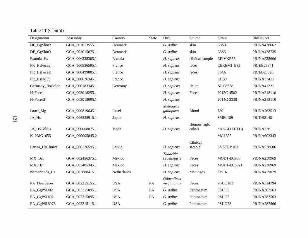
121
Table 11 (Cont’d)
Designation Assembly Country State Host Source Strain BioProject
DE_GgSkin2 GCA_003013555.1 Denmark G. gallus skin L3S3 PRJNA438662
DE_GgSkin3 GCA_003015075.1 Denmark G. gallus skin L5S5 PRJNA438735
Estonia_Hs GCA_006238365.1 Estonia H. sapiens clinical sample EEIVKB55 PRJNA528606
FR_HsFeces GCA_900536595.1 France H. sapiens feces CEREMI_E32 PRJEB28341
FR_HsFeces1 GCA_900499885.1 France H. sapiens feces 884A PRJEB28020
FR_HsIAI39 GCA_000026345.1 France H. sapiens IAI39 PRJNA33411
Germany_HsColon GCA_000183345.1 Germany H. sapiens Ileum NRC857c PRJNA41221
HsFeces GCA_003018255.1
H. sapiens Feces 2012C-4502 PRJNA218110
HsFeces2 GCA_003018095.1
H. sapiens
2014C-3338 PRJNA218110
Israel_Mg GCA_000819645.1 Israel
Meleagris
gallopavo Blood 789 PRJNA262513
JA_Hs GCA_006535915.1 Japan
H. sapiens
SMEc189 PRJDB8148
JA_HsColitis GCA_000008875.1 Japan
H. sapiens
Hemorrhagic
colitis SAKAI (EHEC) PRJNA226
K12MG1655 GCA_000005845.2
MG1655 PRJNA603343
Latvia_HsClinical GCA_006236595.1 Latvia
H. sapiens
Clinical
sample LVSTRB103 PRJNA528606
MX_Bat GCA_002456375.1 Mexico
Tadarida
brasiliensis Feces MOD1-EC908 PRJNA230969
MX_Hs GCA_002485345.1 Mexico
H. sapiens Feces MOD1-EC6621 PRJNA230969
Netherlands_Hs GCA_002888415.1 Netherlands
H. sapiens Meninges SP-16 PRJNA429029
PA_DeerFeces GCA_002215155.1 USA PA
Odocoileus
virginianus Feces PSUO103 PRJNA314794
PA_GgPSU02 GCA_002215095.1 USA PA G. gallus Peritoneum PSUO2 PRJNA287563
PA_GgPSUO2 GCA_002215095.1 USA PA G. gallus Peritoneum PSUO2 PRJNA287563
PA_GgPSUO78 GCA_002215115.1 USA G. gallus Peritoneum PSUO78 PRJNA287566
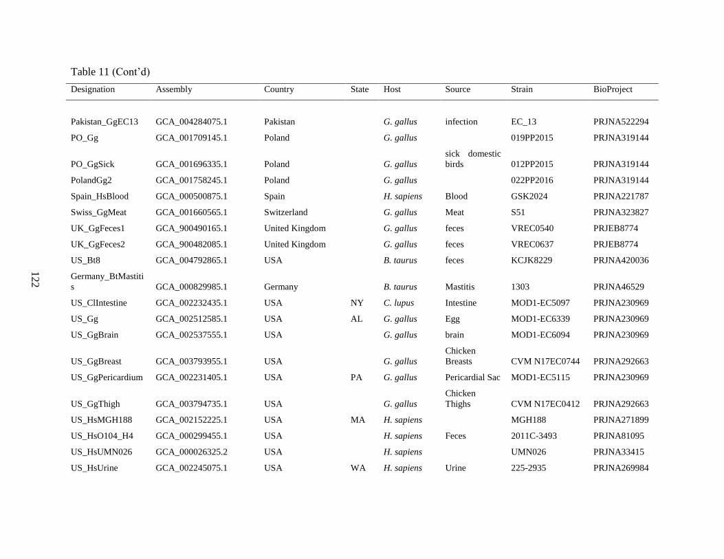
122
Table 11 (Cont’d)
Designation Assembly Country State Host Source Strain BioProject
Pakistan_GgEC13 GCA_004284075.1 Pakistan G. gallus infection EC_13 PRJNA522294
PO_Gg GCA_001709145.1 Poland
G. gallus
019PP2015 PRJNA319144
PO_GgSick GCA_001696335.1 Poland
G. gallus
sick domestic
birds 012PP2015 PRJNA319144
PolandGg2 GCA_001758245.1 Poland
G. gallus
022PP2016 PRJNA319144
Spain_HsBlood GCA_000500875.1 Spain
H. sapiens Blood GSK2024 PRJNA221787
Swiss_GgMeat GCA_001660565.1 Switzerland
G. gallus Meat S51 PRJNA323827
UK_GgFeces1 GCA_900490165.1 United Kingdom
G. gallus feces VREC0540 PRJEB8774
UK_GgFeces2 GCA_900482085.1 United Kingdom
G. gallus feces VREC0637 PRJEB8774
US_Bt8 GCA_004792865.1 USA
B. taurus feces KCJK8229 PRJNA420036
Germany_BtMastiti
s GCA_000829985.1 Germany
B. taurus Mastitis 1303 PRJNA46529
US_ClIntestine GCA_002232435.1 USA NY C. lupus Intestine MOD1-EC5097 PRJNA230969
US_Gg GCA_002512585.1 USA AL G. gallus Egg MOD1-EC6339 PRJNA230969
US_GgBrain GCA_002537555.1 USA
G. gallus brain MOD1-EC6094 PRJNA230969
US_GgBreast GCA_003793955.1 USA
G. gallus
Chicken
Breasts CVM N17EC0744 PRJNA292663
US_GgPericardium GCA_002231405.1 USA PA G. gallus Pericardial Sac MOD1-EC5115 PRJNA230969
US_GgThigh GCA_003794735.1 USA
G. gallus
Chicken
Thighs CVM N17EC0412 PRJNA292663
US_HsMGH188 GCA_002152225.1 USA MA H. sapiens
MGH188 PRJNA271899
US_HsO104_H4 GCA_000299455.1 USA H. sapiens Feces 2011C-3493 PRJNA81095
US_HsUMN026 GCA_000026325.2 USA H. sapiens UMN026 PRJNA33415
US_HsUrine GCA_002245075.1 USA WA H. sapiens Urine 225-2935 PRJNA269984

123
Table 11 (Cont’d)
Designation Assembly Country State Host Source Strain BioProject
US_MgAPEC01 GCA_000014845.1 USA IA M. gallopavo Lung APEC 01 PRJNA16718
US_MgGround GCA_003774815.1 USA M. gallopavo ground meat CVM N17EC1100 PRJNA292663
US_Ss GCA_002474525.1 USA SD S. scrofa Ileum MOD1-EC5757 PRJNA230969
US_SsIntestine GCA_002464015.1 USA S. scrofa Jejunum MOD1-EC6458 PRJNA230969
US_Ss GCA_002474525.1 USA SD S. scrofa Ileum MOD1-EC5757 PRJNA230969
US_SsIntestine GCA_002464015.1 USA
S. scrofa Jejunum MOD1-EC6458 PRJNA230969
USA_AZH2O GCA_002534895.1 USA AZ
water MOD1-EC5915 PRJNA230969
WA_HsUTI GCA_000778565.1 USA WA H. sapiens Urine UPEC-208 PRJNA248737
Table 12. Proteome differences in four genomes of S. aureus infecting chickens. The SEED viewer was used to identify genes present
in 1519 where the predicted polypeptide had a % identity less than 50% in one or more of the genomes for the indicated isolates. Gene
is the RAST 1519 annotation gene number, Length is for the 1519 polypeptide, and function is the annotation from RAST.
1519
B4-
59C Ch21
ED9
8 Gene
Lengt
h function
0 100 100 6 148 hypothetical protein
0 100 100 7 83 hypothetical protein
0 100 100 16 112 hypothetical protein
0 0 0 32 182 DUF1541 domain-containing protein
36.39 35.91 36.39 33 688
Lead, cadmium, zinc and mercury transporting ATPase (EC 3.6.3.3) (EC 3.6.3.5); Copper-
translocating P-type ATPase (EC 3.6.3.4)
0 0 0 117 43 hypothetical protein
0 0 0 153 38 FIG01108408: hypothetical protein
100 0 0 296 97 hypothetical protein
100 34.45 0 327 235 Toxic shock syndrome toxin 1 (TSST-1)
100 0 0 328 55 hypothetical protein

124
Table 12 (Cont’d)
1519
B4-
59C Ch21
ED9
8 Gene
Lengt
h function
100 0 0 331 140 Phage protein
100 0 0 332 390 Phage protein
0 0 0 421 40 Phosphoglycerate kinase (EC 2.7.2.3)
100 45.9 45.9 500 48 hypothetical protein
100 100 0 562 49 Secretory antigen SsaA-like protein transposon-related
0 0 0 595 44 hypothetical protein
100 36.02 36.02 729 222 hypothetical protein
100 26.09 26.09 732 242 hypothetical protein
100 0 0 733 71 Phage protein
0 100 100 1021 128 hypothetical protein
100 100 0 1197 45 hypothetical protein
0 100 100 1210 39 hypothetical protein
0 0 0 1289 64 hypothetical protein
100 100 0 1421 44 hypothetical protein
0 98.63 100 1553 74 Hypothetical protein, phi-ETA orf24 homolog [SA bacteriophages 11, Mu50B]
0 0 0 1554 72 Phage protein
37.38 36.1 37.38 1555 414 Phage DNA helicase
0 0 0 1556 119 Phage protein
0 0 0 1557 255 Phage replication initiation protein
0 99.53 100 1560 213 Phage-associated recombinase
0 100 100 1561 160 ORF027
39.53 97.67 98.84 1563 87 Hypothetical protein, PVL orf39 homolog [SA bacteriophages 11, Mu50B]
0 98.63 97.26 1565 74 Hypothetical protein, PV83 orf12 homolog [SA bacteriophages 11, Mu50B]
0 98.21 98.21 1566 57 Uncharacterized protein pCM2_0059
0 100 100 1567 97 phage protein
0 81.35 100 1568 251 Phage antirepressor protein
0 100 100 1569 62 hypothetical protein

125
Table 12 (Cont’d)
1519
B4-
59C Ch21
ED9
8 Gene
Lengt
h function
0 100 100 1570 201 Phage protein
0 0 0 1572 65 Phage protein
0 0 0 1573 257 Phage antirepressor protein
0 0 0 1574 79 hypothetical protein
0 93.44 100 1576 110 Phage protein
0 100 99.69 1577 553 DNA adenine methylase (EC 2.1.1.72)
25.71 100 100 1578 350 Phage integrase
0 0 0 1719 41 hypothetical protein
100 0 0 1760 52 hypothetical protein transposon-related
0 0 0 1862 158 hypothetical protein
0 0 0 1863 76 hypothetical protein
0 0 0 1864 47 hypothetical protein
0 0 0 1865 65 hypothetical protein
0 0 0 1866 56 hypothetical protein
0 0 0 1867 88 hypothetical protein
0 0 0 1868 77 hypothetical protein
0 0 0 1869 157 hypothetical protein
0 0 0 1870 99 hypothetical protein
0 0 0 1871 186 hypothetical protein
0 0 0 1872 54 hypothetical protein
0 0 0 1916 52 Adhesin of unknown specificity SdrC
0 0 0 1917 205
Type I restriction-modification system, DNA-methyltransferase subunit M (EC 2.1.1.72) / Type
I restriction-modification system, specificity subunit S
24.35 24.35 24.35 1919 178 Aminoglycoside N6'-acetyltransferase (EC 2.3.1.82)
0 0 0 1921 144 hypothetical protein
31.82 31.82 31.82 1922 110 Transcriptional regulator, ArsR family
0 0 45 1923 173 Plasmid recombination, MobE mobilization protein

126
Table 12 (Cont’d)
1519
B4-
59C Ch21
ED9
8 Gene
Lengt
h function
100 0 0 2179 95 Hypothetical SAV0793 homolog in superantigen-encoding pathogenicity islands SaPI
100 30.08 30.08 2180 121 Hypothetical SAV0792 homolog in superantigen-encoding pathogenicity islands SaPI
100 0 0 2181 486 Hypothetical SAV0791 homolog in superantigen-encoding pathogenicity islands SaPI
100 0 0 2189 45 hypothetical protein
100 29.38 29.38 2191 193 hypothetical protein
100 0 0 2192 59 hypothetical protein
0 0 0 2358 280 hypothetical protein
0 0 0 2369 205 hypothetical protein
0 0 0 2384 146 Phage holin
0 100 100 2688 39 hypothetical protein

127
CHAPTER 3
Investigation of Embryo Lethality Assay for Assessing Virulence of Isolates from Bacterial
Chondronecrosis with Osteomyelitis in Lame Broilers.

128
Investigation of Embryo Lethality Assay for Assessing Virulence of Isolates from Bacterial
Chondronecrosis with Osteomyelitis in Lame Broilers.
Abstract
We used an embryo lethality assay (ELA) to assess virulence for different isolates from cases of
bacterial chondronecrosis with osteomyelitis (BCO) in broilers. ELA has been previously used to
measure virulence and lethal dosage of Enterococcus faecalis and Enterococcus cecorum. We
hypothesized that ELA could substitute for more laborious and costly assessments of BCO
isolate pathogenicity using live birds. We evaluated two different levels of bacteria injected into
eggs from layer and commercial broiler embryos. Significant findings include a) Escherichia coli
from neighboring farms operated by the same integrator had very different embryo lethality, b)
isolate Staphylococcus agnetis 908 had low virulence in ELA, even though this isolate can
induce more than 50% BCO lameness, c) Enterococcus cecorum 1415 also had low
pathogenicity; even though it was recovered from severe bilateral tibial dyschondroplasia, and d)
human and chicken isolates of S. aureus had significant pathogenicity. Therefore, ELA may not
be an effective measure for assessing virulence with respect to BCO.
Keywords: Embryo lethality; Lameness; Virulence; Bacterial chondronecrosis with osteomyelitis

129
Introduction
Bacterial chondronecrosis with osteomyelitis (BCO) is the leading cause of lameness in rapidly
growing broilers (1-4). Lameness in broilers is significant as an animal welfare issue, and as a
financial cost, in the poultry industry (2). Our research group isolated and characterized an
isolate of S. agnetis, designated 908, from lame broilers on our research farm (1). S. agnetis 908
can induce greater than 50% BCO lameness by 56 days of age when administered in a single
dose in drinking water at 104 to 105 CFU/mL on day 20 (1, 3, 5, 6). Our current model for
lameness etiology is that stress can lead to increased leakage (translocation) of bacteria across
the gut and pulmonary epithelia into the blood system (1-3, 7-9). Particular species are then able
to colonize the growth plate, a vulnerable niche in the blood system of the rapidly growing leg
bones of fast-growing broilers (2, 3, 10). Therefore, significant lameness can be induced by
specific bacterial pathogens. Distinct bacterial species have been isolated from lame birds
including Staphylococcus aureus, Enterococcus cecorum, and Escherichia coli (11-31).
However, there are few comparisons of different BCO-associated species, or isolates, for
pathogenicity (17, 32). In this study, we investigated the pathogenicity of S. agnetis,
Staphylococcus chromogens, E. coli, E. cecorum, and S. aureus isolates using an embryo
lethality assay (ELA). The isolates were obtained from BCO lesions on our research farm or
commercial broiler farms in Arkansas. ELA has been used to correlate the expression frequency
of nine virulence-associated E. coli genes with embryo mortality (33). Borst et al. (32) used this
technique to compare the virulence of E. cecorum isolated from broiler spinal lesions (kinky
back) to non-pathogenic E. cecorum strains isolated from ceca of unaffected birds. Blanco et al.
(34) used ELA to determine the virulence and the lethal dose of Enterococcus faecalis.

130
Methods
Microbiology
Isolation and handling of the isolates has been described (1, 35). Media included: CHROMagar
Orientation (CO; DRG International, Springfield Township, NJ), tryptic soy broth (Difco brand,
Becton, Dickinson and Company, Franklin Lakes, NJ); and Luria broth (LB; per liter 10 g
tryptone, 5 g yeast extract, 5 g NaCl).
ELA
Fertilized eggs were obtained from leghorns (LCL) and cobb700 commercial broilers (BCL) on
the University of Arkansas research farm. The eggs were washed with warm soapy water
containing a small amount of household bleach. Eggs were incubated (NatureForm™ Hatchery
Systems, Jacksonville, FL, USA) at 37.5oC, relative humidity of 56%, on autorotate. On day 12,
stationary-phase bacterial cultures were washed with 1xphosphate buffer saline (1xPBS; 150mM
NaCl, 10 mM KHPO4 pH 7.2), CFU concentration estimated by spectrophotometry (A650)
using pre-calibrated standard curves for each isolate, and then diluted in 1xPBS to the required
concentration. eggs were candled, and fertile eggs were injected using a tuberculin syringe and
20G needle (Becton, Dickinson, and Company) with 100µL of the appropriate bacterial
suspension, or vehicle control, into the allantois cavity. The opening was sealed with transparent
cello tape. Inoculated embryos were scored for mortality every day for 4 days after bacteria
administration (32, 34).
Electro-transformation
Overnight cultures of E. coli in LB were diluted 20-fold with warm LB and incubated for 4 hours
at 37 °C with good aeration. The cells were pelleted (5000 x g; 5 min; 4°C) then resuspended in
an equal volume of ice-cold sterile deionized H2O, then pelleted. The pellet was again

131
resuspended in 0.5 volumes of ice-cold sterile deionized H2O, then pelleted. The pellet was
resuspended in 0.01 volumes of ice-cold sterile 10% glycerol. Then 100 ul of cells were mixed
with DNA and transferred to a 1 mm gap electroporation cuvette. The cuvette was precooled on
ice, then electroporated in a BIO-RAD Gene Pulser™ set to 200 Ohms, 25 μFD, and 1.75 kvolts.
The cells were diluted into 1 ml pre-warmed LB and incubated for 45 minutes with shaking at 37
oC. Then 106 CFUs were injected into the allantois cavity of a 12-day old fertilized LCL embryo,
as described above. After 5 days, a live embryo was sampled after surface sterilization to collect
100 µl of yolk with a tuberculin syringe through the same opening as for injection. The sample
was added to X ml of LB and grown overnight with shaking at 37 oC. An aliquot was spread on
CHROMagar Orientation for chromogenic verification as pure E. coli. The injection and
recovery were sequentially repeated for a total of three times.
Statistical Analysis
The results of the ELA were analyzed with either Pearson’s Chi-squared (χ2) or Fisher’s Exact
(FE) analysis using SAS and R software (SAS Institute. 2011; RStudio Team. 2016). Significant
differences between Phosphate saline vehicle and treatments were accepted at P < 0.05.
Results
Embryo Lethality Assay with BCO isolates
To establish a suitable assay for comparing different isolates, we first injected E. coli 1413 at a
range of amounts (103 – 108 CFUs) in sterile 1xPBS to estimate the lethal dosage for Leghorn
Chicken Line (LCL) embryos (Figure 24). For doses above 105 CFUs of 1413 had significant
embryo lethality with doses 105, 107-8 CFUs compared with 1XPBS control. We, therefore,
assessed different BCO isolates at 105 and 106 CFUs (Table 11). We included S. agnetis 908
recovered from a femoral BCO lesion on our research facility as this isolate can induce lameness

132
≥50% by day 56 when administered in drinking water for two days to 20-day old broilers (1, 3,
5, 6). Surprisingly, 908 injections of even 106 CFUs resulted in only 14% embryo lethality, a
level not statistically different from phosphate saline control treatment (Figure 25A). For the
methicillin-sensitive human S. aureus isolate 1302, originally retrieved from a wound (Table 1),
injections of 105 or 106 CFUs resulted in 80% embryo lethality (Figure 25B). Staphylococcus
chromogens 1401 was recovered from an infected T4 vertebra of a chicken with “Kinky back”
(Table 11). Injections of 106 CFUs resulted in only 7% embryo death, less than the 1xPBS
control for that experiment (Figure 25C). E. coli 1409 was recovered from a tibial head necrosis
lesion (Table 11). Injection of 105 to 106 CFUs resulted in no lethality through day 4 (Figure
25D). E. coli 1413 was isolated from the blood of a lame bird with bilateral BCO of the tibiae
and femorae, where E. coli was also recovered from multiple lesions (Table 11). As before,
injections of 105 or 106 CFUs into LCL resulted approximately 80% embryo lethality (Figure
25E). E. cecorum 1415 was isolated from a tibial head abscess in a case of bilateral tibial
dyschondroplasia (Table 11). ELA results for 105 CFU actually showed more lethality than 106
CFUs but neither was statistically different from the PBS control (Figure 25F). We used two
isolates (1510 & 1514) of S. aureus obtained from BCO lesions from two different birds in a
commercial broiler house lameness outbreak where draft genome assemblies were highly related
(Table 11; Ekesi 2020). The isolates showed different ELA results with 1510 lethality of 60%
for 106 CFU, while 1514 produced 47% but only the 1510 results were statistically significant
(Figure 25G & 25I). E. coli 1512 and 1527 were recovered from the left and right femoral
lesions of the same bird (Table 11; Ekesi, 2020). Draft genome assemblies for both 1512 and
1527 were determined to be virtually identical. ELA results for 1512 yielded 87% lethality for
106 CFUs and 52% lethality for 105, but only the 106 results were statistically different from the

133
PBS control (Figure 25H). Therefore, only the human isolate S. aureus 1302, and chicken
isolates E. coli 1413, and S. aureus 1510 and E. coli 1512, were found to cause significant
lethality using LCL embryos.
We then chose to extend the analyses by comparing the results for ELA with layer embryos to
ELA using Broiler Chicken Line (BCL) embryos. As shown in Figure 26, significant embryo
lethality was obtained with S. aureus isolates1302 and 1514, plus E. coli 1413 and 1512. S.
agnetis 908, S. chromogenes 1401, E. coli 1409, and E. cecorum 1415, showed no virulence for
either CFU quantity. We did note that for all four isolates that showed lethality for BCL, both the
105 and 106 CFU injections showed significant embryo mortality (Figure 26 panels B, E, G and
H). For LCL the 105 injections were only different from the controls for S. aureus isolate 1302
and E. coli 1413. However, for the other two isolates we might reach significance for the 105
CFU injections with more embryos. We also note that lethality was more rapid in the BCL than
with LCL embryos (Figure 25 and 26).
Is ELA Virulence Readily Transferable by Electroporation?
To study the potential mechanism(s) of bacterial virulence acquisition, we used electroporation
to transfer DNA from E. coli 1413 into E. coli 1409 to produce 1540T. We then passaged these
cells through three rounds of selection by ELA in LCL embryos. After each round bacteria were
recovered from one of the living embryos and verified on chromogenic media as E. coli. The
rescued bacteria from each round were designated 1541P, 1544P, and 1547P (Table 11). We then
repeated the ELA assay to compare the lethality of the transformant and recovered populations to
1409. Isolate 1409 inoculated into 106 CFUs 1409 into LCL embryos produced higher embryo
lethality that previously for LCL embryos and was statistically different from 1xPBS control (P
= 0.002). Injection of 106 CFU of 1540T resulted in a 27% lethality of LCL that was not different
134
from the negative control (Figure 27A). The bacterial culture recovered from live embryos was
designated E. coli 1541P and confirmed as E. coli on chromogenic CO plates (Table 11).
Injection of 106 CFU of 1541P into LCL embryos resulted in 20% lethality which was not
different from the PBS control (Figure 27B). The culture recovered from live embryos was
designated 1544P and confirmed as E. coli as before. When we injected 1544P injected into LCL
at 106 CFU there was 40% embryo lethality which was statistically different from the PBS
control (Figure 27C). The recovery from live embryos was repeated and designated E. coli
1547P. Injection of 106 CFUs of 1547P into LCL embryos resulted in 73% lethality (Figure
27D).
Discussion
We performed ELA with different bacterial isolates isolated from lame broilers to estimate
relative pathogenicity. We observed that S. agnetis 908 is not pathogenic in the ELA even
though we have shown that this isolate readily infects young broilers when administered at 10 to
105 CFU/ml in drinking water at 20 and 21 days of age (1, 3, 5, 6). Those broilers then begin to
develop lameness by 41 days of age and by 56 days of age 50% of the birds will be clinically
lame with BCO lesions in proximal femoral and tibial heads. Many of the birds develop
bacteremia with hundreds to thousands of CFU/ml. Additionally, the infected birds spread the
infection to birds within the same room and 30-40% of those birds will be lame by 56 days of
age. ELA has been used to compare virulence of E. cecorum from BCO birds (primarily kinky
back) and E. cecorum from feces (32). We used an E. cecorum we collected from an infected
vertebrae in a kinky back bird (35) but it showed no significant virulence in the ELA. We
compared three E. coli isolates from BCO lame birds (35) and found they had very different
apparent ELA virulence. Even though these three isolates were from three different commercial

135
broiler farms in Arkansas that were experiencing BCO outbreaks, we have shown that all 3 are
very different based on whole genome comparisons (35). This is surprising given that 1409 and
1413 were isolated on the same day from two different farms within 5 km of each other that were
operated by the same integrator and supplied from the same hatchery. S. aureus isolates showed
virulence in the ELA, including an isolate from a human infection, and isolates from a BCO
outbreak on a different farm operated by a different integrator. We sampled 11 lame birds from
that farm and determined that 7 of the birds were infected with S. aureus. Genome analysis
showed that the S. aureus isolates were highly related and very closely related to numerous S.
aureus isolates obtained from diseased birds or broiler meat dating back to the 1970s in Europe
(35, 36). The clade has been isolated multiple times in Arkansas and Oklahoma for at least a
decade. The clade appears to be exclusively associated with poultry so virulence in the ELA is
not surprising. The genomic comparisons led us to propose that E. coli association with BCO is
not exclusively poultry specific and that this species appears to be more of a generalist, whereas
S. aureus and S. agnetis appear to be specialists and do not readily jump back and forth infecting
different host species.
References
Al-Rubaye AAK, Couger MB, Ojha S, Pummill JF, Koon JA, II, Wideman RF, Jr., Rhoads DD.
2015. Genome analysis of Staphylococcus agnetis, an agent of lameness in broiler chickens.
PLoS One 10:e0143336.
Wideman RF. 2016. Bacterial chondronecrosis with osteomyelitis and lameness in broilers: a
review. Poult Sci 95 325-344.
Al-Rubaye AAK, Ekesi NS, Zaki S, Emami NK, Wideman RF, Rhoads DD. 2017.
Chondronecrosis with osteomyelitis in broilers: Further defining a bacterial challenge model
using the wire flooring model. Poult Sci 96:332-340.
Weimer SL, Wideman RF, Scanes CG, Mauromoustakos A, Christensen KD, Vizzier-Thaxton
Y. 2019. The utility of infrared thermography for evaluating lameness attributable to bacterial
chondronecrosis with osteomyelitis. Poult Sci 98:1575-1588.

136
Alrubaye A, Ekesi NS, Hasan A, Koltes DA, Wideman Jr R, Rhoads D. 2020. Chondronecrosis
with osteomyelitis in broilers: Further defining a bacterial challenge model using standard litter
flooring and protection with probiotics. Poult Sci In Review.
Alrubaye AAK, Ekesi NS, Hasan A, Elkins E, Ojha S, Zaki S, Dridi S, Wideman RF, Rebollo
MA, Rhoads DD. 2020. Chondronecrosis with Osteomyelitis in Broilers: Further Defining
Lameness-Inducing Models with Wire or Litter Flooring, to Evaluate Protection with Organic
Trace Minerals. Poult Sci In Review.
Wideman RF, Prisby RD. 2013. Bone circulatory disturbances in the development of
spontaneous bacterial chondronecrosis with osteomyelitis: A translational model for the
pathogenesis of femoral head necrosis. Front Endocrinol (Lausanne) 3:183.
Jiang T, Mandal RK, Jr. RFW, Khatiwara A, Pevzner I, Kwon YM. 2015. Molecular survey of
bacterial communities associated with bacterial chondronecrosis with osteomyelitis (BCO) in
broilers. PLoS One 10:e0124403.
Mandal RK, Jiang T, Al-Rubaye AA, Rhoads DD, Wideman RF, Zhao J, Pevzner I, Kwon YM.
2016. An investigation into blood microbiota and its potential association with bacterial
chondronecrosis with osteomyelitis (BCO) in broilers. Sci Rep 6:25882.
Wideman RF, Al-Rubaye A, Gilley A, Reynolds D, Lester H, Yoho D, Hughes JM, Pevzner I.
2013. Susceptibility of 4 commercial broiler crosses to lameness attributable to bacterial
chondronecrosis with osteomyelitis. Poult Sci 92:2311-2325.
Carnaghan RBA. 1966. Spinal cord compression due to spondylitis caused by Staphylococcus
pyogenes. J Comp Pathol 76:9-14.
Duff SR. 1984. Dyschondroplasia of the caput femoris in skeletally immature broilers. Res Vet
Sci 37:293-302.
Emslie KR, Fenner LM, Nade S. 1984. Acute haematogenous osteomyelitis: II. The effect of a
metaphyseal abscess on the surrounding blood supply. J Pathol 142:129-134.
Duff S, Randall C. 1987. Observations on femoral head abnormalities in broilers. Res Vet Sci
42:17-23.
Duff S. 1989. Disturbed endochondral ossification in the axial skeleton of young fowls. J Comp
Pathol 101:399-409.
Duff SRI. 1989. Physeal clefts and disturbed endochondral ossification in broiler fowls. J Comp
Pathol 101:75-86.
Braga JFV, Chanteloup NK, Trotereau A, Baucheron S, Guabiraba R, Ecco R, Schouler C. 2016.
Diversity of Escherichia coli strains involved in vertebral osteomyelitis and arthritis in broilers in
Brazil. BMC Veterinary Research 12:140.

137
Nairn ME, Watson ARA. 1972. Leg weakness of poultry—a clinical and pathological
characterisation. Aust Vet J 48:645-656.
McCaskey PC, Rowland GN, Page RK, Minear LR. 1982. Focal Failures of Endochondral
Ossification in the Broiler. Avian Dis 26:701-717.
Mutalib A, Riddell C, Osborne AD. 1983. Studies on the pathogenesis of Staphylococcal
osteomyelitis in chickens. II. Role of the respiratory tract as a route of infection. Avian Dis
27:157-160.
Riddell C. 1983. Pathology of the skeleton and tendons of broiler chickens reared to roaster
weights. I. crippled chickens. Avian Dis 27:950-962.
Griffiths G, Hopkinson W, Lloyd J. 1984. Staphylococcal necrosis of the head of the femur in
broiler chickens. Aust Vet J 61:293-293.
Duff SRI. 1990. Diseases of the musculoskeletal system. In Jordan F (ed), Poultry Diseases 3rd
ed. Bailliere, Tindall. UK.
Hocking PM. 1992. Musculo-skeletal disease in heavy breeding birds., p 297-309. In Whitehead
CC (ed), Bone Biology and Skeletal Disorders in Poultry. Carfax Publishing Company,
Abingdon, United Kingdom.
Tate CR, Mitchell WC, Miller RG. 1993. Staphylococcus hyicus associated with turkey stifle
joint osteomyelitis. Avian Dis 37:905-907.
Thorp BH. 1994. Skeletal disorders in the fowl: a review. Avian Pathol 23:203-236.
Skeeles KJ. 1997. Staphylococcosis, p 247-253. In B.W. Calnek, Barnes HJ, Beard CW,
McDougald LR, Saif YM (ed), Diseases of Poultry 10 ed. Iowa State University Press, Ames,
IA, USA.
Thorp B, Waddington D. 1997. Relationships between the bone pathologies, ash and mineral
content of long bones in 35-day-old broiler chickens. Res Vet Sci 62:67-73.
McNamee PT, Smyth JA. 2000. Bacterial chondronecrosis with osteomyelitis ('femoral head
necrosis') of broiler chickens: a review. Avian Pathol 29:477-495.
Joiner KS, Hoerr FJ, van Santen E, Ewald SJ. 2005. The avian major histocompatibility complex
influences bacterial skeletal disease in broiler breeder chickens. Vet Pathol 42:275-281.
Gaußmann B, Hess C, Grafl B, Kovacs M, Troxler S, Stessl B, Hess M, Paudel S. 2018.
Escherichia coli isolates from femoral bone marrow of broilers exhibit diverse pheno- and
genotypic characteristics that do not correlate with macroscopic lesions of bacterial
chondronecrosis with osteomyelitis. Avian Pathol 47:271-280.
138
Borst LB, Suyemoto MM, Keelara S, Dunningan SE, Guy JS, Barnes HJ. 2014. A Chicken
Embryo Lethality Assay for Pathogenic Enterococcus cecorum. Avian Dis 58:244-248.
Oh JY, Kang MS, Yoon H, Choi HW, An BK, Shin EG, Kim YJ, Kim MJ, Kwon JH, Kwon YK.
2012. The embryo lethality of Escherichia coli isolates and its relationship to the presence of
virulence-associated genes. Poult Sci 91:370-375.
Blanco AE, Cavero D, Icken W, Voss M, Schmutz M, Preisinger R, Sharifi AR. 2018. Genetic
approach to select against embryo mortality caused by Enterococcus faecalis infection in laying
hens. Poult Sci 97:4177-4186.
Ekesi NS, Hasan A, Alrubaye A, Rhoads D. 2020. Genomic comparisons of bacterial isolates
from lame broilers. manuscript in preparation.
Lowder BV, Guinane CM, Ben Zakour NL, Weinert LA, Conway-Morris A, Cartwright RA,
Simpson AJ, Rambaut A, Nübel U, Fitzgerald JR. 2009. Recent human-to-poultry host jump,
adaptation, and pandemic spread of Staphylococcus aureus. PNAS USA 106:19545-19550.

139
Appendix
Table 13. Sources (strain designation for species, and host) of bacterial isolates utilized for ELA.
In Isolate Source Lame indicates a bird number from BCO sampling. Abbreviations include LT:
Left Tibia, RT: Right tibia, LF: Left femur, and RF: Right femur.
Species Designation Host Isolate Source Citation
S. agnetis 908 Broiler Femoral BCO; UA Research Farm Al-Rubaye, et al.,
2015
S. chromogens 1401 Broiler Thoracic Vertebrae; Lame3 Ekesi, 2020
E. cecorum 1415 Broiler LT/RT; Lame5 Ekesi, 2020
E. coli 1409 Broiler RT; Lame3 Ekesi, 2020
1413 Broiler Blood; Lame12 Ekesi, 2020
1512 Broiler LF; Lame18 Ekesi, 2020
1527 Broiler RF; Lame18 Ekesi, 2020
1540T 1409 transformed with 1413
Plasmids
This work
1541P Recovered from live embryo
injected with 1540T
This work
1544P Recovered from live embryo
injected with 1541P
This work
1547P Recovered from live embryo
injected with 1544P
This work
S. aureus 1510 Broiler LT Lame14 Ekesi, 2020
1514 Broiler RF Lame15 Ekesi, 2020
1302 Human Wound ATCC-29213

140
Figure 24. Embryo lethality assay in layer chicken line embryos to estimate the lethal dosage of
bacterial isolates. The number of live embryos (Y-axis) for different quantities of E. coli 1413 is
plotted over four days post-injection (X-axis). Isolate abbreviations and their sources are
described in Table 11. For each treatment n = 5. Asterisks (*) indicates that particular treatment
was significantly different from 1xPBS (P <0.05).

141
Figure 25. Layer chicken line embryo lethality for injection of 105 or 106 CFU of bacterial isolates vs. phosphate saline vehicle. Isolates were: A. 908; n=14, B. 1302; n=15, C. 1401; n=14, D. 1409; n=15, E. 1413; n=15, F. 1415; n=9, G. 1510; n =15, H.1512; n=15, and I. 1514, n=15. Details are as in Figure 24. Asterisks (*) indicates that particular treatment was significantly different from 1xPBS (P <0.05).

142
Figure 26. Broiler chicken line embryo lethality for injections of 105 or 106 CFU of bacterial
isolates vs. phosphate saline vehicle. Isolates were: A. 908, B. 1302, C. 1401, D. 1409, E. 1413,
F. 1415, G. 1514, and H.1527. Details are as in Figures 24 and 25. For all trials n=15 per
treatment.
Figure 27. Embryo lethality is transferable from E. coli 1413 to 1409. Embryo lethality assays
for 15 embryos per treatment for the transformant 1540T (Panel A), and recovered E. coli
cultures after 1 passage, 1541P (Panel B), 2 passages, 1544P (Panel C), and 3 passages, 1547P
(Panel D). All four panels compare to the 1xPBS control and 1409. Additional details are as in
Figures 24 and 25.
143
CHAPTER 4
Chondronecrosis with Osteomyelitis in Broilers: Further Defining Lameness-Inducing
Models with Wire or Litter Flooring, to Evaluate Protection with Organic Trace Minerals

144
Disclaimer
In this trial, I was part of the animal trial at the farm where we administered Availa-ZMC the
commercial product in broiler feed. I was part of the histopathological evaluation of the intestinal
villi. I was part of the sample collection team for the assay of intestinal gene expression. In the
phagocytosis assay, I collected the blood and helped purify the PBMCs.

145
Chondronecrosis with Osteomyelitis in Broilers: Further Defining Lameness-Inducing
Models with Wire or Litter Flooring, to Evaluate Protection with Organic Trace Minerals
Abstract
The feed additive Availa-ZMC was investigated for the ability to reduce lameness in
broilers using two alternative models for inducing lameness. The mixture of organic trace
minerals was effective in reducing lameness by 20% in the wire-flooring model and 25% in the
bacterial challenge on litter flooring model. Lameness in both models is overwhelmingly
attributable to bacterial chondronecrosis with osteomyelitis. The reduction in lameness was
associated, at least in part, with enhanced intestinal barrier integrity mediated by elevated
expression of tight junction proteins and stimulation of bactericidal killing of adherent peripheral
blood monocytes obtained from the birds on Availa-ZMC. Lameness is a major animal welfare
concern in broiler production. The wire-flooring and bacterial challenge on litter-flooring
models are effective models for evaluation of management strategies for mitigating infectious
causes of lameness.
Keywords: Broiler; Lameness; Chondronecrosis; Staphylococcus; Organic trace mineral

146
Introduction
Lameness is one of the most significant animal welfare issues in the broiler industry, resulting in
annual losses of millions of dollars (Siegel, et al., 2019; Wideman, 2016). A wire-flooring model
has been shown to induce a high incidence of lameness in broilers (Wideman, 2016; Wideman,
et al., 2013; Wideman, et al., 2014; Wideman, et al., 2012; Wideman and Prisby, 2013).
Lameness induced in this system is overwhelmingly bacterial chondronecrosis with osteomyelitis
(BCO) of the proximal tibiae and femora (Wideman, 2016; Wideman, et al., 2013; Wideman, et
al., 2012; Wideman and Prisby, 2013). The predominant isolates from BCO lesions using the
wire floor model on our research farm are Staphylococcus agnetis and the BCO lameness is
sometimes associated with a significant bacteremia (Al-Rubaye, et al., 2015). The type strain, S.
agnetis 908, when administered in drinking water can induce high levels of lameness for birds
grown on wire or on litter (Al-Rubaye, et al., 2015; Alrubaye, et al., 2020). The BCO lameness
model has demonstrated: i) translocation of bacteria into the blood for birds on litter, with higher
translocation in birds on wire flooring (Al-Rubaye, et al., 2017); ii) transmission of BCO-
inducing pathogens within a flock (Al-Rubaye, et al., 2017) or within a facility (Alrubaye, et al.,
2020); and iii) protection against BCO inducing pathogens by probiotics and prebiotics
(Alrubaye, et al., 2020; Wideman, 2016; Wideman, et al., 2015; Wideman, et al., 2012). We now
extend these investigations to the investigation of the commercial, complex organic trace
mineral, Availa-ZMC. Organic zinc is reported to enhance epithelial integrity, gut health, and
immune function (Hudson, et al., 2004; Star, et al., 2012; Zakaria, et al., 2017). The data
reported herein demonstrate that this product can reduce lameness in both the wire-flooring
model, and the bacterial challenge on litter-flooring model. Additionally, we observed that
Availa-ZMC shows a dose-dependent enhancement of bacterial killing activity by adherent

147
peripheral blood monocytes cultured from treated birds. These data extend the range of products
that can be used to reduce BCO lameness, highlight the importance of organic trace minerals in
improving animal well-being, and provide further validation of the two models we have
developed to investigate treatments and management strategies for reducing BCO lameness in
broiler operations.
Materials and Methods
Lameness Trials
All animal experiments were approved by the University of Arkansas Institutional Animal Care
and Use Committee under protocols 18010, and 18075. One day (d) old chicks representing
surplus males from a female broiler-breeder product were kindly provided by Cobb-Vantress
(Siloam Springs, AR). Chicks were placed in 5 x 10 ft. pens on either suspended wire flooring
(Wideman, 2016; Wideman, et al., 2012) or on standard wood-shaving litter at 60 per pen.
Nipple water lines were supplied with city tap water on one side of the pen and two feeders were
placed on the opposite side. Feed was standard starter through d35 and finisher through d56.
Computer controllers regulated the temperature, photoperiod and ventilation. Tunnel ventilation
and evaporative cooling pads were automatically activated when needed. The photoperiod was
set for 23 h light:1 h dark for the duration of the experiment. Thermoneutral temperature targets
were as follows: 90 °F for d1 to d3, 88 °F for d4 to d6, 85 °F for d7 to d10, 80 °F for d11 to d14,
and 75 °F thereafter. On d19 all pens were culled to 50 birds. For pens challenged with S.
agnetis in the drinking water the tap water supplying the nipple waterer was replaced with a
gravity flow from an elevated 20L carboy of tap water. The bacteria (stationary overnight
culture) were mixed into tap water in the carboy to 104 CFU/ml (colony forming units per ml).
After d21 the nipple supply was returned to the tap water. All water lines were flushed with

148
dilute bleach and fresh tap water prior to each experiment. Beginning on d20 all birds were
encouraged twice per d to move using standard kitchen brooms. Any bird that was reticent to
move was marked with spray paint. Birds that continued to be unwilling or unable to walk were
diagnosed as “clinically lame” and euthanized. All birds that died or were diagnosed as clinical
lame were recorded by date, and pen number. Necropsy for BCO lameness was as described
(Wideman, 2016) to categorize as either: N = Normal proximal femur head or proximal tibia
head; KB = Kinky Back (Spondylolisthesis); FHS = Proximal Femoral Head Separation
(epiphyseolysis); FHT = Proximal Femoral Head Transitional degeneration; FHN = Proximal
Femoral Head Necrosis; THN = Proximal Tibial Head Necrosis; Other = symptoms other than
BCO, and Total Lame included all birds with any FHS, FHT, FHN, THN, or KB lesions.
For administration of Availa-ZMC the commercial product was added to the feed (Table 12)
prior to pelleting. Samples of the pelleted feed were shipped to the supplier for verification of
proper mixing prior to any experiment. Details on the feed formulations and treatment groups
are described in Table 12.
Histological Evaluation of Intestinal Villi
Intestinal samples (3 cm section) for histopathology were the distal jejunum (1 cm proximal to
Meckel’s diverticulum) and proximal ileum (1 cm distal to Meckel’s diverticulum). Samples
from freshly euthanized birds were flushed with 1 x PBS and fixed in phosphate buffered
formalin. The fixed samples were processed through the histology laboratory in the Department
of Poultry Science at the University of Arkansas. Hematoxylin-Eosin stained sections were
imaged on an Olympus inverted scope at 400x using a CCD camper to display on an LCD
monitor. Villus length was measured on a 21-inch diagonal LCD monitor with a flexible ruler.
Calibration was based on a stage micrometer. For villus length and pathology, at least 4 sections

149
were examined for each tissue for each bird (8 to 10 birds per treatment). For each section, villus
length was measured and gross pathology (villus tip integrity) was scored, on four sides (top,
left, right and bottom). For some sections, villus length and tip integrity could not be measured
on all four sides owing to tissue damage in sectioning.
Assay of Intestinal Gene Expression
One μg of total RNA was extracted from tissue samples by homogenization using Trizol Reagent
(ThermoFisher Scientific, Rockford, IL) in accordance with the manufacturer’s recommendations.
RNA concentration, quality, and integrity were assessed by the ratio of absorbance (260/280), and
electrophoresis in 1% agarose gels using a Take 3 micro volume plate and the Synergy HT multi-
mode microplate reader (BioTek, Winooski, VT). RNAs were treated with DNAseI, and reverse
transcribed via qScript cDNA SuperMix (Quanta Biosciences, Gaithersburg, MD). The cDNA was
then amplified by real-time quantitative PCR (Applied Biosystems 7500 Real-Time PCR system)
with Power SYBR green Master Mix (ThermoFisher Scientific, Rockford, IL) in triplicate 20 µL
per reactions. Oligonucleotide primers specific for chicken occludin (OCLN): forward 5’-
CGCAGATGTCCAGCGGTTA-3’ and reverse 5’- GTAGGCCTGGCTGCACATG; claudin 1
(CLDN1); forward 5’- CCCACGTTTTCCCCTGAAA-3’ and reverse 5’-
GCCAGCCTCACCAGTGTTG-3’ ; gap junction protein alpha 1 (GJA1): forward 5’-
TGGCAGCACCATCTCCAA -3’ and reverse 5’- GGTGCTCATCGGCGAAGT-3’; and catenin
beta 1 (CTNNB1): forward 5’- TGCCCCACTGCGTGAAC-3’ and reverse 5’-
TGCTCTAACCAGCAGCTGAACT-3’. Primers for the reference, housekeeping gene r18S have
been published previously (Dhamad, et al., 2019; Greene, et al., 2019; Lassiter, et al., 2015;
Piekarski-Welsher, et al., 2018). The cycling conditions were 50°C for 2 min, 95°C for 10 min
followed by 40 cycles of 95°C for 15 s and 58°C for 1 min with plate read. Post PCR, melting
150
curve analysis was applied using the dissociation protocol from the Sequence Detection system to
exclude samples with non-specific products. PCR products were also confirmed for one specific
size band by agarose gel electrophoresis. Negative controls lacked cDNA input as template for the
PCR and were verified for absence of gel bands. Relative expression of target genes were
determined by the 2-ΔΔCt method using r18S as the reference and the control group as the calibrator
(Livak and Schmittgen, 2001; Schmittgen and Livak, 2008).
Phagocytosis Assay
Blood (1 ml) was collected from a wing vein using a Vacutainer containing EDTA (Becton,
Dickinson and Company, Franklin Lakes, NJ). Monocytes were enriched and cultured using
published protocols (Dawes, et al., 2014; Drechsler, et al., 2013). Medium was RPMI (VWR)
with 1x GlutaMax (Life Technologies) and 10% low endotoxin Fetal Bovine Serum. After 5 d in
culture (37 oC; 5% CO2) we challenged the adherent cells in triplicate with an approximate
multiplicity of infection (MOI) of 1:1 with S. agnetis 908 for 2 d following published methods
(Campbell, et al., 1994; Drevets, et al., 2015). Specifically, the bacteria were added to the
medium for 2 h, then the medium was replaced with media supplemented with gentamycin (50
µg/ml) for 6 h to kill non-internalized bacteria. The medium was replaced with antibiotic-free
medium. After 2 d in culture the adherent cells are lysed by addition of pure water and a dilution
series of the lysate was plated on Luria Broth agar plates for viable bacterial cell counts.
Statistical Analyses
Data were compared using either the T-test function in Microsoft Excel or a Generalized Linear
Model (GLM) module in R.3.4.2 to produce P-values between treatments, as indicated. Gene
expression data were analyzed by One-way ANOVA. If ANOVA revealed significant effects, the
means were compared by Tukey’s multiple range test using the Graph Pad Prism version 6.00 for

151
Windows (Graph Pad Software, La Jolla California, USA). Significant difference was accepted
at P ≤ 0.05. Data are expressed as the mean ± SEM.
Results
Experiment 1 evaluated whether Availa-ZMC could reduce lameness for birds raised on wire-
flooring to induce lameness. Chicks (1d old) were raised to d56 on wire-flooring with no direct
administration of a bacterial challenge. There were four pens in each treatment group, the
Control treatment received standard feed formulations (Table 12), whereas the Availa-ZMC
normal treatment received the product at 1000 mg/Kg of feed, and the Availa-ZMC high
treatment group received the product at 1500 mg/Kg of feed. Feed formulations were continuous
through d56, the end of the experiment. Lameness began to appear in all three treatments on d37,
but the trajectory of lameness accumulation was higher for the birds on standard feed (Figure
28). The final cumulative lameness for the Control was 66%, but the Availa-ZMC normal
treatment had 47% lameness, and Availa-ZMC high had 57% lameness. Comparison of the
lameness data by Generalized Linear Model (GLM) with the individual bird as the experimental
unit showed that the Availa-ZMC normal treatment was statistically different from Control (P =
0.0003) and Availa-ZMC high (P = 0.03). Control and Availa-ZMC high were not statistically
different (P = 0.15). Pen-to-pen variability for the three treatments in experiment 1 reveals a
degree of variability in the total lame per pen within a treatment (Table 13). Loss of birds to
mortalities unrelated to lameness, and final body weights were not different between treatments.
Supplementation of feed with Availa-ZMC at either level had no discernable effect on the
severity of BCO lesions for proximal tibiae and femorae from birds diagnosed as lame through
the course of the experiment (Figure 29).

152
Experiment 2 evaluated whether Availa-ZMC could reduce lameness for birds raised on litter-
flooring when a bacterial challenge is imposed. The feed supplementation was the same, but
lameness was induced by the transmission of the hypervirulent strain S. agnetis 908 from birds
challenged with the bacterium in drinking water for d20 and d21. There were 3 pens of birds on
litter-flooring on standard feed that were the Source population. These three pens were
“upwind,” relative to the exhaust fans, of the treatment pens (Table 12). There were four pens
for each of the three treatments: Control, Availa-ZMC normal, and Availa-ZMC high, arrayed in
a randomized block design and separated by at least 3 meters from the Source pens. Lameness
began to appear on Day-36 but lameness accumulation was accentuated in the Source (Figure
30). Accumulation of lameness in the three treatment groups lagged behind that for the Source
population by about 3 to 4 d through d48 where the Control cumulative lameness continued to
parallel that for the Source but the lameness accumulation is reduced for both Availa-ZMC
treatments. Final percent lameness was Source 83%, Control 65%, Availa-ZMC normal 49%,
and Availa-ZMC high 52%. GLM based comparisons of the lameness data with the individual
bird as the experimental unit showed that the percent lameness was statistically higher in the
Control treatment than in the Availa-ZMC normal (P = 0.002) and Availa-ZMC high (P =
0.006), treatments. Pen-to-pen total lame was more uniform in this experiment compared to
experiment 1, and losses due to mortalities unrelated to lameness were lower (Table 13). Final
body weights were comparable between experiments 1 and 2, but the body weights for Availa-
ZMC normal were lower (T-test, P = 0.02) compared to the other two treatments in experiment
2. There was no clear difference in the distribution of BCO lesions between any of the four
treatment groups in experiment 2 (Figure 31), however we have no explanation for the rather
high percentage of Normal left femoral head diagnoses in the Source population.

153
Sections of distal jejunum and proximal ileum were collected from five apparently healthy birds
on d57 from the Control and Availa-ZMC treatments. Villus length was assessed for multiple
sections from each bird and the average villus lengths were computed (Figure 32). Student T-
test of the villus length data indicate that villus length in the ileum was reduced in the Availa-
ZMC treatments (P < 0.001) but villus length in the jejunum increased (P < 0.001). There was no
difference in villus length between the two different levels of Availa-ZMC. In terms of important
determinants of villus integrity, we examined expression of critical tight junction genes using
reverse transcription quantitative polymerase chain reaction (RT-qPCR). Ileum and jejunum
from the Availa-ZMC high samples showed significantly upregulated expression for CLDN1,
OCLN, GJA-1, and CTNB1, compared to the Control (Figure 33). Complimentary data for
histology and expression from experiment 1 is not provided as the stress imposed by the wire-
flooring appears to significantly impact intestinal development and villus formation (A. Hasan,
unpublished).
Wing vein blood was collected from the same birds examined for intestinal histopathology.
Monocytes were enriched and adherent cells cultured for 5 d. The cells were then used in
phagocytosis assays against S. agnetis 908 at an approximate MOI of 1:1. Bacterial survival was
assessed after 2 d (Table 14). The bactericidal activity was variable between birds within each
treatment. The most variation was in the Availa-ZMC normal birds where the adherent cells for
bird 3 were highly active in killing S. agnetis 908. For the lowest dilution plated, 10-2, there were
only 5 colonies from one of the three triplicate wells. Therefore, verifying that bacteria were
added but the bacterial survival was very low within the monocytes from this broiler. The high
variability for the five birds from Availa-ZMC normal meant that this treatment group was not
statistically different from either the Control or Availa-ZMC high cells. Bactericidal activity of

154
cells from the Availa-ZMC high birds was higher than the activity of cells from the Control
treatment (T-test, P = 0.00085).
Discussion
In our previous research publications we had reported that S. agnetis 908 can induce lameness at
80-90% when administered to birds raised on wire flooring (Al-Rubaye, et al., 2015; Al-Rubaye,
et al., 2017). We also demonstrated that S. agnetis 908 can induce lameness of 50 to 80% when
administered in drinking water to birds raised on litter flooring (Alrubaye, et al., 2020). Further,
the lameness can be transmitted from the birds challenged with bacteria to unchallenged birds
within the same broiler house. We also demonstrated that specific probiotics in the feed can
protect broilers when raised on wire-flooring (Al-Rubaye, et al., 2017; Wideman, et al., 2012),
but different probiotics in the feed can protect the unchallenged birds in the litter-flooring with
bacterial challenge model (Alrubaye, et al., 2020). The combination of organic zinc, manganese,
and copper has been reported to improve poultry health, reduce bacterial pathogen colonization,
and reduce femoral head necrosis (McKnight, et al., 2020; Sirri, et al., 2016). We therefore
hypothesized that supplementation with Availa-ZMC complexed trace minerals could be
efficacious in reducing BCO lameness in our two models for inducing BCO lameness. The
results from experiments 1 and 2 confirm that Availa-ZMC is effective in reducing lameness in
both models for inducing lameness. When birds were raised on wire-flooring with no direct
bacterial challenge, the mineral supplement reduced lameness by 14 to 29% (Figure 25) and
appears to have reduced mortality due to causes not attributable to lameness (Table 13). In the
litter-flooring with bacterial challenge model the Availa-ZMC similarly reduced lameness by 20
to 25% relative to the Control treatment (Figure 27). Most importantly, this latter model
employs the contagious spread of the infection observed in some broiler operations. Although
155
our model uses a hypervirulent bacterial strain, the reduction could be greater against less
virulent species/strains. Substituting complexed organic trace minerals for inorganic minerals
appears to have reduced villus length in the ileum and increased villus length in the jejunum
(Table 13). Further investigations are warranted to determine whether these changes arising from
the difference in source of trace minerals alters any aspect of assimilation or feed conversion
rates in infectious models. In these studies, the final body weights did not seem to have been
impacted (Table 13). It is important to consider the influence of different stocking density
induced by different mortality rates. Interestingly, the organic trace mineral upregulated the
expression of the genes for tight junction (CLDN1, OCLN), gap junction (GJA-1), and
desmosome (CTNB1), consistent with improved gut barrier integrity. Although the exact
functions of the individual tight junction proteins remain elusive, in avian species, occludin has
been reported to be an integral component in tight junction barrier function (Balda, et al., 2000).
Studies conducted in occludin-deficient mice showed gut inflammation and defective epithelial
barrier function (Schulzke, et al., 2005). Similarly, it has been reported that down regulation of
CLDN1 can drastically reduce barrier integrity (Zeissig, et al., 2007). Upregulation of these
genes is consistent with Availa-ZMC enhancing barrier functions and reducing translocation of
bacteria into the blood, a critical first step in the progression of BCO lameness (Al-Rubaye, et
al., 2017; Wideman, 2016; Wideman and Prisby, 2013). Further, the organic trace minerals
appear to enhance the bacterial killing activity of adherent peripheral blood monocytes (Table
14). Availa-ZMC has been reported to improve intestinal health, epithelial integrity, and
immune function (Hudson, et al., 2004; Star, et al., 2012; Zakaria, et al., 2017). The reduction in
bacterial lameness in both models could result from either, or both, of enhanced barrier function
and enhanced bactericidal activity of phagocytes. Growth on wire-flooring increases
156
translocation of bacteria into the blood relative to growth on litter-flooring (Al-Rubaye, et al.,
2017). The bacterial challenge on litter-flooring model involves non-contact spread of the
infection from the Source population and we have speculated on whether the infection is through
the pulmonary or gastro-intestinal path (Alrubaye, et al., 2020). The adherent peripheral blood
monocyte phagocytosis results suggest that the Availa-ZMC reduces lameness in part by
enhanced killing of bacteria that translocate into the blood on either type of flooring (Al-Rubaye,
et al., 2017). The enhanced gene expression data for gut integrity markers argues that both
immunity and barrier functions have been enhanced for the Availa-ZMC treated birds on litter-
flooring. Most intriguing is the high bactericidal activity of the monocytes from bird 3 from the
Availa-ZMC normal treatment. Only one of 15 birds displayed such superior activity. However,
we cannot discern whether the activity for cells from this bird were inherent to that bird or
resulted from stimulation by the Availa-ZMC supplementation. There could be a small
percentage of birds with superior innate immunity, or a small percentage of birds with immune
systems that are highly activated by organic trace minerals. Regardless, identification of these
birds would provide a major new tool for improving animal welfare.
The work presented here and our recent work demonstrating that certain probiotics can also
reduce BCO lameness (Alrubaye et al., 2020) strongly support investigations pairing probiotics
with Availa-ZMC to determine whether the protective effects are overlapping, additive or
synergistic. The bacterial challenge on litter flooring model provides an excellent system for
evaluation of these interactions. Development of effective management strategies that can be
employed in the broiler industry will improve productivity and reduce animal welfare concerns.

157
References
Al-Rubaye, A. A. K., M. B. Couger, S. Ojha, J. F. Pummill, J. A. Koon, II, R. F. Wideman, Jr.,
and D. D Rhoads. 2015. Genome analysis of Staphylococcus agnetis, an agent of lameness in
broiler chickens.PLoS One 10:e0143336. doi 10.1371/journal.pone.0143336
Al-Rubaye, A. A. K., N. S. Ekesi, S. Zaki, N. K. Emami, R. F. Wideman, and D. D. Rhoads.
2017.Chondronecrosis with osteomyelitis in broilers: Further defining a bacterial challenge
model using the wire flooring model. Poult. Sci. 96:332-340. doi 10.3382/ps/pew299
Alrubaye, A., N. S. Ekesi, A. Hasan, D. A. Koltes, R. Wideman Jr, and D. Rhoads. 2020.
Chondronecrosis with osteomyelitis in broilers: Further defining a bacterial challenge model
using standard litter flooring and protection with probiotics. Poult. Sci. In Review.
Balda, M. S., C. Flores-Maldonado, M. Cereijido, and K. Matter. 2000. Multiple domains of
occludin are involved in the regulation of paracellular permeability. J. Cell. Biochem. 78:85-96.
doi 10.1002/(sici)1097-4644(20000701)78:1<85::Aid-jcb8>3.0.Co;2-f
Burin Jr, A. M., N. L. M. Fernandes, A. Snak, A. Fireman, D. Horn, and J. I. M. Fernandes.
2019. Arginine and manganese supplementation on the immune competence of broilers immune
stimulated with vaccine against Salmonella Enteritidis. Poult Sci. 98:2160-2168. doi
10.3382/ps/pey570
Campbell, P. A., B. P. Canono, and D. A. Drevets. 1994. Measurement of Bacterial Ingestion
and Killing by Macrophages. Curr. Protoc. Immunol. 12:14.16.11-14.16.13. doi
10.1002/0471142735.im1406s12
Dawes, M. E., L. M. Griggs, E. W. Collisson, W. E. Briles, and Y. Drechsler. 2014. Dramatic
differences in the response of macrophages from B2 and B19 MHC-defined haplotypes to
interferon gamma and polyinosinic:polycytidylic acid stimulation. Poult. Sci. 93:830-838. doi
10.3382/ps.2013-03511
Dhamad, A. E., E. Greene, M. Sales, P. Nguyen, L. Beer, R. Liyanage, and S. Dridi. 2019. 75-
kDa glucose336 regulated protein (GRP75) is a novel molecular signature for heat stress
response in avian species. Am. J.Physiol. Cell Physiol. 318:C289-C303. doi
10.1152/ajpcell.00334.2019
Drechsler, Y., S. Tkalcic, M. D. Saggese, H. L. Shivaprasad, D. K. Ajithdoss, and E. W.
Collisson. 2013. A DNA Vaccine Expressing ENV and GAG Offers Partial Protection Against
Reticuloendotheliosis Virus in the Prairie Chicken (Tympanicus cupido). J. Zoo Wildl. Med.
44:251-261. doi 10.1638/2011-0229R1.1
Drevets, D. A., B. P. Canono, and P. A. Campbell. 2015. Measurement of Bacterial Ingestion
and Killing by Macrophages. Curr. Protoc. Immunol. 109:14.16.11-14.16-17. doi
10.1002/0471142735.im1406s109
Favero, A., S. Vieira, C. Angel, A. Bos-Mikich, N. Lothhammer, D. Taschetto, R. Cruz, and T.
Ward. 2013. Development of bone in chick embryos from Cobb 500 breeder hens fed diets
supplemented with zinc, manganese, and copper from inorganic and amino acid-complexed
sources. Poult. Sci. 92:402-411. Doi https://doi.org/10.3382/ps.2012-02670

158
Greene, E., J. Flees, A. Dhamad, A. Alrubaye, S. Hennigan, J. Pleimann, M. Smeltzer, S.
Murray, J. Kugel, J. Goodrich, A. Robertson, R. Wideman, D. Rhoads, and S. Dridi. 2019.
Double-stranded RNA Is a novel molecular target in osteomyelitis pathogenesis: a translational
avian model for human bacterial chondronecrosis with osteomyelitis. Am. J. Pathol. 189:1897.
doi https://doi.org/10.1016/j.ajpath.2019.06.013
Hudson, B. P., W. A. Dozier, J. L. Wilson, J. E. Sander, and T. L. Ward. 2004. Reproductive
Performance and Immune Status of Caged Broiler Breeder Hens Provided Diets Supplemented
with Either Inorganic or Organic Sources of Zinc from Hatching to 65 wk of Age. J. Appl. Poult.
Res. 13:349-359. doi https://doi.org/10.1093/japr/13.2.349
Hurley, L. S., and C. L. Keen. 1987. Manganese. Pages 185–223 in Trace Elements in Human
and Animal Nutrition. W. Mertz ed. Academic Press, Orlando, Fla. Kidd, M. T. 2004. Nutritional
modulation of immune function in broilers. Poult. Sci. 83:650-657. doi
https://doi.org/10.1093/ps/83.4.650
Lassiter, K., E. Greene, A. Piekarski, O. B. Faulkner, B. M. Hargis, W. Bottje, and S. Dridi.
2015. Orexin system is expressed in avian muscle cells and regulates mitochondrial dynamics.
Am. J. Physiol. Regul.Integr. Comp. Physiol. 308: R173-R187. doi 10.1152/ajpregu.00394.2014
Livak, K. J., and T. D. Schmittgen. 2001. Analysis of Relative Gene Expression Data Using
Real-Time Quantitative PCR and the 2−ΔΔCT Method. Methods 25:402-408. doi
10.1006/meth.2001.1262
McKnight, L., G. Page, and Y. Han. 2020. Effect of replacing in-feed antibiotics with synergistic
organic acids, with or without trace minerals and/or water acidification, on growth performance
and health of broiler chickens under a Clostridium perfringens type A challenge. Avian Dis.
online:null. Doi 368 10.1637/aviandiseases-D-19-0011
Piekarski-Welsher, A., E. Greene, K. Lassiter, B. C. Kong, S. Dridi, and W. Bottje. 2018.
Enrichment of Autophagy and Proteosome Pathways in Breast Muscle of Feed Efficient Pedigree
Male Broilers. Front.Physiol. 9. doi 10.3389/fphys.2018.01342
Schmittgen, T. D., and K. J. Livak. 2008. Analyzing real-time PCR data by the comparative CT
method.Nat. Protoc. 3:1101-1108. doi 10.1038/nprot.2008.73
Schulzke, J. D., A. H. Gitter, J. Mankertz, S. Spiegel, U. Seidler, S. Amasheh, M. Saitou, S.
Tsukita, and M. Fromm. 2005. Epithelial transport and barrier function in occludin-deficient
mice. BBA. Biomembranes 1669:34-42. doi https://doi.org/10.1016/j.bbamem.2005.01.008
Siegel, P. B., K. Barger, and F. Siewerdt. 2019. Limb Health in Broiler Breeding: History Using
Genetics to Improve Welfare. J. Appl. Poult. Res. 28:785-790. doi 10.3382/japr/pfz052
Sirri, F., G. Maiorano, S. Tavaniello, J. Chen, M. Petracci, and A. Meluzzi. 2016. Effect of
different levels of dietary zinc, manganese, and copper from organic or inorganic sources on
performance, bacterial chondronecrosis, intramuscular collagen characteristics, and occurrence
of meat quality defects of broiler chickens. Poult. Sci. 95:1813-1824. doi 10.3382/ps/pew064
Star, L., J. Van der Klis, C. Rapp, and T. Ward. 2012. Bioavailability of organic and inorganic
zinc sources in male broilers. Poult. Sci. 91:3115-3120.

159
Wideman, R. F. 2016. Bacterial chondronecrosis with osteomyelitis and lameness in broilers: a
review. Poult. Sci. 95 325-344. doi 10.3382/ps/pev320
Wideman, R. F., A. Al-Rubaye, A. Gilley, D. Reynolds, H. Lester, D. Yoho, J. M. Hughes, and I.
Pevzner. 2013. Susceptibility of 4 commercial broiler crosses to lameness attributable to
bacterial chondronecrosis with osteomyelitis. Poult. Sci. 92:2311-2325. doi 10.3382/ps.2013-
03150
Wideman, R. F., A. Al-Rubaye, Y. M. Kwon, J. Blankenship, H. Lester, K. N. Mitchell, I. Y.
Pevzner, T. Lohrmann, and J. Schleifer. 2015. Prophylactic administration of a combined
prebiotic and probiotic, or therapeutic administration of enrofloxacin, to reduce the incidence of
bacterial chondronecrosis with osteomyelitis in broilers. Poult. Sci. 94:25-36. doi
10.3382/ps/peu025
Wideman, R. F., A. Al-Rubaye, D. Reynolds, D. Yoho, H. Lester, C. Spencer, J. D. Hughes, and
I. Y. Pevzner. 2014. Bacterial chondronecrosis with osteomyelitis in broilers: Influence of sires
and straight-run versus sex-separate rearing. Poult. Sci. 93:1675-1687. doi 10.3382/ps.2014-
03912
Wideman, R. F., K. R. Hamal, J. M. Stark, J. Blankenship, H. Lester, K. N. Mitchell, G.
Lorenzoni, and I. Pevzner. 2012. A wire-flooring model for inducing lameness in broilers:
Evaluation of probiotics as a prophylactic treatment. Poult. Sci. 91:870-883. doi
10.3382/ps.2011-01907
Wideman, R. F., and R. D. Prisby. 2013. Bone circulatory disturbances in the development of
spontaneous bacterial chondronecrosis with osteomyelitis: A translational model for the
pathogenesis of femoral head necrosis. Front. Endocrinol. (Lausanne) 3:183. doi
10.3389/fendo.2012.00183
Zakaria, H., M. Jalal, H. AL-Titi, and A. Souad. 2017. Effect of Sources and Levels of Dietary
Zinc on the Performance, Carcass Traits and Blood Parameters of Broilers. Braz. J. Poultry Sci.
19:519-526.
Zeissig, S., N. Bürgel, D. Günzel, J. Richter, J. Mankertz, U. Wahnschaffe, A. J. Kroesen, M.
Zeitz, M. Fromm, and J. D. Schulzke. 2007. Changes in expression and distribution of claudin 2,
5 and 8 lead to discontinuous tight junctions and barrier dysfunction in active Crohn’s disease.
Gut 56:61. doi408 10.1136/gut.2006.094375

160
Appendix
Table 14. Feed supplementation for the three treatments
Treatment
Inorganic Sources,
SO4a
Availa-ZMCab
Zn Mn Cu mg/Kg of
feed
Zn Mn Cu
Control 100 100 20.5 0 0 0 0
Availa-ZMC
normal
40 40 10 1000 40 40 7
Availa-ZMC
high
40 40 10 1500 60 60 10.5
a-Values are in parts per million, with the same levels in starter and finisher feeds. b-Availa-ZMC: Zn, Mn and Cu amino acid complex; Zinpro Corporation, 10400 Viking Drive,
Eden Prairie, MN 55344
Table 15. Lame, and Mortality by pen, and ending BW for three treatments in
Experiment 1 and 2
Experiment
Count Lame Count
Mortalitya
BW (kg)c
Pen 1 2 3 4 Avgc 1 2 3 4 N Avgc
1 Control 36 24 27 40 31.8 ± 3.2 2 2 4 0 10 4.27 ± 0.17
1 Availa-ZMC
normal
28 25 23 16 23.0 ± 2.2 0 1 0 2 12 4.27 ± 0.07
1 Availa-ZMC
high
26 25 43 19 28.3 ± 4.5 0 0 0 0 10 4.23 ± 0.09
2 Control 32 35 32 30 32.3 ± 0.9 0 1 0 0 5 4.37 ± 0.05
2 Availa-ZMC
normal
22 23 31 26 25.5 ± 1.8 0 0 0 0 5 4.18 ± 0.05
2 Availa-ZMC
high
26 26 21 28 25.3 ± 1.3 0 0 0 0 5 4.40 ± 0.07
a-Mortality from issues other than lameness
b-BW for apparently healthy birds (N) at the end of the experiment (d56) c-Average (Avg) ± SEM
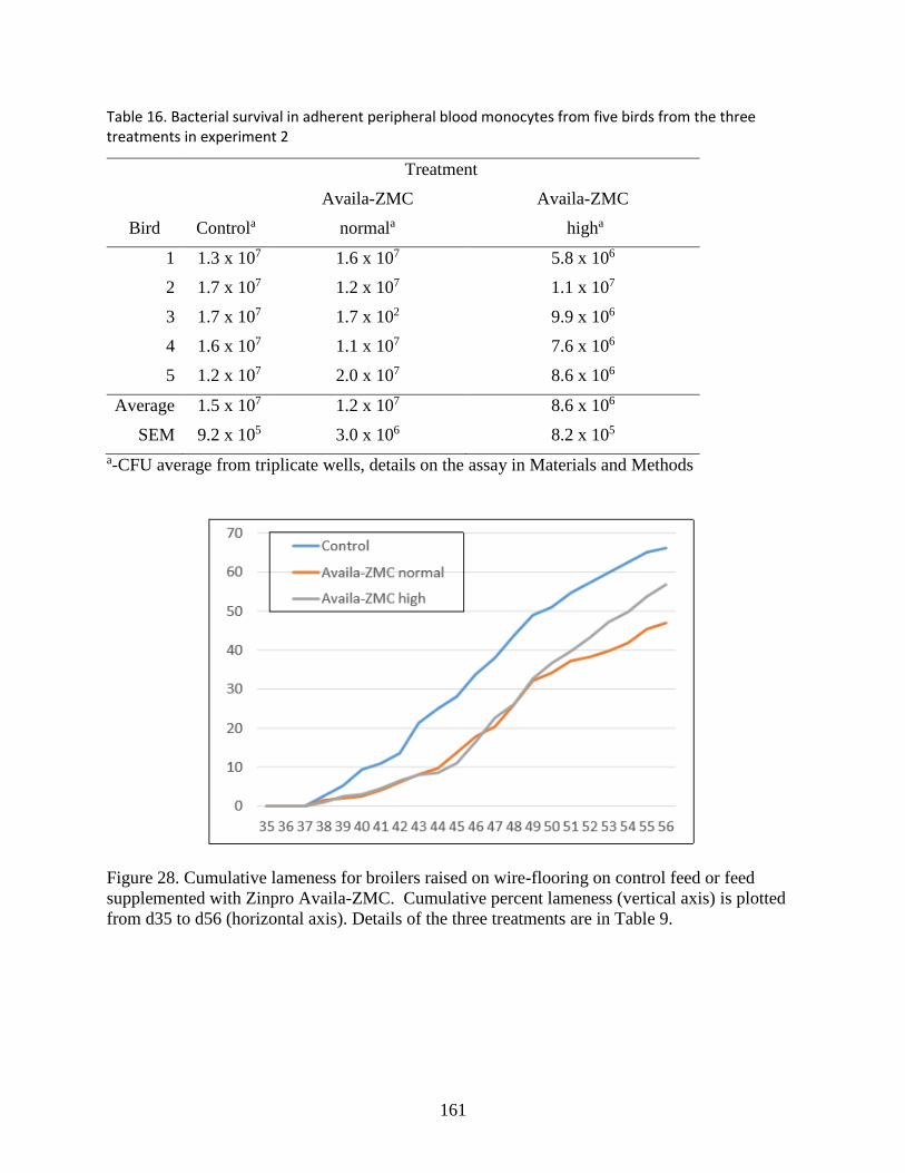
161
Table 16. Bacterial survival in adherent peripheral blood monocytes from five birds from the three treatments in experiment 2
Treatment
Bird Controla
Availa-ZMC
normala
Availa-ZMC
higha
1 1.3 x 10⁷ 1.6 x 10⁷ 5.8 x 10⁶
2 1.7 x 10⁷ 1.2 x 10⁷ 1.1 x 10⁷
3 1.7 x 10⁷ 1.7 x 10² 9.9 x 10⁶
4 1.6 x 10⁷ 1.1 x 10⁷ 7.6 x 10⁶
5 1.2 x 10⁷ 2.0 x 10⁷ 8.6 x 10⁶
Average 1.5 x 10⁷ 1.2 x 10⁷ 8.6 x 10⁶
SEM 9.2 x 10⁵ 3.0 x 10⁶ 8.2 x 10⁵
a-CFU average from triplicate wells, details on the assay in Materials and Methods
Figure 28. Cumulative lameness for broilers raised on wire-flooring on control feed or feed
supplemented with Zinpro Availa-ZMC. Cumulative percent lameness (vertical axis) is plotted
from d35 to d56 (horizontal axis). Details of the three treatments are in Table 9.

162
Figure 29. Tibial (upper) and femoral (lower) lesion diagnoses for all lame birds raised on wire-
flooring in experiment 1. Proximal heads were diagnosed at necropsy for R- right; L- left leg
bones, for each of the three treatments.

163
Figure 30. Cumulative lameness for broilers with a bacterial challenged, raised on litter-flooring,
on control feed or feed supplemented with Zinpro Availa-ZMC in experiment 2. Cumulative
percent lameness (vertical axis) is plotted from d35 to d56 (horizontal axis). Source was the same
as Control but was challenged with S. agnetis 908 at 104 CFU/ml in drinking water for d20 and
d21.
Figure 31. Tibial (upper panel) and femoral (lower panel) lesion diagnoses for all lame birds
raised on litter-flooring in experiment 2. Proximal heads were diagnosed at necropsy for R- right;
L- left leg bones, for each of the four treatments.

164
Figure 32. Villus length at d57 for apparently healthy birds from three treatment groups in
experiment 2. Average villus length (µm; vertical axis) for distal jejunum and proximal ileum
for five birds from each treatment. Error bars indicate SEM.
Figure 33: Expression of intestinal barrier integrity-related genes from three treatment groups in
experiment 2. The relative expression of ileal and jejunal OCLN, CLDN1, GJA-1, and CTNB1
was determined by qPCR and analyzed by 2–ΔΔCt method using control group as the calibrator.
Data are presented as mean ± SEM (n = 6/group). Different letters indicate significant difference
at P < 0.05.

165
CHAPTER 5
Typhoid Mary Experiment: BCO incidence variation due to bird-to-bird intra-pen
transmission when administering Staphylococcus agnetis in the water

166
Disclaimer
In this trial, I was part of a team that performed the animal trials in this project.
167
Typhoid Mary Experiment: BCO incidence variation due to bird-to-bird intra-pen
transmission when administering Staphylococcus agnetis in the water
Summary
In our series of BCO trials, we have observed that some pens seem to show lameness earlier than
others. We contended that this is because of one of two alternatives: i) a variable incidence of
some additional infectious agent (virus, bacteria, coccidian), or ii) a carrier infecting other pen-
mates. Since we have determined that S. agnetis is a causative agent of BCO and lameness, we
tested whether this pathogen can be easily transmitted in a floor pen environment. S. agnetis
administered in drinking water at Day 20 to birds reared on wire flooring results in a 40-80%
incidence of lameness. Without bacterial administration we see 30-50% lameness depending on
the experiment, and the major (78%) bacterial species recovered from BCO lesions on our farm
is S. agnetis. In this experiment we wanted to determine if broilers challenged with S. agnetis can
transmit the bacterium to birds in the same pen, thereby spreading BCO and lameness. We
discovered that young broilers exposed to S. agnetis at an early age of Day-20 may harbor the
bacterium and if mixed with unexposed birds 10 days later can transmit the bacterium to their
pen mates via unknown mechanism(s).
Materials and methods
Animal Housing, Care, and Treatment
Animal procedures were approved by the University of Arkansas Institutional Animal Care and
Use Committee (Protocol #11002). The experiments were conducted using pens in A365W at the
University of Arkansas Poultry Research Farm. Carboys and nipple waterers were flushed with
dilute bleach (5%) followed by a tap water flush to remove bacterial biofilms at least two days
before the chicks were placed. All birds were on 23 hours of light per day,

168
Thermoneutral/Optimal brooding and growing temperatures throughout (32.2 oC for d1 to d3,
31.1 oC for d4 to d6, 29.4 oC for d7 to d10, 26.7 oC for d11 to d14, and 23.9 oC), and Optimal
ventilation throughout. The starter diet was a commercial corn and soybean meal-based chick
starter (crumbles), and on d35 (10/14/2014) all birds were switched to a pelleted commercial
corn and soybean meal-based finisher diet. Feed was formulated without meat or animal
byproducts to meet or exceed minimum National Research Council (1994) standards for all
ingredients. Starter and finisher feeds were provided ad libitum. Body weights were not recorded
during the experiments, to avoid imposing additional stress on birds that already were under
significant stress due to the wire flooring (Wideman & Pevzner., 2012; Wideman & Prisby.,
2012).
Cobb500 surplus fast feathering breeder male chicks from the Fayetteville Hatchery were placed
at 70 chicks per pen on suspended wire flooring as described (Wideman et al., 2012). Bird
densities initially were approximately 1.65 ft2/chick in all pens. A365W is equipped with
computer controllers to regulate the temperature, photoperiod, and ventilation. On day 14 all
pens were culled to 60 birds per pen. Beginning on day 15 all birds were “walked” daily by
being prompted with a broom. For days 20 and 21 the birds in pens 11, 12, 16-21, 23, and 24
were treated with S. agnetis isolate 908 at 105 CFU/ml in their drinking water from 20 L carboys
then the lines were flushed well with tap water and returned to city tap water. On day 30, we
reciprocally exchanged half of the birds between pens 4&16, 5&17, 6&18, 7&19, 8&20, and
9&21. Beginning on day 22 we began recording lameness per pen and the actual birds
diagnosed as lame. All broilers that died or that developed clinical lameness were recorded by
date, gender, and tag number, and then necropsied to assess BCO lesion distributions.

169
Clinical Diagnosis of Lameness
The birds were “walked” and observed for lameness every two days beginning on Day 15.
Affected broilers had difficulty standing, exhibited an obvious limping gait while dipping one or
both wingtips and, if not removed, became completely immobilized within 48h. Birds were
humanely euthanized as soon as the onset of lameness was noticed, and were necropsied within
20 min post-mortem. Lame birds were humanely euthanized with CO2 gas or by cervical
dislocation. Birds succumbing to BCO can die quickly because they have difficulty accessing
food and water, and they can be trampled by their flock mates. Therefore, birds found dead also
were necropsied to ascertain the cause of death and assess leg lesions. All broilers that died or
that developed clinical lameness were recorded by date, gender, and wing band, and they were
necropsied to assess BCO lesion distributions.
Birds that were unable to walk were diagnosed as “clinically lame” and humanely euthanized.
All birds that died or developed clinical lameness were recorded by date, tag number, and pen
number. They were necropsied and assigned to one of the following categories:
Normal = Femur head and proximal tibia appear entirely normal
Cull = Runts and individuals that failed to thrive or appeared to be clinically ill
U = Unknown cause of death
NE = Necrotic Enteritis
SDS = Sudden Death Syndrome (Flip over, Heart Attacks)
PHS = Pulmonary Hypertension Syndrome, Ascites
KB = Kinky Back (Spondylolisthesis)
TW = Twisted Leg or Slipped Tendon (perosis)
TD = Tibial Dyschondroplasia
Lame-UNK = Lameness for undetermined reasons
FHS = Proximal Femoral Head Separation (epiphyseolysis)
FHT = Proximal Femoral Head Transitional degeneration
FHN = Proximal Femoral Head Necrosis (bacterial chondronecrosis with osteomyelitis,
BCO)
THN = Mild Proximal Tibial Head Necrosis, a sub-category of BCO in the tibiotarsus
THNS = Proximal Tibial Head Necrosis Severe, THN in which the growth plate was
imminently threatened or damaged
170
THNC = Proximal Tibial Head Necrosis Caseous, THN in which caseous exudates or
bacterial sequestrate were macroscopically evident
Total Lame = FHS + FHT + FHN + THN + THNS + THN
Percent Lame = Total Lame/ (Total Birds - [sick or dead from other reasons])
Previously published photographs illustrate typical BCO lesions of the proximal femora and
tibiae (Wideman & Prisby., 2012; Wideman et al., 2012). Proximal femoral head lesions (FHS,
FHT, FHN) and tibial head lesions (THN, THNs, THNc) were categorized separately to
emphasize the progressive development of BCO (Wideman etal.,2012).On day 56 representative
surviving birds were euthanized and necropsied to assess sub-clinical lesion incidences: Normal
proximal femoral head; Femoral Head Separation; Femoral Head Transitional degeneration;
Femoral Head Necrosis; Tibial Head Necrosis, and Tibial Dyschondroplasia.
Results
The layout of the pens for the experiment is presented in Figure 34 and the protocol followed is
in Table 15. There were four treatment groups assigned for this experiment that was classified as
Y: Challenged with S. agnetis; mixed with no challenge; N: No challenge; mixed with S. agnetis
challenged; C+: S. agnetis challenged and not mixed; and C-: No challenge and not mixed.
Death due to causes other than BCO lameness was minimal for all four treatments (Y: 3; N: 2;
C+: 1; N: 3). The cumulative percentage of lameness per treatment throughout the experiment is
presented in Figure 35. The % lameness for treatments Y, N, and C+ diverges from the
unchallenged unmixed control (C-) after Day 42, and then continue to near 70% with C-
finishing at 50%. The Y and C+ S. agnetis challenged groups had nearly identical total %
lameness of 73.4 and 75.5% lameness, respectively. Unchallenged and mixed treatment (N)
were slightly less at 69%, but N was closed to Y+ and C+ challenged groups. As the
accumulation of lameness in the unchallenged N treatment group appears to follow a similar
accumulation to the Y and C+ treatment groups which were challenged with S. agnetis, this

171
strongly suggests that the Y treatment birds transmitted the bacterium to their N treatment
unchallenged pen-mates. Table 16 presents the average lameness per pen for the treatment
groups. In Table 16, we pooled each of the Y and N birds for the pens that were mixed to
maintain approximately 60 birds per pen (we summed the lame birds in the Y treatment for pens
4 and 16, and we summed the lame birds in the N treatment for pens 4 and 16). The Student T-
tests on the lameness counts for treatment groups by pen showed no significant difference
between treatments Y, N, and C+ (P≥0.187). The three treatments were all significantly different
from C- (P<0.013). Therefore, this experiment demonstrates that if we expose broiler chicks to a
two-day inoculation with S. agnetis at day 20, and 10 days later mix those exposed birds with
unexposed birds then the incidence of lameness in the mixed birds is equivalent throughout the
pen. Thus, the exposed birds transmit the bacterium to their unexposed pen-mates. The
mechanism of transmittal could be direct physical contact, contamination of the waterers from
exposed birds, or vectored transmittal. The exposure should not be from residual S. agnetis on
the nipple waterers because those were flushed after the challenge administration and we did not
see any difference in lameness incidence between pens 4-9 and 14-21 (pens 14-21 were the pens
with S. agnetis administration).
Assessment of all types of femoral and tibial lesions diagnosed for each treatment group showed
little or no difference in the range of lesions or particular proximal leg bone head afflicted
(Figure 35) except for perhaps a lower incidence of tibial lesions in the C- group.
Discussion
The Typhoid Mary experiment shows that young broilers exposed to S. agnetis at an early age
(20 days) harbor the bacterium and if mixed with unexposed birds 10 days later can transmit the
bacterium to their pen mates via some mechanism, most likely direct physical transfer,

172
contaminating the waterers, or other vectors. The transfer must be rapid because as the incidence
of BCO lameness begins to accumulate around 40-42 days that is only 10-12 days after the
mixing of the two treatment groups. We have long suspected the Typhoid Mary aspect of BCO
lameness because in the absence of our bacterial challenge we often experienced a few pens
where BCO lameness appeared early and reached much higher levels than other wire flooring
pens. We suspected there might be one or more pre-disposing subclinical infectious agents
(mycoplasma, virus, etc.) but this experiment suggests it is more likely a carrier of the eliciting
bacterial species, in this case, S. agnetis. One caveat is that we did not do microbial surveys of
the BCO lesions in this experiment. Therefore, we cannot explicitly state that the lesions in the
N treatment birds contained predominantly S. agnetis. However, the data are most consistent
with the Y treatment birds transmitting the S. agnetis to the N group of birds. In prior
experiments, we have shown that S. agnetis is the predominant species isolated from BCO
lesions on our research farm in multiple facilities, and that if a non-BCO pathogen is
administered (human S. aureus) we still recover S. agnetis from the BCO lesions (Al-Rubaye et
al., 2015). One major suspect for horizontal transmission is mites. Chickens are known to have
several different types of endemic mites. Metagenomics has detected the presence of S. agnetis
DNA in the guts of sheep scab mites (Hogg & Lehane., 1999). That does not mean there are
viable organisms, but the presence of the DNA suggests at least a transient presence. Intense
poultry production systems have been known to harbor more than 30 mite genera (Horn et al.,
2015) and molecular surveys of poultry red mites have detected multiple chicken viruses and
mycoplasma (Huong et al., 2014). Therefore, chicken mites should be suspect for vectoring the
bacterium. As the C- treatment was in the same room but separated by an unused pen the vector
does not appear to transmit readily over 8-10 feet which argues against vectoring by flies or

173
gnats. Although the C- pens which were closest to the challenged pens did have a slightly higher
percentage of lameness. Our experimental data is consistent with the spread of BCO lameness
within a broiler house or within a region of a broiler house that conforms with anecdotal
narratives from growers. Future experiments should be designed to examine whether birds in
neighboring pens can transmit BCO lameness when direct physical contact is reduced and there
are no shared nipple waterers. There should also be concern about vertical transmission of
agents of lameness through eggs or egg facilities. In conclusion, the bacterial species we have
identified as intimately involved in BCO lameness in young broilers is communicable between
broilers raised in a shared pen. And further work on the mechanism of horizontal transmission of
the bacterium is warranted.
Pen 1
Control; No Challenge
Pen 13
Control; No Challenge
Pen 2
Control; No Challenge
Pen 14
Control; No Challenge
Pen 3: empty Pen 15: empty
Pen 4
No Challenge mix 50:50 with 16
Pen 16
Challenge mix 50:50 with 4
Pen 5
No Challenge mix 50:50 with 17
Pen 17
Challenge mix 50:50 with 5
Pen 6
No Challenge mix 50:50 with 18
Pen 18
Challenge mix 50:50 with 6
Pen 7
No Challenge mix 50:50 with 19
Pen 19
Challenge mix 50:50 with 7
Pen 8
No Challenge mix 50:50 with 20
Pen 20
Challenge mix 50:50 with 8
Pen 9
No Challenge mix 50:50 with 21
Pen 21
Challenge mix 50:50 with 9
Pen 10: empty Pen 22: empty
Pen 11
Challenge positive control
Pen 23
Challenge positive control
Pen 12
Challenge positive control
Pen 24
Challenge positive control
Figure 34. Pen Setup in A364 for the Typhoid Mary experiment.

174
Figure 35. Cumulative % lameness per treatment from day 34 through day 56.
0.00%
10.00%
20.00%
30.00%
40.00%
50.00%
60.00%
70.00%
80.00%
34 35 36 37 38 39 40 41 42 43 44 45 46 47 48 49 50 51 52 53 54 55 56
Cumulative % Lame by Day
Y N C+ C-

175
Figure 36. Tibial and Femoral BCO lesion diagnoses for the four treatment groups. Percent
incidence in lame birds is presented for RF right femur, LF- left femur, RT- right tibia; and LT-
left tibia. Lesion type are as in Materials and Methods
Table 17. Protocol for Typhoid Mary Experiment
Day of Age Comments
1 Place chicks on wire flooring. Cobb starter. All chicks tagged
and recorded
14 cull to 60 birds per pen
20 Administer S. agnetis at 105 cfu/ml in drinking water for 2 days to
pens 11,12,16-21,23,24
22 Begin recording all deaths, lame and infirmed
30 Swap birds 50:50 between pens
4&16,5&17,6&18,7&19,8&20,9&21
35 Switch to Cobb finisher
56 Complete experiment. Weigh all remaining birds and necropsy 5
apparently healthy birds from each pen
0.0%
10.0%
20.0%30.0%
40.0%
50.0%
60.0%
70.0%80.0%
N
FHS
FHT
FHN N
FHS
FHT
FHN N
THN
THN
C
THN
S N
THN
THN
C
THN
S
RF RF RF RF LF LF LF LF RT RT RT RT LT LT LT LT
Experiment 19: Typhoid Mary Diagnoses
Y N C+ C-

176
Table 18. Total and percent lameness for the four treatment groups along with average lame
birds per pen. Note that for the Y and N treatment groups the per pen averages were for the two
pens that were mixed so that all averages are for the same number of treated birds. Treatments
that are different (P < 0.05) have Average ± std with superscript letters that are different.
Treatment Group Y N C+ C-
Final Count 357 358 239 237
% Lame 73.4% 69.0% 75.7% 51.5%
Average lame ± std per pen 43.7±6.8a 41.2±7.8a 45.3±5.9a 30.5±4.8b
References
Wideman RF, Pevzner I. Dexamethasone triggers lameness associated with necrosis of the proximal
tibial head and proximal femoral head in broilers. Poult Sci. 2012;91(10):2464-74. doi:
10.3382/ps.2012-02386.
Wideman RF, Prisby RD. Bone circulatory disturbances in the development of spontaneous
bacterial chondronecrosis with osteomyelitis: A translational model for the pathogenesis of
femoral head necrosis. Front Endocrinol (Lausanne). 2013;3. doi:
10.3389/fendo.2012.00183.
Wideman RF, Hamal KR, Stark JM, Blankenship J, Lester H, Mitchell KN, et al. A wire-
flooring model for inducing lameness in broilers: Evaluation of probiotics as a prophylactic
treatment. Poult Sci. 2012;91(4):870-83. doi: 10.3382/ps.2011-01907.
Al-Rubaye AAK, Couger MB, Ojha S, Pummill JF, Koon JA, II, Wideman RF, Jr., et al.
Genome Analysis of Staphylococcus agnetis, an Agent of Lameness in Broiler Chickens.
PLoS ONE. 2015;10(11): e0143336. doi: 10.1371/journal.pone.0143336.
Hogg JC, Lehane MJ. Identification of bacterial species associated with the sheep scab mite
(Psoroptes ovis) by using amplified genes coding for 16S rRNA. Appl Environ Microbiol.
1999;65(9):4227-9.
Horn T, Körbes J, Granich J, Senter M, Ferla N. Influence of laying hen systems on the mite
fauna (Acari) community of commercial poultry farms in southern Brazil. Parasitol Res.
2015:1-12. doi: 10.1007/s00436-015-4756-9.
Huong CTT, Murano T, Uno Y, Usui T, Yamaguchi T. Molecular Detection of Avian
Pathogens in Poultry Red Mite (Dermanyssus gallinae) Collected in Chicken Farms. The
Journal of Veterinary Medical Science. 2014;76(12):1583-7. doi: 10.1292/jvms.14-0253.
PubMed PMID: PMC4300372.

177
CHAPTER 6
Conclusions

178
Conclusions
The poultry industry has seen significant intensification since the 1940s. This has led to shift in
poultry production from a small scale “Backyard” farming to a more intensified and integrated
production structure. Poultry industry has been successful due to its ability to produce
marketable products. Post 1940s the introduced poultry breeding programmes employed
numerous technologies to select important production traits. Today, broilers grow faster, have
improved meat quality, have reduced feed conversion ratio, reduced mortality, and their
production is more eco-friendly compared to production of pork or beef. These gains seem to
come at a price, as the poultry industry is faced with the issue of lameness. Numerous factors
combine to cause the incidence of lameness in broilers. However, Bacterial chondronecrosis with
osteomyelitis is the leading cause of the disease. The full etiology of the ailment is not
understood, but we have hypothesized that bacteria from vertical and horizontal transfers
translocate through compromised gut, integuments and respiratory pathways into birds that
develop lameness. These bacteria survive and colonize the long bones and T4 vertebrae causes
various forms of necrosis that have been described. We studied lameness the Wideman’s wire-,
litter-flooring, or a combination of both. We have isolated S. agnetis as a major BCO agent in
lame birds on our research facility, that can cause 50% incidence of lameness. Other BCO
species like E. coli and S. aureus are frequent causative agents in other farms.
In this dissertation, we covered the analysis of genomes of E. coli and S. aureus that were
isolated from three farms in Arkansas area. We found that the E. coli were more generalist in
their pattern of infections regardless of geolocation. S. aureus seemed were closely related to
isolates from Europe and were more specialized to infect chickens. Further analysis of their

179
genomes indicates the S. aureus we isolated have only been in the area for a few decades and
may have acquired virulence factors important for its adaptation and disease mechanism.
We used embryo lethality assay to access the pathogenicity of BCO isolates and for the different
reasons highlight in chapter 3, we do not trust that ELA is an efficient mechanism for
determining pathogenicity of BCO isolates. And although this work is on-going, we find that a
non-pathogenic E. coli electroporated with plasmids from a more pathogenic strain and passaged
over a few generations, acquired embryo lethality properties.
We used the Staphylococcus-Lameness model to test the efficacy of Zimpro Micronutrients for
reducing lameness in broilers and found that at certain levels these products can significantly
reduce the incidence of BCO lameness. We also analysed the levels of expression of mucin and
gut integral proteins. While there are certain gut integral proteins expressed in a differentially
important manner to the incidence of BCO, we cannot say the same of Mucin genes as our
analysis was inconclusive. The gut length and integrity for birds that received treatments of
Zinpro Avalia micronutrients were also significantly improved. We therefore recommend Zinpro
Avalia for it beneficial effects in commercial broiler productions.
Finally, we used the wire-flooring model to determine whether BCO can be transmittable
between birds of the same pen. We transferred birds exposed to S. agnetis in drinking water for
two days on Days 20, after found that birds were capable of transferring bacteria to their pen-
mates. The mechanism of this transfer is still not known. Research is on-going in many aspects
of BCO to better characterize the etiology of this disease, as therein lies the remedial.

180
Chapter 7
Appendix

181

182