exercise and osteoarthritis - usc davis school of...
TRANSCRIPT
J. Anat.
(2009)
214
, pp197–207 doi: 10.1111/j.1469-7580.2008.01013.x
© 2009 The Authors Journal compilation © 2009 Anatomical Society of Great Britain and Ireland
Blackwell Publishing Ltd
REVIEW
Exercise and osteoarthritis
David J. Hunter
1,2
and Felix Eckstein
3,4
1
Division of Research, New England Baptist Hospital, Boston, MA, USA
2
Boston University School of Medicine, Boston, MA, USA
3
Institute of Anatomy & Musculoskeletal Research, Paracelsus Medical University
4
Chondrometrics GmbH, Ainring, Germany
Abstract
Exercise remains an extremely popular leisure time activity in many countries throughout the western world. It iswidely promoted in the lay press as having salutory benefits for weight control, disease management advantagesfor cardiovascular disease and diabetes, in addition to improving psychological well-being amongst an array ofother benefits. In contrast, however, the lay press and community perception is also that exercise is potentiallydeleterious to one’s joints. The purpose of this review is to consider what osteoarthritis (OA) is and providean overview of the epidemiology of OA focusing on validated risk factors for its development. In particular the roleof both exercise and occupational activity in OA will be described as well as the role of exercise to the joints’ tissues(particularly cartilage) and the role of exercise in disease management. Despite the common misconception thatexercise is deleterious to one’s joints, in the absence of joint injury there is no evidence to support this notion.Rather it would appear that exercise has positive salutory benefits for joint tissues in addition to its other health benefits.
Key words
diarthrodial joint; exercise; osteoarthritis.
Introduction
Exercise remains an extremely popular leisure-time activityin many countries throughout the Western world and hasfor many become part of the modern lifestyle. It is widelypromoted in the lay press as having salutory benefits forweight control, disease management advantages forcardiovascular disease and diabetes, and for improvingpsychological well being amongst an array of other bene-fits. In contrast, however, the lay press and communityperception is also that exercise is potentially deleterious toone’s joints, in particular those of the lower extremities. Aprevious review in this journal has focused on the effectsof exercise on healthy articular cartilage, specifically itsfunctional adaptation to loading, both short and longterm (Eckstein et al. 2006). The purpose of the currentreview is to consider the potential effect of exercise on theonset and progression of joint disease, specifically to:
1
address the question of what osteoarthritis (OA) is;
2
provide an overview of the epidemiology of OA, focusingon validated risk factors for its development, in particularthe role of both exercise and occupational activity;
3
consider the role of exercise on the joint tissues (particularlycartilage) and the role of exercise in disease management.
We hope by the end that the reader will have a clearperception of the role of exercise in OA that will dispel thecommon misconception that exercise is damaging to joints.This is a narrative review in which the authors haveselected and considered the pivotal articles in constructingthis manuscript. The literature on this topic predominantlypertains to the knee and this is reflected in our review.
What is osteoarthritis?
Early investigators tended to regard OA as an isolateddisease of articular cartilage. Although cartilage loss is aprominent feature of OA, contemporary models recognizethat the entire joint organ is affected by OA. OA can beviewed as the clinical and pathological outcome of a rangeof disorders that result in
structural and functional failureof synovial joints
with loss and erosion of articular cartilage,subchondral bone alteration, meniscal degeneration, asynovial inflammatory response, and bone and cartilageovergrowth (osteophytes) (Nuki, 1999). OA occurs when thedynamic equilibrium between the breakdown and repairof joint tissues becomes unbalanced (Eyre, 2004). Thisprogressive joint failure may cause pain and disability(Guccione et al. 1994), although many persons with struc-tural changes consistent with OA are asymptomatic(Hannan et al. 2000). The source of pain is not particularlywell understood and is best framed in a biopsychosocial
Correspondence
David J. Hunter, 125 Parker Hill Ave, Division of Research, New England Baptist Hospital, Boston, MA 02120, USA. E: [email protected]
Accepted for publication
21 October 2008

Exercise and osteoarthritis, D. J. Hunter and F. Eckstein
© 2009 The AuthorsJournal compilation © 2009 Anatomical Society of Great Britain and Ireland
198
framework (posits that biological, psychological and socialfactors all play a significant role in pain in OA) (Dieppe &Lohmander, 2005). The etiology of pain, however, is notthe focus of this review. OA can occur in any synovial jointin the body but is most common in the knees, hips andhands.
In epidemiological investigations, OA is typically definedusing radiographs, and less frequently self report. Thecharacteristic features of OA scored on radiographs areosteophytes (osteocartilaginous growths), sclerosis andjoint space narrowing (Fig. 1).
Understanding this definition becomes important whenwe consider the impact of exercise on development of OA,as osteophytes (typically the first feature identified onradiographs) are not necessarily a deleterious finding andmay represent an effort on the part of the joint to pro-mote stability. They are important, however, if they repre-sent a source of symptoms, and yet most of the positiveevidence that suggests an association between exerciseand OA is based on the presence of self-reported OA orradiographic osteophytes and not on symptomatic OA(the concomitant presence of pain and radiographicfeatures). It is the presence of symptomatic OA that isimportant clinically, not simply the radiographic identifi-cation of osteophyte formation or self-reported OA (wheremisclassification is even more problematic than thecommonly used radiographic OA definition).
Epidemiology of OA
OA is the leading cause of disability in older people(Centers for Disease Control and Prevention (CDC) 2001).The reported prevalence of OA varies according to themethod used to evaluate it. In most epidemiologicalstudies it is commonly assessed by radiography. Marked
osteoarthritic damage must be present, however, to detectcharacteristic changes with plain radiographs, and theyare therefore not sensitive diagnostic tests. About 6%of adults aged > 30 years (Hunter & Felson, 2006) and 13%of persons aged 60 and over (Lawrence et al. 1998) havefrequent knee pain and radiographic OA. Although OA iscommon in the knee, it is even more prevalent in thehands, especially the distal (DIP) and proximal (PIP) inter-phalangeal joints and the base of the thumb. Whensymptomatic, especially so for the base of thumb joint,hand OA is associated with functional impairment(Cunningham & Kelsey, 1984; Zhang et al. 2002). OA of thethumb carpo-metacarpal joint is a common condition thatcan lead to substantial pain, instability, deformity, and lossof motion (Armstrong et al. 1994). Over the age of 70years, approximately 5% of women and 3% of men havesymptomatic OA affecting this joint with impairment ofhand function (Zhang et al. 2002). The prevalence of hipOA is about 9% in Caucasian populations (Felson & Zhang,1998). The prevalence of symptomatic hip OA is approxi-mately 4% (Lawrence et al. 1998).
The prevalence of OA is expected to increase as the popu-lation ages and the prevalence of obesity rises (this beingan important risk factor; see below). By 2020, it is expectedthat the number of people with OA may have doubled(Centers for Disease Control and Prevention (CDC), 1994;Badley & DesMeules, 2003).
Risk factors for OA
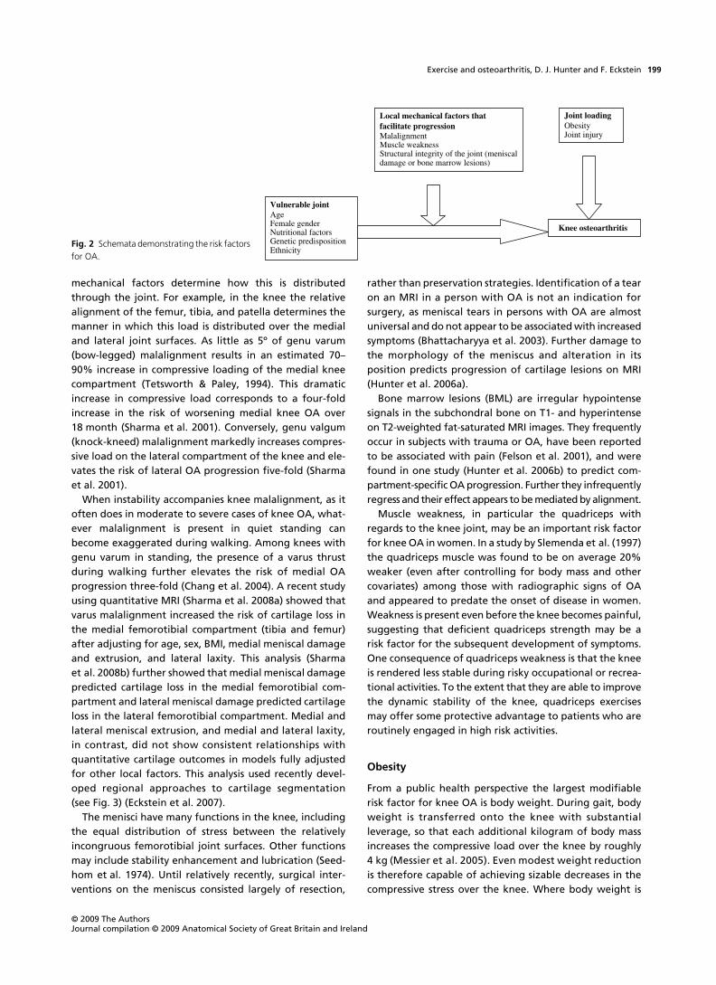
OA is perhaps best understood as resulting from excessivemechanical stress applied in the context of systemic sus-ceptibility (see Fig. 2 for an overview).
Susceptibility to OA may be increased in part by
geneticinheritance
(a positive family history increases risk),
age,ethnicity, diet
and
female gender
(Felson, 2004a).In persons vulnerable to the development of knee OA,
local mechanical factors such as abnormal joint congruity,malalignment (varus- or valgus deformity), muscle weak-ness or alterations in the structural integrity of the jointenvironment (such as meniscal damage or ligament rup-ture) facilitate the progression of OA. Loading can also beaffected by obesity and joint injury (either acutely as in asporting injury or after repetitive overuse such as occupa-tional exposure), both of which can increase the likelihoodof development or progression of OA.
Mechanical factors increasing risk for progression
Local mechanical factors such as the
adduction moment
,
malalignment
,
meniscal damage
,
bone marrow lesions
,and
altered quadriceps strength
potentially put the kneejoint at increased risk of progression of OA (Felson, 2004b).Local mechanical factors also mediate the impact on theknee of more systemic factors such as obesity (Felson et al.2004). Body weight and habitual activity determine howmuch overall load the joint must routinely sustain and the
Fig. 1 A weight-bearing plain radiograph of the knee depicting the characteristic features seen in OA: joint space narrowing, osteophytosis and subchondral sclerosis.
Exercise and osteoarthritis, D. J. Hunter and F. Eckstein
© 2009 The Authors Journal compilation © 2009 Anatomical Society of Great Britain and Ireland
199
mechanical factors determine how this is distributedthrough the joint. For example, in the knee the relativealignment of the femur, tibia, and patella determines themanner in which this load is distributed over the medialand lateral joint surfaces. As little as 5º of genu varum(bow-legged) malalignment results in an estimated 70–90% increase in compressive loading of the medial kneecompartment (Tetsworth & Paley, 1994). This dramaticincrease in compressive load corresponds to a four-foldincrease in the risk of worsening medial knee OA over18 month (Sharma et al. 2001). Conversely, genu valgum(knock-kneed) malalignment markedly increases compres-sive load on the lateral compartment of the knee and ele-vates the risk of lateral OA progression five-fold (Sharmaet al. 2001).
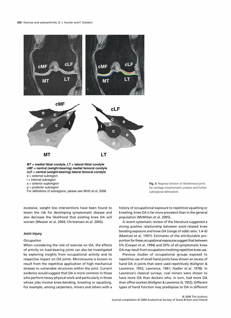
When instability accompanies knee malalignment, as itoften does in moderate to severe cases of knee OA, what-ever malalignment is present in quiet standing canbecome exaggerated during walking. Among knees withgenu varum in standing, the presence of a varus thrustduring walking further elevates the risk of medial OAprogression three-fold (Chang et al. 2004). A recent studyusing quantitative MRI (Sharma et al. 2008a) showed thatvarus malalignment increased the risk of cartilage loss inthe medial femorotibial compartment (tibia and femur)after adjusting for age, sex, BMI, medial meniscal damageand extrusion, and lateral laxity. This analysis (Sharmaet al. 2008b) further showed that medial meniscal damagepredicted cartilage loss in the medial femorotibial com-partment and lateral meniscal damage predicted cartilageloss in the lateral femorotibial compartment. Medial andlateral meniscal extrusion, and medial and lateral laxity,in contrast, did not show consistent relationships withquantitative cartilage outcomes in models fully adjustedfor other local factors. This analysis used recently devel-oped regional approaches to cartilage segmentation(see Fig. 3) (Eckstein et al. 2007).
The menisci have many functions in the knee, includingthe equal distribution of stress between the relativelyincongruous femorotibial joint surfaces. Other functionsmay include stability enhancement and lubrication (Seed-hom et al. 1974). Until relatively recently, surgical inter-ventions on the meniscus consisted largely of resection,
rather than preservation strategies. Identification of a tearon an MRI in a person with OA is not an indication forsurgery, as meniscal tears in persons with OA are almostuniversal and do not appear to be associated with increasedsymptoms (Bhattacharyya et al. 2003). Further damage tothe morphology of the meniscus and alteration in itsposition predicts progression of cartilage lesions on MRI(Hunter et al. 2006a).
Bone marrow lesions (BML) are irregular hypointensesignals in the subchondral bone on T1- and hyperintenseon T2-weighted fat-saturated MRI images. They frequentlyoccur in subjects with trauma or OA, have been reportedto be associated with pain (Felson et al. 2001), and werefound in one study (Hunter et al. 2006b) to predict com-partment-specific OA progression. Further they infrequentlyregress and their effect appears to be mediated by alignment.
Muscle weakness, in particular the quadriceps withregards to the knee joint, may be an important risk factorfor knee OA in women. In a study by Slemenda et al. (1997)the quadriceps muscle was found to be on average 20%weaker (even after controlling for body mass and othercovariates) among those with radiographic signs of OAand appeared to predate the onset of disease in women.Weakness is present even before the knee becomes painful,suggesting that deficient quadriceps strength may be arisk factor for the subsequent development of symptoms.One consequence of quadriceps weakness is that the kneeis rendered less stable during risky occupational or recrea-tional activities. To the extent that they are able to improvethe dynamic stability of the knee, quadriceps exercisesmay offer some protective advantage to patients who areroutinely engaged in high risk activities.
Obesity
From a public health perspective the largest modifiablerisk factor for knee OA is body weight. During gait, bodyweight is transferred onto the knee with substantialleverage, so that each additional kilogram of body massincreases the compressive load over the knee by roughly4 kg (Messier et al. 2005). Even modest weight reductionis therefore capable of achieving sizable decreases in thecompressive stress over the knee. Where body weight is
Fig. 2 Schemata demonstrating the risk factors for OA.
Exercise and osteoarthritis, D. J. Hunter and F. Eckstein
© 2009 The AuthorsJournal compilation © 2009 Anatomical Society of Great Britain and Ireland
200
excessive, weight loss interventions have been found tolessen the risk for developing symptomatic disease andalso decrease the likelihood that existing knee OA willworsen (Messier et al. 2004; Christensen et al. 2005).
Joint injury
Occupation
When considering the role of exercise on OA, the effectsof activity on load-bearing joints can also be investigatedby exploring insights from occupational activity and itsrespective impact on OA joints. Microtrauma is known toresult from the repetitive application of high mechanicalstresses to vulnerable structures within the joint. Currentevidence would suggest that OA is more common in thosewho perform heavy physical work and particularly in thosewhose jobs involve knee-bending, kneeling or squatting.For example, among carpenters, miners and others with a
history of occupational exposure to repetitive squatting orkneeling, knee OA is far more prevalent than in the generalpopulation (McMillan et al. 2005).
A recent systematic review of the literature suggested astrong positive relationship between work-related kneebending exposure and knee OA (range of odds ratio: 1.4–6)(Maetzel et al. 1997). Estimates of the attributable pro-portion for these occupational exposures suggest that between5% (Cooper et al. 1994) and 20% of all symptomatic kneeOA may result from occupations involving repetitive knee use.
Previous studies of occupational groups exposed torepetitive use of small hand joints have shown an excess ofhand OA in joints that were used repetitively (Kellgren &Lawrence, 1952; Lawrence, 1961; Hadler et al. 1978). InLawrence’s classical surveys, coal miners were shown tohave more OA than dockers who, in turn, had more OAthan office workers (Kellgren & Lawrence JS, 1952). Differenttypes of hand function may predispose to OA in different
Fig. 3 Regional division of tibiofemoral joints for cartilage morphometric analysis and further subregional delineation.

Exercise and osteoarthritis, D. J. Hunter and F. Eckstein
© 2009 The Authors Journal compilation © 2009 Anatomical Society of Great Britain and Ireland
201
articular areas, with studies of repeated pincer grip andgenerally increased workload demonstrating a predisposi-tion to distal interphalangeal joint OA (Hadler et al. 1978;Bergenudd et al. 1989; Nakamura et al. 1993).
Joint injury from sports
Consideration of the role of exercise in OA needs to payheed to the potential overlap with sports injury. Amongboth genders, a past history of injury to the stabilizing orload-bearing structures of the knee renders the jointhighly vulnerable to OA in subsequent years. This is distinctfrom the degenerate meniscal tears that occur frequentlyas part of the OA disease process that was described earlier.
Knee injury/trauma has been identified as the mostimportant modifiable risk factor for subsequent knee OAin men, and is second only to obesity in women (Felsonet al. 2000). It typically occurs in the younger populationand as such leads to prolonged disability and economiccost (Yelin & Callahan, 1995), largely due to work loss. Alarge prospective cohort study revealed that joint injurysubstantially increased the risk for subsequent knee OA[relative risk, 5.17 (CI, 3.07–8.71)] (Gelber et al. 2000).
The high incidence of knee OA in the years followinganterior cruciate ligament (ACL) (Roos et al. 1995; Loh-mander et al. 2004) or meniscal injury (Roos et al. 1995;Englund et al. 2003) is well-documented, and credibleevidence suggests that current arthroscopic procedures,including ACL reconstruction and meniscectomy, are notsufficient to fully restore normal joint mechanics orneutralize the long-term risk of OA (Englund & Lohmander,2004; Andriacchi et al. 2006).
Meniscal injury and subsequent meniscectomy is oftenaccompanied by cartilage degeneration and the onset ofOA because of the high focal stresses imposed on articularcartilage and subchondral bone due to excision of themeniscus (Tapper & Hoover, 1969; Johnson et al. 1974). Anumber of recent publications have documented long-termfollow-up of radiographic changes after meniscectomy(Roos et al. 1998; Macnicol & Thomas, 2000; McNicholaset al. 2000). Whilst the observed incidence of secondaryOA varies, Roos et al. (1998) reported mild radiographicchanges in 71% of knees, with more advanced changes,comparable with a Kellgren and Lawrence grade 2 orhigher in 48%. Further analysis suggested that surgicalremoval of a meniscus following knee injury representeda significant risk factor for radiographic tibiofemoral OA,with a relative risk of 14.0 after 21 years. Older age at thetime of trauma or surgery appears to predict a more rapiddeterioration to OA of the involved knee.
Certain competitive athletes, including hockey players,whose sporting activities repetitively stress the stabilizingstructures of the knee and who are at great risk for kneeinjury also have very high rates of early-onset knee OA(Sandmark et al. 1999).
Risk of OA from sports participation
Recent years have witnessed an enormous increase inthe popularity of recreational exercise. There is a plethoraof evidence supporting participation in regular exercise,including recreational activities or competitive sports, as itimproves general health and may increase longevity.Individuals with normal joints frequently ask whethertheir exercise programs increase the risk of developing OA.
The initial studies that evaluated the relationshipbetween regular recreational weight-bearing exercise andOA of the knee generally found no ill effects on the jointsfrom exercise participation (Lane et al. 1986; Panush et al.1986). More recent studies that assessed the longitudinaleffects of aging and exercise on OA of the hip and kneeafter 5 and 8 years of follow-up also found no increasedrisk of developing OA in runners, compared with age-similarcontrols (Hannan et al. 1993; Panush et al. 1995). What isclear from the data is that the risk of subsequent OArelates more to the intensity of the level of participation,the performance level (e.g. elite vs. recreational) and theconcomitant presence and or likelihood of joint injury. Inthis light we have considered recreational vs. eliteseparately, paying particular attention to the presence orconsideration of joint injury.
A study of female physical education teachers comparedwith population-based controls found no increase in ratesof radiographic hip or knee OA, and actually found lowerrates of knee OA (White et al. 1993). Potentially in contrastto the former study, 571 graduates from a single Swedishtraining facility had higher rates of self-reported OA kneeand knee injury than the comparator group (Sandmark &Sandmark, 2000). The graduates had a current median ageof about 57 and were matched with people from a popu-lation register. However, radiographs were not taken inthis study and therefore the prevalence of radiographicOA is not certain. Further, the concomitant presence ofjoint injury likely inflated the risk of subsequent OA.
A number of population-based studies have studied theeffects of participation in sport on rates of OA. Thesestudies provide a clearer picture of overall associations incommunities but tend to rely upon recall of sportingparticipation. A British team of investigators (Cooper et al.2000) examined a large cohort with knee radiographstaken 5 years apart and a median age at follow-up of 75.8years. They found that a history of regular sport parti-cipation was associated with an increased risk of incidence(but not progression) of knee OA. The risks for hip jointreplacement from retrospective case-control studies ofSwedish male (Vingard et al. 1993) and female (Vingardet al. 1998) population-based cohorts found that in thosewith the most hours of recalled sporting activities (over800 h in total based on recall of all sporting involvementup till the age of 50), men had a relative risk of 4.5 andwomen 2.3 of developing OA of the hip, compared with

Exercise and osteoarthritis, D. J. Hunter and F. Eckstein
© 2009 The AuthorsJournal compilation © 2009 Anatomical Society of Great Britain and Ireland
202
the lowest activity groups. Importantly, men who hadperformed jobs with high joint loading as well as highlevels of sport had almost double the relative risk. Laneet al. (1999) examined the association of reported exerciseand radiographic OA hip in a population of 5818 womenwith a mean age of about 72. Women who reportedregular recreational physical activities (defined as one tofive times a week) as teenagers, at age 30, and at age 50had significantly greater odds of moderate to severe hipOA than those who performed no recreational activities.In all of these population-based studies, a major confound-ing issue has been the separation of the impact loadingaspects of the sport from the associated injuries whichundoubtedly predispose to OA. A retrospective, case-controlstudy from the UK used 216 subjects (drawn from a largernational survey of 4316 persons) with self-reported OAknee and matched each of them with four controls (Suttonet al. 2001). Extensive data on lifetime exercise involvementwas obtained. The only strong risk factor for knee OA wasa reported prior knee injury (odds ratio 8.0); most of theseknee injuries were related to sport.
Therefore, the results from these studies show thatindividuals who had normal joints and participated inlow-impact exercises did not have an increased risk ofdeveloping OA of the knee or hip as they aged, independentof joint injury. Whilst there is some evidence suggesting anincreased risk with activity this has not been adequatelydisentangled from injury. In this light there is
no good evi-dence supporting a deleterious effect of exercise on jointsin the setting of normal joints and moderate activity
.In contrast, there does seem to be an association between
elite sports participation and an increased risk of OA.However, the nature of the sport is very important to thedegree of risk. The sports with major risk are those thatinvolve repetitive, high intensity, high impact forces throughthe affected joints, especially where there is a high associ-ated risk of injury. Categorizing exercise into different levelsof impact is somewhat arbitrary but relates to the extentof compressive loading during the activity. Common examplesof high-impact exercise include running, dance exercise,tennis, racquetball, and squash. This is in contrast to low- tomoderate-impact exercises such as walking, swimming,stair climbing, rowing, and cross-country skiing.
A Finnish group examined hospital admission rates over21 years for OA (hip, knee or ankle) for over 2000 maleex-athletes (Kujala et al. 1994). They divided the cohortinto endurance (long-distance running, cross-countryskiing), team (soccer, ice hockey, basketball, track and field),and power (boxing, wrestling, weight-lifting, throwing)sports. All three groups had higher incidences of OA-relatedadmissions, with power and team sports having admis-sions at an earlier age. Soccer players and weight-liftershad higher risk, which was due at least partly to kneeinjuries in the former and high body mass index at age 20in the latter (Kujala et al. 1995). The former power-sport
competitors had the highest odds ratios for hip disability,whereas only the team-sport players had a high risk forknee disability, again perhaps reflecting the injury-pronenature of these sports (Kettunen et al. 2001).
A Swedish retrospective cohort reported both hip andknee radiographic OA associations in 71 elite and 215non-elite players with a mean age of 55 and comparedthe rates with those of 572 age-matched controls (Lindberget al. 1993; Roos et al. 1994). For the hip, the prevalence ofOA was 14% for elite players compared with 4.2% innon-elite and control groups (Lindberg et al. 1993). For theknee, the prevalence of OA was 15.5%, 4.2%, and 1.6% inelite, non-elite, and control groups, respectively (Roos et al.1994).
The data from studies of runners indicate that thedistance run and intensity may play a role. In a retrospectivecohort study, examinations were performed in 1973 and1988 on a number of former athletes: 27 long-distancerunners (averaging 60 miles per week in 1973), ninebobsleigh riders, and 23 controls (Marti et al. 1989). Theathletes had a mean age of 42 years at the secondexamination. Radiographic hip OA was found in 19% ofthe runners, but in neither of the other groups. Age andnumber of miles run per week in 1973 were the positivepredictors of radiographic OA. Spector et al. (1996) com-pared 67 female elite middle- and long-distance runnersand 14 female tennis players (aged 40–65) with a largematched control group. Radiographic hip and knee OArates were significantly higher in the former athletescompared with controls of a similar age, with a tendencyto more patellofemoral OA in the runners. No clear riskfactors were seen within the ex-athlete groups, althoughthe tennis players tended to have more osteophytes at thetibiofemoral joints and hip, but the runners had morepatellofemoral joint disease. In contrast, in a retrospectivecohort study, the rates of radiographic hip OA in 60 ex-marathon runners were not higher than in controls,although the timing of radiographs differed for the twogroups (Puranen et al. 1975). A study comparing 504university level cross-country runners with similar-levelswimmers found no difference in levels of hip and kneepain but x
-
ray examinations were not performed (Sohnet al. 1985). A small prospective study of 17 male runners(nine were marathon runners) compared with controlsfound no difference in radiographic OA at the hip, knee,ankle or feet (Panush et al. 1986).
Lane et al. (1986, 1993, 1998) have presented a series ofreports from a well-described, prospective cohort of sub-jects from a long-distance running club – perhaps closer torecreational runners than the elite athletes studied above.At baseline, 41 runners aged 50–72 years and averaging25 miles a week were compared with controls matched forage, sex, years of education, and occupation (Lane et al.1986). There were no differences in clinical and radiographicOA findings in the knee and lumbar spine. Follow-up of

Exercise and osteoarthritis, D. J. Hunter and F. Eckstein
© 2009 The Authors Journal compilation © 2009 Anatomical Society of Great Britain and Ireland
203
this cohort at 5 (Lane et al. 1993) and 9 (Lane et al. 1998)years showed significant within-group progression of bothosteophytes and total knee radiographic scores (
P
= 0.01for runners and
P
= 0.05 for non-runners) and joint spacenarrowing in non-runners (
P
= 0.01), with no significantbetween-group differences. For the runners, regressionanalysis disclosed that the predictors of progression ofradiographic knee OA were baseline radiographic scoreand a faster pace per mile. Hip radiographs taken 9 yearslater also showed no differences in OA between the groups(Lane et al. 1998). Another study of 30 long-distance runners,90% of whom averaged 12.5–25 miles a week over a medianof 40 years, found no differences in rates of radiographicOA at the hips, knees or ankles when compared withcontrols matched for age, body mass index, and occupa-tion (Konradsen et al. 1990).
Thus elite athletes who perform their activities withhigh impact and high stress to the joints appear to have anincreased risk for OA in the hips and knees compared withage-matched controls
(Kujala et al. 1995; Spector et al.1996). Again the concomitant presence or likelihood ofjoint injury increases the risk of developing OA.
Exercise and cartilage
When considering the impact of weight-bearing activityon the joint tissues there is a preponderant focus on carti-lage as this pertains to OA. In general, though, the effectof exercise on load-bearing joints extends beyond carti-lage, and exercise is known to have advantageous trophiceffects on periarticular bone and muscle in particular, andalso tendon, at least in men (Magnusson et al. 2007).
Hyaline articular cartilage provides the articulatingsurface of synovial joints. Its complex composition facilitatesthe even transfer of forces from one subchondral plate tothe other (Mow & Setton, 1998). Many studies suggest thatarticular cartilage is mechano-adaptive; that is, the biosyn-thetic activity of chondrocytes is responsive to mechanicalstimuli and can alter the morphology and composition ofcartilage (Carter et al. 2004). Other studies suggest thatexcessive mechanical force can have a deleterious effecton the prevalence of OA (Kujala et al. 1995; Spector et al.1996). Due to the pluripotent effects of mechanical load-ing on articular cartilage, physical activities may play animportant role in either the causation of or protectionagainst OA. Prolonged immobilization in animals leads toreductions in articular cartilage thickness, although itdoes not necessarily become osteoarthritic (surface remainssmooth and no osteophytes or erosions develop) (Van-wanseele et al. 2002b). Similarly, in humans the absence ofnormal joint loading due to spinal cord injury results inrates of cartilage thinning that are higher than thoseobserved in persons with OA (Vanwanseele et al. 2002a,2003). Thus cartilage undergoes atrophy in the absence ofmechanical stimulation.
In animal studies, physical activity has been shown tohave varying effects on articular cartilage (Jurvelin et al.1986; Lammi et al. 1993; Newton et al. 1997). Overexercisedanimals have been found to develop glycosaminoglycandepletion (Komulainen et al. 1999). In humans, there aredata supporting both directions of effect of physical activity,and it is not understood how both could be true. To test thiseffect in humans, Jones and colleagues studied 92 childrenranging in age from 9–18 and evaluated their cartilagethickness on MRI initially cross-sectionally, followed by a lon-gitudinal assessment of 74 of the same cohort (Jones et al.2000, 2003). They reported that self-reported activity priorto the MR examination was related to articular cartilagevolume and that the effect of physical activity was mediated,in part, by its relation to muscle strength. The longitudinalobservation suggested that participants who were abovethe median for average intensity of sport gained more car-tilage than did those below the median. It should be notedthat these observations were in children and that analysisof cartilage volume does not permit separation of the effecton cartilage development (thickness) from bone growth(epiphyseal joint area). Vigorous self-reported activity inchildren was associated with greater accrual of cartilage intibial but not patellar cartilage compared with childrenwith no reports of vigorous activity (Jones et al. 2003).
What may be more relevant to disease occurrence iswhether activity in middle or older years affects cartilagethickness at a time when the person is at highest risk ofdisease. Eckstein et al. (2002) has demonstrated that in 18triathletes their knee joint surface area was larger andcartilage thickness the same as 18 controls who had neverbeen physically active for more than 2 h per week, butwere not obese. This may suggest that load bearing mayhave a greater influence on the articular surface area thanon cartilage thickness. Gratzke et al. (2007) reported noincreased cartilage thickness (but also no increasedjoint surface areas) in weight-lifting and sprinting topathletes compared with non-athletic controls. Animalstudies corroborate the evidence that weight-bearingphysical activity appears to protect against the develop-ment of OA (Otterness et al. 1998) but, as stated above,human studies investigating the influence of physicalactivity on cartilage thickness have reported somewhatconflicting findings.
A cross-sectional study in athlete runners, occasionalrunners and sedentary controls found an increased DelayedGadolinium Enhanced MRI of Cartilage (dGEMRIC) indexin knee cartilage in those who exercised (Tiderius et al.2004) and a brief exercise intervention in persons at highrisk for the development of OA was reported to have afavorable effect on the dGEMRIC index, indicating increasedproteoglycan, compared with sedentary individuals notparticipating in exercise (Roos et al. 2005). dGEMRIC relieson intravenous injection of a negatively charged MR contrastagent and the acquisition of a T1 map after equilibration

Exercise and osteoarthritis, D. J. Hunter and F. Eckstein
© 2009 The AuthorsJournal compilation © 2009 Anatomical Society of Great Britain and Ireland
204
of the contrast agent in the cartilage, to estimate theglycosaminoglycan distribution within cartilage.
This finding may suggest a protective effect on thedevelopment of OA in persons at high risk of developingOA, but another study found no significant difference inthe
in vivo
deformational behavior of cartilage betweenathletes (weight-lifters, sprinters) and non-athletic con-trols (Eckstein et al. 2005). In a longitudinal study of youngercommunity-based participants, Foley et al. (2007) identi-fied that physical work capacity was negatively associatedwith cartilage loss and thus appeared to be protective.
Exercise as a tool in the therapeutic armoury
The benefits of recreational exercise are not distinct fromthat prescribed as part of a therapeutic intervention.Exercise has been a central component of any effort toconservatively manage OA. Exercises can be prescribed tofacilitate weight loss, preserve joint range of motion,improve strength, improve functional performance, andreduce symptoms (Ettinger et al. 1997; Bennell et al. 2005).Persons with OA capable of exercise have been recom-mended to be encouraged to partake in a low-impactaerobic exercise program (walking, biking, swimmingor other aquatic exercise) (Roddy et al. 2005b). Aquaticexercise is preferable to land-based exercise as the body’sbuoyancy greatly limits the compressive load that the kneemust sustain. Seated bicycling can similarly partially unloadthe knee and keep it stable while it is exercised through alarge range of motion. The rationale behind the promo-tion of low-impact exercise is that it will encourage thebenefits of exercise whilst avoiding the potentially damag-ing influences of high-impact activities.
Quadriceps weakness is common among patients withknee OA, in whom it had been believed to be a manifestationof disuse atrophy which develops because of unloading ofthe painful extremity (Hurley, 1999). Some studies, however,have indicated that quadriceps weakness may be presentin persons with radiographic changes of OA who have nohistory of knee pain, and in whom lower extremity musclemass is increased, rather than decreased (Slemenda et al.1997). Quadriceps weakness was hence considered a riskfactor for the development of knee OA, presumably bydecreasing stability of the knee joint and reducing theshock-attenuating capacity of the muscle (Slemenda et al.1998; Hurley, 1999). The role of exercise therapy was thesubject of a systematic review that concluded that there isevidence of beneficial effects of exercise therapy in patientswith OA of the knee (van Baar et al. 1999). Quadricepsstrengthening exercises were found to lead to improvementsin pain and function. Most strengthening exercise regimenswere recommended to begin with isometric exercises andthen advance to isotonic resistance exercises as tolerated.
It is important to individualize exercise therapy for hipor knee OA, particularly considering individual patient
preference, and ensure that adequate advice and educa-tion to promote increased physical activity is provided(American Geriatrics Society Panel on Exercise and Osteo-arthritis 2001, 2003; Roddy et al. 2005a). As adherence isthe main predictor of long-term outcome from exercise inhip or knee OA, strategies to improve adherence should beadopted, such as long-term monitoring. Similarly, patientsshould do exercise they enjoy to promote long-term par-ticipation. Some exercises are likely to be harmful in thelong term, particularly those that involve high velocityimpact (running, step aerobics, etc.) on an already injuredjoint surface; thus these should be actively discouraged.
Concluding remarks
Based upon current evidence, individuals with normaljoints and no joint injury should be actively encouraged toexercise regularly both for benefits as they pertain to thejoints and other health benefits. There is no strong evidenceto suggest that vigorous low-impact exercise is associatedwith an accelerated rate of development of OA. The currentevidence in persons who participate in elite sports activity,particularly in sporting groups susceptible to joint injury,suggests that these groups are at increased risk for OA as aresult of their participation, but it is unclear whether par-ticipation in the absence of injury is harmful. When con-sidering the individual risk of OA development it is importantto consider the type of sports participation, its intensity andextent of joint impact, the existence of concomitant joint injury,family history of OA and body weight, as well as occupationalrisk. Exercise has, and will continue to play, an importantrole in both the pathogenesis and management of OA.
References
American Geriatrics Society Panel on Exercise and Osteoarthritis
(2003) Work group recommendations: 2002 Exercise and PhysicalActivity Conference, St. Louis, Missouri. Session V: evidence ofbenefit of exercise and physical activity in arthritis.
ArthritisRheum
49
, 453–454.
American Geriatrics Society Panel on Exercise and Osteoarthritis
(2001) Exercise prescription for older adults with osteoarthritispain: consensus practice recommendations. A supplement tothe AGS Clinical Practice Guidelines on the management ofchronic pain in older adults. [Erratum appears in
J Am GeriatrSoc
2001 Oct; 49: 1400].
J Am Geriatr Soc
49
, 808–823.
Andriacchi TP, Briant PL, Bevill SL, et al.
(2006) Rotational changesat the knee after ACL injury cause cartilage thinning.
Clin OrthopRelat Res
442
, 39–44.
Armstrong AL, Hunter JB, Davis TR
(1994) The prevalence ofdegenerative arthritis of the base of the thumb in post-menopausalwomen.
J Hand Surg Br
19
,
3
, 340–341.
Badley E, DesMeules M
(2003)
Arthritis in Canada: an OngoingChallenge
. Ottawa: Health Canada.
Bennell KL, Hinman RS, Metcalf BR, et al.
(2005) Efficacy of physio-therapy management of knee joint osteoarthritis: a randomised,double blind, placebo controlled trial.
Ann Rheumatic Dis
64
,906–912.

Exercise and osteoarthritis, D. J. Hunter and F. Eckstein
© 2009 The Authors Journal compilation © 2009 Anatomical Society of Great Britain and Ireland
205
Bergenudd H, Lindgarde F, Nilsson B
(1989) Prevalence andcoincidence of degenerative changes of the hands and feet inmiddle age and their relationship to occupational work load,intelligence, and social background.
Clin Orthop Relat Res
239
,306–310.
Bhattacharyya T, Gale D, Dewire P, et al.
(2003) The clinical impor-tance of meniscal tears demonstrated by magnetic resonanceimaging in osteoarthritis of the knee. [Comment].
J Bone JointSurg Am
85-A
, 4–9.
Carter DR, Beaupre GS, Wong M, Smith RL, Andriacchi TP,Schurman DJ
(2004) The mechanobiology of articular cartilagedevelopment and degeneration.
Clin Orthop Relat Res
427
(Suppl.), S69–S77.
Centers for Disease Control and Prevention (CDC)
(1994) Arthritisprevalence and activity limitations – United States, 1990.
MMWRMorb Mortal Wkly Rep
43
, 433–438.
Centers for Disease Control and Prevention (CDC)
(2001) Preva-lence of disabilities and associated health conditions among adults– United States, 1999. [erratum appears in MMWR Morb MortalWkly Rep 2001 Mar 2; 50(8): 149.].
MMWR Morb Mortal WklyRep
50
, 120–125.
Chang A, Hayes K, Dunlop D, et al.
(2004) Thrust during ambula-tion and the progression of knee osteoarthritis.
Arthritis Rheum
50
, 3897–3903.
Christensen R, Astrup A, Bliddal H, Christensen R, Astrup A,Bliddal H
(2005) Weight loss: the treatment of choice for kneeosteoarthritis? A randomized trial.
Osteoarthr Cartilage
13
, 20–27.
Cooper C, McAlindon T, Coggon D, Egger P, Dieppe P
(1994) Occu-pational activity and osteoarthritis of the knee.
Ann Rheum Dis
53
, 90–93.
Cooper C, Snow S, McAlindon TE, et al.
(2000) Risk factors for theincidence and progression of radiographic knee osteoarthritis.
Arthritis Rheum
43
, 995–1000.
Cunningham LS, Kelsey JL
(1984) Epidemiology of musculoskeletalimpairments and associated disability.
Am J Public Health
74
,574–579.
Dieppe PA, Lohmander LS
(2005) Pathogenesis and managementof pain in osteoarthritis.
Lancet
365
, 965–973.
Eckstein F, Faber S, Muhlbauer R, et al.
(2002) Functional adapta-tion of human joints to mechanical stimuli.
Osteoarthr Cartilage
10
, 44–50.
Eckstein F, Lemberger B, Gratzke C, et al.
(2005) In vivo cartilagedeformation after different types of activity and its dependenceon physical training status.
Ann Rheum Dis
64
, 291–295.
Eckstein F, Hudelmaier M, Putz R, Eckstein F, Hudelmaier M,Putz R
(2006) The effects of exercise on human articular cartilage.
J Anat
208
, 491–512.
Eckstein F, Maschek S, Wirth W, et al.
(2007) Change in femorotibialcartilage volume and subregional cartilage thickness over 1year-data from the osteoarthritis initiative progression subcohort.
Arthritis Rheum
56
, S283.
Englund M, Lohmander LS
(2004) Risk factors for symptomaticknee osteoarthritis fifteen to twenty-two years after meniscec-tomy.
Arthritis Rheum
50
, 2811–2819.
Englund M, Roos EM, Lohmander LS
(2003) Impact of type ofmeniscal tear on radiographic and symptomatic knee osteoarthritis:a sixteen-year followup of meniscectomy with matched controls.
Arthritis Rheum
48
, 2178–2187.
Ettinger WHJ, Burns R, Messier SP, et al.
(1997) A randomized trialcomparing aerobic exercise and resistance exercise with a healtheducation program in older adults with knee osteoarthritis. TheFitness Arthritis and Seniors Trial (FAST).
JAMA
27
, 25–31.
Eyre DR (2004) Collagens and cartilage matrix homeostasis.Clin Orthop Relat Res 427 (Suppl.), S118–S122.
Felson DT (2004a) An update on the pathogenesis and epidemi-ology of osteoarthritis. Radiologic Clin North Am 42, 1–9.
Felson DT (2004b) Risk factors for osteoarthritis: understandingjoint vulnerability. Clin Orthop Relat Res 427 (Suppl.), S16–S21.
Felson DT, Zhang Y (1998) An update on the epidemiology of kneeand hip osteoarthritis with a view to prevention. Arthritis Rheum41, 1343–1355.
Felson DT, Lawrence RC, Dieppe PA, et al. (2000) Osteoarthritis:new insights. Part 1: the disease and its risk factors. Ann InternMed 133, 635–646.
Felson DT, Chaisson CE, Hill CL, et al. (2001) The association ofbone marrow lesions with pain in knee osteoarthritis. Ann InternMed 134, 541–549.
Felson DT, Goggins J, Niu J, Zhang Y, Hunter DJ (2004) The effectof body weight on progression of knee osteoarthritis is dependenton alignment. Arthritis Rheum 50, 3904–3909.
Foley S, Ding C, Cicuttini F, et al. (2007) Physical activity and kneestructural change: a longitudinal study using MRI. Med Sci SportsExerc 39, 426–434.
Gelber AC, Hochberg MC, Mead LA, Wang NY, Wigley FM, Klag MJ(2000) Joint injury in young adults and risk for subsequent kneeand hip osteoarthritis. Ann Intern Med 133, 321–328.
Gratzke C, Hudelmaier M, Hitzl W, et al. (2007) Knee cartilagemorphologic characteristics and muscle status of professionalweight lifters and sprinters: a magnetic resonance imaging study.Am J Sports Med 35, 1346–1353.
Guccione AA, Felson DT, Anderson JJ, et al. (1994) The effects ofspecific medical conditions on the functional limitations of eldersin the Framingham Study. Am J Public Health 84, 351–358.
Hadler NM, Gillings DB, Imbus HR, et al. (1978) Hand structure andfunction in an industrial setting. Arthritis Rheum 21, 210–220.
Hannan MT, Felson DT, Anderson JJ, Naimark A (1993) Habitualphysical activity is not associated with knee osteoarthritis: theFramingham Study. J Rheumatol 20, 704–709.
Hannan MT, Felson DT, Pincus T (2000) Analysis of the discordancebetween radiographic changes and knee pain in osteoarthritisof the knee. J Rheumatol 27, 1513–1517.
Hunter D, Felson D (2006) Osteoarthritis. Br Med J 332, 639–642.
Hunter DJ, Zhang YQ, Niu JB, et al. (2006a) The association ofmeniscal pathologic changes with cartilage loss in symptomaticknee osteoarthritis. Arthritis Rheum 54, 795–801.
Hunter D, Zhang Y, Niu J, et al. (2006b) Increase in bone marrowlesions is associated with cartilage loss: a longitudinal MRI studyin knee osteoarthritis. Arthritis Rheum 54, 1529–1535.
Hurley MV (1999) The role of muscle weakness in the pathogenesisof osteoarthritis. Rheum Dis Clin North Am 25, 283–298.
Johnson RJ, Kettelkamp DB, Clark W, Leaverton P (1974) Factorseffecting late results after meniscectomy. J Bone Joint Surg Am56, 719–729.
Jones G, Glisson M, Hynes K, Cicuttini F (2000) Sex and site differ-ences in cartilage development: a possible explanation forvariations in knee osteoarthritis in later life. Arthritis Rheum 43,2543–2549.
Jones G, Ding C, Glisson M, et al. (2003) Knee articular cartilagedevelopment in children: a longitudinal study of the effect ofsex, growth, body composition, and physical activity. PediatricRes 54, 230–236.
Jurvelin J, Kiviranta I, Tammi M, et al. (1986) Effect of physicalexercise on indentation stiffness of articular cartilage in the canineknee. Int J Sports Med 7, 106–110.
Exercise and osteoarthritis, D. J. Hunter and F. Eckstein
© 2009 The AuthorsJournal compilation © 2009 Anatomical Society of Great Britain and Ireland
206
Kellgren JH, Lawrence JS (1952) Rheumatism in miners. Part II.X-ray study. Br J Ind Med 9, 197–207.
Kettunen JA, Kujala UM, Kaprio J, et al. (2001) Lower-limb func-tion among former elite male athletes. Am J Sports Med 29, 2–8.
Komulainen M, Kroger H, Tuppurainen MT, et al. (1999) Preven-tion of femoral and lumbar bone loss with hormone replacementtherapy and vitamin D3 in early postmenopausal women: apopulation-based 5-year randomized trial. J Clin EndocrinolMetab 84, 546–552.
Konradsen L, Hansen EM, Sondergaard L, Konradsen L, Hansen EM,Sondergaard L (1990) Long distance running and osteoarthrosis.Am J Sports Med 18, 379–381.
Kujala UM, Kaprio J, Sarna S, Kujala UM, Kaprio J, Sarna S (1994)Osteoarthritis of weight bearing joints of lower limbs in formerelite male athletes.[Erratum appears in Br Med J 1994 Mar 26;308, 819]. Br Med J 308, 231–234.
Kujala UM, Kettunen J, Paananen H, et al. (1995) Knee osteoarthritisin former runners, soccer players, weight lifters, and shooters.Arthritis Rheum 38, 539–546.
Lammi MJ, Hakkinen TP, Parkkinen JJ, et al. (1993) Effects oflong-term running exercise on canine femoral head articularcartilage. Agents Actions – Suppl 39, 95–99.
Lane NE, Bloch DA, Jone’s HH, Marshall WHJ, Wood PD, Fries JF(1986) Long-distance running, bone density, and osteoarthritis.JAMA 255, 1147–1151.
Lane NE, Michel B, Bjorkengren A, et al. (1993) The risk of osteoar-thritis with running and aging: a 5-year longitudinal study. J Rheu-matol 20, 461–468.
Lane NE, Oehlert JW, Bloch DA, Fries JF (1998) The relationship ofrunning to osteoarthritis of the knee and hip and bone mineraldensity of the lumbar spine: a 9 year longitudinal study.J Rheumatol 25, 334–341.
Lane NE, Hochberg MC, Pressman A, et al. (1999) Recreationalphysical activity and the risk of osteoarthritis of the hip inelderly women. J Rheumatol 26, 849–854.
Lawrence J (1961) Rheumatism in cotton operatives. Br J Ind Med18, 270–276.
Lawrence RC, Helmick CG, Arnett FC, et al. (1998) Estimates of theprevalence of arthritis and selected musculoskeletal disorders inthe United States. Arthritis Rheum 41, 778–799.
Lindberg H, Roos H, Gardsell P, Lindberg H, Roos H, Gardsell P(1993) Prevalence of coxarthrosis in former soccer players. 286players compared with matched controls. Acta Orthop Scand 64,165–167.
Lohmander LS, Ostenberg A, Englund M, et al. (2004) High pre-valence of knee osteoarthritis, pain, and functional limitations infemale soccer players twelve years after anterior cruciate ligamentinjury. Arthritis Rheum 50, 3145–3152.
Macnicol MF, Thomas NP (2000) The knee after meniscectomy[Editorial]. J Bone Joint Surg Br 82, 157–159.
Maetzel A, Makela M, Hawker G, Bombardier C (1997) Osteo-arthritis of the hip and knee and mechanical occupational exposure– a systematic overview of the evidence. J Rheumatol 24, 1599–1607.
Magnusson SP, Hansen M, Langberg H, et al. (2007) The adapta-bility of tendon to loading differs in men and women. Int J ExpPathol 88, 237–240.
Marti B, Knobloch M, Tschopp A, et al. (1989) Is excessive runningpredictive of degenerative hip disease? Controlled study of formerelite athletes. Br Med J 299, 91–93.
McMillan G, Nichols L, McMillan G, Nichols L (2005) Osteoarthritisand meniscus disorders of the knee as occupational diseases ofminers. Occupat Environ Med 62, 567–575.
McNicholas MJ, Rowley DI, McGurty D, et al. (2000) Total menis-cectomy in adolescence. A thirty-year follow-up. J Bone JointSurg Br 82, 217–221.
Messier SP, Loeser RF, Miller GD, et al. (2004) Exercise and dietaryweight loss in overweight and obese older adults with kneeosteoarthritis: the Arthritis, Diet, and Activity Promotion Trial.Arthritis Rheum 50, 1501–1510.
Messier SP, Gutekunst DJ, Davis C, et al. (2005) Weight loss reducesknee-joint loads in overweight and obese older adults with kneeosteoarthritis. Arthritis Rheum 52, 2026–2032.
Mow V, Setton, L. (1998) Mechanical properties of normal andosteoarthritis articular cartilage. In: Osteoarthritis (eds Brandt K,Doherty M, Lohmander LS), pp. 108–122. NY: Oxford MedicalPublications.
Nakamura R, Ono Y, Horii E, Tsunoda K, Takeuchi Y (1993) Theaetiological significance of work-load in the development ofosteoarthritis of the distal interphalangeal joint. J Hand Surg Br18, 540–542.
Newton PM, Mow VC, Gardner TR, et al. (1997) Winner of the1996 Cabaud Award. The effect of lifelong exercise on caninearticular cartilage. Am J Sports Med 25, 282–287.
Nuki G (1999) Osteoarthritis: a problem of joint failure. Z Rheum58, 142–147.
Otterness IG, Eskra JD, Bliven ML, et al. (1998) Exercise protectsagainst articular cartilage degeneration in the hamster. ArthritisRheum 41, 2068–2076.
Panush RS, Schmidt C, Caldwell JR, et al. (1986) Is running associ-ated with degenerative joint disease? JAMA 255, 1152–1154.
Panush R, Hanson C, Caldwell J, Longley S, Stork J, Thoburn R(1995) Is running associated with osteoarthritis? An eight yearfollow-up study. J Clin Rheumatol 1, 35–39 (Abstract).
Puranen J, Ala-Ketola L, Peltokallio P, Saarela J, Puranen J,Ala-Ketola L, Peltokallio P, Saarela J (1975) Running and primaryosteoarthritis of the hip. Bri Med J 2 (5968), 424–425 (Abstract).
Roddy E, Zhang W, Doherty M, et al. (2005a) Evidence-basedrecommendations for the role of exercise in the management ofosteoarthritis of the hip or knee – the MOVE consensus. Rheu-matology 44, 67–73.
Roddy E, Zhang W, Doherty M, Roddy E, Zhang W, Doherty M(2005b) Aerobic walking or strengthening exercise for osteoar-thritis of the knee? A systematic review. Ann Rheum Dis 64,544–548.
Roos H, Lindberg H, Gardsell P, et al. (1994) The prevalence ofgonarthrosis and its relation to meniscectomy in former soccerplayers. Am J Sports Med 22, 219–222.
Roos H, Adalberth T, Dahlberg L, et al. (1995) Osteoarthritis of theknee after injury to the anterior cruciate ligament or meniscus:the influence of time and age. Osteoarthr Cartilage 3, 261–267.
Roos H, Lauren M, Adalberth T, Roos EM, Jonsson K, LohmanderLS (1998) Knee osteoarthritis after meniscectomy: prevalence ofradiographic changes after twenty-one years, compared withmatched controls. Arthritis Rheum 41, 687–693.
Roos EM, Dahlberg L, Roos EM, Dahlberg L (2005) Positive effectsof moderate exercise on glycosaminoglycan content in kneecartilage: a four-month, randomized, controlled trial in patientsat risk of osteoarthritis. Arthritis Rheum 52, 3507–3514.
Sandmark H, Sandmark H (2000) Musculoskeletal dysfunction inphysical education teachers. Occupat Environ Med 57, 673–677.
Sandmark H, Vingard E, Sandmark H, Vingard E (1999) Sports andrisk for severe osteoarthrosis of the knee. Scand J Med Sci Sports9, 279–284.
Seedhom BB, Dowson D, Wright V (1974) Proceedings: Functionsof the menisci. A preliminary study. Ann Rheum Dis 33, 111.

Exercise and osteoarthritis, D. J. Hunter and F. Eckstein
© 2009 The Authors Journal compilation © 2009 Anatomical Society of Great Britain and Ireland
207
Sharma L, Eckstein F, Song J, et al. (2008a) Relationship of meniscaldamage, meniscal extrusion, malalignment, and joint laxity tosubsequent cartilage loss in osteoarthritic knees. Arthritis Rheum58, 1716–1726.
Sharma L, Song J, Felson DT, Cahue S, Shamiyeh E, Dunlop DD(2001) The role of knee alignment in disease progression andfunctional decline in knee osteoarthritis. [Erratum appears inJAMA 2001 Aug 15; 286, 792.]. JAMA 286, 188–195.
Slemenda C, Brandt KD, Heilman DK, et al. (1997) Quadriceps weak-ness and osteoarthritis of the knee. Ann Intern Med 127, 97–104.
Slemenda C, Heilman DK, Brandt KD, et al. (1998) Reducedquadriceps strength relative to body weight: a risk factor forknee osteoarthritis in women? Arthritis Rheum 41, 1951–1959.
Sohn RS, Micheli LJ, Sohn RS, Micheli LJ (1985) The effect ofrunning on the pathogenesis of osteoarthritis of the hips andknees. Clin Orthop Relat Res 198, 106–109.
Spector TD, Harris PA, Hart DJ, et al. (1996) Risk of osteoarthritisassociated with long-term weight-bearing sports: a radiologicsurvey of the hips and knees in female ex-athletes and popula-tion controls. Arthritis Rheum 39, 988–995.
Sutton AJ, Muir KR, Mockett S, et al. (2001) A case-control studyto investigate the relation between low and moderate levels ofphysical activity and osteoarthritis of the knee using datacollected as part of the Allied Dunbar National Fitness Survey.Ann Rheum Dis 60, 756–764.
Tapper EM, Hoover NW (1969) Late results after meniscectomy.J Bone Joint Surg Am 51, 517–526.
Tetsworth K, Paley D (1994) Malalignment and degenerativearthropathy. Orthop Clin North Am 25, 367–377.
Tiderius CJ, Svensson J, Leander P, et al. (2004) dGEMRIC (delayedgadolinium-enhanced MRI of cartilage) indicates adaptive capacityof human knee cartilage. Magn Reson Med 51, 286–290.
van Baar M, Assendelft WJ, Dekker J, Oostendorp RA, Bijlsma JW(1999) Effectiveness of exercise therapy in patients with osteo-arthritis of the hip or knee: a systematic review of randomizedclinical trials. Arthritis Rheum 42, 1361–1369.
Vanwanseele B, Eckstein F, Knecht H, et al. (2003) Longitudinalanalysis of cartilage atrophy in the knees of patients with spinalcord injury. Arthritis Rheum 48, 3377–3381.
Vanwanseele B, Eckstein F, Knecht H, et al. (2002a) Knee cartilageof spinal cord-injured patients displays progressive thinning inthe absence of normal joint loading and movement. ArthritisRheum 46, 2073–2078.
Vanwanseele B, Lucchinetti E, Stussi E, Vanwanseele B, Lucchinetti E,Stussi E (2002b) The effects of immobilization on the characteristicsof articular cartilage: current concepts and future directions.Osteoarthr Cartilage 10, 408–419.
Vingard E, Alfredsson L, Goldie I, et al. (1993) Sports and osteo-arthrosis of the hip. An epidemiologic study. Am J Sports Med 21,195–200.
Vingard E, Alfredsson L, Malchau H, Vingard E, Alfredsson L,Malchau H (1998) Osteoarthrosis of the hip in women and itsrelationship to physical load from sports activities. Am J SportsMed 26, 78–82.
White JA, Wright V, Hudson AM, White JA, Wright V, Hudson AM(1993) Relationships between habitual physical activity andosteoarthrosis in ageing women. Public Health 107, 459–470.
Yelin E, Callahan LF (1995) The economic cost and social and psy-chological impact of musculoskeletal conditions. National ArthritisData Work Groups. Arthritis Rheum 38, 1351–1362.
Zhang Y, Niu J, Kelly-Hayes M, Chaisson CE, Aliabadi P, Felson DT(2002) Prevalence of symptomatic hand osteoarthritis and itsimpact on functional status among the elderly: The FraminghamStudy. Am J Epidemiol 156, 1021–1027.