exogastric mature teratoma in an infant - koreamed · found at other sites (1). to our knowledge,...
TRANSCRIPT

Gastric teratoma is an extremely rare neoplasm. Thistumor has clinical characteristics such as a male pre-dominance and a benign nature. These characteristics ofgastric teratoma are not shared with the teratomasfound at other sites (1). To our knowledge, 102 cases ofgastric teratomas have been reported worldwide (2, 3).In the most current literature, 2 cases occurring in a pe-diatric patient with an immature gastric teratoma havebeen reported (1, 4, 5). We report here on a case of exo-gastric mature teratoma that occurred in a 12-month-oldboy.
Case Report
A 12-month-old boy was admitted because of a palpa-ble mass and abdominal distension that he had experi-
enced for the previous 2 months. He did not have evi-dence of hematemesis, melena and fever. The abdomi-nal radiograph showed a large soft tissue mass with ir-regular central calcifications; the mass was in the leftupper quadrant of the abdomen, which displaced thestomach and bowel gas to the right side (Fig. 1A). US re-vealed a large, mixed-echoic, multi-septated cystic masswith central echogenic solid areas that showed acousticshadowing (Fig. 1B). Contrast enhanced abdominal CTat the level of the stomach showed about a 16×12×10cm heterogenous exogastric, multi-septated cystic masswith irregular shaped calcifications and fat components,and the mass arose from the anterior wall of the bodyportion of the stomach (Fig. 1C). The mass was in con-tact with abdominal wall anteriorly and it displaced thestomach to the posterior and right lateral side. The char-acteristic finding of the tethering point on the anteriorgastric wall suggested the stalk of the mass. Our first im-pression was germ cell tumor such as gastric teratoma,and the differential diagnosis with a low probability wasneuroblastoma or stromal tumor of the stomach withnecrosis.
A pre-operative diagnosis of gastric teratoma wasbased on the presence of intratumoral calcification, fat
J Korean Radiol Soc 2006;55:149-152
─ 149 ─
Exogastric Mature Teratoma in an Infant: A Case Report1
Jung Wook Seo, M.D., Yoon Joon Hwang, M.D., Su Young Kim, M.D., Gham Hur, M.D., Soon Joo Cha, M.D.
1Department of Radiology, Ilsan Paik Hospital, Inje University School ofMedicineThis work was supported by a 2004 Inje University research grantReceived June 24, 2006 ; Accepted November 17, 2006Address reprint requests to : Jung Wook Seo, M.D., Department ofDiagnostic Radiology, Inje University Ilsan Paik Hospital2240 Daehwa-dong, Ilsanseo-gu, Goyang, Gyeonggi 411-706, Korea.Tel. 82-31-910-7389 Fax. 82-31-910-7369E-mail: [email protected]
Gastric teratomas are extremely rare neoplasms that have clinical characteristicssuch as a male predominance and a benign nature. We experienced a case of maturegastric teratoma occurring in a 12-month-old boy, who presented with an exogastriccystic mass, and this was confirmed by complete excision with subsequent pathologicexamination. We report here on the radiologic and pathologic findings of an exogastricmature teratoma in an infant.
Index words : Stomach, neoplasmsTeratomaInfants, gastrointestinal tract
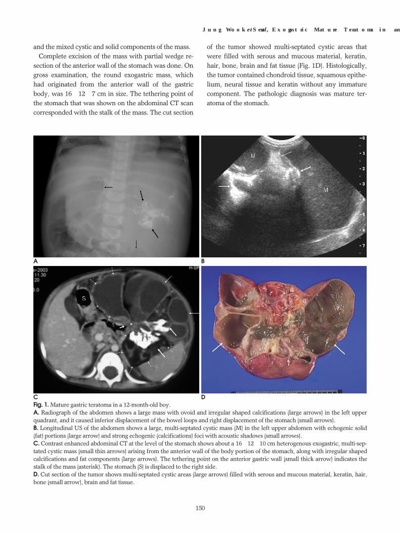
and the mixed cystic and solid components of the mass.Complete excision of the mass with partial wedge re-
section of the anterior wall of the stomach was done. Ongross examination, the round exogastric mass, whichhad originated from the anterior wall of the gastricbody, was 16×12×7 cm in size. The tethering point ofthe stomach that was shown on the abdominal CT scancorresponded with the stalk of the mass. The cut section
of the tumor showed multi-septated cystic areas thatwere filled with serous and mucous material, keratin,hair, bone, brain and fat tissue (Fig. 1D). Histologically,the tumor contained chondroid tissue, squamous epithe-lium, neural tissue and keratin without any immaturecomponent. The pathologic diagnosis was mature ter-atoma of the stomach.
Jung Wook Seo, et al : Exogastric Mature Teratoma in an Infant
─ 150 ─
A B
C DFig. 1. Mature gastric teratoma in a 12-month-old boy.A. Radiograph of the abdomen shows a large mass with ovoid and irregular shaped calcifications (large arrows) in the left upperquadrant, and it caused inferior displacement of the bowel loops and right displacement of the stomach (small arrows).B. Longitudinal US of the abdomen shows a large, multi-septated cystic mass (M) in the left upper abdomen with echogenic solid(fat) portions (large arrow) and strong echogenic (calcifications) foci with acoustic shadows (small arrows).C. Contrast enhanced abdominal CT at the level of the stomach shows about a 16×12×10 cm heterogenous exogastric, multi-sep-tated cystic mass (small thin arrows) arising from the anterior wall of the body portion of the stomach, along with irregular shapedcalcifications and fat components (large arrows). The tethering point on the anterior gastric wall (small thick arrow) indicates thestalk of the mass (asterisk). The stomach (S) is displaced to the right side.D. Cut section of the tumor shows multi-septated cystic areas (large arrows) filled with serous and mucous material, keratin, hair,bone (small arrow), brain and fat tissue.
Discussion
Teratomas may arise from the gonads or from extrago-nadal sites (2). There are various sites of origin for ter-atoma of infancy and childhood. The more commonsites of origin are the sacrococcygeal area (60-65%) andthe gonads (10-20%) and the relative uncommon sitesare the mediastinum (5-10%), the presacral area (5%),the head, neck and the retroperitoneum (<5%) (3). Theincidence of gastric teratoma in infancy and childhood isapproximately 1% of all pediatric teratomas. Since thefirst case of gastric teratoma was reported in 1922, only102 such cases have been reported in the literature (1-3). In the most current literature, 2 cases occurring inpediatric patients (a 3-month-old boy and a neonatemale) with immature gastric teratomas have been re-ported (1, 4, 5), but ours is a case of exogastric matureteratoma that developed in an infant.
Although gastric teratomas can occur at any age, mostcases were reported in infants or neonates (approxi-mately 94%) (2, 6). The most common gender for all gas-tric teratomas is male (approximately 90%) (7). Almostall gastric teratomas have been benign, except for onecase involving malignant transformation that was re-ported by Matsukama et al. (6). Where histological ex-amination indicates that a tumor is immature, malignan-cy may or may not ensue, although neither metastasisnor recurrence has been reported.
Teratoma is different from dermoids in that teratomashows derivatives of all three germ layers, and dermoidsare neoplasms containing derivatives of only two germlayers (7). The etiology of gastric teratoma is not yetknown. The accepted histogenesis of gastric teratomainvolves germ cell theory: extragonadal teratomas origi-nate from migrated totipotential germ cells (6).
The clinical symptoms of gastric teratoma are palpablemass (75% of reported cases) and/or abdominal disten-sion (56%). It causes respiratory distress in infants,whereas it causes epigastic discomfort, hematemesisand/or melena in adults (2, 3). These symptoms were re-lated to the large polypoid, exo/endogastric growth pat-tern and surface ulceration. The tumor can be classifiedby the growth pattern. The most common growth formis exogatric (65%), followed by exo/endogastric (26%)and endogastric (9%) (1). In our case, only an exogastricmass with symptoms of palpable mass and abdominaldistension was disclosed.
Although the plain radiographic findings such as a soft
tissue mass with calcification are nonspecific, it can sug-gest the possibility of teratoma. US and CT are morespecific tools that show not only a heterogeneous masscontaining varying amounts of cystic and solid compo-nents, but they can also show fat and calcification, sug-gesting a diagnosis of gastric teratoma. In our case therewas definite evidence of characteristic findings such asfat and calcification, and also the mixed cystic and solidcomponents of the mass suggested teratoma. Tetheringto the anterior wall of the stomach on CT was a helpfulfinding to evaluate the origin site of teratoma.
When imaging studies show a solid and cystic masswith calcifications in the left upper quadrant in an in-fant, neuroblastoma should be included in the differen-tial diagnosis. Neuroblastoma is a malignant tumor ofprimitive neural crest cells that occurs more often inchildren under 2 years of age. The median age at diagno-sis is 22 months, and more than 95% of cases are diag-nosed by 10 years of age (8). This tumor shows as a het-erogeneously echogenic mass in the suprarenal areawith anechoic areas on US; a large heterogeous solidmass with calcifications (about 80-90%) (9) and low at-tenuation areas of necrosis or hemorrhage are seen onCT (10). It frequently shows uncommon cystic changein neonates, but it tends to be solid even in the olderchild (11).
This gastric teratoma gave us several diagnostic cluesthat were different from neuroblastoma. It was an in-traperitoneal mass that displaced the bowel loops andstomach posteriorly without vascular encasement, andit was a multi-septated cystic mass with irregular largecalcifications and fat components. Based on the imagingfindings, the possibility of other tumors such as Wilms’tumor, hepatoblastoma, rhabdomyosarcoma and li-posarcoma was low (12).
In conclusion, gastric teratoma is an extremely rare,benign tumor in childhood. It most commonly occurs inmale infants as a palpable abdominal mass. Radiologicevaluation can demonstrate the gastric origin of themass and the characteristic findings such as fat, calci-fication and the mixed cystic and solid component of themass, which are all helpful to exclude other masses inan infant from the differential diagnosis.
References
1. Munoz NA, Takehara H, Komi N, Hizawa K. Immature gastric ter-atoma in an infant. Acta Paediatr Japon 1992;34:483-488
2. Joo M, Kang YK, Lee HK, Lee HS, Yum HK, Bang SW, et al.Intrapulmonary and gastric teratoma: report of two cases. J Korean
J Korean Radiol Soc 2006;55:149-152
─ 151 ─

Med Sci 1999;14:330-3343. Shirodkar NP, Chopra PS, Marker M, Murphy KD, Dhamoon A,
Kwon OJ. Conjoined gastric and mediastinal benign cystic ter-atomas: case report of a rare occurrence and a review of the litera-ture. Clin Imaging 1997;21:340-345
4. Yoon SE, Goo HW, Jun S, Lee IC, Yoon CH. Immature gastric ter-atoma in an infant: a case report. Korean J Radiology 2000;1:226-228
5. Gore MD, Fernbach SK. Case 52: gastric teratoma. Radiology 2002;225:497-499
6. Matsukuma S, Wada R, Daibou M, Watanabe N, Kuwabara N,Abe H, et al. Adenocarcinoma arising from gastric immature ter-atoma: report of a case in an adult and a review of the literature,Cancer 1995;75:2663-2668
7. Bowen B, Ros PR, McCarthy MJ, Olmsted WW, Hjermstad BM.
Gastrointestinal teratomas: CT and US appearance with pathologiccorrelation. Radiology 1987;162:431-433
8. Brossard J, Bernstein ML, Lemieux B. Neuroblastoma: an enigmat-ic disease. Br Med Bull 1996;52:787-801
9. Stark DD, Moss AA, Brasch RC, delLorimier AA, Albin AR,London DA, et al. Neuroblastoma: diagnostic imaging and staging.Radiology 1983;148:101-105
10. Lonergan GJ, Schwab CM, Suarez ES, Carlson CL.Neuroblastoma, ganglioneuroblastoma, and ganglioneuroma: radi-ologic-pathologic correlation. Radiographics 2002;22:911-934
11. Bousvaros A, Kirks DR, Grossman H. Imaging of neuroblastoma:an overview. Pediatr Radiol 1986;16:89-106
12. Dunlap JP, James CA, Maxson RT, Bell JM, Wagner CW. Gastricteratoma with intramural extension. Pediatr Radiol 1995;25:383-384
Jung Wook Seo, et al : Exogastric Mature Teratoma in an Infant
─ 152 ─
대한영상의학회지 2006;55:149-152
영아 위장에서 발생한 외위장 성숙 기형종: 증례 보고1
1인제대학교 의과대학 일산백병원 진단방사선과학교실
서정욱·황윤주·김수영·허 감·차순주
위장에서 발생한 기형종은 매우 드문 종양으로, 남아에서 흔하고 양성 성향이 있다. 저자들은 완전 절제 후 조직
검사에서 확진된 12개월 남아의 외위장 성숙 낭성 기형종 1예를 경험하였기에 방사선학적 소견과 조직병리학적 소
견을 함께 보고한다.