exosomes decrease in vitro infectivity of hiv-1 preparations:...
TRANSCRIPT

7
Exosomes Decrease in vitro Infectivity of HIV-1 Preparations: Implication for CD4+T
Lymphocyte Depletion in vivo
Subra Caroline, Burelout Chantal, Proulx Sophie, Simard Sébastien and Gilbert Caroline
Centre de Recherche en Rhumatologie et Immunologie (CRRI), Département de Médecine, Faculté de Médecine, Université Laval, Québec,
Canada
1. Introduction
1.1 Nature and role of exosomes
Since their identification by Johnstone et al (Johnstone et al., 1987), exosomes have gained importance in understanding many biological processes. Exosomes are vesicles expelled by cells into the extracellular milieu. They originate from internal endocytic compartments called multivesicular bodies (MVB) and are released following fusion of MVB with the plasma membrane (Stoorvogel et al., 2002). Numerous cell types, including tumour, foetal, epithelial and haematopoietic cells share the characteristics of releasing exosomes upon activation by cytokines (Abusamra et al., 2005; Ahn and Johnstone, 1993; Altieri, Khan, and Tomasi, 2004; Peche et al., 2006; Segura, Amigorena, and Thery, 2005; Taylor, Akyol, and Gercel-Taylor, 2006; van Niel and Heyman, 2002). Initially associated with the elimination of obsolete proteins during reticulocyte maturation, exosomes are now known to play several roles in intercellular communication (for reviews, (Chaput and Thery, 2010) and (Record et al., 2011)). Based on the presence of various molecules within the vesicle membrane or lumen, it has been proposed that exosomes are particularly involved in regulation of the immune response, for example tolerance induction (Admyre et al., 2006; Frangsmyr et al., 2005; Kapsogeorgou et al., 2005; Karlsson et al., 2001; Kim, Morse, and Choi, 2006; Larregina et al., 2004; Mallegol, van Niel, and Heyman, 2005; Ostman, Taube, and Telemo, 2005; Peche et al., 2003; Peche et al., 2006; Quah and O'Neill, 2005a; Segura, Amigorena, and Thery, 2005; Taylor, Akyol, and Gercel-Taylor, 2006; Van Niel et al., 2003), antigen presentation (Andre et al., 2004; Chaput et al., 2004; Clayton et al., 2003; Kleijmeer et al., 2001; Peche et al., 2003; Raposo et al., 1996; Thery et al., 2002), cancer immunotherapy (Amigorena, 2000; Andre et al., 2001; Mignot et al., 2006; Quah and O'Neill, 2000; Zitvogel et al., 1998), control of receptor expression (Ahn and Johnstone, 1993; Hawari et al., 2004; Levine, 2004), mechanisms involved in cell death (Abusamra et al., 2005; Farsad, 2002; Iero et al., 2008; Lenassi et al., 2010; Zhang et al., 2006) and control of inflammation (Abusamra et al., 2005; Kim et al., 2006; Levine, 2004). Exosomes may also contain functional miRNA (Pegtel et al., 2010) or deliver bioactive lipids (Esser et al., 2010; Subra et al., 2010). Depending on the function and on
www.intechopen.com

Understanding HIV/AIDS Management and Care – Pandemic Approaches in the 21st Century 100
the activation state of the secreting cells, exosomes thus regulate multiple pathways in neighbouring cells in autocrine, paracrine and juxtacrine fashion. The mechanism by which molecules are sorted in exosomes involves a recycling process that is influenced by molecular lateral mobility within lipid domains (de Gassart et al., 2003; de Gassart et al., 2004). MHC-II, co-stimulatory molecules, enzymes (Alonso et al., 2007; Baynes et al., 1991) and heat-shock proteins (HSP) (Lancaster and Febbraio, 2005) are among the proteins associated with exosomes (Segura, Amigorena, and Thery, 2005; Segura et al., 2005; Skokos et al., 2001). Exosomes are similar to retroviruses not only in terms of size but also the molecules they incorporate and their ability to activate immune cells. Exosomes are slightly smaller and more heterogeneous in size (30-100 nm) than HIV-1 particles (100 nm). The most obvious similarity between these two types of particles is the presence of molecules of host origin. For example, incorporation of MHC-I and MHC-II by virions and by exosomes has been described (Cantin, Fortin, and Tremblay, 1996; Cantin, Martin, and Tremblay, 2001; Gansuvd et al., 2003; Raposo et al., 2002; Vincent-Schneider et al., 2002). In addition, several cell-surface molecules such as LFA-1 integrins (CD11a, CD18), co-stimulatory molecules (CD28, CD54) and complement-neutralizing molecules (CD55, CD59) are associated with both particles (Cantin, Methot, and Tremblay, 2005; Nguyen et al., 2003; Thery et al., 2001; Thery et al., 1999). Finally, the buoyant density of exosomes ranges from 1.13 to 1.21 g/l, while that of HIV-1 particles ranges from 1.16 to 1.18 g/l (Thery et al., 2001; Wang et al., 1999). Similar protein and lipid composition as well as buoyant densities render the separation of exosomes from virions quite difficult using standard techniques such as density-gradient centrifugation. These problems prompted us to use an Optiprep™-based velocity gradient method (Cantin et al., 2008), which has allowed us to show clearly that exosomes can be separated completely from viruses, based on detection of exosome marker (acetylcholinesterase) and HIV-1 marker (capsid protein p24). The relationship between exosome biogenesis and retrovirus assembly has not yet been described in satisfactory detail. Although considerable evidence points to the takeover of the intracellular machinery responsible for MVB biogenesis (located at the cytoplasmic membrane) in the case of HIV-1 budding from CD4+ T lymphocytes (CD4TL), virions are found in endosomes of macrophages and dendritic cells (DCs), suggesting an internal budding process (Booth et al., 2006; Gould, Hildreth, and Booth, 2004; Morita and Sundquist, 2004; Nguyen et al., 2003). Comparative studies of exosomes and HIV-1 particle production pathways (Nguyen et al., 2003) based on observations of similar viral budding (Derse et al., 1987) and uptake by cells (Izquierdo-Useros et al., 2010) indicate that retroviruses evolved by exploiting the exosome release pathway. The highly varied exosome composition and content suggest crucial roles for these vesicles in intercellular communication (Thery, Zitvogel, and Amigorena, 2002), transport of genetic material (mRNA or microRNA) (Valadi et al., 2007) and exchange of proteins (Andre et al., 2004; Thery, Zitvogel, and Amigorena, 2002), or in inflammation by carrying bioactive lipids (Esser et al.; Subra et al., 2010). Numerous studies indicate more efficient T cell activation by exosomes released from mature (mDCs) than from immature DCs (iDCs) (Admyre et al., 2006; Chaput and Thery, 2010; Segura, Amigorena, and Thery, 2005). In other studies, an inhibitory role for exosomes in the immune response has been described and particularly in the induction of T cell death via either FasL or galactin-9 by tumour-derived exosomes (Abusamra et al., 2005; Alonso et al., 2007; Chaput and Thery, 2010; Klibi et al., 2009; Ren et al.; Xie et al., 2010) (Andreola et al., 2002; Monleon et al., 2001). Compelling evidence for a
www.intechopen.com

Exosomes Decrease in vitro Infectivity of HIV-1 Preparations: Implication for CD4+T Lymphocyte Depletion in vivo 101
role of the HIV-1 protein Nef (released in association with exosomes) in inducing apoptosis of bystander CD4TL has been published recently (Lenassi et al., 2010). All of these data have led us to examine the involvement of exosomes in CD4TL depletion during HIV-1 infection.
1.2 Rapid depletion of CD4TL during primary infection
HIV-1-caused disease is characterized by a state of chronic immune activation due to
sustained inflammation and immune hyperactivation that persists even under antiretroviral
therapy (HAART) (Imami et al., 2001). Several observations from non-pathogenic simian
immunodeficiency virus (SIV) infection, HIV-1 infected “elite controllers”, “elite suppressors”
or long-term non-progressors reveal a good correlation between low level of activation of
the immune system and absence of clinical signs of AIDS (Bailey et al., 2008; Fontaine et al.,
2011; Milush et al., 2007; Shacklett, 2010; Silvestri et al., 2003). In contrast, strongly increased
immune activation characterized by dysregulated neutrophil and macrophage functions
(Roilides et al., 1990; Torre et al., 2002), polyclonal B cell activation (Aberg et al., 2005),
increased T cell turnover (Aberg et al., 2005), increased numbers of T cells with an activated
phenotype (Aberg et al., 2005) and increased levels of pro-inflammatory molecules are
hallmarks of disease progression in pathogenic infections by primate (HIV/SIV) lentiviruses
(Ascher and Sheppard, 1988), (Giorgi et al., 1999; Liu et al., 1997). More significant is that a
major rapid loss of mucosal CD4TL occurs in the gut-associated lymphoid tissues quite
early in HIV-1 infection (Brenchley, Price, and Douek, 2006; Brenchley et al., 2004;
Mehandru et al., 2004). At this stage, both mucosal lymph node destruction (which initiates
immune dysfunction) and loss of integrity of the gut epithelium allow microbial products to
cross the intestinal barrier. This translocation phenomenon produces high levels of
circulating bacterial lipopolysaccharides (Brenchley, Price, and Douek, 2006) and thus
contributes to the maintenance of the inflammatory state and systemic immune activation
observed in chronic HIV-1-infected patients (Brenchley, Price, and Douek, 2006; Marchetti et
al., 2008). These studies all point to early events in HIV-1 infection as decisive determinants
of the irreversible damage inflicted on immune cells. It is well established that dendritic cells
(DCs) are involved early in HIV-1 transmission (Granelli-Piperno et al., 1998; Manel et al.,
2010; Tsunetsugu-Yokota et al., 1997). It is also known that CD4TL, more particularly the
Th17 mucosal subset (Cecchinato and Franchini, 2010; Cecchinato et al., 2008; Elhed and
Unutmaz, 2010; Favre et al., 2009; Milush et al., 2011; Paiardini, 2010) are dysregulated
(Elbim et al., 2009; Hofman et al., 1999; Okada, Takei, and Tashiro, 1997; Okada, Takei, and
Tashiro, 1998; Pitrak et al., 1996; Roilides et al., 1993; Roilides et al., 1990; Szelc et al., 1992;
Thorsen, Busch-Sorensen, and Sondergaard, 1989) in pathogenic HIV-1/SIV infection
(Brenchley et al., 2008; Elbim et al., 2009; Elbim et al., 2008; Favre et al., 2009).
A rapid decrease thus occurs in the numbers of both infected and uninfected CD4TL within the very first weeks of infection. CD4TL are known to play a pivotal role in orchestrating the immune response as well as in the development, maturation and maintenance of cytotoxic T cells (Matloubian, Concepcion, and Ahmed, 1994; Zajac et al., 1998), the development of a humoral response and B cell antibody class switching (Tsuji et al., 1994), control of the bactericidal activity of macrophages and induction of HIV-1-specific CD4 and CD8 T cell responses. In fact, HIV-1 infection and the specific immune response to it depend largely on CD4TL functionnality and depletion of these cells during primary infection constitutes major interference, perhaps explaining the long-term inability of the host immune response to control the infection. Different mechanisms have been proposed to explain the significant
www.intechopen.com

Understanding HIV/AIDS Management and Care – Pandemic Approaches in the 21st Century 102
depletion of CD4TL in the gut-associated lymphatic tissues. Among these, direct infection of CD4TL by the virus (Arnoult et al., 2003), cytotoxic activity of CD8 T cells against infected cells (Sewell et al., 2000) and cytopathic effects on bystander cells or abortive infection (Doitsh et al., 2010) are the most plausible. However, additional factors, including HIV-1 proteins such as Vpr, Tat, Nef, VpU, proteases and gp120 (Varbanov, Espert, and Biard-Piechaczyk, 2006; Wan and Chen, 2010), mechanisms such as activation-induced cell death (AICD) mediated by Fas, TNF and TRAIL/APO2 (Lichtner et al., 2004) or dysregulation of cytokine/chemokine production (Saelens et al., 2004) can contribute to CD4TL death. Moreover, the detection of Nef in exosomes and the known involvement of this viral protein in apoptosis add support to the potential role of exosomes in bystander cell viability (Lenassi et al., 2010). We therefore propose another mechanism involving the release, from HIV-1-loaded iDCs, of exosomes that can induce functional defects in CD4TL and contribute to their elimination.
1.3 The role of dendritic cells in HIV-1 primary infection
The weakening of the immune system begins soon after the virus enters the body, which it does principally via the mucosal tissues. Following transmission of HIV-1, the virus crosses the mucosal barrier and is met by DCs, which are among the first cells to encounter the virus (Hladik and McElrath, 2008). A major immune system cell type involved in capturing and internalizing HIV-1 is the iDCs, which then migrates principally to the lymph nodes of the gastrointestinal tract, a site of HIV-1 replication during acute infection. Despite the progress that has been made in understanding iDC/HIV-1 interactions as well as virion sequestration and transmission to CD4TL, several fundamental questions surrounding the near total depletion of memory CD4TL observed during the acute infection (Brenchley et al., 2004; Guadalupe et al., 2003; Li et al., 2005; Mattapallil et al., 2005; Mehandru et al., 2004) remain unanswered. It is well known that both cells play a pivotal role in the dissemination of HIV-1, in the establishment of infection and also in anti-HIV-1 immunity. It is now well established that HIV-1 entry into CD4TL is mediated by cellular chemokine receptors such as CCR5 or CXCR4. However, we, along with others, have found that additional factors, such as the DC-SIGN and DCIR lectins can mediate virus attachment to DCs and its subsequent endocytosis (Cambi et al., 2009; Geijtenbeek et al., 2000; Lambert et al., 2008; Permanyer, Ballana, and Este, 2010). Indeed, several recent studies have shown virions concentrated in late endocytic compartments also called multivesicular bodies (MVBs) or MHC class II compartments in mature DCs (Garcia et al., 2005; Izquierdo-Useros et al., 2009; Kwon et al., 2002), where they are sheltered both from the action of antiviral drugs and the immune response. Mature DCs are capable of stocking viral particles and migrating via the lymphatic network to lymph nodes and thus constitute reservoirs of virions. At this stage, it is thought that mDCs can transmit virions to T cells through two sequential routes: an early route known as trans-infection, via passive transfer through late endosomes, or a later route called cis-infection following productive infection (Turville et al., 2004). Fusion of late endosomes with the DC plasma membrane releases large numbers of virions into intercellular space called the virological synapse, which can infect nearby target cells (Moir, Chun, and Fauci, 2010; Piguet and Steinman, 2007). Endosomes contain the intraluminal vesicles that become exosomes when delivered into this space at the same time as the viral particles contained in HIV-1-loaded DCs. Exosomes and virions thus pass via the late endosome across the cell to be exchanged with other cells (Izquierdo-Useros et al., 2010).
www.intechopen.com

Exosomes Decrease in vitro Infectivity of HIV-1 Preparations: Implication for CD4+T Lymphocyte Depletion in vivo 103
After their migration to the lymph nodes, iDCs likely transfer HIV-1 to CD4TL with great efficiency and simultaneously release exosomes. The ability of exosomes to activate CD4TL, thereby enhancing HIV-1 replication, or to induce T cell apoptosis directly, could contribute
Fig. 1. Potential Role of exosomes in HIV-1 infection
www.intechopen.com

Understanding HIV/AIDS Management and Care – Pandemic Approaches in the 21st Century 104
to the massive depletion of CD4TL. Indeed, our preliminary observations show that HIV-1
increases exosome release from iDCs. Some studies have shown that exosomes can activate
T cells and consequently are involved in the regulation of the immune response (Admyre et
al., 2006; Segura et al., 2005; Thery et al., 2002). Furthermore, the Fas-ligand on exosomes can
also induce apoptosis of both CD4 and CD8 T cells (Abusamra et al., 2005; Alonso et al.,
2007; Alonso et al., 2005; Segura, Amigorena, and Thery, 2005). It should be noted that the
capacity of exosomes to regulate the viability of CD4 T cell sub-populations has not yet been
fully investigated in the context of HIV-1 infection. In order to answer this important
fundamental question, new methods are needed for separating mixtures of exosomes and
HIV-1 to purity.
HIV-1 is a retrovirus that causes a slow but sustained depletion of CD4TL during the
chronic stage of infection, leading to progressive failure of the immune system. The
principal immune cell type that captures and internalizes HIV-1 is the iDC. HIV-1-loaded
iDCs migrate to the lymph nodes of the gastrointestinal tract, a major site of HIV-1
replication during acute infection. Given that CD4TL play a pivotal role in the orchestration
of the immune response, the rapid and sustained disappearance of mucosal CD4TL (within
the first 15 days) compromises the development of both the cellular and humoral responses
to HIV-1 infection. Exosomes release by DCs or CD4TL can contribute to elimination of
CD4TL as well as other cell deregulations characterizing this crucial phase.
2. Protocol to study the role of exosomes in CD4TL viability in the context of HIV-1 infection
2.1 Cell purification
Experiments were performed using human primary cells, DCs and CD4TL. Cells were
isolated from peripheral blood mononuclear cells (PBMCs) obtained from anonymous and
healthy volunteer donors. PBMCs were prepared by centrifugation on a lymphocyte
separation medium from Wisent Inc. (St Bruno, QC, Canada). CD14+ cells were then isolated
using a monocyte positive selection kit according to the manufacturer’s instructions
(Stemsep Human CD14 positive selection Kit, STEMCell Technologies, Vancouver, BC,
Canada) using an AutoMacs (Miltenyl Biotech, Auburn, CA, USA) and a previously
established procedure (Bounou et al., 2004; Gilbert et al., 2007a; Gilbert et al., 2007b). CD14+
cells were cultured in six-well plates at a concentration of 1 X 10⁶ cells/ml. To generate iDCs,
purified monocytes were cultured in complete culture medium supplemented every two
days with granulocyte-macrophage colony-stimulating factor (1,000 U/ml) from Genscript
(Cedarlane Laboratories, Burlington, ON, Canada) and IL-4 (200 U/ml) from R&D Systems
(Minneapolis, MN, USA) for 6 to 7 days. Expression of CD3 and CD19 was measured to
assess contamination with T and B cells respectively. Expression of HLA-DR, CD86, DC-
SIGN, CD83 and CD14 was monitored to verify the immature phenotypes of DCs. In the
immature state, DCs express a high level of DC-SIGN and low level of CD83, whereas
mature DCs express CD83 and high levels of ICAM-1, HLA-DR and CD86.
CD4TL were isolated using a negative selection kit according to the manufacturer’s instructions (Stemsep Human CD4 T cell enrichment kit, STEMCell Technologies). In some experiments, these cells were activated with phytohemagglutinin-L (1 µg/ml) to obtain mitogen-stimulated cells and maintained at a density of 2 x 10⁶ cells/ml in RPMI supplemented with IL-2 (30 U/ml) obtained through the AIDS Repository Reagent Program
www.intechopen.com

Exosomes Decrease in vitro Infectivity of HIV-1 Preparations: Implication for CD4+T Lymphocyte Depletion in vivo 105
(Germantown, MD, USA). Experiments were performed with cell preparations that were devoid of contamination (i.e. DC purity > 95%; CD4TL purity > 98%). In all culture media, bovine exosomes from foetal bovine serum (FBS) were eliminated by O/N ultracentrifugation at 100,000xg. Complete RPMI 1640 culture medium contains FBS, penicillin G, streptomycin, glutamine from Wisent and primocin and plasmocin from Invivogen (San Diego, CA, USA).
2.2 Virus production and purification
Virions were produced by transient transfection in human embryonic kidney 293T cells
(HEK293T) as previously described (Cantin et al., 1997). Plasmids used include pJR-CSF (R5-
tropic), pNLAD8 (R5-tropic), pNL4-3balenv (R5-tropic) and pNL4-3 (X4-tropic). The pNL4-
3balenv vector (provided by R. Pomerantz, Thomas Jefferson University, Philadelphia, PA,
USA) was generated by replacing the env gene of the T-tropic HIV-1 strain, NL4-3, with that
of the macrophage-tropic HIV-1 Bal strain, thus resulting in an infectious molecular clone
with R5-tropic properties (Dornadula et al., 1999). Other plasmids were obtained from the
AIDS Repository Reagent Program (Germantown, MD, USA). Several viral preparations
were obtained from primary cells. Peripheral blood from healthy donors was centrifuged on
lymphocyte separation medium (Wisent) to obtain PBMCs. NL4-3Balenv virions were
propagated by acute infection of PBMCs (1.5x10⁷ cells/ml) with virus (500 ng/ml) for six
days.
2.3 Separation of exosomes and virions by velocity gradient
Exosomes, microvesicles and virions contained in cell-free supernatants were passed through a 0.22 µm filter or centrifuged, 10 min at 10,000xg to eliminate micro-particles. Filtered virus and/or exosomes from 293T cells or PBMCs were concentrated by ultracentrifugation in an Optima L-90K Beckman Coulter centrifuge (Fullerton, CA) for 45 min at 31,500 rpm (100,000xg) in a 70 Ti rotor. The pellet containing virions and microvesicles/exosomes was re-suspended in 500 µl of PBS. HIV-1 viral particles were then centrifuged through a 6-18% Optiprep™ (60% iodixanol) separation gradient from Sigma Aldrich® (Winston Park, CA, USA) as previously described (Dettenhofer and Yu, 1999). The densities of each gradient fraction were below 1.13 g/ml, as shown in Figure 2. The virus preparations were then centrifuged using the Optima L-90K centrifuge for 75 min at 52,000 rpm (250,000xg) in a NVT65 rotor. Gradient fractions were collected from the top. Optiprep™ separation medium possesses intrinsic properties such as neutral pH and physiological osmolarity (Dettenhofer and Yu, 1999; Ford, Graham, and Rickwood, 1994; Van Veldhoven, Baumgart, and Mannaerts, 1996) that make it highly suitable for efficient separation of viral entities from cellular debris and microvesicles (Dettenhofer and Yu, 1999; Hermens et al., 1999; Moller-Larsen and Christensen, 1998; Zolotukhin et al., 1999). This technique, identical to the velocity gradient adapted by Dettenhofer (Dettenhofer and Yu, 1999) for HIV-1 separation, is also very efficient for separating microvesicle contaminants from HIV-1 preparations. The results depicted in Figure 3A (black rectangles) express the separation efficiency in terms of quantity of viral p24gag protein, which is concentrated in fraction 15.6. Electron microscopy, performed at the Armand Frappier Institute Microscopy Laboratory using standard protocol (Alain et al., 1987; Hammond et al., 1981), confirmed that fractions 15.6 and 16.8 contain concentrated and homogenous viral particles (1011 per ml). Virion contents were normalized by means of an in-house sensitive double-antibody
www.intechopen.com

Understanding HIV/AIDS Management and Care – Pandemic Approaches in the 21st Century 106
sandwich enzyme-linked immunosorbent assay (ELISA) specific for the viral p24gag protein (Bounou, Leclerc, and Tremblay, 2002). The distribution of fractions containing exosomes along the gradient was evaluated by
measuring acetylcholinesterase (AChE) activity, which is a commonly used specific exosome
marker (Fig. 3A, open rectangles) (Gastpar et al., 2005; Rieu et al., 2000). This enzyme
activity was measured following a procedure previously described (Cantin et al., 2008).
Briefly, 30 µl of standard or sample were mixed with PBS pH 8 containing acetylthiocholine
and PBS pH 7 containing 5,5-dithio-bis-(2-nitrobenzoic acid) to obtain final reagent
concentrations of respectively 1.25 mM and 0.1 mM in a volume of 200 µl and held at room
temperature. Changes in absorption at 450 nm were monitored for 10 min with a plate
reader spectrophotometer (ELX808, BIO-TEK instruments, Winooski, VT, USA).
Glycosylphosphatidylinositol-anchored AChE as well as other GPI proteins are localized in
the MVB and are part of the exosome membrane (Gastpar et al., 2005; Johnstone et al., 1987).
The results illustrated in Figure 3 show that AChE activity is concentrated in fractions 8.4 to
12 and microscopic observations showed that 108 particles/ml are present in these fractions.
To confirm the efficiency of this velocity gradient method for separating HIV-1 from
exosomes, we also checked for the presence of infectious particles in each gradient fraction
(i.e. infectivity on TZM-bl cells (Cantin et al., 2008) or measure of spliced TAT on CD4TL).
Infectivity assays using the TZM-bl indicator cell line showed that fractions 14.4 to 16.8
contained fully infectious virus while fractions 8.4 to 12 contained insignificant amounts.
These results thus clearly indicate that our procedures can isolate exosomes and HIV-1
particles differentially and independently. We are satisfied that fractions 8.4 to 12 of the
Optiprep™ gradients contained exosomes exclusively, while fractions 14.4 to 16.8 contained
infectious viruses.
Fig. 2. Density of gradient fractions
The particularity of velocity gradient separation resides in its capacity to isolate particles of similar density. The density range of 6 to 18 % of Optiprep™ is 1.03 to 1.13 g/l.
www.intechopen.com

Exosomes Decrease in vitro Infectivity of HIV-1 Preparations: Implication for CD4+T Lymphocyte Depletion in vivo 107
2.4 Rapid purification of exosomes
A quick alternative procedure for eliminating exosomes prior to contacting the cells with HIV-1 preparation consisted of immuno-depleting AChE-bearing vesicles directly from 100,000xg pellet (starting material) as shown in Figure 3B. Briefly, ultracentrifuged cells supernatants were diluted in PBS and incubated with protein A/G beads pre-coated with anti-AChE (AE-1 from ATCC) or with isotypic control antibodies (IgG1). The beads (exosomes) and final supernatants (infectious viruses without exosomes) were kept for viral Titer determination (data not shown) and AChE assay (Fig. 3B). The results show that 95% of the AChE activity was recovered using the anti-AChE beads after the first round of precipitation. Exosomes can be eluted from the beads in two minutes using 50 µl of 0.2M glycine in 0.1M KH2PO4 pH 3. Acidic pH was then neutralized by adding 100 µl of 0.1M K2HPO4 (pH 8.8). It should be noted that this treatment does not affect AChE activity.
2.5 Analysis of protein contained in each gradient fraction
Several proteins including LFA-1 are known to be present on both particles and were quantified in exosome/HIV-1 preparations by means of an in-house enzymatic sandwich-type immunoassay. Plates were initially coated with anti-LFA-1 (MEM-25) (50 µg/ml) in carbonate buffer. After three washes with PBS/0.1% Tween 20, the non-specific sites were blocked with PBS/0.1% Tween 20/1% BSA for 1 hr at RT. The plates were washed and 75 µl of each gradient fraction mixed with 0.5% Triton X-100 to cause lysis were added and the plates were then held overnight at 4°C. The plates were then washed three times and biotinylated anti-LFA-1 (TSI/22) (0.5 µg/ml) was added in blocking solution for 1 hr at RT. After three more washings, streptavidin-peroxidase conjugate was added for 30 min at RT. The plates were washed and the detection was performed by the addition of the TMB-S substrate followed by the addition of H3PO4. Absorbance at 450 nm was read to quantify LFA-1 in each fraction. Figure 3C shows that LFA-1 is present on both vesicles (exosomes and fully infectious virions) and also in uncharacterized particles from fractions 13.2 and 14.4. Both methods (Optiprep™ gradient and immunodepletion) were used to purify exosomes. These methods were employed to eliminate exosomes from all viral or mock preparations before contact with cells and results depicted in Figure 3D show that depletion of exosomes from the HIV-1 preparation increased cell viability.
2.6 Electron microscopy procedure and examination of negative-stain specimens The protocol described by Hammond et al. and Alain et al was performed with each analytical fraction (Alain et al., 1987; Hammond et al., 1981). Briefly, microvesicle preparation was fixed with an equal volume of 2% paraformaldehyde and 50–200 µL in a micro-ultracentrifuge tube with a formvar-coated electron microscopy grid at the bottom were concentrated for 5 minutes at 120,000xg (20 psig) using an Airfuge ultracentrifuge (Beckman, Palo Alto, CA, USA). The grids were dried on bibulous paper and stained for 1 min with a drop of 3% phosphotungstic acid (pH 6.0). The concentration, shape and overall appearance of the microvesicles were examined with a Hitachi 7100 (Hitachi, Japan) transmission electron microscope (see Fig. 3A).
2.7 Virus infection assays
Indicator cell line TZM-bl, which carries a stably integrated luciferase reporter gene under the control of the HIV-1 regulatory element known as the long terminal repeat (Thibault et
www.intechopen.com

Understanding HIV/AIDS Management and Care – Pandemic Approaches in the 21st Century 108
Fig. 3. Exosome and virus purification The upper panel illustrates the purification protocols based on Optiprep™ velocity gradient and on immunocapture. Panel A shows velocity gradient purification of NL4-3balenv virions produced on HEK293T cells by transient transfection. Gradients were overlaid with 100,000xg centrifugal pellet. Viral protein p24gag (black rectangles) was measured by ELISA. AChE activity (x106 OD/min, open rectangles) was evaluated in each fraction by colorimetry. Each fraction was examined by electron microscopy for particle quality and quantity. Panel B represents the results obtained by immunocapture with AChE. Panel C shows the presence of LFA-1 in each gradient fraction. Finally, panel D shows that depletion of exosomes increased cell viability. These results show that we are able to separate exosomes from HIV-1 particles.
www.intechopen.com
Exosomes Decrease in vitro Infectivity of HIV-1 Preparations: Implication for CD4+T Lymphocyte Depletion in vivo 109
al., 2007; Wei et al., 2002; Zhao et al., 2005), was used to quantify infectious viral particles
after exosomes depletion. Each well contained 1.5 x 10⁴ cells plus 100 µl of fraction in a final
volume of 200 µl. Plates were incubated for 48 hrs. All experimental points were done in
triplicate. Luciferase activity was determined following a modified version of a known
protocol (Barbeau et al., 1997; Berube et al., 1996). Briefly, cell-free supernatant (100 µl)
withdrawn from each well and mixed with solution containing 25 mM Tris phosphate pH
7.8, 2 mM dithiothreitol, 1% Triton X-100 and 10% glycerol (25 µl) to cause vesicle lysis was
held at room temperature for 30 min. A 20-µl aliquot of this mixture was then mixed with
100 µl of luciferase assay buffer (20 mM Tricine, 1.07 mM (MgCO3)4·Mg(OH)2·5 H2O,
2.67 mM MgSO4, 0.1 mM EDTA, 270 µM coenzyme A, 470 µM luciferin, 530 µM ATP and
33.3 mM dithiothreitol) and the luciferase reaction was monitored on a Dynex MLX
microplate luminometer for 20s/well after a 2-5s delay.
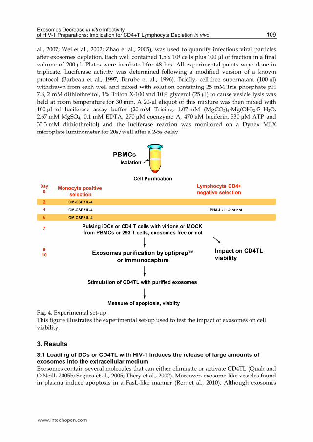
Fig. 4. Experimental set-up This figure illustrates the experimental set-up used to test the impact of exosomes on cell viability.
3. Results
3.1 Loading of DCs or CD4TL with HIV-1 induces the release of large amounts of exosomes into the extracellular medium
Exosomes contain several molecules that can either eliminate or activate CD4TL (Quah and O'Neill, 2005b; Segura et al., 2005; Thery et al., 2002). Moreover, exosome-like vesicles found in plasma induce apoptosis in a FasL-like manner (Ren et al., 2010). Although exosomes
www.intechopen.com

Understanding HIV/AIDS Management and Care – Pandemic Approaches in the 21st Century 110
release by DCs have been studied extensively (Chaput et al., 2006; Izquierdo-Useros et al., 2009; Thery et al., 2001; Thery et al., 1999), the release mechanism and the nature of the exosomes produced by HIV-1-loaded cells (DCs or CD4TL) have not been thoroughly investigated. To begin to answer this question, the experimental set up proposed in Figure 4 and methods presented in Figure 3 were used. DCs and CD4TL pulsed with NL4-3balenv and washed several times were cultured for respectively 2 or 5 days. Exosomes and virions were isolated initially by differential centrifugation and exosome levels were determined by measuring exosomal AChE activity (Cantin et al., 2008). These results, presented in Figure 5, confirmed higher levels of exosomes secreted by iDCs and CD4TL pulsed with purified HIV-1 (1.4 fold and 1.9 respectively, Fig. 5A, B). Using Optiprep™ velocity gradients to separate exosomes and HIV-1, we processed the pellet obtained following sedimentation centrifugation. As
Fig. 5. Exosomes released by DCs and CD4TL after pulsing with HIV-1 I DCs (A, C) or CD4TL (B, D) were incubated for 2h with exosome-free NL4-3balenv HIV-1 virus or mock preparation and cultured for an additional 72h. Cell-free supernatants were obtained by centrifugation and exosomes in the pellets were quantified by measuring AChE activity (x106 DO/min). Exosomes were then separated from HIV-1 on an Optiprep gradient and the exosome content (based on AchE activity) of each fraction was determined. Data are representative of five independent donors. These results show that HIV-1 induced exosome release (in fractions 9.6 through 12) by both cell types (mean increases of 1.4-fold for DCs and 1.9-fold for CD4TL, based on at least 5 independent experiments).
www.intechopen.com

Exosomes Decrease in vitro Infectivity of HIV-1 Preparations: Implication for CD4+T Lymphocyte Depletion in vivo 111
expected, exosomes were concentrated in iodixanol fractions 8.4-12.0% on the Optiprep™ gradient (Fig. 5C, D). Large amounts of exosomes produced by HIV-1-loaded cells accumulated in fractions starting at 9.6% iodixanol (in comparison to the control condition, open bar). The velocity method thus allows efficient separation of exosomes which accumulate in iodixanol fractions 8.4 to 12 % as illustrated in Figure 3. Immature DCs release exosomes and are highly relevant to HIV-1 primary infection since they are involved in the capture of HIV-1 in mucosal tissues and play a crucial role in the subsequent transmission of the virus to CD4TL in the lymph nodes (Gilbert et al., 2007a; Gilbert et al., 2007b; Turville et al., 2004) as illustrated in Figure 1.
3.2 Impact of exosome depletion on CD4TL p24 production and infectivity
The large increase in exosome release by HIV-1-pulsed cells, combined with the results showing that exosomes from these cells induced apoptosis, is particularly relevant in the context of HIV-1 infection for several reasons. These results could explain in part the severe depletion of mucosal CD4TL, a cell type very susceptible to HIV-1. In addition, these cells play a pivotal role in orchestrating immune response and their decline during the early phase of infection undoubtedly delays the specific response to HIV-1. Furthermore, most laboratory preparations of HIV-1 contain exosomes, which may explain in vitro observations such as cytokine release, apoptosis, atypical gene expression, infectivity and so on. Since it became clear that exosomes play a major role in several aspects of the immune response to HIV-1, we sought to evaluate their impact on HIV-1 p24 production and infectivity. NL4-3balenv produced by transfection of 293T cells was made free of exosomes by immunocapture with anti-AE-1. Activated CD4TL were pre-incubated for 2h at 37°C with either exosome-free or exosome-containing HIV-1 preparation washed and then incubated for up to 5 days. ELISA was used to determine viral protein p24 in culture supernatants. Figure 6A shows that infection in the presence of exosomes is transient and less efficient
Fig. 6. Impact of exosomes on p24 production and HIV-1 infectivity Panel A) P24 production was evaluated in the supernatants of CD4TL cultured for up to 5 days after pulsing with NL4-3balenv preparation either free of exosomes (Balenv AE-1) or not (Balenv IgG1). Panel B) TZM-bl cells were incubated with several dilutions of HIV-1 preparation either immunodepleted (Balenv AE-1) or not (Balenv IgG1) and maintained in culture for 48 hrs before lysis. Results are representative of two independent experiments.
www.intechopen.com

Understanding HIV/AIDS Management and Care – Pandemic Approaches in the 21st Century 112
than in their absence (solid bar). To evaluate virion infectivity, indicator cell line TZM-bl was incubated with several dilutions of HIV-1 preparation either exosome-depleted or not. Panel B of Figure 6 shows that the exosome-depleted preparation is more infectious than the non-depleted preparation. All these results provide additional evidence that exosomes derived from HIV-1-pulsed cells influence cell viability (figure 3D) and indirectly p24 production and infectivity. They suggest that the presence of exosomes in culture supernatants of HIV-1-stimulated cells should be considered in all laboratory experiments with HIV-1.
4. Conclusion
Exosome biogenesis and the HIV-1 virion assembly pathway converge in a common intracellular compartment. Moreover, both types of vesicle can be released during the trans-infection process in DCs (Izquierdo-Useros et al., 2009). However, exosome secretion in the context of HIV-1 infection has not been properly investigated, due primarily to lack of effective methods of separating the two types of vesicles. Their separation using antibodies directed against specific membrane antigens is often suboptimal since exosomes and HIV-1 display approximately the same antigen expression pattern in addition to several other surface molecules. This is why flow cytometry, ELISA or bead capture techniques based on specific markers are not sufficiently discriminating for the separation of exosomes, extraneous micro-particles and HIV-1. Alternatively, immunocapture with anti-CD45 (Chertova et al., 2006; Trubey et al., 2003), used to separate only micro-particles derived from leucocyte plasma membranes, does not eliminate exosomes originating from the endosomal membrane and cannot be used to separate exosomes from HIV-1. We have shown that AChE appears essentially excluded from the HIV-1 fraction, since the major portion of its activity is recovered in the early Optiprep™ fractions (8.4 to 12), in which no virus is detected (Cantin et al., 2008). Based on this observation, depletion of 100,000xg pellets with protein-A/G-bound anti-AChE on agarose beads appears to provide excellent means of rapidly purifying virions or capturing exosomes (Cantin et al., 2008). Using these methods, we have observed that HIV-1 contact with DCs or CD4TL enhances extracellular exosome release and that these exosomes can affect the viability of nearby cells such as CD4TL. These results are in agreement with observations concerning the pro-apoptotic role of Nef accessory proteins. Using Optiprep™ gradients, recent work has shown that the viral protein Nef is enclosed in exosomes, conferring to it the capacity to trigger apoptosis of uninfected bystander T cells (Lenassi et al., 2010). In summary, the results of the present study show that relatively simple methods of purifying both exosomes and HIV-1 contained in the same cell supernatant are now available. Achieving very highly purified exosomes from HIV-1 preparations is a definite advantage in studying the respective roles of both vesicles as well as the links between them. These methods could also provide the opportunity for specific isolation of exosomes secreted by a variety of cell types and could prove useful in experiments that require highly purified exosome preparations to study their roles in various biological processes. Indeed, these purification steps are crucial in studies involving mixtures of exosomes and HIV-1 (or for that matter, other retroviruses) as starting material. We may anticipate that these methods will constitute a significant contribution to the use of exosomes for vaccination or gene therapy. In addition, we strongly believe that an improved and standardized method of exosome purification should lead to more comparable results among different
www.intechopen.com

Exosomes Decrease in vitro Infectivity of HIV-1 Preparations: Implication for CD4+T Lymphocyte Depletion in vivo 113
laboratories and lessen discrepancies such as those seen among several studies in recent years as well as facilitate the interpretation of new results to be published in this subject area.
5. Acknowledgments
The authors wish to thank Dr Lahlou Hadji for his technical assistance in editing this manuscript and for constructive comments. We also thank Dr. Stephen Davids for proofreading. We are also grateful to M. Robert Alain for his assistance with the electron microscopy analyses and to Odette Simard for excellent technical contributions. This work was supported by start-up funds to CG from the Centre Hospitalier Universitaire de Québec and an operating grant to CG from the Canadian Institutes of Health Research (CIHR) (MOP-188726). CG is recipient of a level-1 junior researcher award from the Fonds de la Recherche en Santé du Québec and a New Investigator Award from CIHR. C.S. is the recipient of a fellowship award from the Canadian Institutes of Health Research HIV/AIDS Research Program.
6. References
Aberg, J. A., Zackin, R. A., Brobst, S. W., Evans, S. R., Alston, B. L., Henry, W. K., Glesby, M. J., Torriani, F. J., Yang, Y., Owens, S. I., and Fichtenbaum, C. J. (2005). A randomized trial of the efficacy and safety of fenofibrate versus pravastatin in HIV-infected subjects with lipid abnormalities: AIDS Clinical Trials Group Study 5087. AIDS Res Hum Retroviruses 21(9), 757-67.
Abusamra, A. J., Zhong, Z., Zheng, X., Li, M., Ichim, T. E., Chin, J. L., and Min, W. P. (2005). Tumor exosomes expressing Fas ligand mediate CD8+ T-cell apoptosis. Blood Cells Mol Dis 35(2), 169-73.
Admyre, C., Johansson, S. M., Paulie, S., and Gabrielsson, S. (2006). Direct exosome stimulation of peripheral human T cells detected by ELISPOT. Eur J Immunol 36(7), 1772-81.
Ahn, J., and Johnstone, R. M. (1993). Origin of a soluble truncated transferrin receptor. Blood 81(9), 2442-51.
Alain, R., Nadon, F., Seguin, C., Payment, P., and Trudel, M. (1987). Rapid virus subunit visualization by direct sedimentation of samples on electron microscope grids. J Virol Methods 16(3), 209-16.
Alonso, R., Mazzeo, C., Merida, I., and Izquierdo, M. (2007). A new role of diacylglycerol kinase alpha on the secretion of lethal exosomes bearing Fas ligand during activation-induced cell death of T lymphocytes. Biochimie 89(2), 213-21.
Alonso, R., Rodriguez, M. C., Pindado, J., Merino, E., Merida, I., and Izquierdo, M. (2005). Diacylglycerol kinase alpha regulates the secretion of lethal exosomes bearing Fas ligand during activation-induced cell death of T lymphocytes. J Biol Chem 280(31), 28439-50.
Altieri, S. L., Khan, A. N., and Tomasi, T. B. (2004). Exosomes from plasmacytoma cells as a tumor vaccine. J Immunother 27(4), 282-8.
Amigorena, S. (2000). Cancer immunotherapy using dendritic cell-derived exosomes. Medicina (B Aires) 60 Suppl 2, 51-4.
www.intechopen.com

Understanding HIV/AIDS Management and Care – Pandemic Approaches in the 21st Century 114
Andre, F., Andersen, M., Wolfers, J., Lozier, A., Raposo, G., Serra, V., Ruegg, C., Flament, C., Angevin, E., Amigorena, S., and Zitvogel, L. (2001). Exosomes in cancer immunotherapy: preclinical data. Adv Exp Med Biol 495, 349-54.
Andre, F., Chaput, N., Schartz, N. E., Flament, C., Aubert, N., Bernard, J., Lemonnier, F., Raposo, G., Escudier, B., Hsu, D. H., Tursz, T., Amigorena, S., Angevin, E., and Zitvogel, L. (2004). Exosomes as potent cell-free peptide-based vaccine. I. Dendritic cell-derived exosomes transfer functional MHC class I/peptide complexes to dendritic cells. J Immunol 172(4), 2126-36.
Andreola, G., Rivoltini, L., Castelli, C., Huber, V., Perego, P., Deho, P., Squarcina, P., Accornero, P., Lozupone, F., Lugini, L., Stringaro, A., Molinari, A., Arancia, G., Gentile, M., Parmiani, G., and Fais, S. (2002). Induction of lymphocyte apoptosis by tumor cell secretion of FasL-bearing microvesicles. J Exp Med 195(10), 1303-16.
Arnoult, D., Petit, F., Lelievre, J. D., and Estaquier, J. (2003). Mitochondria in HIV-1-induced apoptosis. Biochem Biophys Res Commun 304(3), 561-74.
Ascher, M. S., and Sheppard, H. W. (1988). AIDS as immune system activation: a model for pathogenesis. Clin Exp Immunol 73(2), 165-7.
Bailey, J. R., O'Connell, K., Yang, H. C., Han, Y., Xu, J., Jilek, B., Williams, T. M., Ray, S. C., Siliciano, R. F., and Blankson, J. N. (2008). Transmission of human immunodeficiency virus type 1 from a patient who developed AIDS to an elite suppressor. J Virol 82(15), 7395-410.
Barbeau, B., Bernier, R., Dumais, N., Briand, G., Olivier, M., Faure, R., Posner, B. I., and Tremblay, M. (1997). Activation of HIV-1 long terminal repeat transcription and virus replication via NF-kappaB-dependent and -independent pathways by potent phosphotyrosine phosphatase inhibitors, the peroxovanadium compounds. J Biol Chem 272(20), 12968-77.
Baynes, R. D., Shih, Y. J., Hudson, B. G., and Cook, J. D. (1991). Characterization of transferrin receptor released by K562 erythroleukemia cells. Proc Soc Exp Biol Med 197(4), 416-23.
Berube, P., Barbeau, B., Cantin, R., Sekaly, R. P., and Tremblay, M. (1996). Repression of human immunodeficiency virus type 1 long terminal repeat-driven gene expression by binding of the virus to its primary cellular receptor, the CD4 molecule. J Virol 70(6), 4009-16.
Booth, A. M., Fang, Y., Fallon, J. K., Yang, J. M., Hildreth, J. E., and Gould, S. J. (2006). Exosomes and HIV Gag bud from endosome-like domains of the T cell plasma membrane. J Cell Biol 172(6), 923-35.
Bounou, S., Giguere, J. F., Cantin, R., Gilbert, C., Imbeault, M., Martin, G., and Tremblay, M. J. (2004). The importance of virus-associated host ICAM-1 in human immunodeficiency virus type 1 dissemination depends on the cellular context. Faseb J 18(11), 1294-6.
Bounou, S., Leclerc, J. E., and Tremblay, M. J. (2002). Presence of host ICAM-1 in laboratory and clinical strains of human immunodeficiency virus type 1 increases virus infectivity and CD4(+)-T-cell depletion in human lymphoid tissue, a major site of replication in vivo. J Virol 76(3), 1004-14.
Brenchley, J. M., Paiardini, M., Knox, K. S., Asher, A. I., Cervasi, B., Asher, T. E., Scheinberg, P., Price, D. A., Hage, C. A., Kholi, L. M., Khoruts, A., Frank, I., Else, J., Schacker, T.,
www.intechopen.com

Exosomes Decrease in vitro Infectivity of HIV-1 Preparations: Implication for CD4+T Lymphocyte Depletion in vivo 115
Silvestri, G., and Douek, D. C. (2008). Differential Th17 CD4 T-cell depletion in pathogenic and nonpathogenic lentiviral infections. Blood 112(7), 2826-35.
Brenchley, J. M., Price, D. A., and Douek, D. C. (2006). HIV disease: fallout from a mucosal catastrophe? Nat Immunol 7(3), 235-9.
Brenchley, J. M., Schacker, T. W., Ruff, L. E., Price, D. A., Taylor, J. H., Beilman, G. J., Nguyen, P. L., Khoruts, A., Larson, M., Haase, A. T., and Douek, D. C. (2004). CD4+ T cell depletion during all stages of HIV disease occurs predominantly in the gastrointestinal tract. J Exp Med 200(6), 749-59.
Cambi, A., Beeren, I., Joosten, B., Fransen, J. A., and Figdor, C. G. (2009). The C-type lectin DC-SIGN internalizes soluble antigens and HIV-1 virions via a clathrin-dependent mechanism. Eur J Immunol 39(7), 1923-8.
Cantin, R., Diou, J., Belanger, D., Tremblay, A. M., and Gilbert, C. (2008). Discrimination between exosomes and HIV-1: purification of both vesicles from cell-free supernatants. J Immunol Methods 338(1-2), 21-30.
Cantin, R., Fortin, J. F., Lamontagne, G., and Tremblay, M. (1997). The acquisition of host-derived major histocompatibility complex class II glycoproteins by human immunodeficiency virus type 1 accelerates the process of virus entry and infection in human T-lymphoid cells. Blood 90(3), 1091-100.
Cantin, R., Fortin, J. F., and Tremblay, M. (1996). The amount of host HLA-DR proteins acquired by HIV-1 is virus strain- and cell type-specific. Virology 218(2), 372-81.
Cantin, R., Martin, G., and Tremblay, M. J. (2001). A novel virus capture assay reveals a differential acquisition of host HLA-DR by clinical isolates of human immunodeficiency virus type 1 expanded in primary human cells depending on the nature of producing cells and the donor source. J Gen Virol 82(Pt 12), 2979-87.
Cantin, R., Methot, S., and Tremblay, M. J. (2005). Plunder and stowaways: incorporation of cellular proteins by enveloped viruses. J Virol 79(11), 6577-87.
Cecchinato, V., and Franchini, G. (2010). Th17 cells in pathogenic simian immunodeficiency virus infection of macaques. Curr Opin HIV AIDS 5(2), 141-5.
Cecchinato, V., Trindade, C. J., Laurence, A., Heraud, J. M., Brenchley, J. M., Ferrari, M. G., Zaffiri, L., Tryniszewska, E., Tsai, W. P., Vaccari, M., Parks, R. W., Venzon, D., Douek, D. C., O'Shea, J. J., and Franchini, G. (2008). Altered balance between Th17 and Th1 cells at mucosal sites predicts AIDS progression in simian immunodeficiency virus-infected macaques. Mucosal Immunol 1(4), 279-88.
Chaput, N., Flament, C., Viaud, S., Taieb, J., Roux, S., Spatz, A., Andre, F., LePecq, J. B., Boussac, M., Garin, J., Amigorena, S., Thery, C., and Zitvogel, L. (2006). Dendritic cell derived-exosomes: biology and clinical implementations. J Leukoc Biol 80(3), 471-8.
Chaput, N., Taieb, J., Schartz, N. E., Andre, F., Angevin, E., and Zitvogel, L. (2004). Exosome-based immunotherapy. Cancer Immunol Immunother 53(3), 234-9.
Chaput, N., and Thery, C. (2010). Exosomes: immune properties and potential clinical implementations. Semin Immunopathol.
Chertova, E., Chertov, O., Coren, L. V., Roser, J. D., Trubey, C. M., Bess, J. W., Jr., Sowder, R. C., 2nd, Barsov, E., Hood, B. L., Fisher, R. J., Nagashima, K., Conrads, T. P., Veenstra, T. D., Lifson, J. D., and Ott, D. E. (2006). Proteomic and biochemical analysis of purified human immunodeficiency virus type 1 produced from infected monocyte-derived macrophages. J Virol 80(18), 9039-52.
www.intechopen.com

Understanding HIV/AIDS Management and Care – Pandemic Approaches in the 21st Century 116
Clayton, A., Harris, C. L., Court, J., Mason, M. D., and Morgan, B. P. (2003). Antigen-presenting cell exosomes are protected from complement-mediated lysis by expression of CD55 and CD59. Eur J Immunol 33(2), 522-31.
de Gassart, A., Geminard, C., Fevrier, B., Raposo, G., and Vidal, M. (2003). Lipid raft-associated protein sorting in exosomes. Blood 102(13), 4336-44.
de Gassart, A., Geminard, C., Hoekstra, D., and Vidal, M. (2004). Exosome secretion: the art of reutilizing nonrecycled proteins? Traffic 5(11), 896-903.
Derse, D., Dorn, P. L., Levy, L., Stephens, R. M., Rice, N. R., and Casey, J. W. (1987). Characterization of equine infectious anemia virus long terminal repeat. J Virol 61(3), 743-7.
Dettenhofer, M., and Yu, X. F. (1999). Highly purified human immunodeficiency virus type 1 reveals a virtual absence of Vif in virions. J Virol 73(2), 1460-7.
Doitsh, G., Cavrois, M., Lassen, K. G., Zepeda, O., Yang, Z., Santiago, M. L., Hebbeler, A. M., and Greene, W. C. (2010). Abortive HIV infection mediates CD4 T cell depletion and inflammation in human lymphoid tissue. Cell 143(5), 789-801.
Dornadula, G., Zhang, H., Shetty, S., and Pomerantz, R. J. (1999). HIV-1 virions produced from replicating peripheral blood lymphocytes are more infectious than those from nonproliferating macrophages due to higher levels of intravirion reverse transcripts: implications for pathogenesis and transmission. Virology 253(1), 10-6.
Elbim, C., Monceaux, V., Francois, S., Hurtrel, B., Gougerot-Pocidalo, M. A., and Estaquier, J. (2009). Increased neutrophil apoptosis in chronically SIV-infected macaques. Retrovirology 6, 29.
Elbim, C., Monceaux, V., Mueller, Y. M., Lewis, M. G., Francois, S., Diop, O., Akarid, K., Hurtrel, B., Gougerot-Pocidalo, M. A., Levy, Y., Katsikis, P. D., and Estaquier, J. (2008). Early divergence in neutrophil apoptosis between pathogenic and nonpathogenic simian immunodeficiency virus infections of nonhuman primates. J Immunol 181(12), 8613-23.
Elhed, A., and Unutmaz, D. (2010). Th17 cells and HIV infection. Curr Opin HIV AIDS 5(2), 146-50.
Esser, J., Gehrmann, U., D'Alexandri, F. L., Hidalgo-Estevez, A. M., Wheelock, C. E., Scheynius, A., Gabrielsson, S., and Radmark, O. Exosomes from human macrophages and dendritic cells contain enzymes for leukotriene biosynthesis and promote granulocyte migration. J Allergy Clin Immunol 126(5), 1032-40, 1040 e1-4.
Esser, J., Gehrmann, U., D'Alexandri, F. L., Hidalgo-Estevez, A. M., Wheelock, C. E., Scheynius, A., Gabrielsson, S., and Radmark, O. (2010). Exosomes from human macrophages and dendritic cells contain enzymes for leukotriene biosynthesis and promote granulocyte migration. J Allergy Clin Immunol 126(5), 1032-40, 1040 e1-4.
Farsad, K. (2002). Exosomes: novel organelles implicated in immunomodulation and apoptosis. Yale J Biol Med 75(2), 95-101.
Favre, D., Lederer, S., Kanwar, B., Ma, Z. M., Proll, S., Kasakow, Z., Mold, J., Swainson, L., Barbour, J. D., Baskin, C. R., Palermo, R., Pandrea, I., Miller, C. J., Katze, M. G., and McCune, J. M. (2009). Critical loss of the balance between Th17 and T regulatory cell populations in pathogenic SIV infection. PLoS Pathog 5(2), e1000295.
Fontaine, J., Chagnon-Choquet, J., Valcke, H. S., Poudrier, J., and Roger, M. (2011). High expression levels of B lymphocyte stimulator (BLyS) by dendritic cells correlate with HIV-related B-cell disease progression in humans. Blood 117(1), 145-55.
www.intechopen.com

Exosomes Decrease in vitro Infectivity of HIV-1 Preparations: Implication for CD4+T Lymphocyte Depletion in vivo 117
Ford, T., Graham, J., and Rickwood, D. (1994). Iodixanol: a nonionic iso-osmotic centrifugation medium for the formation of self-generated gradients. Anal Biochem 220(2), 360-6.
Frangsmyr, L., Baranov, V., Nagaeva, O., Stendahl, U., Kjellberg, L., and Mincheva-Nilsson, L. (2005). Cytoplasmic microvesicular form of Fas ligand in human early placenta: switching the tissue immune privilege hypothesis from cellular to vesicular level. Mol Hum Reprod 11(1), 35-41.
Gansuvd, B., Hagihara, M., Higuchi, A., Ueda, Y., Tazume, K., Tsuchiya, T., Munkhtuvshin, N., Kato, S., and Hotta, T. (2003). Umbilical cord blood dendritic cells are a rich source of soluble HLA-DR: synergistic effect of exosomes and dendritic cells on autologous or allogeneic T-Cell proliferation. Hum Immunol 64(4), 427-39.
Garcia, E., Pion, M., Pelchen-Matthews, A., Collinson, L., Arrighi, J. F., Blot, G., Leuba, F., Escola, J. M., Demaurex, N., Marsh, M., and Piguet, V. (2005). HIV-1 trafficking to the dendritic cell-T-cell infectious synapse uses a pathway of tetraspanin sorting to the immunological synapse. Traffic 6(6), 488-501.
Gastpar, R., Gehrmann, M., Bausero, M. A., Asea, A., Gross, C., Schroeder, J. A., and Multhoff, G. (2005). Heat shock protein 70 surface-positive tumor exosomes stimulate migratory and cytolytic activity of natural killer cells. Cancer Res 65(12), 5238-47.
Geijtenbeek, T. B., Kwon, D. S., Torensma, R., van Vliet, S. J., van Duijnhoven, G. C., Middel, J., Cornelissen, I. L., Nottet, H. S., KewalRamani, V. N., Littman, D. R., Figdor, C. G., and van Kooyk, Y. (2000). DC-SIGN, a dendritic cell-specific HIV-1-binding protein that enhances trans-infection of T cells. Cell 100(5), 587-97.
Gilbert, C., Barat, C., Cantin, R., and Tremblay, M. J. (2007a). Involvement of Src and Syk Tyrosine Kinases in HIV-1 Transfer from Dendritic Cells to CD4+ T Lymphocytes. J Immunol 178(5), 2862-2871.
Gilbert, C., Cantin, R., Barat, C., and Tremblay, M. J. (2007b). Human Immunodeficiency Virus Type 1 Replication in Dendritic Cell-T-Cell Cocultures Is Increased upon Incorporation of Host LFA-1 due to Higher Levels of Virus Production in Immature Dendritic Cells. J Virol 81(14), 7672-82.
Giorgi, J. V., Hultin, L. E., McKeating, J. A., Johnson, T. D., Owens, B., Jacobson, L. P., Shih, R., Lewis, J., Wiley, D. J., Phair, J. P., Wolinsky, S. M., and Detels, R. (1999). Shorter survival in advanced human immunodeficiency virus type 1 infection is more closely associated with T lymphocyte activation than with plasma virus burden or virus chemokine coreceptor usage. J Infect Dis 179(4), 859-70.
Gould, S. J., Hildreth, J. E., and Booth, A. M. (2004). The evolution of alloimmunity and the genesis of adaptive immunity. Q Rev Biol 79(4), 359-82.
Granelli-Piperno, A., Delgado, E., Finkel, V., Paxton, W., and Steinman, R. M. (1998). Immature dendritic cells selectively replicate macrophagetropic (M-tropic) human immunodeficiency virus type 1, while mature cells efficiently transmit both M- and T-tropic virus to T cells. J Virol 72(4), 2733-7.
Guadalupe, M., Reay, E., Sankaran, S., Prindiville, T., Flamm, J., McNeil, A., and Dandekar, S. (2003). Severe CD4+ T-cell depletion in gut lymphoid tissue during primary human immunodeficiency virus type 1 infection and substantial delay in restoration following highly active antiretroviral therapy. J Virol 77(21), 11708-17.
www.intechopen.com
Understanding HIV/AIDS Management and Care – Pandemic Approaches in the 21st Century 118
Hammond, G. W., Hazelton, P. R., Chuang, I., and Klisko, B. (1981). Improved detection of viruses by electron microscopy after direct ultracentrifuge preparation of specimens. J Clin Microbiol 14(2), 210-21.
Hawari, F. I., Rouhani, F. N., Cui, X., Yu, Z. X., Buckley, C., Kaler, M., and Levine, S. J. (2004). Release of full-length 55-kDa TNF receptor 1 in exosome-like vesicles: a mechanism for generation of soluble cytokine receptors. Proc Natl Acad Sci U S A 101(5), 1297-302.
Hermens, W. T., ter Brake, O., Dijkhuizen, P. A., Sonnemans, M. A., Grimm, D., Kleinschmidt, J. A., and Verhaagen, J. (1999). Purification of recombinant adeno-associated virus by iodixanol gradient ultracentrifugation allows rapid and reproducible preparation of vector stocks for gene transfer in the nervous system. Hum Gene Ther 10(11), 1885-91.
Hladik, F., and McElrath, M. J. (2008). Setting the stage: host invasion by HIV. Nat Rev Immunol 8(6), 447-57.
Hofman, P., Fischer, F., Far, D. F., Selva, E., Battaglione, V., Bayle, J., and Rossi, B. (1999). Impairment of HIV polymorphonuclear leukocyte transmigration across T84 cell monolayers: an alternative mechanisms for increased intestinal bacterial infections in AIDS? Eur Cytokine Netw 10(3), 373-82.
Iero, M., Valenti, R., Huber, V., Filipazzi, P., Parmiani, G., Fais, S., and Rivoltini, L. (2008). Tumour-released exosomes and their implications in cancer immunity. Cell Death Differ 15(1), 80-8.
Imami, N., Hardy, G., Burton, C., Pires, A., Pido-Lopez, J., Moss, R., Gazzard, B., and Gotch, F. (2001). Immune responses and reconstitution in HIV-1 infected individuals: impact of anti-retroviral therapy, cytokines and therapeutic vaccination. Immunol Lett 79(1-2), 63-76.
Izquierdo-Useros, N., Naranjo-Gomez, M., Archer, J., Hatch, S. C., Erkizia, I., Blanco, J., Borras, F. E., Puertas, M. C., Connor, J. H., Fernandez-Figueras, M. T., Moore, L., Clotet, B., Gummuluru, S., and Martinez-Picado, J. (2009). Capture and transfer of HIV-1 particles by mature dendritic cells converges with the exosome-dissemination pathway. Blood 113(12), 2732-41.
Izquierdo-Useros, N., Naranjo-Gomez, M., Erkizia, I., Puertas, M. C., Borras, F. E., Blanco, J., and Martinez-Picado, J. (2010). HIV and mature dendritic cells: Trojan exosomes riding the Trojan horse? PLoS Pathog 6(3), e1000740.
Johnstone, R. M., Adam, M., Hammond, J. R., Orr, L., and Turbide, C. (1987). Vesicle formation during reticulocyte maturation. Association of plasma membrane activities with released vesicles (exosomes). J Biol Chem 262(19), 9412-20.
Kapsogeorgou, E. K., Abu-Helu, R. F., Moutsopoulos, H. M., and Manoussakis, M. N. (2005). Salivary gland epithelial cell exosomes: A source of autoantigenic ribonucleoproteins. Arthritis Rheum 52(5), 1517-21.
Karlsson, M., Lundin, S., Dahlgren, U., Kahu, H., Pettersson, I., and Telemo, E. (2001). "Tolerosomes" are produced by intestinal epithelial cells. Eur J Immunol 31(10), 2892-900.
Kim, H. P., Morse, D., and Choi, A. M. (2006). Heat-shock proteins: new keys to the development of cytoprotective therapies. Expert Opin Ther Targets 10(5), 759-69.
www.intechopen.com

Exosomes Decrease in vitro Infectivity of HIV-1 Preparations: Implication for CD4+T Lymphocyte Depletion in vivo 119
Kim, S. H., Bianco, N., Menon, R., Lechman, E. R., Shufesky, W. J., Morelli, A. E., and Robbins, P. D. (2006). Exosomes derived from genetically modified DC expressing FasL are anti-inflammatory and immunosuppressive. Mol Ther 13(2), 289-300.
Kleijmeer, M. J., Escola, J. M., UytdeHaag, F. G., Jakobson, E., Griffith, J. M., Osterhaus, A. D., Stoorvogel, W., Melief, C. J., Rabouille, C., and Geuze, H. J. (2001). Antigen loading of MHC class I molecules in the endocytic tract. Traffic 2(2), 124-37.
Klibi, J., Niki, T., Riedel, A., Pioche-Durieu, C., Souquere, S., Rubinstein, E., Le Moulec, S., Guigay, J., Hirashima, M., Guemira, F., Adhikary, D., Mautner, J., and Busson, P. (2009). Blood diffusion and Th1-suppressive effects of galectin-9-containing exosomes released by Epstein-Barr virus-infected nasopharyngeal carcinoma cells. Blood 113(9), 1957-66.
Kwon, D. S., Gregorio, G., Bitton, N., Hendrickson, W. A., and Littman, D. R. (2002). DC-SIGN-mediated internalization of HIV is required for trans-enhancement of T cell infection. Immunity 16(1), 135-44.
Lambert, A. A., Gilbert, C., Richard, M., Beaulieu, A. D., and Tremblay, M. J. (2008). The C-type lectin surface receptor DCIR acts as a new attachment factor for HIV-1 in dendritic cells and contributes to trans- and cis-infection pathways. Blood 112(4), 1299-307.
Lancaster, G. I., and Febbraio, M. A. (2005). Exosome-dependent trafficking of HSP70: a novel secretory pathway for cellular stress proteins. J Biol Chem 280(24), 23349-55.
Larregina, A. T., Morelli, A. E., Tkacheva, O., Erdos, G., Donahue, C., Watkins, S. C., Thomson, A. W., and Falo, L. D., Jr. (2004). Highly efficient expression of transgenic proteins by naked DNA-transfected dendritic cells through terminal differentiation. Blood 103(3), 811-9.
Lenassi, M., Cagney, G., Liao, M., Vaupotic, T., Bartholomeeusen, K., Cheng, Y., Krogan, N. J., Plemenitas, A., and Peterlin, B. M. (2010). HIV Nef is secreted in exosomes and triggers apoptosis in bystander CD4+ T cells. Traffic 11(1), 110-22.
Levine, S. J. (2004). Mechanisms of soluble cytokine receptor generation. J Immunol 173(9), 5343-8.
Li, Q., Duan, L., Estes, J. D., Ma, Z. M., Rourke, T., Wang, Y., Reilly, C., Carlis, J., Miller, C. J., and Haase, A. T. (2005). Peak SIV replication in resting memory CD4+ T cells depletes gut lamina propria CD4+ T cells. Nature 434(7037), 1148-52.
Lichtner, M., Maranon, C., Vidalain, P. O., Azocar, O., Hanau, D., Lebon, P., Burgard, M., Rouzioux, C., Vullo, V., Yagita, H., Rabourdin-Combe, C., Servet, C., and Hosmalin, A. (2004). HIV type 1-infected dendritic cells induce apoptotic death in infected and uninfected primary CD4 T lymphocytes. AIDS Res Hum Retroviruses 20(2), 175-82.
Liu, Z., Cumberland, W. G., Hultin, L. E., Prince, H. E., Detels, R., and Giorgi, J. V. (1997). Elevated CD38 antigen expression on CD8+ T cells is a stronger marker for the risk of chronic HIV disease progression to AIDS and death in the Multicenter AIDS Cohort Study than CD4+ cell count, soluble immune activation markers, or combinations of HLA-DR and CD38 expression. J Acquir Immune Defic Syndr Hum Retrovirol 16(2), 83-92.
Mallegol, J., van Niel, G., and Heyman, M. (2005). Phenotypic and functional characterization of intestinal epithelial exosomes. Blood Cells Mol Dis 35(1), 11-6.
www.intechopen.com

Understanding HIV/AIDS Management and Care – Pandemic Approaches in the 21st Century 120
Manel, N., Hogstad, B., Wang, Y., Levy, D. E., Unutmaz, D., and Littman, D. R. (2010). A cryptic sensor for HIV-1 activates antiviral innate immunity in dendritic cells. Nature 467(7312), 214-7.
Marchetti, G., Bellistri, G. M., Borghi, E., Tincati, C., Ferramosca, S., La Francesca, M., Morace, G., Gori, A., and Monforte, A. D. (2008). Microbial translocation is associated with sustained failure in CD4+ T-cell reconstitution in HIV-infected patients on long-term highly active antiretroviral therapy. AIDS 22(15), 2035-8.
Matloubian, M., Concepcion, R. J., and Ahmed, R. (1994). CD4+ T cells are required to sustain CD8+ cytotoxic T-cell responses during chronic viral infection. J Virol 68(12), 8056-63.
Mattapallil, J. J., Douek, D. C., Hill, B., Nishimura, Y., Martin, M., and Roederer, M. (2005). Massive infection and loss of memory CD4+ T cells in multiple tissues during acute SIV infection. Nature 434(7037), 1093-7.
Mehandru, S., Poles, M. A., Tenner-Racz, K., Horowitz, A., Hurley, A., Hogan, C., Boden, D., Racz, P., and Markowitz, M. (2004). Primary HIV-1 infection is associated with preferential depletion of CD4+ T lymphocytes from effector sites in the gastrointestinal tract. J Exp Med 200(6), 761-70.
Mignot, G., Roux, S., Thery, C., Segura, E., and Zitvogel, L. (2006). Prospects for exosomes in immunotherapy of cancer. J Cell Mol Med 10(2), 376-88.
Milush, J. M., Mir, K. D., Sundaravaradan, V., Gordon, S. N., Engram, J., Cano, C. A., Reeves, J. D., Anton, E., O'Neill, E., Butler, E., Hancock, K., Cole, K. S., Brenchley, J. M., Else, J. G., Silvestri, G., and Sodora, D. L. (2011). Lack of clinical AIDS in SIV-infected sooty mangabeys with significant CD4+ T cell loss is associated with double-negative T cells. J Clin Invest.
Milush, J. M., Reeves, J. D., Gordon, S. N., Zhou, D., Muthukumar, A., Kosub, D. A., Chacko, E., Giavedoni, L. D., Ibegbu, C. C., Cole, K. S., Miamidian, J. L., Paiardini, M., Barry, A. P., Staprans, S. I., Silvestri, G., and Sodora, D. L. (2007). Virally induced CD4+ T cell depletion is not sufficient to induce AIDS in a natural host. J Immunol 179(5), 3047-56.
Moir, S., Chun, T. W., and Fauci, A. S. (2010). Pathogenic Mechanisms of HIV Disease. Annu Rev Pathol.
Moller-Larsen, A., and Christensen, T. (1998). Isolation of a retrovirus from multiple sclerosis patients in self-generated Iodixanol gradients. J Virol Methods 73(2), 151-61.
Monleon, I., Martinez-Lorenzo, M. J., Monteagudo, L., Lasierra, P., Taules, M., Iturralde, M., Pineiro, A., Larrad, L., Alava, M. A., Naval, J., and Anel, A. (2001). Differential secretion of Fas ligand- or APO2 ligand/TNF-related apoptosis-inducing ligand-carrying microvesicles during activation-induced death of human T cells. J Immunol 167(12), 6736-44.
Morita, E., and Sundquist, W. I. (2004). Retrovirus budding. Annu Rev Cell Dev Biol 20, 395-425.
Nguyen, D. G., Booth, A., Gould, S. J., and Hildreth, J. E. (2003). Evidence that HIV budding in primary macrophages occurs through the exosome release pathway. J Biol Chem 278(52), 52347-54.
Okada, H., Takei, R., and Tashiro, M. (1997). HIV-1 Nef protein-induced apoptotic cytolysis of a broad spectrum of uninfected human blood cells independently of CD95(Fas). FEBS Lett 414(3), 603-6.
www.intechopen.com

Exosomes Decrease in vitro Infectivity of HIV-1 Preparations: Implication for CD4+T Lymphocyte Depletion in vivo 121
Okada, H., Takei, R., and Tashiro, M. (1998). Inhibition of HIV-1 Nef-induced apoptosis of uninfected human blood cells by serine/threonine protein kinase inhibitors, fasudil hydrochloride and M3. FEBS Lett 422(3), 363-7.
Ostman, S., Taube, M., and Telemo, E. (2005). Tolerosome-induced oral tolerance is MHC dependent. Immunology 116(4), 464-76.
Paiardini, M. (2010). Th17 cells in natural SIV hosts. Curr Opin HIV AIDS 5(2), 166-72. Peche, H., Heslan, M., Usal, C., Amigorena, S., and Cuturi, M. C. (2003). Presentation of
donor major histocompatibility complex antigens by bone marrow dendritic cell-derived exosomes modulates allograft rejection. Transplantation 76(10), 1503-10.
Peche, H., Renaudin, K., Beriou, G., Merieau, E., Amigorena, S., and Cuturi, M. C. (2006). Induction of tolerance by exosomes and short-term immunosuppression in a fully MHC-mismatched rat cardiac allograft model. Am J Transplant 6(7), 1541-50.
Pegtel, D. M., Cosmopoulos, K., Thorley-Lawson, D. A., van Eijndhoven, M. A., Hopmans, E. S., Lindenberg, J. L., de Gruijl, T. D., Wurdinger, T., and Middeldorp, J. M. (2010). Functional delivery of viral miRNAs via exosomes. Proc Natl Acad Sci U S A 107(14), 6328-33.
Permanyer, M., Ballana, E., and Este, J. A. (2010). Endocytosis of HIV: anything goes. Trends Microbiol 18(12), 543-51.
Piguet, V., and Steinman, R. M. (2007). The interaction of HIV with dendritic cells: outcomes and pathways. Trends Immunol 28(11), 503-10.
Pitrak, D. L., Tsai, H. C., Mullane, K. M., Sutton, S. H., and Stevens, P. (1996). Accelerated neutrophil apoptosis in the acquired immunodeficiency syndrome. J Clin Invest 98(12), 2714-9.
Quah, B., and O'Neill, H. C. (2000). Review: the application of dendritic cell-derived exosomes in tumour immunotherapy. Cancer Biother Radiopharm 15(2), 185-94.
Quah, B. J., and O'Neill, H. C. (2005a). The immunogenicity of dendritic cell-derived exosomes. Blood Cells Mol Dis 35(2), 94-110.
Quah, B. J., and O'Neill, H. C. (2005b). Maturation of function in dendritic cells for tolerance and immunity. J Cell Mol Med 9(3), 643-54.
Raposo, G., Moore, M., Innes, D., Leijendekker, R., Leigh-Brown, A., Benaroch, P., and Geuze, H. (2002). Human macrophages accumulate HIV-1 particles in MHC II compartments. Traffic 3(10), 718-29.
Raposo, G., Nijman, H. W., Stoorvogel, W., Liejendekker, R., Harding, C. V., Melief, C. J., and Geuze, H. J. (1996). B lymphocytes secrete antigen-presenting vesicles. J Exp Med 183(3), 1161-72.
Record, M., Subra, C., Silvente-Poirot, S., and Poirot, M. (2011). Exosomes as intercellular signalosomes and pharmacological effectors. Biochem Pharmacol.
Ren, Y., Yang, J., Xie, R., Gao, L., Yang, Y., Fan, H., and Qian, K. Exosomal-like vesicles with immune-modulatory features are present in human plasma and can induce CD4+ T-cell apoptosis in vitro. Transfusion.
Ren, Y., Yang, J., Xie, R., Gao, L., Yang, Y., Fan, H., and Qian, K. (2010). Exosomal-like vesicles with immune-modulatory features are present in human plasma and can induce CD4+ T-cell apoptosis in vitro. Transfusion.
Rieu, S., Geminard, C., Rabesandratana, H., Sainte-Marie, J., and Vidal, M. (2000). Exosomes released during reticulocyte maturation bind to fibronectin via integrin alpha4beta1. Eur J Biochem 267(2), 583-90.
www.intechopen.com

Understanding HIV/AIDS Management and Care – Pandemic Approaches in the 21st Century 122
Roilides, E., Holmes, A., Blake, C., Pizzo, P. A., and Walsh, T. J. (1993). Impairment of neutrophil antifungal activity against hyphae of Aspergillus fumigatus in children infected with human immunodeficiency virus. J Infect Dis 167(4), 905-11.
Roilides, E., Mertins, S., Eddy, J., Walsh, T. J., Pizzo, P. A., and Rubin, M. (1990). Impairment of neutrophil chemotactic and bactericidal function in children infected with human immunodeficiency virus type 1 and partial reversal after in vitro exposure to granulocyte-macrophage colony-stimulating factor. J Pediatr 117(4), 531-40.
Saelens, X., Festjens, N., Vande Walle, L., van Gurp, M., van Loo, G., and Vandenabeele, P. (2004). Toxic proteins released from mitochondria in cell death. Oncogene 23(16), 2861-74.
Segura, E., Amigorena, S., and Thery, C. (2005). Mature dendritic cells secrete exosomes with strong ability to induce antigen-specific effector immune responses. Blood Cells Mol Dis 35(2), 89-93.
Segura, E., Nicco, C., Lombard, B., Veron, P., Raposo, G., Batteux, F., Amigorena, S., and Thery, C. (2005). ICAM-1 on exosomes from mature dendritic cells is critical for efficient naive T-cell priming. Blood 106(1), 216-23.
Sewell, A. K., Price, D. A., Oxenius, A., Kelleher, A. D., and Phillips, R. E. (2000). Cytotoxic T lymphocyte responses to human immunodeficiency virus: control and escape. Stem Cells 18(4), 230-44.
Shacklett, B. L. (2010). Immune responses to HIV and SIV in mucosal tissues: 'location, location, location'. Curr Opin HIV AIDS 5(2), 128-34.
Silvestri, G., Sodora, D. L., Koup, R. A., Paiardini, M., O'Neil, S. P., McClure, H. M., Staprans, S. I., and Feinberg, M. B. (2003). Nonpathogenic SIV infection of sooty mangabeys is characterized by limited bystander immunopathology despite chronic high-level viremia. Immunity 18(3), 441-52.
Skokos, D., Le Panse, S., Villa, I., Rousselle, J. C., Peronet, R., Namane, A., David, B., and Mecheri, S. (2001). Nonspecific B and T cell-stimulatory activity mediated by mast cells is associated with exosomes. Int Arch Allergy Immunol 124(1-3), 133-6.
Stoorvogel, W., Kleijmeer, M. J., Geuze, H. J., and Raposo, G. (2002). The biogenesis and functions of exosomes. Traffic 3(5), 321-30.
Subra, C., Grand, D., Laulagnier, K., Stella, A., Lambeau, G., Paillasse, M., De Medina, P., Monsarrat, B., Perret, B., Silvente-Poirot, S., Poirot, M., and Record, M. (2010). Exosomes account for vesicle-mediated transcellular transport of activatable phospholipases and prostaglandins. J Lipid Res 51(8), 2105-20.
Szelc, C. M., Mitcheltree, C., Roberts, R. L., and Stiehm, E. R. (1992). Deficient polymorphonuclear cell and mononuclear cell antibody-dependent cellular cytotoxicity in pediatric and adult human immunodeficiency virus infection. J Infect Dis 166(3), 486-93.
Taylor, D. D., Akyol, S., and Gercel-Taylor, C. (2006). Pregnancy-associated exosomes and their modulation of T cell signaling. J Immunol 176(3), 1534-42.
Thery, C., Boussac, M., Veron, P., Ricciardi-Castagnoli, P., Raposo, G., Garin, J., and Amigorena, S. (2001). Proteomic analysis of dendritic cell-derived exosomes: a secreted subcellular compartment distinct from apoptotic vesicles. J Immunol 166(12), 7309-18.
www.intechopen.com

Exosomes Decrease in vitro Infectivity of HIV-1 Preparations: Implication for CD4+T Lymphocyte Depletion in vivo 123
Thery, C., Duban, L., Segura, E., Veron, P., Lantz, O., and Amigorena, S. (2002). Indirect activation of naive CD4+ T cells by dendritic cell-derived exosomes. Nat Immunol 3(12), 1156-62.
Thery, C., Regnault, A., Garin, J., Wolfers, J., Zitvogel, L., Ricciardi-Castagnoli, P., Raposo, G., and Amigorena, S. (1999). Molecular characterization of dendritic cell-derived exosomes. Selective accumulation of the heat shock protein hsc73. J Cell Biol 147(3), 599-610.
Thery, C., Zitvogel, L., and Amigorena, S. (2002). Exosomes: composition, biogenesis and function. Nat Rev Immunol 2(8), 569-79.
Thibault, S., Tardif, M. R., Barat, C., and Tremblay, M. J. (2007). TLR2 signaling renders quiescent naive and memory CD4+ T cells more susceptible to productive infection with X4 and R5 HIV-type 1. J Immunol 179(7), 4357-66.
Thorsen, S., Busch-Sorensen, M., and Sondergaard, J. (1989). Reduced neutrophil production of leukotriene B4 associated with AIDS. Aids 3(10), 651-3.
Torre, D., Gennero, L., Baccino, F. M., Speranza, F., Biondi, G., and Pugliese, A. (2002). Impaired macrophage phagocytosis of apoptotic neutrophils in patients with human immunodeficiency virus type 1 infection. Clin Diagn Lab Immunol 9(5), 983-6.
Trubey, C. M., Chertova, E., Coren, L. V., Hilburn, J. M., Hixson, C. V., Nagashima, K., Lifson, J. D., and Ott, D. E. (2003). Quantitation of HLA class II protein incorporated into human immunodeficiency type 1 virions purified by anti-CD45 immunoaffinity depletion of microvesicles. J Virol 77(23), 12699-709.
Tsuji, T., Nibu, R., Iwai, K., Kanegane, H., Yachie, A., Seki, H., Miyawaki, T., and Taniguchi, N. (1994). Efficient induction of immunoglobulin production in neonatal naive B cells by memory CD4+ T cell subset expressing homing receptor L-selectin. J Immunol 152(9), 4417-24.
Tsunetsugu-Yokota, Y., Yasuda, S., Sugimoto, A., Yagi, T., Azuma, M., Yagita, H., Akagawa, K., and Takemori, T. (1997). Efficient virus transmission from dendritic cells to CD4+ T cells in response to antigen depends on close contact through adhesion molecules. Virology 239(2), 259-68.
Turville, S. G., Santos, J. J., Frank, I., Cameron, P. U., Wilkinson, J., Miranda-Saksena, M., Dable, J., Stossel, H., Romani, N., Piatak, M., Jr., Lifson, J. D., Pope, M., and Cunningham, A. L. (2004). Immunodeficiency virus uptake, turnover, and 2-phase transfer in human dendritic cells. Blood 103(6), 2170-9.
Valadi, H., Ekstrom, K., Bossios, A., Sjostrand, M., Lee, J. J., and Lotvall, J. O. (2007). Exosome-mediated transfer of mRNAs and microRNAs is a novel mechanism of genetic exchange between cells. Nat Cell Biol 9(6), 654-9.
van Niel, G., and Heyman, M. (2002). The epithelial cell cytoskeleton and intracellular trafficking. II. Intestinal epithelial cell exosomes: perspectives on their structure and function. Am J Physiol Gastrointest Liver Physiol 283(2), G251-5.
Van Niel, G., Mallegol, J., Bevilacqua, C., Candalh, C., Brugiere, S., Tomaskovic-Crook, E., Heath, J. K., Cerf-Bensussan, N., and Heyman, M. (2003). Intestinal epithelial exosomes carry MHC class II/peptides able to inform the immune system in mice. Gut 52(12), 1690-7.
www.intechopen.com

Understanding HIV/AIDS Management and Care – Pandemic Approaches in the 21st Century 124
Van Veldhoven, P. P., Baumgart, E., and Mannaerts, G. P. (1996). Iodixanol (Optiprep), an improved density gradient medium for the iso-osmotic isolation of rat liver peroxisomes. Anal Biochem 237(1), 17-23.
Varbanov, M., Espert, L., and Biard-Piechaczyk, M. (2006). Mechanisms of CD4 T-cell depletion triggered by HIV-1 viral proteins. AIDS Rev 8(4), 221-36.
Vincent-Schneider, H., Stumptner-Cuvelette, P., Lankar, D., Pain, S., Raposo, G., Benaroch, P., and Bonnerot, C. (2002). Exosomes bearing HLA-DR1 molecules need dendritic cells to efficiently stimulate specific T cells. Int Immunol 14(7), 713-22.
Wan, Z. T., and Chen, X. L. (2010). Mechanisms of HIV envelope-induced T lymphocyte apoptosis. Virol Sin 25(5), 307-15.
Wang, J. J., Horton, R., Varthakavi, V., Spearman, P., and Ratner, L. (1999). Formation and release of virus-like particles by HIV-1 matrix protein. AIDS 13(2), 281-3.
Wei, X., Decker, J. M., Liu, H., Zhang, Z., Arani, R. B., Kilby, J. M., Saag, M. S., Wu, X., Shaw, G. M., and Kappes, J. C. (2002). Emergence of resistant human immunodeficiency virus type 1 in patients receiving fusion inhibitor (T-20) monotherapy. Antimicrob Agents Chemother 46(6), 1896-905.
Xie, Y., Zhang, H., Li, W., Deng, Y., Munegowda, M. A., Chibbar, R., Qureshi, M., and Xiang, J. (2010). Dendritic cells recruit T cell exosomes via exosomal LFA-1 leading to inhibition of CD8+ CTL responses through downregulation of peptide/MHC class I and Fas ligand-mediated cytotoxicity. J Immunol 185(9), 5268-78.
Zajac, A. J., Blattman, J. N., Murali-Krishna, K., Sourdive, D. J., Suresh, M., Altman, J. D., and Ahmed, R. (1998). Viral immune evasion due to persistence of activated T cells without effector function. J Exp Med 188(12), 2205-13.
Zhang, H. G., Liu, C., Su, K., Yu, S., Zhang, L., Zhang, S., Wang, J., Cao, X., Grizzle, W., and Kimberly, R. P. (2006). A membrane form of TNF-alpha presented by exosomes delays T cell activation-induced cell death. J Immunol 176(12), 7385-93.
Zhao, C., Cantin, R., Breton, M., Papadopoulou, B., and Tremblay, M. J. (2005). DC-SIGN-mediated transfer of HIV-1 is compromised by the ability of Leishmania infantum to exploit DC-SIGN as a ligand. J Infect Dis 191(10), 1665-9.
Zitvogel, L., Regnault, A., Lozier, A., Wolfers, J., Flament, C., Tenza, D., Ricciardi-Castagnoli, P., Raposo, G., and Amigorena, S. (1998). Eradication of established murine tumors using a novel cell-free vaccine: dendritic cell-derived exosomes. Nat Med 4(5), 594-600.
Zolotukhin, S., Byrne, B. J., Mason, E., Zolotukhin, I., Potter, M., Chesnut, K., Summerford, C., Samulski, R. J., and Muzyczka, N. (1999). Recombinant adeno-associated virus purification using novel methods improves infectious titer and yield. Gene Ther 6(6), 973-85.
www.intechopen.com

Understanding HIV/AIDS Management and Care - PandemicApproaches in the 21st CenturyEdited by Dr. Fyson Kasenga
ISBN 978-953-307-603-4Hard cover, 384 pagesPublisher InTechPublished online 14, December, 2011Published in print edition December, 2011
InTech EuropeUniversity Campus STeP Ri Slavka Krautzeka 83/A 51000 Rijeka, Croatia Phone: +385 (51) 770 447 Fax: +385 (51) 686 166www.intechopen.com
InTech ChinaUnit 405, Office Block, Hotel Equatorial Shanghai No.65, Yan An Road (West), Shanghai, 200040, China
Phone: +86-21-62489820 Fax: +86-21-62489821
Like any other book on the subject of HIV/AIDS, this book is not a substitute or exhausting the subject inquestion. It aims at complementing what is already in circulation and adds value to clarification of certainconcepts to create more room for reasoning and being part of the solution to this global pandemic. It is furtherexpected to complement a wide range of studies done on this subject, and provide a platform for the moreupdated information on this subject. It is the hope of the authors that the book will provide the readers withmore knowledge and skills to do more to reduce HIV transmission and improve the quality of life of those thatare infected or affected by HIV/AIDS.
How to referenceIn order to correctly reference this scholarly work, feel free to copy and paste the following:
Subra Caroline, Burelout Chantal, Proulx Sophie, Simard Se ́bastien and Gilbert Caroline (2011). ExosomesDecrease In Vitro Infectivity of HIV-1 Preparations: Implication for CD4+T Lymphocyte Depletion In Vivo,Understanding HIV/AIDS Management and Care - Pandemic Approaches in the 21st Century, Dr. FysonKasenga (Ed.), ISBN: 978-953-307-603-4, InTech, Available from:http://www.intechopen.com/books/understanding-hiv-aids-management-and-care-pandemic-approaches-in-the-21st-century/exosomes-decrease-in-vitro-infectivity-of-hiv-1-preparations-implication-for-cd4-t-lymphocyte-deplet

© 2011 The Author(s). Licensee IntechOpen. This is an open access articledistributed under the terms of the Creative Commons Attribution 3.0License, which permits unrestricted use, distribution, and reproduction inany medium, provided the original work is properly cited.