extrusion without a motor: a new take on the loop...
TRANSCRIPT
Full Terms & Conditions of access and use can be found athttp://www.tandfonline.com/action/journalInformation?journalCode=kncl20
Nucleus
ISSN: 1949-1034 (Print) 1949-1042 (Online) Journal homepage: http://www.tandfonline.com/loi/kncl20
Extrusion without a motor: a new take on the loopextrusion model of genome organization
C. A. Brackley, J. Johnson, D. Michieletto, A. N. Morozov, M. Nicodemi, P. R.Cook & D. Marenduzzo
To cite this article: C. A. Brackley, J. Johnson, D. Michieletto, A. N. Morozov, M. Nicodemi, P. R.Cook & D. Marenduzzo (2018) Extrusion without a motor: a new take on the loop extrusion modelof genome organization, Nucleus, 9:1, 95-103, DOI: 10.1080/19491034.2017.1421825
To link to this article: https://doi.org/10.1080/19491034.2017.1421825
© 2018 The Author(s). Published withlicense by Taylor & Francis Group, LLC© C.A. Brackley, J. Johnson, D. Michieletto, A. N.Morozov, M. Nicodemi, P. R. Cook and D.Marenduzzo
Accepted author version posted online: 04Jan 2018.Published online: 18 Jan 2018.
Submit your article to this journal
Article views: 210
View related articles
View Crossmark data

Extrusion without a motor: a new take on the loop extrusion model of genomeorganization
C. A. Brackley a, J. Johnsona, D. Michielettoa, A. N. Morozova, M. Nicodemib, P. R. Cookc and D. Marenduzzoa
aSUPA, School of Physics and Astronomy, University of Edinburgh, Peter Guthrie Tait Road, Edinburgh, EH9 3FD, UK; bDipartimento di Fisica,Universita’ di Napoli Federico II, INFN Napoli, CNR, SPIN, Complesso Universitario di Monte Sant’Angelo, Naples, Italy; cSir William Dunn Schoolof Pathology, University of Oxford, South Parks Road, Oxford, OX1 3RE, UK
ARTICLE HISTORYReceived 8 November 2017Accepted 5 December 2017
ABSTRACTChromatin loop extrusion is a popular model for the formation of CTCF loops and topologicaldomains. Recent HiC data have revealed a strong bias in favour of a particular arrangement of theCTCF binding motifs that stabilize loops, and extrusion is the only model to date which can explainthis. However, the model requires a motor to generate the loops, and although cohesin is a strongcandidate for the extruding factor, a suitable motor protein (or a motor activity in cohesin itself) hasyet to be found. Here we explore a new hypothesis: that there is no motor, and thermal motionwithin the nucleus drives extrusion. Using theoretical modelling and computer simulations we askwhether such diffusive extrusion could feasibly generate loops. Our simulations uncover aninteresting ratchet effect (where an osmotic pressure promotes loop growth), and suggest, bycomparison to recent in vitro and in vivo measurements, that diffusive extrusion can in principlegenerate loops of the size observed in the data. Extra View on : C. A. Brackley, J. Johnson, D.Michieletto, A. N. Morozov, M. Nicodemi, P. R. Cook, and D. Marenduzzo “Non-equilibriumchromosome looping via molecular slip-links”, Physical Review Letters 119 138101 (2017)
KEYWORDSChromatin; loop extrusion;cohesin; CTCF; polymerphysics
The development of a high-throughput version ofchromosome conformation capture experiments(HiC) has led to some paradigm-shifting discover-ies about the three-dimensional (3D) organisationof chromosomes within the nucleus. First, it wasfound that the genome can be split into two “com-partments” [1], where active chromatin preferen-tially interacts with other active regions, andinactive chromatin preferentially interacts withother inactive regions. Active and inactive regionsare normally called A and B compartments respec-tively. Next came the identification of topologi-cally-associating domains, or “TADs” [2,3]. A TADis defined in a HiC contact map as a genomicblock in which interactions between loci within ablock are enriched compared to those between lociin neighbouring blocks. More recently [4], HiCexperiments have led to the discovery of “loopdomains”, which are a subset of TADs that areenclosed within a loop (i.e., there is a direct inter-action between the two boundaries). Such loops are
normally anchored by the CCCTC-binding factor(CTCF) bound at its cognate sites.
The CTCF loops have an intriguing property. Asthe CTCF binding motif is non-palindromic, it has adirection on the DNA and can be thought of as anarrow pointing along the chromatin fibre. A pair ofsites on the same chromosome can therefore be in oneof four possible arrangements: the two motifs couldpoint towards each other or away from each other,both could point forward, or both could point back-wards. High-resolution HiC data [4] revealed that inover 90% of CTCF loops, the motifs point towardseach other (they are convergent). This striking obser-vation is difficult to explain, as it requires that largescale information on the nature of a genomic loop issomehow transmitted to a protein complex containingCTCF. A simple picture of loop formation mightentail a thermodynamic model where two CTCF sitescome into contact through random 3D diffusion, andthen stick together thanks to some biochemical affin-ity. But then how could such a pair of sites “know”
CONTACT C. A. Brackley [email protected] View on : C. A. Brackley, J. Johnson, D. Michieletto, A. N. Morozov, M. Nicodemi, P. R. Cook, and D. Marenduzzo “Nonequilibrium chromosome loopingvia molecular slip-links”, Physical Review Letters 119 138101 (2017). doi:10.1103/PhysRevLett.119.138101.
© 2018 C. A. Brackley, J. Johnson, D. Michieletto, A. N. Morozov, M. Nicodemi, P. R. Cook and D. Marenduzzo. Published with license by Taylor & Francis Group, LLCThis is an Open Access article distributed under the terms of the Creative Commons Attribution-NonCommercial-NoDerivatives License (http://creativecommons.org/licenses/by-nc-nd/4.0/), which permits non-commercial re-use, distribution, and reproduction in any medium, provided the original work is properly cited, and is not altered, transformed, or built upon inany way.
NUCLEUS, 2018VOL. 9, NO. 1, 95–103https://doi.org/10.1080/19491034.2017.1421825

about the large scale arrangement, and determinewhether they should bind or not?
One way in which information about genome orga-nisation could be transferred along the chromosome,is through a tracking mechanism where a proteinbinds at one point, and then tracks along the chroma-tin to reach another. The loop extrusion model is apopular idea proposed by several groups [5–8] toexplain the CTCF bias: some loop-extruding factorbinds to the chromatin at a single point, folds it into aloop, and then tracks along it in opposite directions togrow, or “extrude”, this loop. Thus, information aboutthe direction of the loop is transmitted down the fibre;in the model the factor is halted when it meets aCTCF bound to a site with its motif oriented towardsit, but continues extruding if it meets a CTCF pointingthe other way. This naturally explains the looping bias.
Loop extrusion is an appealing model: as well asexplaining the motif orientation bias, computer simu-lations have shown that it can also give a very goodprediction of the TAD structure observed in HiC data(though this requires a constant flux of extruders anddepends on the choice of parameters [8]). Since themotif bias was first discovered, disruption of CTCFbinding (using genome editing to remove, or evenreverse the orientation of binding sites) has beenshown to alter domain organisation and affect pro-moter-enhancer interactions [7,9,10]: loop extrusioncan successfully predict many of these observations. Astrong candidate for the loop-extruding factor is theSMC complex cohesin [11]. This ring-like complextopologically embraces DNA and chromatin [12–14],and is found with CTCF at loop anchors (showing abias to be found to one side of the CTCF motif – justi-fying the assumption that the interaction betweenCTCF and extruders is directional). Additionally,cohesin has been observed to translocate away fromits loading sites to become enriched elsewhere [15,16].
While extrusion can seemingly predict many of theinteraction patterns observed in HiC data, the idearemains controversial. One crucial requirement of themodel is a motor activity, needed to push cohesin andgenerate the loop. Which protein is the motor? Howmuch biochemical energy is required? How is thedirection of extrusion maintained to promote loopgrowth (and not shrinking)? These are all as yet unan-swered questions. Though cohesin does have anATPase activity, this is thought to be involved in ringopening and closing, and not directional motion.
Interestingly, the related condensin complex hasrecently been shown to be able move unidirectionallyalong DNA in the presence of ATP [17], but undersimilar conditions cohesin only shows diffusivemotion [14,18]. An alternative possibility is that cohe-sin is pushed by another motor. In any case the motormust generate loops of 100-1000 kbp within the resi-dence time of cohesin on chromatin (about 20-25 min [19–21]). This means that the motor musttravel, at the very least, at speeds of 2-20 kbp/min.While some bacterial translocases can travel evenfaster, the required speed far outstrips that of RNApolymerase (1 kbp/min) which is one of the mostprocessive motors active in interphase.
The loop extrusion hypothesis has inspired manyrecent publications on CTCF and cohesin, so it wouldseem that the search is on for the mystery motorwhich does the extrusion. Here, however, we consideran alternative. What if there is no motor at all? Whatif a cohesin ring encircles the chromatin fibre in a waythat allows it to diffuse freely along that fibre? Can thethermal energy in the nucleus provide enough diffu-sive motion to extrude loops without a motor? Weexplore this possibility using theoretical modelling,computer simulations, and the latest in vitro and invivo data.
Diffusive extrusion: a non-equilibrium model.
We consider a simple picture where a pair of cohesincomplexes are loaded at adjacent positions on a chro-matin fibre in a handcuff configuration [Fig. 1(a)].This is one easy-to-visualize arrangement – everythingbelow also holds for a single ring encircling the fibre attwo points [various alternative arrangements areshown in Fig 1(c)]. We then assume that the handcuffcan diffuse by sliding along the fibre(s), and a loopwill grow and shrink diffusively. Then, sometime later,the cohesin will be unloaded from the chromatin.Importantly, even though the motion is diffusive, thisis still a “non-equilibrium”, or active system. In thelanguage of statistical physics, detailed balance is bro-ken since cohesin is always loaded at adjacent pointson the chromatin, a loop can grow or shrink, andcohesin can be unloaded (but not loaded) where thereis a finite-sized loop (i.e., the system is not time revers-ible). Biologically, it is thought that chemical energy isrequired both to load and unload cohesin from thefibre (requiring both ATP hydrolysis and specific
96 C. A. BRACKLEY ET AL.

loading/unloading factors [22]): this provides a mech-anistic justification for considering a non-equilibriummodel. If, when a diffusing cohesin meets a DNA-bound CTCF protein, it either forms a complex withCTCF or it reflects off it (i.e. just diffuses away again)depending on the CTCF orientation, then this explainsthe bias for convergent CTCF motifs in loops. Diffu-sive extrusion is in many ways similar to the activeextrusion model discussed above.
Now the question is whether diffusion can generateloops of the required size within the allowed time (themean residence time of cohesin on DNA). In the
active extrusion case the motor would either have totrack along the DNA contour (negotiating nucleo-somes and other obstacles along the way), or it wouldhave to step along the nucleosomal fibre while main-taining a fixed direction of motion. In the diffusivecase, the cohesin ring instead diffuses over whateverfibre structure is present in vivo. The important quan-tities are therefore the effective diffusion constant for1D motion along the fibre, and the linear compactionof that fibre [e.g., the number of bp per nanometre(nm)]. A simple theoretical model (full details aregiven in Ref. [23]) can put some limits on what these
Figure 1. Cartoon describing the diffusive loop extrusion model. (a) Cohesin is loaded onto chromatin fibre at two adjacent points. Herea pair of cohesins is shown as a handcuff. The cohesin and chromatin are then able to diffuse such that the rings slide along the fibre; aloop can grow and shrink. Later the cohesin is unloaded; since loading can only occur at adjacent positions, but unloading can occurwhile there is a loop, the process is not time reversible. The handcuff is unloaded stochastically with rate koff, and an unbound handcuffis reloaded with rate kon. This geometry is the same as the active loop extrusion case, but no motor action is required to grow the loop.(b) If cohesin interacts directionally with CTCF, binding only when it is pointing towards it, then convergent CTCFs form an absorbingboundary whereas divergent CTCFs form a reflecting boundary. Only for the convergent orientation will a stable CTCF loop form, inagreement with HiC experiments. (c) Cartoons showing alternative cohesin loading configurations which could accommodate diffusiveloop extrusion: (i) shows a pair of cohesins as a handcuff; (ii) and (iii) show possible configurations for a single cohesin ring.
NUCLEUS 97

quantities can be in order that diffusive extrusion isviable. For example if we need to generate 100 kbploops within 25 min, the theory tells us that a 1D dif-fusion constant of at least 10 kbp2/s is required: ifchromatin exists as a 30 nm fibre with about 100 bp/nm, this equates to D » 0.001mm2/s as a minimumdiffusion constant. If a more conservative estimate of20 bp/nm is used (corresponding to a relatively openfibre), then diffusive extrusion is viable if D »0.025mm2/s or above. Recent in vitro experiments ofacetylated cohesin diffusing on chromatin fibresreconstituted in Xenopus egg extract found D = 0.2525§ 0.0031mm2/s; although this was on stretched chro-matin in a dilute solution, if the in vivo value is any-where near this, diffusive extrusion may well befeasible. Other recent in vitro work [13] studied cohe-sin on DNA with nucleosome-like obstacles: theyfound that cohesin did not translocate over obstacleslarger than 20 nm, and extrapolating crossing timesfor smaller obstacles suggested that cohesin would beable to travel 7 kbp in 1 hour (this would correspondto D = 0.0003 mm2/s and a compaction of 3.4 bp/nm,only suitable for naked DNA, hence this extrapolationis in practice a lower bound). If diffusion in vivo iscloser to that estimate, then diffusive extrusion wouldseem less feasible (but see below).
3D simulations of diffusing extruders
As well as theoretical modelling, we also performed3D Brownian dynamics simulations (full details aregiven in Ref. [23]) to assess whether diffusive extru-sion can generate loops, rearranging large stretches ofchromatin within the crowded nuclear environment.In these polymer-physics based simulations (whichare similar to those in previous studies [25–27], butwith some additions described below) the chromatinfibre is represented as a simple chain of beads con-nected by springs. Each bead represents 3 kbp of chro-matin (though similar results are obtained withdifferent values), and we simulate stochastic diffusivemotion of the chain. In previous works on the activeextrusion model [7,8], extruding factors were repre-sented by extra springs which move actively along thefibre. Here we explicitly simulate a pair of molecularhandcuffs (made up of beads similar to the chromatin)which can slide diffusively on the chromatin. Thehandcuffs are attached to, and removed from, the fibreat time intervals according to a Poisson process
(having a mean residence time t = koff¡1); they are
always loaded as a pair onto two adjacent chromatinbeads. These dynamics mimic an active, ATP-depen-dant, loading-unloading process which drives the sys-tem away from equilibrium. Figure 2(a) shows asnapshot of part of a simulated fibre.
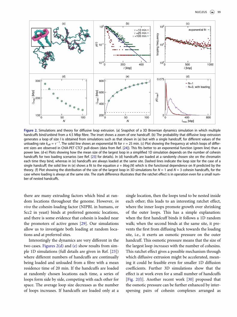
Figure 2(b) shows a plot of the probability that thesimulation generates a loop of a given size, for differ-ent values of the unloading rate. There is a significantprobability of finding loops of several hundred kbp,implying that the diffusive extrusion mechanism islikely capable of the rearrangement of the chromatinfibre necessary to form a loop. The diffusion constantfor cohesin sliding is D = 2.3 £ 10¡3~mm2s¡1 (whicharises naturally from the geometry of our simple bead-based model); this is much smaller than the in vitrovalue for chromatin in Xenopus extract quoted above,so the simulations provide a conservative estimate ofthe probability to form loops (still, this value is suffi-cient to create large loops diffusively).
An interesting feature of the plot in Fig. 2(b) is thatthe probability of forming a loop is approximatelyan exponential function of loop length (theoreticalmodelling predicts an exponential decay with apower-law correction). Standard equilibrium polymerphysics would predict that the probability of forminga loop of length l is a simple power-law function ofl [28], so the non-equilibrium binding/un-bindingkinetics have indeed altered the looping behaviour.HiC data shows that in vivo the probability of two lociinteracting decreases with a power-law function oftheir genomic separation on average [1]; however,ChIA-PET data [24] obtained using an antibody tar-geting CTCF (which therefore only includes interac-tions between CTCF bound loci), fit better to anexponential decay [see Fig. 2(c)]. Though there arelikely many other factors affecting these data, this sug-gests that different mechanisms are at play for CTCFloop formation to those behind chromosome interac-tions in general.
A ratchet effect promotes loop growth overshrinking
In the simulations and theory discussed so far we haveconsidered only a single bound cohesin handcuff,whereas in vivo we might expect many bound cohesinsto form a complicated pattern of loops. In the activeloop extrusion simulations presented in Refs. [7,8]
98 C. A. BRACKLEY ET AL.
there are many extruding factors which bind at ran-dom locations throughout the genome. However, invivo the cohesin-loading factor (NIPBL in humans, orScc2 in yeast) binds at preferred genomic locations,and there is some evidence that cohesin is loaded nearthe promoters of active genes [29]. Our simulationsallow us to investigate both loading at random loca-tions and at preferred sites.
Interestingly the dynamics are very different in thetwo cases. Figures 2(d) and (e) show results from sim-ple 1D simulations (full details are given in Ref. [23])where different numbers of handcuffs are continuallybeing loaded and unloaded from a fibre with a meanresidence time of 20 min. If the handcuffs are loadedat randomly chosen locations each time, a series ofloops form side by side, competing with each other forspace. The average loop size decreases as the numberof loops increases. If handcuffs are loaded only at a
single location, then the loops tend to be nested insideeach other; this leads to an interesting ratchet effect,where the inner loops promote growth over shrinkingof the outer loops. This has a simple explanation:when the first handcuff binds it follows a 1D randomwalk; when the second binds at the same site, it pre-vents the first from diffusing back towards the loadingsite, i.e., it exerts an osmotic pressure on the outerhandcuff. This osmotic pressure means that the size ofthe largest loop increases with the number of cohesins.This ratchet effect gives a possible mechanism throughwhich diffusive extrusion might be accelerated, mean-ing it could be feasible even for smaller 1D diffusioncoefficients. Further 3D simulations show that theeffect is at work even for a small number of handcuffs[Fig. 2(f)]. Another recent work [30] proposed thatthe osmotic pressure can be further enhanced by inter-spersing pairs of cohesin complexes arranged as
Figure 2. Simulations and theory for diffusive loop extrusion. (a) Snapshot of a 3D Brownian dynamics simulation in which multiplehandcuffs bind/unbind from a 4.5 Mbp fibre. The inset shows a zoom of one handcuff. (b) The probability that diffusive loop extrusiongenerates a loop of size l is obtained from simulations such as that shown in (a) but with a single handcuff, for different values of theunloading rate koff = t¡1. The solid line shows an exponential fit for t = 25 min. (c) Plot showing the frequency at which loops of differ-ent sizes are observed in ChIA-PET CTCF pull-down (data from Ref. [24]). This fits better to an exponential function (green line) than apower law. (d-e) Plots showing how the mean size of the largest loop in a simplified 1D simulation depends on the number of cohesinhandcuffs for two loading scenarios (see Ref. [23] for details). In (d) handcuffs are loaded at a randomly chosen site on the chromatineach time they bind, whereas in (e) handcuffs are always loaded at the same site. Dashed lines indicate the loop size for the case of asingle handcuff; the solid line in (e) shows a fit to the equation a + blog (N) which is the functional dependence on N predicted by thetheory. (f) Plot showing the distribution of the size of the largest loop in 3D simulations for N = 1 and N = 3 cohesin handcuffs, for thecase where loading is always at the same site. The stark difference illustrates that the ratchet effect is in operation even for a small num-ber of nested handcuffs.
NUCLEUS 99

handcuffs with single cohesins which diffuse along thefibre but are not linked at multiple points so do notform loops.
Domains with diffusive extruders
Large-scale Brownian dynamics simulations can also beused to investigate whether diffusing cohesins can gen-erate domains and interaction patterns similar to thoseseen in HiC data. We performed a simulation of a15 Mbp region with realistic chromatin density; 32 pairsof handcuffs were continuously added and removedfrom the fibre with 16 preferred loading sites, and a
mean residence time equivalent to 25 min. CTCF siteswere placed at 750 or 1500 kbp intervals in either con-vergent or divergent arrangements. Eight repeat simula-tions were performed, with CTCF sites populatedstochastically such that each simulation could have adifferent set of sites (to model cell-to-cell variation inCTCF binding); diffusing handcuffs only stick at CTCFspointing towards them, and when there is a CTCFbound at each side of the handcuff, the unbinding rateis reduced by 10 fold. Figure 3(a) shows a snapshot ofthe simulation system, and Fig. 3(b) shows a zoom withthe region between one pair of convergent CTCF sites(i.e. a TAD) highlighted in blue.
Figure 3. Large scale 3D simulation. (a) Simulation snapshot of large polymer representing a 15 Mbp chromatin fibre, with 32 diffusinghandcuffs. Here the polymer is confined within a sphere so as to give a realistic chromatin density (using periodic boundaries with thesame density instead of confinement gives similar results). (b) Zoom of the same snapshot, but with one domain highlighted in blue. (c)A HiC-like interaction map is shown for a 300 kbp region of the simulated fibre. The colour at each point in the map indicates the fre-quency of interaction between the chromatin positions connected by a triangle with its apex at that point. Positions and orientations ofCTCF sites, and positions of the loader sites are indicated.
100 C. A. BRACKLEY ET AL.

Figure 3(c) shows a contact map generated fromthese simulations. As in HiC interaction maps, red tri-angles show domains, and dark spots are seen at theedges of convergent CTCF loop domains. Dark spotsare also seen close to the diagonal at loading sites – afeature not normally seen in HiC (though note thatthere remain few publicly available data sets showinggenomic locations where the loader is enriched, and ithas yet to be confirmed that cohesin is preferentiallyloaded at these sites).
While these simulations show that some aspects ofthe domain structure can indeed be reproduced byour diffusion-based model, we urge caution in expect-ing such a simple model to be able to replicate interac-tion maps exactly. For example, the model does notinclude other DNA-binding proteins that might affectcohesin motion, nor do we attempt to account foractive processes such as transcription. Elongating pol-ymerases generate forces and torques (leading tosupercoiling [31,32]) which may affect cohesin diffu-sion; indeed recent experiments where the WAPLcohesin unloader protein is knocked down in mousenuclei show that cohesin collects preferentiallybetween convergent genes, indicating that polymerasecan push cohesin along the fibre [16,33]. These caveatsapply equally to the active loop extrusion model.
Discussion
In this work we have argued that 1D diffusion of cohe-sin along chromatin can lead to loop extrusion with-out the need to invoke an explicit motor action. Ofcourse experimental verification of this remains a sig-nificant challenge. Nevertheless, we suggest that diffu-sive extrusion cannot be dismissed in favour of anactive extrusion model in the absence of additionalexperimental evidence.
The in vitro experiments mentioned above [13,14]studied the topological loading and diffusion of cohesinrings on stretched DNA templates, and over obstacles.No directed motion was observed, but the diffusivitywas found to strongly depend on ATPase activity, saltconcentration, and on the way in which the cohesincomplex was loaded onto the substrate. Diffusion onreconstituted chromatin in Xenopus egg extracts wasalso measured [18], and it was found that acetylationof the Smc3 sub-unit strongly increased the diffusioncoefficient. Together these results suggest that the poresize and diffusivity of cohesin might be regulated by
ATP hydrolysis and acetylation in vivo. Recent in vivostudies have shown that knocking-down the loaderNIPBL (which leads to loss of chromosome-boundcohesin) leads to loss of looped domains [34], whereasa knock-down of CTCF affects intra-domain interac-tions [35]. All these observations are consistent withboth the active and diffusive extrusion models. A thirdpossibility is that there is some active translocation, butthat the direction is not fixed and the cohesin is“kicked” randomly back and forth along the fibre (theoverall effect would look like diffusive motion, but withan increased diffusion constant).
Unlike cohesin, the condensin complex can performunidirectional active stepping along a stretched DNAtemplate in the presence of ATP [17]. This points tothe possibility that active extrusion may be at work dur-ing mitosis, where condensin plays a central role [5,36].
Active loop extrusion is often cited as a model forthe formation of topological domains, but this is notthe only possible mechanism. Another popular modelis that chromatin interactions are mediated by tran-scription factors (or complexes thereof) which can dif-fuse freely in 3D through the nucleus, and which aremultivalent, meaning they can from molecular bridgesbetween different genomic loci [27,37,38]. This ideahas been extensively studied using molecular dynam-ics and Monte Carlo simulations of simple bead-and-spring polymer physics models (sometimes referred toas the strings-and-binders-switch (SBS) model [26]).Using only limited data about where proteins bind (orusing histone modification data to infer proteinbinding) it is possible to reproduce the TAD patternsobserved in HiC data. For example a model using onlytwo factors, one binding to active and one to inactiveregions, correctly predicted the locations of 85% ofTAD boundaries on chromosome 19 in HUVECs(human umbilical vein endothelial cells) [27].This model naturally describes promoter-enhancerinteractions mediated by polymerase-transcriptionfactor complexes, or heterochromatin and polycombrepressed regions organised by HP1 and PRC com-plexes respectively. It can explain the formation of thedomains which do not have looping between theirboundaries, as well as the larger scale A/B compart-ment formation, and the fact that compartments arepreserved upon loss of chromosome-bound cohesinor CTCF (which is difficult to reconcile with a loopextrusion model). The transcription factor model can-not, however, explain the CTCF motif bias.
NUCLEUS 101

It seems likely then, that a complete explanation ofgenome organisation will require a combination ofloop extrusion and multivalent transcription factormodels. Even so, as noted above, there are many addi-tional processes which are not yet included in either ofthese models, so one should not expect to be able toreproduce, for example, all the features of a HiC inter-action map. The aim of modelling and simulationstherefore should not be to reproduce carbon-copies ofexperimental results, but should rather be to provideinsight, propose new hypothesis, and help direct newexperiments.
Disclosure of potential conflicts of interestNo potential conflict of interest were disclosed.
AcknowledgmentsThis work was supported by ERC (CoG 648050,THREED-CELLPHYSICS), the NIH ID 1U54DK107977-01, CINECAISCRA Grants No. HP10CYFPS5 and HP10CRTY8P, and bythe Einstein BIH Fellowship Award to MN.
FundingEuropean Research Council CoG 648050, National Institutes ofHealth 1U54DK107977-01.
ORCIDC. A. Brackley http://orcid.org/0000-0002-5973-8179
References
[1] Lieberman-Aiden E, et al. Comprehensive mapping oflong-range interactions reveals folding principles of thehuman genome. Science. 2009;326:289.
[2] Dixon, JR, et al. Topological domains in mammaliangenomes identified by analysis of chromatin interactions.Nature. 2012;485:376.
[3] Sexton T, et al. Three-Dimensional folding and func-tional organization principles of the drosophila genome.Cell. 2012;148:458.
[4] Rao, S, et al. A 3D map of the human genome at kilobaseresolution reveals principles of chromatin looping. Cell.2014;159:1665.
[5] Nasmyth K. Disseminating the Genome: joining, Resolv-ing, and Separating sister chromatids during mitosis andmeiosis. Ann. Rev. Genet. 2001;35:673.
[6] Alipour, E, Marko, JF. Self-organization of domain struc-tures by DNA-loop-extruding enzymes. Nucl. Acid. Res.2012;40:11202.
[7] Sanborn, AL, et al. Chromatin extrusion explains keyfeatures of loop and domain formation in wild-type and
engineered genomes. Proc. Nat. Acad. Sci. USA. 2015;112:E6456.
[8] Fudenberg G, et al. Formation of chromosomal domainsby loop extrusion. Cell Report. 2016;15:2038.
[9] de Wit E, et al. CTCF binding polarity determines chro-matin looping. Mol. Cell. 2015;60:676.
[10] Guo Y, et al. CRISPR inversion of CTCF sites altersgenome topology and enhancer/promoter function. Cell.2015;162:900.
[11] Uhlmann F. SMC complexes: from DNA to chromo-somes. Nature Rev. Mol. Cell Biol. 2016;17.
[12] Ivanov D, Nasmyth K. A Topological interaction betweencohesin rings and a circular minichromosome. Cell.2005;122:849.
[13] Stigler J, et al. Single-Molecule imaging reveals a col-lapsed conformational state for DNA-bound cohesin.Cell Report. 2016;15:988.
[14] Davidson IF, et al. Rapid movement and transcriptionalre-localization of human cohesin on DNA. EMBO J.2016;35:2671.
[15] Lengronne A, et al. Cohesin relocation from sites of chro-mosomal loading to places of convergent transcription.Nature. 2004;430:573.
[16] Busslinger GA, et al. Cohesin is positioned in mammaliangenomes by transcription, CTCF and Wapl. Nature.2017;544:503.
[17] Terakawa T, et al. The condensin complex is a mech-anochemical motor that translocates along DNA. Sci-ence. 2017.
[18] Kanke M, et al. Cohesin acetylation and Wapl-Pds5oppositely regulate translocation of cohesin along DNA.EMBO J. 2016;35:2686.
[19] Gerlich D, et al. Live-Cell imaging reveals a stable cohe-sin-chromatin interaction after but not before DNA rep-lication. Curr. Biol. 2006;16:1571.
[20] Ladurner R, et al. Cohesin’s ATPase activity couplescohesin loading onto DNA with Smc3 acetylation. Cur-rent Biol. 2014;24:2228.
[21] Hansen AS, et al. CTCF and cohesin regulate chroma-tin loop stability with distinct dynamics. eLife. 2017;6:e25776.
[22] Murayama Y, Uhlmann F. DNA entry into and exit outof the cohesin ring by an interlocking gate mechanism.Cell. 2015;163:1628.
[23] Brackley CA, et al. Non-equilibrium chromosomelooping via molecular slip-links. Phys. Rev. Lett.2017;119:138101.
[24] Tang Z, et al. CTCF-Mediated human 3D genome archi-tecture reveals chromatin topology for transcription.Cell. 2015;163:1611.
[25] Brackley CA, et al. Nonspecific bridging-induced attrac-tion drives clustering of DNA-binding proteins andgenome organization. Proc. Natl. Acad. Sci. USA.2013;110:E3605.
[26] Barbieri M, et al. Complexity of chromatin folding is cap-tured by the strings and binders switch model. Proc. Natl.Acad. Sci. USA. 2012;109:16173.
102 C. A. BRACKLEY ET AL.
[27] Brackley CA, et al. Simulated binding of transcriptionfactors to active and inactive regions folds human chro-mosomes into loops, rosettes and topological domains.Nucl. Acid. Res. 2016;44:3503.
[28] Gennes P-Gd. Scaling concepts in polymer physics.Ithaca [N.Y.] and London: Cornell University Press;1979.
[29] Kagey MH, et al. Mediator and cohesin connect geneexpression and chromatin architecture. Nature. 2010;467:430.
[30] Yamamoto T, Schiessel H. Osmotic mechanism of theloop extrusion process. Phys. Rev. E. 2017;96:030402(R).
[31] Gilbert N, Allan J. Supercoiling in DNA and chromatin.Curr. Opin. Genet. Devel. 2014;25:15.
[32] Benedetti F, et al. Transcription-induced supercoilingexplains formation of self-interacting chromatin domainsin S. pombe. Nucl. Acid. Res. 2017;45:9850.
[33] Haarhuis JH, et al. The cohesin release factor WAPLrestricts chromatin loop extension. Cell. 2017;169:693.
[34] Schwarzer W, et al. Two independent modes of chromatinorganization revealed by cohesin removal. Nature. 2017.
[35] Nora EP, et al. Targeted degradation of CTCF decoupleslocal insulation of chromosome domains from genomiccompartmentalization. Cell. 2017;169:930.
[36] Goloborodko A, et al. Compaction and segregation ofsister chromatids via active loop extrusion. eLife. 2016;5:e14864.
[37] Brackley CA, et al. Predicting the three-dimensional fold-ing of cis-regulatory regions in mammalian genomesusing bioinformatic data and polymer models. GenomeBiol. 2016;17:59.
[38] Barbieri M, et al. Active and poised promoter states drivefolding of the extended HoxB locus in mouse embryonicstem cells. Nature Struct. Mol. Biol. 2017;24:515.
NUCLEUS 103