facial morphology as a determinant of anchorage control
TRANSCRIPT
University of ConnecticutOpenCommons@UConn
Master's Theses University of Connecticut Graduate School
5-19-2017
Facial Morphology as a Determinant of AnchorageControlJonathan Norman DzingleOrthodontic Resident, [email protected]
This work is brought to you for free and open access by the University of Connecticut Graduate School at OpenCommons@UConn. It has beenaccepted for inclusion in Master's Theses by an authorized administrator of OpenCommons@UConn. For more information, please [email protected].
Recommended CitationDzingle, Jonathan Norman, "Facial Morphology as a Determinant of Anchorage Control" (2017). Master's Theses. 1106.https://opencommons.uconn.edu/gs_theses/1106

i
Facial Morphology as a Determinant of Anchorage Control
Jonathan Norman Dzingle
B.S., University of Michigan, 2010
D.D.S., University of Michigan, 2014
A Thesis
Submitted in Partial Fulfillment of the
Requirements for the Degree of
Masters in Dental Science
At the
University of Connecticut
2017

ii
Copyright by
Jonathan Norman Dzingle
2017

iii
APPROVAL PAGE
Master of Dental Science Thesis
Facial Morphology as a Determinant of Anchorage Control
Presented by
Jonathan Dzingle B.S., D.D.S
Major Advisor________________________________________________
Sumit Yadav B.D.S., M.D.S, Ph.D.
Associate Advisor_____________________________________________
Madhur Upadhyay B.D.S., M.D.S, M.Dent.Sc.
Associate Advisor_____________________________________________
Aditya Tadinada B.D.S, M.D.S
University of Connecticut
2017
iv
Table of Contents
Abstract ...............................................................................................................iv
Introduction and Review of Literature ............................................................01
Facial Morphology and Growth .........................................................01
Morphologic Characteristics and Clinical Manifestation ..................03
Orthodontic Treatment in Different Facial Types .............................06
Orthodontic Extraction and Anchorage Considerations ....................08
Cephalometric Superimposition and Treatment Effects ....................11
Summary ............................................................................................12
Rationale .............................................................................................................13
Specific Aims ......................................................................................................14
Null Hypothesis ..................................................................................................14
Materials and Methods ......................................................................................15
Study Design ............................................................................................15
Subjects ....................................................................................................15
Cephalometric Preparation and Superimposition ....................................17
Crowding..................................................................................................18
Statistical Analysis ...................................................................................19
Results .................................................................................................................20
Discussion............................................................................................................22
Clinical Significance ................................................................................27
Study limitations ......................................................................................28

v
Conclusion ..........................................................................................................29
References ...........................................................................................................29
Appendix .............................................................................................................39

vi
Abstract:
Introduction: Facial morphology is determined early in life and skeletal characteristics
can be used to classify patients into hypodivergent or hyperdivergent phenotypes based on
differences in the vertical dimension. Differences in the vertical dimension can present unique
challenges to orthodontics as deep-bite or open-bite patients. These patients undergo a variety of
treatment modalities, including premolar extraction to obtain ideal overjet and overbite.
Hyperdivergent and hypodivergent patients exhibit differences in facial musculature and alveolar
bone density, which can impact anchorage control during orthodontic space closure. This study
looks at differences in anchorage loss between different skeletal phenotypes to determine
whether facial morphology is a primary determinant of anchorage loss. Materials and
Methods: Male and female orthodontic extraction patients (2,3,4 premolar), ages 8-18, were
categorized into hypodivergent (SN-MP <25), Normodivergent (27</=SN-MP</=37), or
hyperdivergent (SN-MP>39) groups. Cranial base, maxillary, and mandibular superimpositions
were used to measure changes in the mandibular plane angle, molar anchorage loss, and changes
in overbite on lateral cephalograms. 9 linear and 12 angular measurements were taken at T1
(pre-treatment) and T2 (post-treatment) time points. Results: A total of 337 patients (139 males,
198 females) were included in this study. Horizontal anchorage loss was found to be
insignificantly correlated with initial facial morphology (P=0.07). Horizontal anchorage loss was
found to be negatively correlated with age and initial crowding (P=0.02*, P=0.00***).
Horizontal anchorage loss resulted in a significant decrease in the mandibular plane angle
(P=0.00***); whereas, vertical molar extrusion was insignificantly correlated with mandibular
plane angle change (P=0.17). Positive change in overbite was the result of a complex interaction
of incisor extrusion, angulation change, and initial facial morphology. Conclusion: Facial
vii
morphology does not appear to be a primary determinant of anchorage loss in orthodontic
extraction patients. Horizontal anchorage loss was found to have a significant impact on closure
of the mandibular plane angle in favor of the wedge hypothesis. Overbite can be improved in
hyperdivergent patients using a combination of incisor extrusion and angulation change under
the premise of the “drawbridge effect.” While significant correlations indicate real interactions
between the variables studied, their clinical significance should be examined closely due to the
large variation seen within the sample population.
1
Introduction and Literature Review:
Facial Morphology and Growth:
The growth of the human face follows a distinct pattern that is established early in life
and consists of a complex process of modeling and remodeling to underlying skeletal structures 1
2 3. A classical orthodontic study conducted by Bjork in the 1950’s showed how metallic
implants could be used to track maxillary and mandibular growth in relation to a relatively stable
cranial base 4. Facial growth has been found to follow a similar pattern to somatic growth with
completion of neurological tissue, including the brain, occurring before the age of puberty 4 2.
During this time of neural growth, expansion of the brain causes an increase in size of the
cranium and modeling changes to the cranial base 2,4. These changes, in turn, can affect the size
and position of the maxilla and mandible as growth continues into adolescence.
The downward and forward growth of the maxilla and mandible has been shown to be the
product of a complex interaction of primary and secondary displacement of skeletal structures 2.
Primary displacement of the maxilla or mandible occurs through the process of appositional or
sutural growth 3. In the case of the maxilla, primary displacement can occur through growth at
sutural boundaries with other facial structures such as the frontal or zygomatic suture or through
appositional modeling changes in the area of the palate or nasal cavity. Secondary displacement
of the maxilla can occur as a result of growth of intermediary bones, which help to bring the
maxilla downward and forward, or through the expansion of functional matrixes such as the
nasal cavity 5 6. Similarly, the growth and position of the mandible is affected by primary
displacement through appositional growth of the condylar head and posterior ramus 2. The
position of the mandible is also determined by secondary displacement in which growth of the
cranial base and posterior modeling of the glenoid fossa help to determine its final position 6.

2
The size and shape of the mandible has also been said to be influenced by the orofacial capsule
as a functional matrix 5.
The functional matrix theory was proposed by Melvin Moss in 1969 as a way to explain
the primary determinant of oral facial growth 5. Previous theories had hypothesized that
maxillary and mandibular growth was driven by sutural growth or cartilaginous growth within
the oral facial complex 5. Moss hypothesized that oral facial growth was actually determined by
the interaction of skeletal structures with the periosteal and capsular matrixes of the face. The
periosteal matrix represented the influence of muscular attachments to the underlying
periosteum. Moss predicted that facial morphology would be influenced by these periosteal
attachments and growth of the maxilla and mandible would he heavily determined by muscular
function. He also believed that capular matrixes, or enclosed membrane-bound spaces, played a
role in facial development. The oral-pharyngeal and naso-pharyngeal spaces are just two
examples of capsular matrixes that Moss believes play a crucial role in facial development.
Together, the periosteal and capsular matrixes are believed to play a crucial role in the shape and
development of the face from early childhood through adolescence 5.
Aside from the functional matrix theory proposed by Moss, other early researchers of
facial growth such as James Scott believed that cartilaginous structures such as the nasal septum
were responsible for carrying the maxilla downward and forward 6 7. They believed that the
force exerted by chondral growth provided the necessary force to separate the maxilla from other
mid-facial sutures allowing for bony deposition 6 7. Translation of the maxilla was thought to
continue throughout early childhood until about age 7 when the rate of cartilage expansion would
begin to slow and surface apposition would become the primary determinant of facial growth 6.
During this time of appositional growth of the maxilla, Enlow believed that modeling changes

3
followed the “principle of a V” where deposition of bone occurred bilaterally on the posterior
lateral surfaces and resorption occurred on medial surfaces facing away from the direction of
growth. Appositional growth at the maxillary tuberosity based on this principle results in an
increase in length and width of the maxilla with increasing chronological age. Similarly, in the
vertical dimension, the maxilla experiences bony deposition on the palatal surface and resorption
on the nasal surface as the V-shape structure increases in size 6.
Horizontal and vertical growth of mandible is primarily determined by bony apposition
on the posterior ramus and condylar head 8. Typically, bony apposition of the condyle occurs in
a posterior superior position promoting downward and forward growth of the mandible
throughout adolescence. Furthermore, Enlow showed that resorption typically occurs on the
anterior portion of the ramus assisting in horizontal growth and making space for the posterior
dentition 8. Originally, the condyle was thought to function as a primary growth center due to its
histological similarity to epiphyseal growth plates seen throughout the body 9. However,
condylar chondroblasts have been shown to differ in embryonic origin from chondoblasts seen in
growth plates. Condylar Chondroblasts arise from undifferentiated connective tissue cells
whereas hypertrophic chondrocytes seen in epiphyseal growth plates come from resting
chondrocytes 9. Despite fractures or mutations that would have a significant impact on
epiphyseal growth, mandibular growth does not appear to be significantly affected 9. These
observations have lent credibility to the idea that the condyle is not a primary growth center but
rather a secondary growth site and reactive entity that responds to environmental forces 9.
Morphological Characteristics and Clinical Manifestation
Variations in facial growth among different individuals can lead to distinct skeletal
patterns, clinical characteristics, and dental malocclusions. Accordingly, classical orthodontic

4
literature has attempted to identify these characteristics and classify patients exhibiting
physiological extremes that present unique challenges to orthodontic treatment. These patients
have been categorized using many different terms in the literature. In 1964, Schudy introduced
the terms “hyperdivergent” and “hypodivergent” to describe the extremes in facial growth 10. In
1965, he also classified growth of the mandible into “clockwise” or “counterclockwise” rotation
to explain the appearance of an open or deep bite tendency 11. In 1969, Bjork looked at
morphological characteristics of the mandible to determine the type of growth rotation that
would occur 12. Viken Sassouni (1969) looked at the intersection of different facial planes to
classify patients as “skeletal open bite” or “skeletal deep bite” 13. In 1971, Isaacson classified
patients into “high” or “low” angle according to the relationship between their mandible and
cranial base 14. Stephen Schendel et al. (1976) used cephalometic values and clinical
characteristics to define a condition known as “long face syndrome” 15.
Despite being called many different terms, long face, high angle, or skeletal open-bite
patients exhibit similar radiographic, clinical, and dental characteristics. Bjork identified seven
morphological characteristics of the mandible that are commonly seen in skeletal open bite
patients including: backward inclination of condylar head, straight mandibular canal, antegonial
notching, thin cortical and backward inclination of mandibular symphysis, decreased interincisal
angle, decreased intermolar angle, and increased lower facial height 12. From a cephalometric
perspective, hyperdivergent patients exhibit an increase in mandibular plane angle, increased
anterior facial height, tipped up palatal plane, decreased ramus height, and excessive vertical
maxillary growth 15 16 13 17. An increase in skeletal convexity and backward true mandibular
rotation are two other characteristics seen on serial cephalograms that can lead to the assumption
of a vertical growth tendency 18. Clinically, these patients typically exhibit a dolichocephalic

5
facial pattern with greater vertical facial height than width, a narrow alar base, excessive gingival
display, and lip incompetence at rest 15. Dentally, hyperdivergent patients tend to exhibit a
tendency toward decreased transverse molar widths, anterior open bite, and convex sagittal
discrepancies 19 17 20 21.
Hyperdivergent patients also exhibit differences in cortical bone thickness and
masticatory function in comparison to their brachyfacial counterpart. A recent CBCT study
conducted by Horner et al. looking at inter-radicular cortical bone thicknesses found that, on
average, hypodivergent patients had between .08mm and .64mm thicker bone in maxillary and
mandibular posterior segments than hyperdivergent patients 22. These finding were also
corroborated by Masumoto, Ozdemir, Tsunori, and Swasty. 23 24 25 26. The differences in
cortical bone thickness between the two facial types may also be related to the function of the
masticatory muscles. The density and thickness of cortical bone has been found to be a function
of strain created by muscular attachments under masticatory forces 22. Therefore, increased
masticatory forces are thought to increase cortical bone thickness 27 28.
Some studies have found that there may be differences in the muscular profiles between
hyperdivergent patients and normal populations 29. Hyperdivergent patients has been shown to
have smaller muscles of mastication, weaker biting forces, lower EMG activity, and reduced
masticatory efficiency 18 30 31 32. On the other hand, brachyfacial patients were found to have
increased muscular volume, cross-sectional area, and thickness in the muscles of mastication 33.
However, there remains significant controversy in the literature as to whether a significant
correlation actually exists as some other authors have found non-significant or unpredictable
correlations and large individual variation. 34 35. The influence of cortical bone thickness and
muscle morphology on orthodontic treatment can best be appreciated when considered from the
6
perspective of Bioprogressive therapy in orthodontics. The underlying principles behind this
technique, first introduced by Robert Ricketts, are to appreciate the underlying form and function
of facial features to assist in orthodontic treatment 36. In the Bioprogressive philosophy, Ricketts
believes that strong posterior facial musculature in brachyfacial patients and thick cortical bone
can be used as a mechanism to resist tooth movement and extrusive forces experienced during
orthodontic treatment 36 37. In this way, hyperdivergent patients with weaker musculature and
thinner cortical bone would be susceptible to increased anchorage loss and extrusive mechanics
experienced throughout orthodontic therapy 36 37. Interestingly, despite the fact that the rate of
tooth movement has been correlated with sex, age, bone turnover, drug consumption, and a
multitude of other factors, there is a dearth of evidence showing the influence of facial
morphology or cortical bone thickness on the rate of tooth movement 38 39.
Orthodontic Treatment in Different Facial Types
Facial type is often regarded as having a significant impact on the type of orthodontic
treatment prescribed. In general, fixed orthodontic appliances without extraction have been
shown to extrude the posterior dentition and increase the overall vertical dimension of the face 40
41 42 43. Orthodontic extrusion can also lead to downward and backward rotation of the mandible
leading to opening of the bite and can pose significant challenges in hyperdivergent patients
already exhibiting an open bite tendency 44. However, the effects of mandibular skeletal growth
cannot be discounted when compared to dentoalveolar extrusion in overbite analysis. Naumann
et al., 2000, developed a mathematical model to show that mandibular vertical growth and
rotation were twice as important as mandibular dental changes in determining final overbite 45.
Therefore, understanding and controlling the direction of condylar growth seems equally
important as orthodontic treatment selection in controlling the vertical dimension in
7
hyperdivergent patients. Many different treatment modalities have been suggested in the
literature as ways of managing the vertical dimension of hyperdivergent and hypodivergent
patients with non-extraction therapy 46 47 48. Skeletal anchorage, high-pull headgear, and
functional appliances are just few of the strategies that have been studied to limit molar extrusion
during orthodontic therapy and promote favorable mandibular growth 46 47 48. Analogously,
Incisor intrusion and proclination, bite plates, cervical pull head-gear, and reverse curve of spee
wires have all been used as non-extraction techniques to promote vertical molar extrusion and
bite-opening in skeletal deep bite patients 41 49 50.
Orthodontic treatment with premolar extractions has also been explored extensively in
the literature as a way to improve overbite in hyperdivergent patients. Often referred to as the
“wedge hypothesis,” this theory describes the anterior horizontal movement of posterior teeth
away from the terminal hinge axis following premolar extraction to cause closure of the bite 17 51
52. In a study conducted by Aras in 2002, the author showed that extraction of four first molars
or second premolars resulted in more mandibular autorotation when compared to first premolar
extraction patients in support of the wedge hypothesis 53. Another way to increase overbite is by
decreasing the angulation and vertical position of the maxillary and mandibular incisors
following premolar extractions. Incisor retraction is thought to promote greater overlap of the
anterior dentition through a phenomenon known as the “drawbridge effect” 54. On the other
hand, extraction of premolars in low angle, hypodivergent patients has typically received
negative appraisal for the same reasons that extraction in hyperdivergent patients appears to be
so successful 42 50 55. However, many of the claims advocating against extraction in
hypodivergent patients are based on anecdotal evidence citing the drawbridge effect and don’t
take into consideration the possibility for posterior orthodontic extrusion or vertical mandibular

8
growth. Furthermore, stability of deep bite correction following orthodontic therapy has been
found to be reasonably high with the prevalence of vertical relapse among a sample of 61
patients to be less than 11% over a 12 year follow-up period 56.
Despite the fact that extraction of premolar teeth should allow for mesialization of the
posterior dentition and a decrease in facial height based on the wedge hypothesis, significant
controversy exists in the orthodontic literature. In a study of 54 hyperdivergent patients treated
with first or second premolar extractions, Kim et al. did not find a reduction in anterior facial
height in the second premolar extraction group compared to the first even though the former
showed a statistically significant increase in horizontal molar displacement 52. This study did
not, however, correlate the amount vertical molar extrusion with anterior facial height or look at
different facial types. Kocadereli (1999) also found that extraction of four first premolar teeth
did not have an effect on anterior facial height; however, the study sample size was small,
horizontal and vertical molar movement were not quantified, and differences in facial type were
not evaluated 57. So, there appears to be a gap in knowledge about the influence, if any, of the
wedge effect on the vertical dimension in orthodontic extraction patients with different facial
types that fully accounts for the effects of horizontal and vertical molar movement.
Orthodontic Extraction and Anchorage Considerations
During orthodontic treatment, teeth are subjected to a variety of forces and moments
causing them to move in desirable and undesirable directions. Anchorage can be defined simply
as the resistance to unwanted tooth movement 58. Anchorage becomes especially important
during extraction therapies where teeth can be divided into anterior and posterior units and are
exposure to reciprocal forces during space closure 58. Often, resistance of the posterior unit
coming forward is desired to allow for maximum retraction of the anterior unit or possible

9
correction of a sagittal discrepancy. Many different approaches have been utilized over the years
in order to minimize anchorage loss and many factors are believed to play a role. From a
biological standpoint, age, sex, and amount of crowding are all thought to play a role in amount
of anchorage loss in extraction cases 59 60 61. Adolescents have been found to lose more
anchorage than adults, males more than females, and un-crowded more than crowded 59 60 61.
Interestingly, mandibular growth also appears to play an important role in relative anchorage
control and correction of sagittal discrepancies 60. Although McKinney et al. found that
adolescent males tended to have increased anchorage loss compared to age-matched females,
males also had significantly more sagittal mandibular growth throughout treatment, which helped
to correct anterior-posterior molar discrepancies 60.
Aside from biological factors, practitioners have also relied on appliances, biomechanics,
skeletal anchorage, and differential extraction patterns to assist in anchorage control. Headgear,
Nance appliances, Transpalatal arches, and mini-implant supported devices are among the most
popular used in the literature to assist in anchorage control during space closure 62 63 64 65 66 67.
While some appliances show substantial evidence in helping to preserve anchorage, the benefit
of others has been somewhat controversial in the literature 62 63 64 65 66 67. Despite its widespread
use in orthodontics, the transpalatal arch was found to be ineffective in providing anchorage
control in a systematic review conducted by Diar-Bakirly et al., in 2017 64. Analogously, Al-
Awadhi et al., 2014, found that the Nance appliance did provide some anchorage enhancement
compared to control groups but conceded that it was not absolute 63. Extra-oral anchorage has
also been studied in comparison to skeletal anchorage using mini-implants in bimaxillary
protrusion and extraction cases 65 66 67. In these cases, it appears that skeletal anchorage provides
a superior benefit to conventional orthodontic techniques 65 66 67. However, in terms of clinical
10
significance, Sandler et al., 2014, found that the Nance appliance, headgear, or mini-implants
may all provide similar therapeutic benefit 62.
Orthodontic practitioners have also attempted to use biomechanical strategies such as
differential moments, differential extraction patterns, and separate canine retraction to minimize
anchorage loss during orthodontic therapy. Rajcich and Sadowsky, 1997, demonstrated the use
of an intrusion arch during space closure to provide a differential moment and molar tip back to
control anchorage loss during canine retraction 68. Similarly, Kim et al., 2005, found that first
premolar extraction provided better anchorage control than second premolar extraction lending
credibility to the theory that total root surface area is correlated to resistance of tooth movement
52. However, the effect of biology on tooth movement is somewhat unclear within the literature
as Xu demonstrated that 2-step space closure actually lost more anchorage than en-masse
retraction despite having an advantage in root surface area in the posterior unit 69.
Recently, it has become clear that individual biology can have a powerful impact on the
rate of tooth movement. Medications such as bisphosphonates, NSAIDs, and estrogens have
been found to decrease tooth movement; whereas, thyroxin, parathyroid hormone, and Vitamin
D3 seem to increase it 70. While the rate of tooth movement seems to be correlated with cell
turnover and osteoblast and osteoclast activity, there is a lack of evidence to show how bone
density directly correlates with the rate of tooth movement 70. Likewise, there is a dearth of
evidence to show how differences in facial morphology, facial musculature, and subsequent
changes in bone density can affect tooth movement and anchorage loss in extraction cases.
Therefore, it is necessary to examine whether a relationship between facial morphology and
anchorage loss exists, so orthodontic clinicians can make evidence-based decisions concerning
treatment for individual patients.

11
Cephalometric Superimposition and Treatment Effects
Human facial growth and development has been studied extensively within the
orthodontic literature 1 71 72 73. Early researchers such as Broadbent, Brodie, and Steuer showed
that facial growth follows a predictable and definitive pattern that is established early in life 1 71
72. The cranial base has been shown to provide a stable superimposition structure to compare
growth and orthodontic treatment effects in serial cephalograms as nearly 95% of its growth is
completed around the age of six 72. A recent systematic review by Afrand et al. looking at 11
articles published from 1955 to 2009 concerning cranial base changes, showed that the
cribriform plate and pre-sphenoid regions were the least likely to change with time 73. Despite
some modeling changes within sella turcica, dorsum sellae, and several other cranial base
structures, the anterior cranial base has been used widely in the literature for lateral
superimposition. Moreover, a recent CBCT study looking cranial base measurements in 62
adolescent patients aged between 12 and 17 years old verified that cranial base structures remain
stable over time 74.
Historically, two methods have been proposed for superimposition of the maxilla: best fit
and structural superimposition 75. The best fit uses the palatal plane and registration on ANS;
whereas, the structural superimposition uses the anterior portion of the zygomatic process of the
maxilla, nasal floor, and orbital floor according to implant studies conducted by Bjork in 1955 75.
The structural method differs in the way that it takes into account vertical growth of the maxilla
by looking at inferior modeling changes between the palatal roof and nasal floor. Overall,
Nielsen, 1989, showed that the “best fit” superimposition method underestimated the eruption of
maxillary teeth by 30-50% when compared to structural superimposition aided by metallic
implants 75. Similarly, mandibular growth and rotation has been studied using structural

12
superimposition based on the contour of the chin, internal cortical and trabecular structures of the
mandibular symphysis, and contour of the mandibular canal 4 12 76. The validity and repeatability
of this method has been extensively studied in the literature and a recent of CBCT by Ruellas et
al., 2016, further proved how these stable structures can be used to assess growth and the effects
of orthodontic treatment 77.
Summary:
A critical appraisal of the evidence has shown that craniofacial growth is a complex
process that follows a distinct pattern established early in life 1 71 72. Early completion of
neurological development leads to a stable cranial base structure that can be used as a reference
in orthodontic diagnosis and treatment planning 72 73 74. Orofacial growth shows tremendous
diversity in size and shape leading to different patterns of muscular and skeletal growth 2 5 6 10 11
12 13 14 15. Hyperdivergent patients have been shown to display weaker musculature and thinner
cortical bone structure than their hypodivergent counterparts 22 23 24 25 26 18 30 31 32. Clinically,
hyperdivergent patients typically display an increased lower facial height, open bite tendency,
excessive gingival display, and lip incompetence at rest 15 19 17 20 21. These characteristics, in
conjunction with known extrusive effects of orthodontic therapy, lead practitioners to choose
treatment options that often involve extraction of premolar teeth to help close the bite 37 36 17 51 52.
The bioprogressive orthodontic philosophy believes that facial morphology will have an effect
on cortical bone thickness and impact anchorage considerations during orthodontic treatment 37
36. However, despite a wealth of anecdotal evidence, there remains a significant gap in
knowledge as to whether facial morphology will have an impact on posterior anchorage loss in
orthodontic extraction cases. Similarly, there appears to be significant controversy surrounding
the “wedge hypothesis” and whether mesialization of the dentition will lead to closure of the
13
bite, a decrease in anterior facial height, and reduction of the mandibular plane angle 17 51 52 53.
Lastly, it appears unclear whether facial morphology, particularly in hypodivergent patients, is
justification enough for avoiding premolar extractions when they are indicated following
thorough diagnosis 42 50 55.
Rationale and Objectives:
Following a review of the literature, it has been shown that there is a clear lack of well-
designed studies looking at the impact of facial morphology on anchorage control. There
also appears to be a lack of consensus on how premolar extractions affect the vertical
dimension and changes in overbite. Therefore, this study will compare and quantify
anchorage loss during space closure following premolar extraction (4 bicuspid or maxillary
bicuspid) in matched groups of different facial types (hypodivergent, hyperdivergent,
normodivergent). It will also examine the effect of sex, age, time in treatment, and amount
of crowding on anchorage loss. This study will examine whether horizontal anchorage loss
leads to closure of the mandibular plane angle according to the wedge hypothesis. Finally,
it will look at the relationship between facial morphology, change in incisor position,
change in incisor angulation, and posterior molar extrusion on changes in overbite. This
study will reflect an extension of previous work completed in 2015 by Dr. Saleh Alwadei,
but will include a significantly increased sample size and incorporate another approved
data center.
14
Specific Aims/Objectives:
Specific Aim 1: To quantify the amount of horizontal and vertical movement of maxillary
and mandibular molars during space closure of three different facial types.
Specific Aim 2: To quantify the amount of horizontal, vertical, and angular movements of
maxillary and mandibular incisors during space closure of three different facial types.
Specific Aim 3: To measure changes in mandibular plane angles following extractions in
the three facial types.
Hypothesis and Null Hypotheses:
Hypothesis 1: The amount of horizontal anchorage loss will be increased in high
(mandibular plane) angle patients compared to low angle patients in premolar extraction
cases.
Null Hypothesis 1: The amount of horizontal anchorage loss does not depend on the
initial mandibular plane angle.
Hypothesis 2: Horizontal anchorage loss of the posterior teeth will result in the
maintenance or decrease of the mandibular plane angle.
Null Hypothesis 2: There is no relationship between horizontal anchorage loss and
change in the mandibular plane angle.
Hypothesis 3: Vertical extrusion of the posterior teeth will result in an increase in the
mandibular plane angle.
Null Hypothesis 3: There is no relationship between vertical extrusion of posterior teeth
and increase in the mandibular plane angle.

15
Materials and Methods:
Study Design:
This was a retrospective multi-centered cephalometric study reviewed and approved by
the University of Connecticut Health Institutional Review Board (15-040-1). The study design
followed similar methodology used by Dr. Saleh Alwadei in his thesis, The Influence of the
Facial Pattern on Anchorage Control (2015). This study will examine changes in twelve
cephalometric data points from the beginning to end of orthodontic treatment involving the
extraction of either, two maxillary bicuspid teeth or four maxillary and mandibular bicuspid
teeth. The lateral cephalometric x-rays being examined were taken before the start of
orthodontic treatment (T1) and after completion of treatment (T2). Horizontal and vertical
anchorage loss was assessed using maxillary and mandibular regional superimpositions and
changes in the mandibular plane angle were measured using a cranial base superimposition
(Figure 1,2,3). Changes in incisor position and angulation were also measured and compared to
changes in overbite throughout treatment. Similarly, patient’s initial facial morphology was also
compared to changes in overbite after two or four bicuspid extraction therapy. Finally, some
demographic variables including sex, age, time in treatment, and initial crowding were examined
in relation to horizontal anchorage loss in maxillary first molars of all facial types.
A. Subjects:
The study sample consisted of patients who received two or four bicuspid extractions
while undergoing orthodontic treatment at the University of Connecticut Health Center,
Farmington, or from Columbia University, New York City between January 1995 and May 2014.
The sample was collected based on a treatment plan that required the extractions of maxillary
premolars (1st or 2nd) or maxillary and mandibular premolars (1st or 2nd) and complete retraction

16
of the anterior (canine and incisors) segment for each patient. The age range for this study
included patients 8-18 years old and presenting in either late mixed or permanent dentition at the
start of treatment. Since facial morphology was hypothesized to be the primary and dominant
factor in anchorage loss, patients were first divided into categories based on their facial type
before cofactors such as sex, age, time in treatment, and crowding were examined. Similarly,
differences in biomechanical strategies were not examined in this study other than the exclusion
of absolute anchorage using mini-implants, which has been shown to have a significant impact
on reduction of anchorage loss. The patients were divided into 3 groups according to initial
cephalometric measurement of vertical facial patterns using (SN-Go(constructed)Gn).
Hypodivergent Patients were classified as having initial (SN-GoGn) </= 25 degrees.
Normodivergent Patients were classified as (SN-GoGn) >/= 27 degress and </= 37 degrees.
Hyperdivergent Patients were classified as (SN-GoGn) >/= 39 degree.
Summary of Inclusion/Exclusion Criteria
Inclusion Criteria:
a. Two maxillary (1st or 2nd premolar) or four bicuspid (1st or 2nd premolar)
extraction case.
b. Late mixed or permanent dentition with no missing permanent teeth (except third
molars).
c. Initial and Final cephalograms with patient demographic data.
d. One-phase treatment with fixed appliances.
Exclusion:
a. Missing Permanent Teeth (except 3rd molars)
b. Medical condition or medication that could affect tooth movement.
17
c. Surgical Patient or use of skeletal anchorage.
d. Compromised/Incomplete treatment where maxillary or mandibular spaces were
not closed.
B. Cephalometric Preparation and Superimposition
Digital cephalometric films were obtained for each time point (T1 and T2) from each data
source and examined for quality, magnification, and usability. Films that did not have a
calibration ruler or whose quality was too poor to accurately determine the positions of teeth or
cranial base were excluded from this study. Additionally, patients that exhibited missing teeth,
lack of space closure at the end or treatment, or visible evidence of orthognathic surgery were
similarly excluded. Due to the fact that initial and final x-rays were often taken on different
machines, all images were calibrated for magnification error using the calibration ruler and a
calculation using the number of pixels-per-inch. Furthermore, images were enhanced to aid in
landmark identification and printed on high quality glossy photo paper. Images were de-
identified prior to printing and labeled with assigned numerical values, which did not reveal any
information relating to the patient’s facial type. All cephalograms were traced and superimposed
using acetate paper and a 0.5mm black (pre-treatment) or red (post-treatment) mechanical pencil.
Printing magnification accuracy and linear measurements were taken with a digital caliper and
angular measurements with a manual protractor.
Cranial Base, Maxillary, and Mandibular superimpositions were completed on each
patient using criteria set forth by ABO guidelines. A horizontal Sella-Nasion (SN line) was
traced on the (T1) patient cephalogram and transferred to the (T2) cephalogram using best fit of
cranial base structures: anterior sella, walker’s point, greater wing of sphenoid, and planum
sphenoidale (Figure 1). Maxillary superimpositions were completed using the anterior portions

18
of the zygomatic processes, orbital floors, and nasal floors with a horizontal reference line
through the palatal plane (PNS-ANS) and vertical reference line through (PTM). These
horizontal and vertical reference lines were transferred to (T2) films upon structural maxillary
superimposition (Figure 2). Similarly, the mandibular superimposition was completed using the
internal contour of the inferior border of the symphysis, inferior alveolar nerve canal, and third
molar germ. A horizontal reference line was drawn through (Go(constructed)-Gn) and a vertical
reference line was drawn perpendicular to constructed gonion. The mandibular references were
then transferred to (T2) films upon structural mandibular superimposition (Figure 3). The T1 and
T2 (SN-MP) angles were measured from the cranial base and mandibular horizontal reference
lines to determine any change that occurred as a result of horizontal anchorage loss and not the
result of orthodontic vertical extrusion.
Once reference lines were drawn, maxillary and mandibular measurements were taken
and recorded on an excel spreadsheet. Inter-rater and Intra-rater reliability for tracing were
established using the concordant correlation coeffiecient (CCC) between two tracing operators.
Twenty (T1 and T2) randomly chosen cephalograms were traced by each operator and tested for
inter-rater reliability. Similarly, each operator retraced ten sets of cephalograms at least thirty
days after original tracing and tested for intra-rater reliability. Table 1 summarizes the pre and
post-treatment measurements examined in this study.
C. Crowding and Demographics Data
Crowding was measured in this study using an arch space analysis described by Proffit
(4th edition; 2007 195-197) and related to overall horizontal anchorage loss values. Patient’s sex,
age, and length of treatment were also recorded and tested as confounding factors of horizontal
anchorage loss.
19
D. Statistics
Anchorage loss and facial morphology were first related using Pearson’s correlation
coefficient to determine strength of relationship and directionality. Next, patients were separated
into the categorical variables Hypodivergent, Normodivergent, and Hyperdivergent and linear
regression analysis was performed. Facial morphology was also compared to anchorage loss as a
continuous variable in an unadjusted analysis and adjusted analysis taking into consideration age,
sex, initial crowding, and treatment time. Significance values for all statistical tests were set at P
</=0.05. Mean anchorage loss among the three facial groups and between males and females
were also compared using a student T-test. Changes in the mandibular plane angle were tested
against horizontal and vertical anchorage loss using both Pearson’s correlation coefficient and
linear regression. Factors affecting changes in overbite were studied using linear regression in a
full and reduced model and concerns of collinearity of variables were addressed by checking
variance inflation factors. Concordant correlation coefficients were used to evaluate inter-rater
and intra-rater reliability of cephalometric tracing and measurement. A power analysis was also
conducted to determine the sample size needed to detect the anchorage loss differences between
the hypodivergent, hyperdivergent, and normal groups. The power analyses that was conducted
using the low limit of the effect size (based on the ratio of mean difference between conditions
relative to the standard deviation) produced a sample size estimate of 150 participants per group
with a conventional alpha level (p =0 .05) and desired power (1 – ) of 0.80. Larger effect sizes
will, of course, reduce the number of participants needed. The power analysis was performed
with the computer application G-Power.

20
Results:
A total of 355 patients of all facial types were initially included in this study. 18 patients
were eventually excluded because they did not meet the age criteria bringing the total number of
patients to 337. Patients were separated into hypodivergent, normodivergent, and hyperdivergent
facial types with 13, 164, and 150 patients in each category, respectively (Table 2). Based on the
cephalometric criteria for each facial type, 10 patients were excluded when facial type was
examined as a categorical variable because they did not fall into any of the three facial types
based on their initial SN-MP measurement. However, all 337 eligible patients were included for
statistical analysis when facial type was studied as a continuous variable. Table 3 and Figure 4
summarize the demographic data of the sample population. The sample population consisted of
198 females and 139 males.
When facial morphology was coded as a continuous variable, it was negatively associated
with horizontal anchorage loss with Pearson correlation coefficient of -0.121 (Figure 5).
However, horizontal and vertical maxillary anchorage losses were found to be positively
correlated with a Pearson correlation coefficient of 0.243 (Figure 6). The mean anchorage loss
was found to be similar among the three facial types. The mean and standard deviation were as
follows: 3.3±1.99 for hypodivergent, 3.47±1.95 for normodivergent, and 3.01±2.39 for
hyperdivergent (Table 4) (Figure 7). No significant differences in anchorage loss were found
between the three facial groups (Hypo-Normo P=0.95, Hypo-Hyper P=0.89, Normo-Hyper
P=0.14) (Table 4). When facial morphology was coded as a continuous variable in an
unadjusted model, a significant correlation was found in regards to anchorage loss with a P-value
of 0.026 (Table 5). However, in an adjusted model that included parameters of sex, age, time in
treatment, and initial crowding, facial morphology was found not to be a significant factor in

21
anchorage loss P=0.075 (Table 5). Interestingly, increasing age and crowding were found to be
negatively associated with anchorage loss with a significant P-value of 0.018 and 0.000,
respectively (Table 5). The adjusted model had an adjusted R-squared value of 0.11 and residual
error of 2.06 meaning that a number of unexplained factors are participatory in contributing to
anchorage loss. Mean anchorage loss for males and females across all facial types and ages
groups was found to be 3.49±0.18mm and 3.14±0.15mm, respectively. The difference between
males and females was found to be non-significant with a P-value of 0.13 (Table 6).
Next, the validity of the wedge hypothesis was examined to study the effects of
horizontal and vertical anchorage loss on change in the mandibular plane angle in 4 bicuspid
extraction cases. A total of 278 extraction cases were examined and horizontal and vertical
anchorage losses were both found to be negatively correlated with changes in the mandibular
plane angle. The Pearson correlation coefficient for horizontal and vertical anchorage loss were
-0.225 and -0.128, respectively (Figure 8). Three linear regression models were created to study
the effects of horizontal and vertical anchorage loss independently and together. While vertical
loss was found to be significantly associated with a decrease in the mandibular plane angle as an
independent variable (P=0.032), its effect became non-significant when incorporated in a model
that had both horizontal and vertical anchorage loss (P=0.17024). Horizontal anchorage loss, on
the other hand, retained statistical significance in both models (Independent, P=0.00016;
Together, P=0.00068) leading to the conclusion that horizontal anchorage loss plays a bigger role
in changing the mandibular plane angle during four bicuspid extraction cases. However, the
adjusted R-squared value for the combined model was only 0.05, showing that other outside
variables must play a role in influencing changes in the mandibular plane angle. Results from
the independent and combined models are summarized in Table 7.

22
Changes in overbite between T2 and T1 time points were also tested for association
between facial morphology, vertical incisor extrusion, horizontal incisor retraction, changes in
incisor angulation, and posterior molar extrusion (Figure 9). A full model containing all of the
variables was tested for significance and Horizontal incisor retraction was found to non-
significantly associated with changes in overbite (P=0.078). Therefore, a reduced model was
created and horizontal incisor retraction was excluded. Posterior molar extrusion was found to
be negatively correlated changes in overbite with a B-coefficient of -0.30, (-0.36 to -0.23 95%
CI) (Table 8). Facial morphology, Vertical incisor extrusion, and a decrease in incisor
angulation were all found to increase overbite (B-coefficient 0.06, 0.33, and -0.05 respectively)
(Table 8). Since facial morphology was evaluated as a continuous variable in this regression
analysis, changes in overbite were found to be the highest in hyperdivergent population (2.41mm
at 39 degrees SN-MP) and significantly lower in hypodivergent patients (1.54mm at 25 degree
SN-MP).
Interclass and Intraclass concordant correlation coefficients (CCC) and 95% confidence
intervals were also calculated (Table 9). The average Interclass CCC was 0.88 (Range 0.83-
0.98) and Interclass CCC was 0.93 (Range 0.87-0.99) and 0.96 (Range 0.83-0.99) measured
across 12 cephalometric variables (Table 9).
Discussion and Clinical Significance:
In this multi-center, retrospective, cephalometric study, it was hypothesized that facial
morphology would play a significant role in anchorage preservation during extraction therapy.
The basis of this hypothesis was rooted in the observation that differences in growth patterns
among individuals can lead to significant variations in the vertical dimension 7 17 21. These
variations can be measured using both cephalometric analysis and clinical observation. The
23
hyperdivergent phenotype has received a great deal of attention in the literature because of its
open bite its tendency that can be exacerbated during orthodontic treatment 54 78 79. Orthodontic
clinicians employ a variety of techniques, including premolar extractions, to mitigate the effect
of extrusive mechanics and improve overbite throughout treatment 51 80. However, a question
that has remained unanswered is how the morphological characteristics of either phenotype
extreme influence anchorage control during space closure. The literature has shown that
hyperdivergent and hypodivergent patients display different patterns of musculature and cortical
bone, which may affect the rate of tooth movement on a biological level more that differences in
orthodontic technique 18 22 29 81 82. In this study, patients were separated into three phenotype
categories and anchorage loss was measured using maxillary first molars and structural
superimposition.
The results showed there were no significant differences in anchorage loss between facial
types when patients were separated into the three categories (hypodivergent; 3.3±1.99,
normodivergent; 3.47±1.95, and hyperdivergent; 3.01±2.39). However, despite a large sample
size from two academic institutions, only 13 extraction patients that met the inclusion criteria
(SN-MP </=25 degrees) could be analyzed in the hypodivergent category despite the power
analysis showing that 150 patients would be needed for 80% power and small effect size. One
explanation for this observation could be the fact that orthodontic practitioners tend to avoid
extractions in this patient population out of fear that it will further deep the bite. Therefore, facial
morphology was alternatively coded as a continuous variable based on the patient’s initial
mandibular plane value. The unadjusted model for anchorage loss with facial morphology as a
continuous variable showed that they were negatively correlated with -0.121 as the Pearson
correlation coefficient. A linear regression analysis showed that this relationship was
24
statistically significant with a p-value of 0.02. However, in the unadjusted model, the patients
sex, age, time in treatment, or amount of crowding were not taken into consideration and could
have played a role in anchorage loss values among patients with different facial morphologies.
Moreover, the B-coefficient and R-squared value in this linear regression were very low (-0.04
and 0.01) showing that a statistically significant relationship might not indicate clinical
relevance.
Next, an adjustment model was created to analyze how multiple factors in addition to
facial morphology could be affecting anchorage loss values. In this model, facial morphology
was not significantly correlated with anchorage loss values but age at the start of treatment and
the amount of maxillary crowding were significantly correlated. Patient age had a negative B-
coefficient meaning that as patient age increased, the amount of anchorage loss significantly
decreased. This observation falls in line with Ohiomoba et al., which shows how density and
maturity of alveolar bone increase with age and may act as a biological mechanism to resist tooth
movement 83. Maxillary crowding was also negatively correlated with a B-coefficient of -0.13
and significance value less than 0.001. This means that increased crowding significantly
decreases anchorage loss during treatment, which can be attributed to a reduced amount of space
closure. Interestingly, time in treatment was not significantly correlated to anchorage loss, but
this result may be affected by the fact that our observation period included the entire time of
treatment and not the time allocated toward space closure. Also, our results did not show any
significant differences in anchorage loss between genders (Males 3.49±0.18mm; Females;
3.14±0.15mm;P-value 0.12) and contradicts Su et al., which showed a significant difference
between genders 61.
25
Horizontal and vertical anchorage loss was also measured in 278 four bicuspid extraction
cases to determine whether anchorage loss leads to closure of the mandibular plane angle in
support of the wedge hypothesis. When the two variables were tested independently, both were
found to be significantly correlated with closure of the mandibular plane angle
(Horizontal;p<0.001 Vertical:P=0.03). In spite of the fact that posterior vertical extrusion is
commonly associated with downward and backward rotation of the mandible, this study found a
decrease in the mandibular plane angle 42. However, despite that fact that this relationship was
found to be statistically significant, it may not be clinically relevant as the B-coefficient
indicated a weak influential relationship (-0.11 angle change for every 1mm of posterior
extrusion) and R-squared value (0.01) showed that several other factors such as growth could be
playing an influential role. Moreover, the average treatment time for patients in this study was
nearly 42 month, which may also be contributing to closure of the mandibular plane angle under
normal observations of growth.
Fascinatingly, the adjusted linear regression model, which incorporated both horizontal
and vertical anchorage loss, showed that horizontal anchorage loss remained significantly
associated with negative change in the mandibular plane angle (P<0.001). These results support
the wedge hypothesis and notion that closure of the mandibular plane angle can be achieved by
anterior positioning of posterior teeth away from the hinge axis. Furthermore, the adjusted
model showed that vertical anchorage loss contributed insignificantly (P=0.17) to change in the
mandibular plane angle when horizontal anchorage loss was also taken into account. This means
that horizontal anchorage loss is significantly more important than vertical posterior extrusion in
terms of changing the mandibular plane angle during extraction treatment.
26
Lastly, changes in overbite were measured against facial morphology, change in incisor
position, angulation, and posterior vertical extrusion. The average initial overbite in the
hyperdivergent group was 1.30mm compared to 2.52mm in the combined hypodivergent and
normodivergent groups (Table 9). The later was combined due to low patient numbers in the
hypodivergent patient group. Lower initial overbite in the hyperdivergent group tends to follows
trends typically seen in this patient population. A full linear regression model was completed to
examine the effects of the aforementioned factors on change in overbite. The initial model
showed that horizontal incisor retraction did not significantly affect overbite throughout the
patient population studied, so it was excluded and a reduced model was created looking at the
effects of facial morphology, incisor extrusion, change in angulation, and posterior molar
extrusion. Due to concerns of collinearity between the variables, each predictor was checked for
its variance inflation factor and found to be less than 10. This means that each factor of the
reduced model played an independent role in affecting the change in overbite with respect to
other variables being present.
Incisor angulation change and posterior molar extrusion were found to be negatively
correlated with change in overbite. This model corroborates the assumptions of the “drawbridge
effect” that decreasing incisor angulation will have a positive effect on increasing end overbite
values 54. In this model, approximately 1mm of additional overbite can be achieved for every
18-degree decrease in incisor angulation in the maxillary and mandibular incisors. On the hand,
posterior molar extrusion had the opposite effect with a negative B-coefficient of -0.30
effectively reducing the overbite by 1mm for each 3.33mm of combined maxillary and
mandibular molar extrusion. Intriguingly, loss of overbite due to posterior extrusion was almost
perfectly balanced with the effect of vertical incisor extrusion, which had a positive correlation
27
with overbite and B-coefficient of 0.33. Despite the fact that facial morphology was
significantly correlated with positive changes in overbite, the impact of this factor may not be
clinically significantly when deciding whether or not to extract in a hypodivergent patient. The
difference in change in overbite in hypodivergent patient (SN-MP=25 degrees) due to facial
morphology alone could be as little as 1mm (low end of 95% B-coefficient confidence interval).
Other factors such as posterior molar extrusion should not be ignored and could potentially offset
any positive changes in overbite seen during extraction in a skeletal deep bite patient.
Clinical Significance:
Based on the extensive analysis completed in this study, it does not appear that initial
facial morphology has any significant impact on horizontal anchorage loss in extraction patients.
Therefore, null hypothesis one cannot be rejected and other factors such as patient age,
crowding, and treatment mechanics should be contemplated when anchorage demands are
increased. However, there does appear to be a significant relationship between horizontal
anchorage loss and closure of the mandibular plane angle in support of the wedge hypothesis.
Thus, the second null hypothesis is rejected. While the result bore statistical significance, the
clinical relevance should not be ignored. This study shows that within the realm of reasonable
anchorage loss (~3-4mm), the decrease in mandibular plane angle is clinically insignificant (~1
degree) and extraction treatment options should not be chosen based solely on the perception that
it will lead to a greater esthetic benefit. On a similar note, it was surprising to find that vertical
molar extrusion did not lead to an increase in the mandibular plane angle during premolar
extractions. Therefore, the third null hypothesis cannot be rejected. One possible explanation for
this surprising finding could be related to the fact that horizontal and vertical anchorage losses
were positively correlated with each other. Therefore, the effects of posterior vertical extrusion
28
may be camouflaged during extraction treatment by anterior movement of the posterior teeth and
mandibular rotation during growth. Finally, change in overbite was found to be a complex
interaction of many different variables that are often occurring at once during treatment.
However, the linear regression model showed that some variables such as decreasing incisor
inclination or incisor extrusion might hold greater potential to increase overbite than some of the
other variables studied.
Study Limitations:
One of the greatest limitations of this retrospective study was the inability to locate the
required number of patients for the hypodivergent patient group. Despite being a multi-center
study, it proved to be extraordinarily difficult to find extraction cases in brachyfacial, deep-bite
patients. Therefore, the three patients groups could not be fairly compared against one another
because of the drastic differences in the group sizes. Also, there appeared to be significant
individual variation with regards to anchorage loss when facial morphology was studied as a
continuous variable and the sample population included patients from late childhood to early
adulthood. Despite that fact that some significant correlations were found with regards to
anchorage loss and other demographic variables, the linear regression analyses showed relatively
low R-squared values when testing each of the hypotheses. This means that the models
generated, although statistically significant, could not account for the large variation seen within
the population. Hence, there must be some other factors present such as treatment biomechanics
or facial growth that could not be accounted for and played a major role in contributing to
anchorage loss.

29
Conclusions:
1. Facial morphology does not have a significant impact on anchorage loss in extraction
case.
2. Horizontal Anchorage loss leads to a statistically significant decrease in the mandibular
plane angle, but the magnitude may not be clinically relevant.
3. Vertical Anchorage loss does not significantly change the mandibular plane angle in
premolar extraction cases.
4. Changes in overbite achieved during orthodontic treatment are the result of a complex
interaction of factors in the anterior and posterior dentition.
5. Individual age, but not gender or time in treatment, appears to significantly impact
anchorage loss.
References:
1. Brodie AG. On the growth pattern of the human head from the third month to eigth year of
life. Am J Anat. 1941;68(2):209-262.
2. Enlow DH. A morphogenetic analysis of facial growth. Am J Orthod. 1966;52(4):283-
299.
3. Enlow DH, Hunter WS. A differential analysis of sutural and remodeling growth in the
human face. Am J Orthod. 1966;52(11):823-830.
4. Bjork A. Facial growth in man, Studied with the Aid of Metallic Implants. Acta Odontol
scandinav. 1955;13(1):9-34.
5. Moss ML, Salentijn L. The capsular matrix. Am J Orthod. 1969;56(5):474-490.
6. Enlow DH, Bang S. Growth and Remodeling of the Human Maxilla. Am J Orthod.
30
1965;51(6):446-464.
7. Scott JH. The analysis of facial growth: I. The Anteroposterior and Vertical Dimensions.
Am J Orthod. 1958;44(7):507-512.
8. Enlow DH, Harris DB. A study of the postnatal growth of the human mandible. Am J
Orthod. 1964;50(1):25-50.
9. Berraquero R, Palacios J, Rodríguez JI. The role of the condylar cartilage in mandibular
growth. A study in thanatophoric dysplasia. Am J Orthod Dentofac Orthop.
1992;102(3):220-226.
10. Schudy FF. Vertical growth versus anterioposterior growth as related to function and
treatment. Angle Orthod. 1964;34(2):75-93.
11. Schudy FF. The Rotation of the Mandible Resulting From Growth: Its implications in
Orthodontic treatment. Angle Orthod. 1965;35(1):36-50.
12. Bjork A. Prediction of mandibular growth rotation. Am J Orthod. 1969;55(6):585-599.
13. Sassouni V. A classification of skeletal facial types. Am J Orthod. 1969;55(2):109-123.
14. Isaacson JR, Isaacson RJ, Speidel TM, Worms FW. Extreme variation in vertical facial
growth and associated variation in skeletal and dental relations. Angle Orthod.
1971;41(3):219-229.
15. Schendel SA, Eisenfeld J, Bell WH, Epker BN, Mishelevich DJ. The long face syndrome:
Vertical maxillary excess. Am J Orthod. 1976;70(4):398-408.
16. Isaacson RJ, Zapfel RJ, Worms FW, Erdman AG. Effects of Rotational Jaw Growth on
the Occlusion and Profile. Am J Orthod. 1977;72(3):276-286.
17. Nanda SK. Growth patterns in subjects with long and short faces. Am J Orthod Dentofac
Orthop. 1990;98(3):247-258.
31
18. Buschang PH, Jacob H, Carrillo R. The morphological characteristics, growth, and
etiology of the hyperdivergent phenotype. Semin Orthod. 2013;19(4):212-226.
19. Nahoum HI. Vertical proportions: A guide for prognosis and treatment in anterior open-
bite. Am J Orthod. 1977;72(2):128-146.
20. Wagner DM, Chung CH. Transverse growth of the maxilla and mandible in untreated girls
with low, average, and high MP-SN angles: A longitudinal study. Am J Orthod Dentofac
Orthop. 2005;128(6):716-723.
21. Fields HW, Proffit WR, Nixon WL, Phillips C, Stanek E. Facial pattern differences in
long-faced children and adults. Am J Orthod. 1984;85(3):217-223.
22. Horner KA, Behrents RG, Kim KB, Buschang PH. Cortical bone and ridge thickness of
hyperdivergent and hypodivergent adults. Am J Orthod Dentofac Orthop.
2012;142(2):170-178.
23. Masumoto T, Hayashi I, Kawamura A, Tanaka K. Relationships among facial type,
buccolingual molar inclination, and cortical bone thickness of the mandible. Eur J Orthod.
2001;23:15-23.
24. Ozdemir F, Tozlu M, Germec-Cakan D. Cortical bone thickness of the alveolar process
measured with cone-beam computed tomography in patients with different facial types.
Am J Orthod Dentofac Orthop. 2013;143(2):190-196.
25. Tsunori M, Mashita M, Kasai K. Relationship between facial types and tooth and bone
characteristics of the mandible obtained by CT scanning. Angle Orthod. 1998;68(6):557-
562.
26. Swasty D, Lee J, Huang JC, et al. Cross-sectional human mandibular morphology as
assessed in vivo by cone-beam computed tomography in patients with different vertical
32
facial dimensions. Am J Orthod Dentofac Orthop. 2011;139(4):e377-e389.
27. Bresin A, Kiliaridis S, Strid KG. Effect of masticatory function on the internal bone
structure in the mandible of the growing rat. Eur J Oral Sci. 1999;107(1):35-44.
28. Mavropoulos A, Kiliaridis S, Bresin A, Ammann P. Effect of different masticatory
functional and mechanical demands on the structural adaptation of the mandibular
alveolar bone in young growing rats. Bone. 2004;35(1):191-197.
29. Proffit WR, Fields HW. Occlusal Forces in Normal and Long-face Children. J Dent Res.
1983;62(5):566-570.
30. Ingervall B, Minder C. Correlation between maximum bite force and facial morphology in
children. Angle Orthod. 1997;67(6):415-424.
31. Throckmorton GS, Ellis EEI, Buschang PH. Morphologic and Biomechanical Correlates
with Maximum Bite Forces in Orthognathic Surgery Patients. J Oral Maxillofac Surg.
2000;58(5):515-524.
32. Gomes SGF, Custodio W, Faot F, Del Bel Cury AA, Garcia RCMR. Masticatory features,
EMG activity and muscle effort of subjects with different facial patterns. J Oral Rehabil.
2010;37(11):813-819.
33. Benington PCM, Gardener JE, Hunt NP. Masseter muscle volume measured using
ultrasonography and its relationship with facial morphology. Eur J Orthod.
1999;21(6):659-670.
34. Tecco S, Caputi S, Tete S, Orsini G, Festa F. Electromyographic Activity of Masticatory,
Neck and Trunk Muscles of Subjects with Different Mandibular Divergence. Angle
Orthod. 2007;77(2):260-265.
35. Lindauer SJ. Relationship between masticatory muscle function and bite force :

33
Morphological and physiological implications. Am J Orthod Dentofac Orthop.
1989;100(1):97.
36. Ricketts RM. The Wisdom of the Bioprogressive Philosophy. Semin Orthod.
1998;4(4):201-209.
37. Pepicelli A, Woods M, Briggs C. The mandibular muscles and their importance in
orthodontics: A contemporary review. Am J Orthod Dentofac Orthop. 2005;128(6):774-
780.
38. Dudic A, Giannopoulou C, Kiliaridis S. Factors related to the rate of orthodontically
induced tooth movement. Am J Orthod Dentofac Orthop. 2013;143(5):616-621.
39. Verna C, Dalstra M, Melsen B. The rate and the type of orthodontic tooth movement is
influenced by bone turnover in a rat model. Eur J Orthod. 2000;22:343-352.
40. Sivakumar A, Valiathan A. Cephalometric assessment of dentofacial vertical changes in
Class I subjects treated with and without extraction. Am J Orthod Dentofac Orthop.
2008;133(6):869-875.
41. Parker CD, Nanda RS, Currier GF. Skeletal and dental changes associated with the
treatment of deep bite malocclusion. Am J Orthod Dentofac Orthop. 1995;107(4):382-
393.
42. Chua AL, Lim JYS, Lubit EC. The effects of extraction versus nonextraction orthodontic
treatment on the growth of the lower anterior face height. Am J Orthod Dentofac Orthop.
1993;104(4):361-368.
43. Xuan LP, Schneider BJ, Sadowsky C, BeGole EA. Effects of orthodontic treatment on
mandibular rotation and displacement in angle class II division 1 malocclusions. Angle
Orthod. 2004;74(2):174-183.
34
44. Yamaguchi K, Nanda RS. The effects of extraction and nonextraction treatment on the
mandibular position. Am J Orthod Dentofac Orthop. 1991;100(5):443-452.
45. Naumann S a, Behrents RG, Buschang PH. Vertical components of overbite change: A
mathematical model. Am J Orthod Dentofac Orthop. 2000;117(4):486-495.
46. Ibitayo AO, Pangrazio-Kulbersh V, Berger J, Bayirli B. Dentoskeletal effects of
functional appliances vs bimaxillary surgery in hyperdivergent Class II patients. Angle
Orthod. 2011;81(2):304-311.
47. Cozza P, Marino A, Franchi L. A nonsurgical approach to treatment of high-angle Class II
malocclusion. Angle Orthod. 2008;78(3):553-560.
48. Ng J, Major PW, Flores-Mir C. True molar intrusion attained during orthodontic
treatment: A systematic review. Am J Orthod Dentofac Orthop. 2006;130(6):709-714.
49. Haralabakis NB, Sifakakis LB. The effect of cervical headgear on patients with high or
low mandibular plane angles and the “myth” of posterior mandibular rotation. Am J
Orthod Dentofac Orthop. 2004;126(3):310-317.
50. Ghafari JG, Macari AT, Haddad R V. Deep bite: Treatment options and challenges. Semin
Orthod. 2013;19(4):253-266.
51. Hans MG, Groisser G, Damon C, Amberman D, Nelson S, Palomo JM. Cephalometric
changes in overbite and vertical facial height after removal of 4 first molars or first
premolars. Am J Orthod Dentofac Orthop. 2006;130(2):183-188.
52. Kim TK, Kim JT, Mah J, Yang WS, Baek SH. First or second premolar extraction effects
on facial vertical dimension. Angle Orthod. 2005;75(2):177-182.
53. Aras A. Vertical changes following orthodontic extraction treatment in skeletal open bite
subjects. Eur J Orthod. 2002;24(4):407-416.

35
54. Beane RA. Nonsurgical Management of the Anterior Open Bite: A review of the Options.
Semin Orthod. 1999;5(4):275-283.
55. Sassouni V, Nanda S. Analysis of dentofacial vertical proportions. Am J Orthod.
1964;50(11):801-823.
56. Danz JC, Greuter C, Sifakakis L, Fayed M, Pandis N, Katsaros C. Stability and relapse
after orthodontic treatment of deep bite cases - A long-term follow-up study. Eur J
Orthod. 2014;36(5):522-530.
57. Kocadereli Ii. The effect of first premolar extraction on vertical dimension in skeletal
Class I patients. Am J Orthod Dentofac Orthop. 1999;116(1):41-45.
58. Feldmann I, Bondemark L. Orthodontic anchorage: A systematic review. Angle Orthod.
2006;76(3):493-501.
59. Geron S, Shpack N, Kandos S, Davidovitch M, Vardimon AD. Anchorage Loss-A
Multifactorial Response. Angle Orthod. 2003;73(6):730-737.
60. McKinney JR, Harris EF. Influence of patient age and sex on orthodontic treatment:
Evaluations of Begg lightwire, standard edgewise, and straightwire techniques. Am J
Orthod Dentofac Orthop. 2001;120(5):530-541.
61. Su H, Han B, Li S, Na B, Ma W, Xu TM. Factors Predisposing to Maxillary Anchorage
loss: A Retrospective Study of 1403 cases. PLoS One. 2014;9(10):1-8.
62. Sandler J, Murray A, Thiruvenkatachari B, Gutierrez R, Speight P, O’Brien K.
Effectiveness of 3 methods of anchorage reinforcement for maximum anchorage in
adolescents: A 3-arm multicenter randomized clinical trial. Am J Orthod Dentofac Orthop.
2014;146(1):10-20.
63. Al-Awadhi EA, Garvey TM, Alhag M, Claffey NM, O’Connell B. Efficacy of the Nance
36
appliance as an anchorage-reinforcement method. Am J Orthod Dentofac Orthop.
2015;147(3):330-338.
64. Diar-Bakirly S, Feres MFN, Saltaji H, Flores-Mir C, El-Bialy T. Effectiveness of the
transpalatal arch in controlling orthodontic anchorage in maxillary premolar extraction
cases: A systematic review and meta-analysis. Angle Orthod. 2017;87(1):147-158.
65. Lee J, Miyazawa K, Tabuchi M, Kawaguchi M, Shibata M, Goto S. Midpalatal
miniscrews and high-pull headgear for anteroposterior and vertical anchorage control:
Cephalometric comparisons of treatment changes. Am J Orthod Dentofac Orthop.
2013;144(2):238-250.
66. Yao CCJ, Lai EHH, Chang JZC, Chen I, Chen YJ. Comparison of treatment outcomes
between skeletal anchorage and extraoral anchorage in adults with maxillary dentoalveolar
protrusion. Am J Orthod Dentofac Orthop. 2008;134(5):615-624.
67. Benson PE, Tinsley D, O’Dwyer JJ, Majumdar A, Doyle P, Sandler PJ. Midpalatal
implants vs headgear for orthodontic anchorage-a randomized clinical trial: Cephalometric
results. Am J Orthod Dentofac Orthop. 2007;132(5):606-615.
68. Rajcich MM, Sadowsky C. Efficacy of intraarch mechanics using differential moments for
achieving anchorage control in extraction cases. Am J Orthod Dentofac Orthop.
1997;112(4):441-448.
69. Xu TM, Zhang X, Oh HS, Boyd RL, Korn EL, Baumrind S. Randomized clinical trial
comparing control of maxillary anchorage with 2 retraction techniques. Am J Orthod
Dentofac Orthop. 2010;138(5):544.e1-544.e9.
70. Bartzela T, Türp JC, Motschall E, Maltha JC. Medication effects on the rate of orthodontic
tooth movement: A systematic literature review. Am J Orthod Dentofac Orthop.
37
2009;135(1):16-26.
71. Broadbent H. The Face of the Normal Child. Angle Orthod. 1937;VII(4):183-208.
72. Steuer I. The cranial base for superimposition of lateral cephalometric radiographs. Am J
Orthod. 1972;61(5):493-500.
73. Afrand M, Ling CP, Khosrotehrani S, Flores-Mir C, Lagravère-Vich MO. Anterior
cranial-base time-related changes: A systematic review. Am J Orthod Dentofac Orthop.
2014;146(1).
74. Lagravere M, Han J, Bogowicz P, Heo G. Cranial base growth in adolescence assessed
through cone-beam computerized tomographies – pilot study. Int Orthod. 2015;13(3):302-
319.
75. Nielsen IL. Maxillary superimposition: A comparison of three methods for cephalometric
evaluation of growth and treatment change. Am J Orthod Dentofac Orthop.
1989;95(5):422-431.
76. Skieller V, Bjork A, Linde-Hansen T. Prediction of mandibular growth rotation evaluated
from a longitudinal implant sample. Am J Orthod. 1984;86(5):359-370.
77. De Oliveira Ruellas AC, Yatabe MS, Souki BQ, et al. 3D mandibular superimposition:
Comparison of regions of reference for voxel-based registration. PLoS One. 2016;11(6):1-
13.
78. Vaden JL, Harris EF, Sinclair PM. Clinical ramifications of posterior and anterior facial
height changes between treated and untreated Class II samples. Am J Orthod Dentofac
Orthop. 1994;105(5):438-443.
79. Aras A. Vertical changes following orthodontic extraction treatment in skeletal open bite
subjects. 2002;24:407-416.

38
80. Staggers JA. Vertical changes following first premolar extractions. Am J Orthod Dentofac
Orthop. 1994;105(1):19-24.
81. Sadek MM, Sabet NE, Hassan IT. Alveolar bone mapping in subjects with different
vertical facial dimensions. Eur J Orthod. 2014;37(2):194-201.
82. Sadek MM, Sabet NE, Hassan IT. Three-dimensional mapping of cortical bone thickness
in subjects with different vertical facial dimensions. Eur J Orthod. 2014;37(2):194-201.
83. Ohiomoba H, Sonis A, Yansane A, Friedland B. Quantitative evaluation of maxillary
alveolar cortical bone thickness and density using computed tomography imaging. Am J
Orthod Dentofac Orthop. 2017;151(1):82-91.
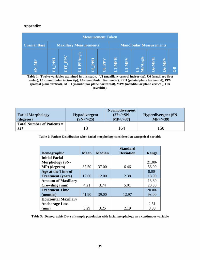
39
Appendix:
Measurement Taken
Cranial Base Maxillary Measurements Mandibular Measurements
SN
_M
P
U1_P
PH
U1T
_P
PV
U1-P
PA
ngle
U6_P
PH
U6_P
PV
L1-M
PH
L1-M
PV
L1-
MP
An
gle
L6-M
PH
L6-M
PV
OB
Table 1: Twelve variables examined in this study. U1 (maxillary central incisor tip), U6 (maxillary first
molar), L1 (mandibular incisor tip), L6 (mandibular first molar), PPH (palatal plane horizontal), PPV
(palatal plane vertical), MPH (mandibular plane horizontal), MPV (mandibular plane vertical), OB
(overbite).
Facial Morphology
(degrees)
Hypodivergent
(SN</=25)
Normodivergent
(27</=SN-
MP</=37)
Hyperdivergent (SN-
MP>/=39)
Total Number of Patients =
327 13 164 150
Table 2: Patient Distribution when facial morphology considered at categorical variable
Demographic Mean Median
Standard
Deviation Range
Initial Facial
Morphology (SN-
MP) (degrees) 37.50 37.00 6.46
21.00-
56.00
Age at the Time of
Treatment (years) 12.60 12.00 2.38
8.00-
18.00
Amount of Maxillary
Crowding (mm) 4.21 3.74 5.01
-13.80-
20.30
Treatment Time
(months) 41.90 39.00 12.97
20.00-
93.00
Horizontal Maxillary
Anchorage Loss
(mm) 3.29 3.25 2.19
-2.51-
8.88
Table 3: Demographic Data of sample population with facial morphology as a continuous variable
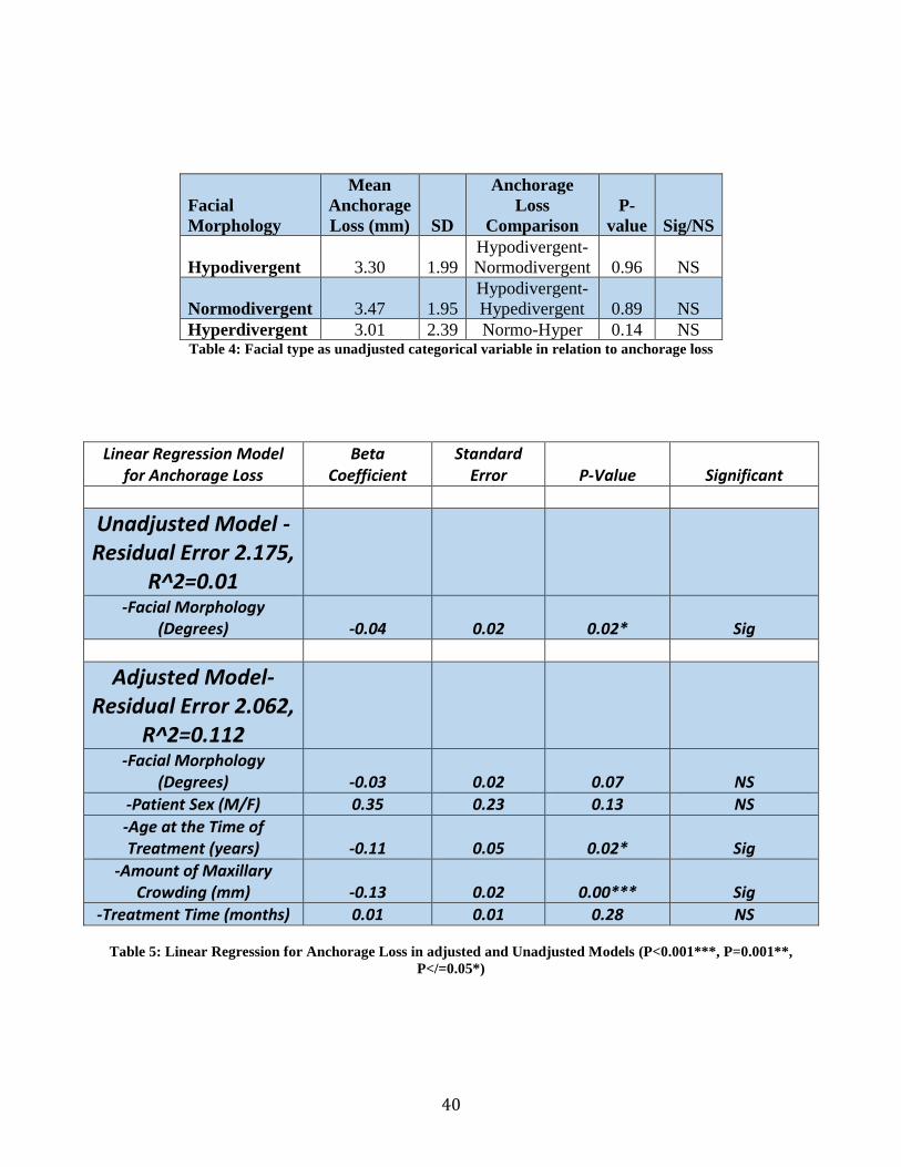
40
Facial
Morphology
Mean
Anchorage
Loss (mm) SD
Anchorage
Loss
Comparison
P-
value Sig/NS
Hypodivergent 3.30 1.99
Hypodivergent-
Normodivergent 0.96 NS
Normodivergent 3.47 1.95
Hypodivergent-
Hypedivergent 0.89 NS
Hyperdivergent 3.01 2.39 Normo-Hyper 0.14 NS Table 4: Facial type as unadjusted categorical variable in relation to anchorage loss
Linear Regression Model for Anchorage Loss
Beta Coefficient
Standard Error P-Value Significant
Unadjusted Model - Residual Error 2.175,
R^2=0.01
-Facial Morphology (Degrees) -0.04 0.02 0.02* Sig
Adjusted Model- Residual Error 2.062,
R^2=0.112
-Facial Morphology (Degrees) -0.03 0.02 0.07 NS
-Patient Sex (M/F) 0.35 0.23 0.13 NS
-Age at the Time of Treatment (years) -0.11 0.05 0.02* Sig
-Amount of Maxillary Crowding (mm) -0.13 0.02 0.00*** Sig
-Treatment Time (months) 0.01 0.01 0.28 NS
Table 5: Linear Regression for Anchorage Loss in adjusted and Unadjusted Models (P<0.001***, P=0.001**,
P</=0.05*)
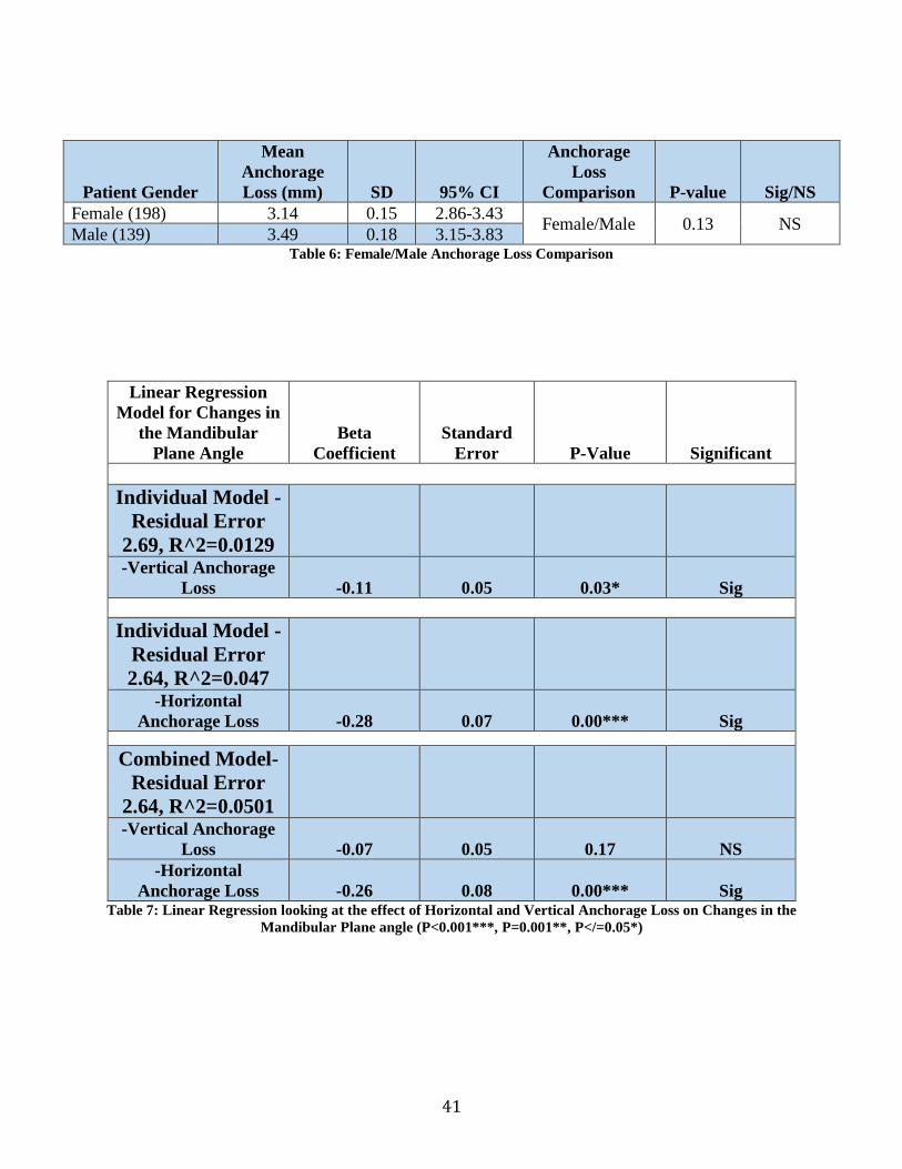
41
Patient Gender
Mean
Anchorage
Loss (mm) SD 95% CI
Anchorage
Loss
Comparison P-value Sig/NS
Female (198) 3.14 0.15 2.86-3.43 Female/Male 0.13 NS
Male (139) 3.49 0.18 3.15-3.83 Table 6: Female/Male Anchorage Loss Comparison
Linear Regression
Model for Changes in
the Mandibular
Plane Angle
Beta
Coefficient
Standard
Error P-Value Significant
Individual Model -
Residual Error
2.69, R^2=0.0129
-Vertical Anchorage
Loss -0.11 0.05 0.03* Sig
Individual Model -
Residual Error
2.64, R^2=0.047 -Horizontal
Anchorage Loss -0.28 0.07 0.00*** Sig
Combined Model-
Residual Error
2.64, R^2=0.0501 -Vertical Anchorage
Loss -0.07 0.05 0.17 NS
-Horizontal
Anchorage Loss -0.26 0.08 0.00*** Sig Table 7: Linear Regression looking at the effect of Horizontal and Vertical Anchorage Loss on Changes in the
Mandibular Plane angle (P<0.001***, P=0.001**, P</=0.05*)

42
Linear Regression Model for Change in Overbite
Beta Coefficient
Standard Error P-Value Significant
Full Model- Residual Error 1.46, R^2=0.442
-Facial Morphology (Degrees) 0.07 0.01 0.0000*** Sig
-Vertical Incisor Extrusion (mm) 0.32 0.03 0.0000*** Sig
Horizontal Incisor Retraction (mm) 0.06 0.04 0.078 NS
-Change in incisor Angulation (Degrees) -0.07 0.01 0.0000*** Sig
-Posterior Molar extrusion (mm) -0.29 0.03 0.0000*** Sig
Reduced Model- Residual Error 1.46,
R^2=0.438
-Facial Morphology (Degrees) 0.06 0.01 0.0000*** Sig
-Vertical Incisor Extrusion (mm) 0.33 0.03 0.0000*** Sig
-Change in incisor Angulation (Degrees) -0.05 0.01 0.0000*** Sig
-Posterior Molar extrusion (mm) -0.30 0.03 0.0000*** Sig
Table 8: Linear Regression for Changes in Overbite tested against four variables (P<0.001***, P=0.001**,
P</=0.05*)
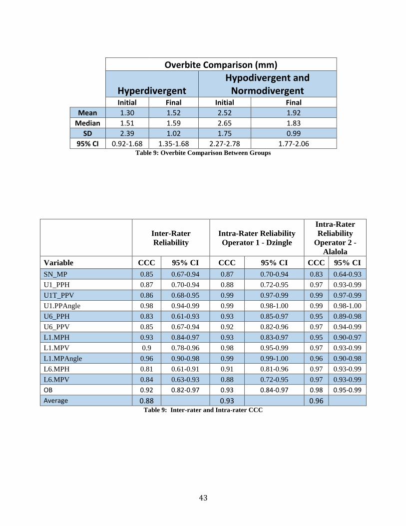
43
Overbite Comparison (mm)
Hyperdivergent Hypodivergent and
Normodivergent Initial Final Initial Final
Mean 1.30 1.52 2.52 1.92
Median 1.51 1.59 2.65 1.83
SD 2.39 1.02 1.75 0.99
95% CI 0.92-1.68 1.35-1.68 2.27-2.78 1.77-2.06 Table 9: Overbite Comparison Between Groups
Inter-Rater
Reliability
Intra-Rater Reliability
Operator 1 - Dzingle
Intra-Rater
Reliability
Operator 2 -
Alalola
Variable CCC 95% CI CCC 95% CI CCC 95% CI
SN_MP 0.85 0.67-0.94 0.87 0.70-0.94 0.83 0.64-0.93
U1_PPH 0.87 0.70-0.94 0.88 0.72-0.95 0.97 0.93-0.99
U1T_PPV 0.86 0.68-0.95 0.99 0.97-0.99 0.99 0.97-0.99
U1.PPAngle 0.98 0.94-0.99 0.99 0.98-1.00 0.99 0.98-1.00
U6_PPH 0.83 0.61-0.93 0.93 0.85-0.97 0.95 0.89-0.98
U6_PPV 0.85 0.67-0.94 0.92 0.82-0.96 0.97 0.94-0.99
L1.MPH 0.93 0.84-0.97 0.93 0.83-0.97 0.95 0.90-0.97
L1.MPV 0.9 0.78-0.96 0.98 0.95-0.99 0.97 0.93-0.99
L1.MPAngle 0.96 0.90-0.98 0.99 0.99-1.00 0.96 0.90-0.98
L6.MPH 0.81 0.61-0.91 0.91 0.81-0.96 0.97 0.93-0.99
L6.MPV 0.84 0.63-0.93 0.88 0.72-0.95 0.97 0.93-0.99
OB 0.92 0.82-0.97 0.93 0.84-0.97 0.98 0.95-0.99
Average 0.88 0.93 0.96 Table 9: Inter-rater and Intra-rater CCC

44
Figure 1: Cranial Base Structural Superimposition with SN-MP Lines Drawn
Figure 2: Maxillary Structural Superimposition with Horizontal and Vertical Reference Lines
Figure 3: Mandibular Structural Superimposition with Horizontal and Vertical Reference Lines
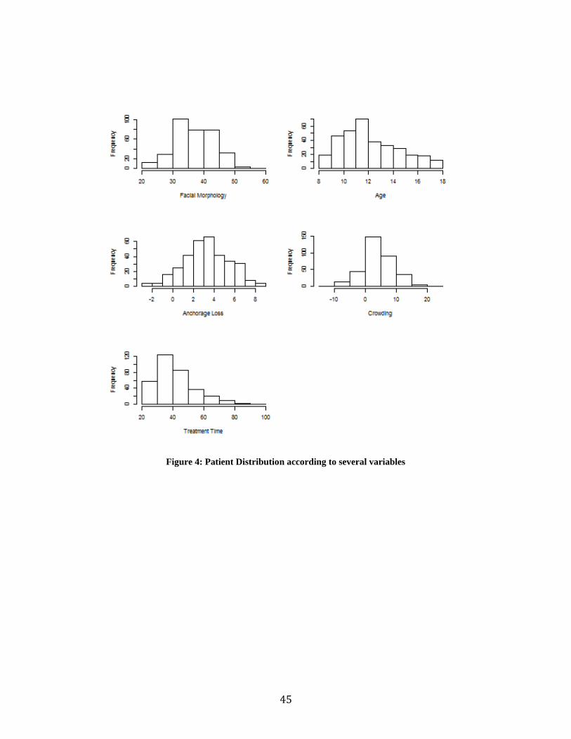
45
Figure 4: Patient Distribution according to several variables

46
Figure 5: Correlation between Facial Morphology and Horizontal Anchorage Loss
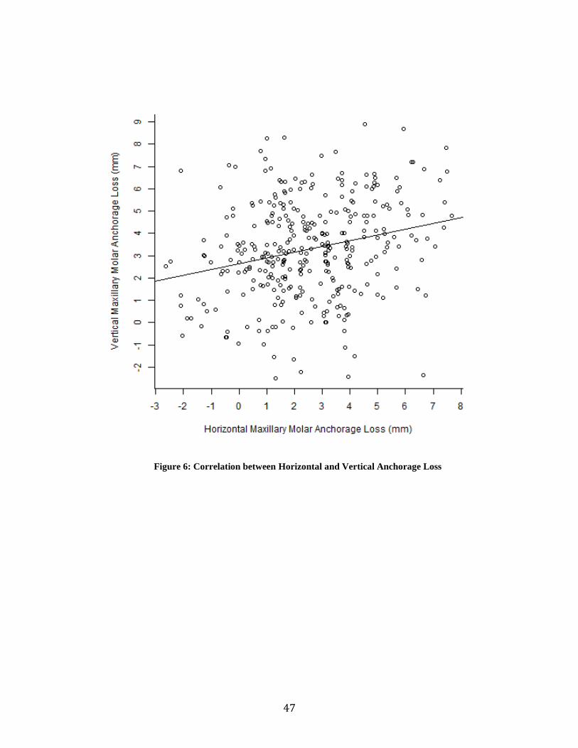
47
Figure 6: Correlation between Horizontal and Vertical Anchorage Loss
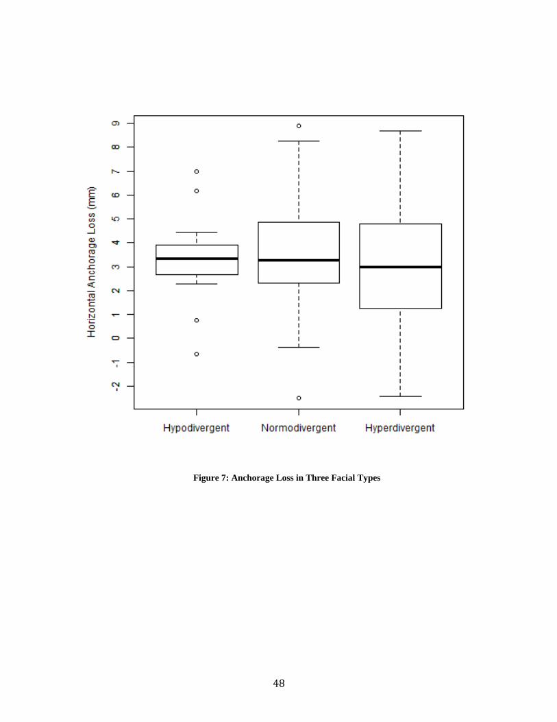
48
Figure 7: Anchorage Loss in Three Facial Types
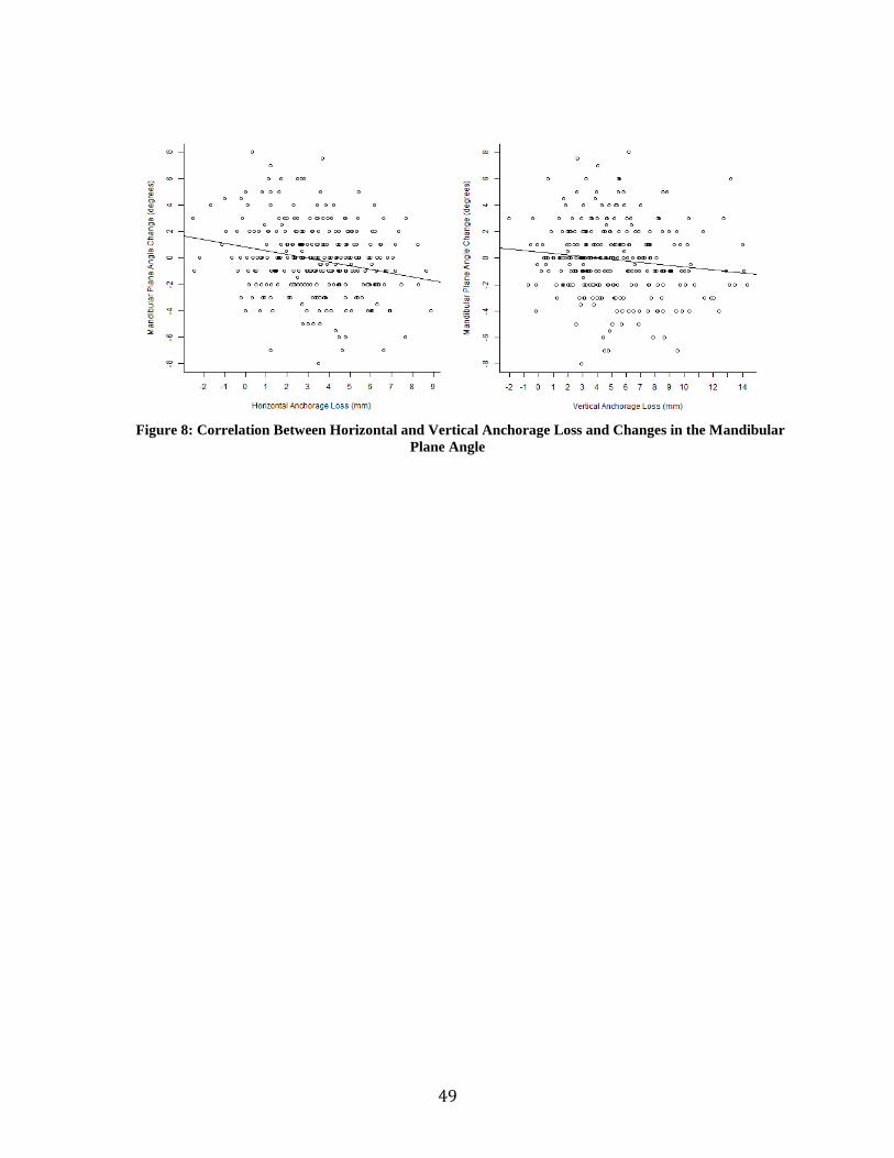
49
Figure 8: Correlation Between Horizontal and Vertical Anchorage Loss and Changes in the Mandibular
Plane Angle
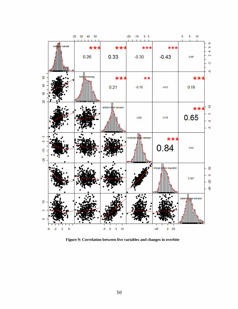
50
Figure 9: Correlation between five variables and changes in overbite