factors involved in passive transfer of …/67531/metadc130713/m2/1/high... · factors involved in...
TRANSCRIPT

FACTORS INVOLVED IN PASSIVE TRANSFER OF
CONTACT HYPERSENSITIVITY
APPROVEDi
mmm essor
tf'i M ,<r • , , • „ „ Minpfr Professor 1
\ \ C
Director o£" ','Depa^^entn" o# feiology
D««ui t)!f Sr«cluat« keto'ooi ^

FACTORS INVOLVED IN PASSIVE TRANSFER OF
CONTACT HYPERSENSITIVITY
THESIS
Presented to the Graduate Council of the
North Texas State University in Partial
Fulfillment of the Requirements
For the Degree of
MASTER OF ARTS
By
Walter L. Ellis, B. A,
Denton, Texas
August 1966

TABLE OF CONTENTS
Page
LIST OF TABLES iv
LIST OF ILLUSTRATIONS * v
Chapter
I. INTRODUCTION 1
II. MATERIALS AND METHODS 7 Animals Chemicals Preparation of Homologous Serum Conjugate Sensitization of Animals Skin Testing Production and Collection of Peritoneal
Exudate Testing for Passive Systemic Anaphylaxis Testing for Presence of Antibody
III. RESULTS 16
Skin Tests of Donor Animals Skin Tests and Passive Systemic
Anaphylaxis of Recipient Animals : im-passive Hemagglutination Test
IV. DISCUSSION 26
V. SUMMARY... 33
BIBLIOGRAPHY 35
xix

LIST OF TABLES
Table Page
I. Skin Tests Reactions . 9
II. Grades of Passive Systemic Anaphylaxis... 11
III. Skin Test Results of Donor Animals..... 17
IV. Skin Test of Lymph Node Recipients 18
V. Skin Tests and Passive Systemic Anaphylaxis of Recipients Receiving Peritoneal Cells After Incubation in Hanks 19
VI. Skin Tests and Passive Systemic Anaphylaxis of Recipients Receiving Supernatant After Incubation in Hanks of Peritoneal Exudate Cells 21
VII. Skin Tests and Passive Systemic Anaphylaxis of Recipients Receiving Peritoneal Exudate Cells After Incubation with Serum Conjugate.. 22
VIII.B Skin Tests and Passive Systemic Anaphylaxis of Recipients Receiving Supernatant After Incubation of Peritoneal Exudate Cells with Serum Conjugate... 2 3
IX. Comparison of Transfers for Schedule A and B..... 21
IV

LIST OF ILLUSTRATIONS
Figure Page
1. Time Schedules for Experimental Procedures
Employed. 8
2. Procedures for Handling Peritoneal Exudate Cells. 12

CHAPTER I
INTRODUCTION
Delayed hypersensitivity is an allergic state which
develops in response to many microbial agents and contact
substances. These include simple chemicals and materials
from plants such as poison oak and poison ivy (29). In an
effort to work out the mechanism of the delayed response,
workers have made many attempts to passively sensitize
normal animals.
Serum has been reported to be effective when taken
from tuberculous rabbits with well-developed lesions (36).
Normal guinea pigs have been passively sensitized to poison
ivy using serum (9). However, transfers with serum have
only occasionally been successful (3). Other workers used
a serum fraction, which they called an alpha-globulin, to
transfer tuberculin sensitivity (8). Unable to repeat this
transfer, another group said it was not true passive trans-
fer (12).
I The most consistently reproducible method of conferring
i
the passive state of delayed hypersensitivity on normal re-
cipients has been by the injection of leucocytes isolated
from peritoneal exudates, blood, spleen, lung, and lymph
nodes of sensitive donors (1,2,4-7,10-11,13-28,30-35).
Many attempts have been made to use cell extracts instead
of whole cells. These have varied, with many being success-
ful (1,11,13,15-17,25,32-31+) and some unsuccessful
(4,20,21,26,31,35).
One such successful report stressed the importance in
guinea pi-gs of the time lapse between donor sensitization
and passive transfers by cells and their extracts (13).
In that work, it was found that there exists a period when
whole peritoneal exudate cells will passively confer de-
layed sensitivity, but a sonic extract from them will not.
Yet, when the cells were collected four days later, it was
found that both whole cells and sonic extracts would trans-
fer sensitivity.
It would appear that in the cell populations from the
two time periods the "transfer factor" is of different
character or is in a different state. It is possible that
the cells, from the group where extracts do not transfer,
have not yet produced the "transfer factor" when collected.
The group which does yield extract transfers may have al-
ready reached this stage of development. This might be
checked by subjecting these cells to contact with the
specific allergen and then testing their passive transfer
capabilities. This investigation was undertaken to study such
differences in cells collected at two different time in-
tervals after initial sensitization of guinea pigs with
1-fluoro-2,4-dinitrobenzene.
CHAPTER BIBLIOGRAPHY
1. Baram, P. arid Mosko, N. M., "A Dialysable Fraction from Tuberculin Sensitive Human While Blood Cells Capable of Inducing Tuberculin-Delayed Hypersensitivity in Negative Recipients," Immunology, LXXXI (May, 1965), 461-474.
2. Blazkovec, A. A., Sorkin, E. and Turk, J. L., "A Study of the Passive Cellular Transfer of Local Cutaneous Hypersensitivity," International Archives of Allergy and Applied Immunology, XXVII (October, 1965);'
3. Burnet, F. M. and Fenner, F., The Production of Anti-bodies , 2nd ed., New,York, New York, Macmillan,1919.
•4. Chase, M. W., "The Cellular Transfer of Cutaneous Hypersensitivity to Tuberculin," Prodeedings: Society of Experimental Biology and Medicine, LIX (June, 1945), 134-135.
5. Chase, M. W., "The Cellular Transfer of Cutaneous Hypersensitivity," Journal of Bacteriology, LI (May, 1946), 643.
6. Chase, M. W., "Experimental Sensitization with Partic-ular Reference to Picryl Chloride," International Archives of Allergy and Applied Immunology, V (April, 1954),163-1917
7. Chase, M. W., Dameshek, W., Haberman, S., Samter, M., and Sawer, T. L., "The Role .of the Formed Elements of the Blood in Allergy and 'Hypersensitivity," Journal of Allergy, XXVI (June, 1955), 219-252.
8. Cole, L. R. and Favoni?, C. B., "Correlations Between , Plasma Protein Fractions, Antibody Titers and the Passive Transfer of Delayed and Immediate Cutaneous Reactivity to Tuberculin PPD and Tuberculopoly-i saccharides," Journal of Experimental Medicine1, CI (April, 1955), 3 91-470". j
j 9. Crepea, S. B., and Cooke, R. A., "Study on the Mechanism
of Dermatitis Venenata in Guinea Pig with a Demon-stration of Skin-sensitizing Antibody by Passive Transfer," Journal of Allergy, XIX (June, 1948), 353-370.

10. Cummings, M. M., Hoyt, M., and Gottshall, R. Y., "Passive Transfer of Tuberculin Sensitivity in the Guinea Pig," Public Health Reports, LXII (December, 1947), 994-998.
11. Cummings, M. M., Patnode, R. A., and Hudgins, P. C., "Passive Transfer of Tuberculin Hypersensitivity in Guinea Pigs Using Cells Disrupted by Sonic Nibration," The American Review of Tuberculosis and Pulmonary Diseases, LXXIII (February, 1956), 2W-250.
12. Ehrenkranz, N. J. and Waksman, B. H., ^Failure to Trans-fer Tuberculin Sensitivity Passively with Plasma Fractions Containing Alpha-Globulin," Journal of Experimental Medicine, CIV (December, 1956), 93^946.
13. Guthrie, R. K., Lowke, G. E., Ferguson, J. K. and Ellis, W. L., "Contact Hypersensitivity to Simple Chemicals. Time After Donor Sensitization as^a Factor in Passive Transfer," Journal of Investi-gative Dermatology, XLVI (February, 1966), 224-229.
14. Jansen, L. H. and Delden, J. V., "Investigation into the Possibility of Passive Transfer <?f Hypersensitivity to Tuberculin (PPD) in Pigs, and from Man to Pig," Dermatologica, CXXVIIK(February, 1964), 202-222.
15. Jeter, W. S., Tremain, M. M., and Seebohm, P. M., "Passive Transfer of Delayed Hypersensitivity to 2,4-Dinitro-chlorobenzene in Guinea Pigs with Leucocytic Extracts," Proceedings: Society of Experimental Bioiogy and Medicine,LLXXXVI (June, 1954), 251-253.
16. Jeter, W. S., Laurence, K. A., and Seebohm, P. M., "Analysis of Leukocytic Extracts from Guinea Pigs Hypersensitive to Tuberculin and 2,4-Dinitrochloro-henzene," Journal of Bacteriology, LXXIV (November, 1957), 680-683.
17. Kind, P. D., Bocobo, F. C., Curtis, A. C., and Bulala, P., "Cellular Passive Transfer of Contact Hyper-sensitivity to Paraphenylenediamine and to 2,4-Dinitrochlorobenzene in Guinea Pigs," Journal of Investigative Dermatology, XLIV,(January^ 1965), 7-11,
18. Kirchheimer, W. F. and Weiser, R. S., "The Tuberculin Reaction. I. Passive Transfer of Tuberculin Sensi-tivity with Cells of Tuberculous Guinea Pigs," ; Proceedings: Society of Experimental Biology and Medicine, LXVI (OctoberT 1947), 166-17TT

19. Landsteiner, K. and Chase, M. W., "Studies of Animals with Simple Chemical Compounds. VII. Skin Sensitization by Intraperitoneal Injections," Journal of Experimental Medicine, LXXI (February, m o ) , 2i7-2HS.
20. Landsteiner, K. and Chase, M. W., ^Experiments on Transfer of Cutaneous Sensitivity to Simple Compounds," Proceedings: Society of Experi-mental Biology and Medicine, XLIX (April, 1942), 688-690.
21. Landsteiner, K., The Specificity of Serological Reactions, Rev. ed., Cambridge, Massachusetts, Harvard Univer-sity Press, 1947.
22. Lawrence, H. S., "The Cellular Transfer of Cutaneous Hypersensitivity to Tuberculin in Man," Proceedings:
q Society of Experimental Biology and Medicine, LXX1~
23. Lawrence, H. S., "The Cellular Transfer in Humans of De-layed Cutaneous Reactivity to Hemolytic Strepto-cocci," Journal of Immunology, LXVII (February, 1952), 159-178.
24. Lawrence, H. S., "The Transfer in Humans of Delayed Skin Sensitivity to Streptococcal M Substance and to Tuberculin with Disrupted Leucocytes," Journal of Clinical Investigation, XXXIV (February, 1955), 219-230.
25. Lawrence, H. S., "Transfer of Delayed Hypersensitivity to Diptheria Toxin in Man," Journal of Experi-mental Medicine, CIV (September, 195677 321-335.
26. Metaxas, M. N. and Metaxas-Buehler, M., "Passive Trans-fer of Local Cutaneous Hypersensitivity to Tuber-culin," Proceedings: Society of Experimental Biology and Medicine, LXIX (October, 1948), 163-165.
27. Metaxas, M. N. and Metaxas-Buehler, M., "Studies on the Cellular Transfer of Tuberculin Sensitivity in the Guinea Pig," Journal of Immunology, LXXV (November, 1955), 333-347. i
28. Najarian, J. S. and Feldman, J. D., "Specificity of Passively Transferred Delayed Hypersensitivity," Journal of Experimental Medicine, CXVII (September, 1563), 31+1-355. I

29. Raffel, S., "Delayed Hypersensitivities," Progress in Allergy, IV (June, 1954), 173-198.
30. Segre, I), and Sharp, J. B., "Quantitation of Delayed Hypersensitivity in Guinea Pigs by a Local Passive Transfer Reaction," International Archives of Allergy and Applied Immunology, XXVII (March, 1965),
31. Stavitsky, A. B., "Passive Cellular Transfer of the Tuberculin Type of Hypersensitivity," Proceedings: Society of Experimental Biology and Medicine, LXvII (February! 1948), 225-2277
32. Tremaine, M. M. and Jeter, W. S., "Passive Cellular Transfer of Hypersensitivity to Serum Antigens in Rabbits," Journal of Immunology, LXXIV (February, 1955), 96-100.
33. Tsuji, S., Oshima, S., Oshiro, M., and Izumi, T., "Studies on the 'Transfer Factor" of Tuberculin Hypersensi-tivity in Animals," Journal of Immunology, XCIII (November, 1964), 838*849.
34. Turk, J. L., "Passive Transfer of Contact Sensitivity to Picryl Chloride in Guinea Pigs, with Subcellular Material," Nature, CXCI (August, 1961), 915-916.
35. Turk, J. L., "Attempts to Transfer Contact Sensitivity Passively with Subcellular Fractions in the Guinea Pig," International Archives of Allergy and Applied Immunology, XXI (June, 1962), "3T1-325.
36. Zinsser,, H. and Mueller, J. H., "On the Nature of Bac-terial Allergies," Journal of Experimental Medicine, XLI (January, 1925), 159-1777"

CHAPTER II
MATERIALS AND METHODS
Animals
Random bred Hartley strain albino guinea pigs were
obtained from Pine Ridge Caviary, Clinton, Tennessee. Only
animals weighing 350-500 grams were used.
Chemicals
DNFB (l-fluoro-2,^-dinitrobenzene) was obtained from
Matheson Coleman and Bell Chemical Company, Norwood, Ohio.
Light mineral oil (viscosity 80/90) and olive oil were
obtained from E. H. Sargent and Company, Dallas, Texas.
Preparation of Homologous Serum Conjugate
Homologous pooled guinea pig serum was conjugated with
DNFB (U) for use in incubation experiments and testing for
passive systemic anaphylaxis.
Sensitization of Animals
All animals were sensitized by rubbing five drops of two
per cent DNFB in absolute ethyl alcohol into the clippedj neck
using a polished glass rod. This application was repeated
at the same time each day for six days (2).

Skin Testing
Actively sensitzed donor guinea pigs were skin-tested
according to either of two schedules: (A) on the ninth day
after initial contact or (B) on the thirteenth day after
initial contact with DNFB. These schedules are shown in
Figure 1. All recipients were skin-tested for passive sensi-
tization forty-eight hours after injection of transfer prep-
arations. For the skin testing of both donors and recipients,
the hair was clipped from each animal's side and at separate
sites on the bare skin was placed one drop of 0.50 and 0.75
per cent DNFB in olive oil. Each drop of reagent was rubbed
gently into the skin using a separate polished glass rod.
These tests were read after twenty-four hours and were graded
as shown in Table I.
Paint Skin Oil Transfer Skin test Read :ipients skin
tests
1-6
1-6
donors
9 10 13 i
13 m J
17
15
19
16
20
Days for schedule A
Days for schedule B
Fig.l—Time schedules for procedures employed on each dayj.

TABLE I
SKIN TESTS REACTIONS*
Description of Reaction Site Grade
Marked homogenous erythema ++++
Homogenous erythema. +++
Patchy erythema ++
Slight erythema +
Questionable reaction +-
No reaction
J *From reference (2).
Production' and Collection of Peritoneal Exudate
At the same time as the reading of donor skin tests, the
animal's abdomens were shaved and fifteen milliliters of
sterile light mineral oil was injected into the peritoneal
cavity of each in order to evoke a peritoneal exudate (2).
Seventy-two hours after oil injection, animals were exsangui-
nated by intracardial puncture. To obtain cells for passive
transfer, the abdominal wall of each was opened and the cavity
washed three times with Hanks balanced salt solution (3)
modified by the addition of 0.1 per cent gelatin (2). |
Collected cells were sedimented by centrifugation at 2,000
revolutions per minute for twenty minutes. Packed cells jwere
10
washed once in Hanks solution, resedimented at 2,000 revo-
lutions per minute for ten minutes, and the volume recorded.
Washed cells were then resuspended to ten milliliters in Hanks
solution and divided into portions to contain a maximum of
0.75 milliliters packed cells. These cells were handled accord-
ing to one of the following procedures:
A) Cells resuspended in Hanks solution and, without
further treatment, injected into the peritoneal cavity
of a recipient guinea pig.
B) Cells resuspended to a volume of five milliliters in
Hanks solution and placed in an incubator at thirty-seven
degrees centrigrade for thirty minutes. After incubation
the cells were sedimented at 2,000 revolutions per
minute for ten minutes. The supernatant was removed and
injected into the peritoneal cavity of a recipient guinea
pig. The packed cells were washed three times with Hanks
solution 1 and then injected into the peritoneal cavity of
a recipient guinea pig.
C) Cells resuspended to a volume of five milliliters in
Hanks solution and an equal volume of homologous serum
conjugate of DNFB added. This suspension was placed in
an incubator at thirty-seven degrees centrigrade for I
thirty minutes. After incubation the cells were sediment-!
ed at 2,000 revolutions per minute for ten minutes.! The j
supernatant was removed and injected into the peritoneal
cavity of a recipient guinea pig.

11
D) Cells resuspended to a volume of five milliliters
in Hanks solution. These cells were disrupted using the
Branson Sonifier until microscopic examination of samples
showejd:.no;>cells intact. To prevent excessive heating,
all treatment with the sonifier was interrupted at inter-
vals. Containers of cells, while being subjected to
sonic treatment, were immersed in tap water. Suspensions
thus treated were centrifuged for twenty minutes at 2,000
revolutions per minute to sediment solid particles. The
supernatant fluid was decanted and injected into the peri-
toneal cavity of a recipient guinea-pig. The sediment
was washed once in Hanks solution, sedimented for twenty
minutes and the volume recorded. The sediment was then
resuspended and injected into the peritoneal cavity of
a recipient guinea pig.
E) Cells incubated as in (B), the supernatant removed
and injected into the peritoneal cavity of a recipient
guinea pig and the cells washed three times in Hanks
solution. These cells were then disrupted as in (D).
The resulting suspension was sedimented as in (D) and
the supernatant was injected into the peritoneal cavity S
of a recipient guinea pig. The sediment was washed: once
in Hanks solution, the volume recorded and then injected
into the peritoneal cavity of a recipient guinea pig. f
F) Cells incubated as in (C) and then treated as in CE). j
These procedures are summarized in Figure 2.

12
CO i H i H 0) o
0) . p
rd * 0 3 X Q)
rd Q) G O
- P
<D • P
G rd H W[
D X3 # n <D G O O &
0 U <D a)
CO i—I *4H rH O 0)
U <D a G
O CD CO <u Jh a l
x * a)
4 J rd £S 3 a co g h g
rd co s c
i H H G 0 ) « H
a
CQ
CO X5 i H 0) rH - P (U rd
a <D
• P
co x j h a> rH +J a) re
o o • H
^ G tu o
n o ^ CO co a)
2 J A
rH CO <D rd
O ^
, p g «d
• p x j rd a>
e s a> <u CLi'f—> 3 G
b 0 « H
X5 CO <D
rH X rH CO <U rd
a 5
• p
c rd
• p x > rd a>
p ^ k o a) <u
3 G (CO - H
X3 a>
• p
CO O rH 0) H * m 0) G
U « H
CO * 0 H 0) H - P 0 ) rd
O O • H
G M O
CO
X5 <U
• P CO o
rH a) i H »r-i Q) p O ' H
CO XJ rH CD rH - P d) rd
a a • H
g p o
co
*v 0
+ j
co o h a> rH « n a> g
O * H
!-P G <D X* £ O H , G
X> 0)
«P O <D
X3 CO XJ •r~i <D rd G G
0 0 & r d « H
- P G <d
• P XJ rd <D
• P E O a> <D P ^ * n 3 G
(GO » H
• P G a) x > 6 <D H , G
XJ CO XJ •r-> a ) rd G G
CO Jjfc fd * H
X> <D
4-1 o CU
• p
G fd
• P x > rd a> G - P U a a ) <u P ^ r ~ > O G
|CO «H
• P G <D X3 6 0 )
•H , G XJ CO XJ 0) rd G
CO 5 rd
X? CD
• P O a)
•r-> G
' H
• P G rd
- P XJ rd 0 ) G «P
a <u <u P u * n
J 3 c
(CO « H
CO rH rH <L> O
<D - p
rd X j P X a)
rH rd a) G O
• P • H u a> a
t>o
G • H rH X? G rd A u o
Mh
CO <D
X3 <D O 0
Ph 1 I
CSi
GO • H

13
In all incubation experiments, one recipient was injected
with a volume of the homologous serum conjugate equal to that
used in the incubation.
Axillary lymph nodes were removed following harvest of
peritoneal cells and the fat was trimmed away. The nodes were
finely minced with scissors and strained through screen wire
to separate cellss from pulp. Strained cells were washed once
in Hanks solution, sedimented for ten minutes, the volume
recorded, and then resuspended in five milliliters of Hanks
solution for injection into the peritoneal cavity of a
recipient guinea pig (2).
All cells, supernatants, and sediments were injected into
the peritoneal cavity of recipient guinea pigs within five
hours of donor exsanguination.
Testing for Passive Systemic Anaphylaxis
After reading skin tests of recipients, each was injected
intracardially with one milliliter of homologous serum conjugate
containing five milligrams per milliliter serum protein and
observed for symptoms of systemic anaphylaxis (1). Reactions
were graded as shown in Table II. The protein content of the
serum was determined by using the Biuret method (1).
Testing for Presence of Antibody
Incubation supernatants and sonicated cell supernatants
were checked for the presence of antibody using the passive
hemagglutination technique (1).

14
TABLE II
GRADES OF PASSIVE SYSTEMIC ANAPHYLAXIS
Reaction Grade in
Animal
Death
Severe. ++ +
Moderate ++
Slight +
Negative

CHAPTER BIBLIOGRAPHY
1. Campbell, D. H., Garey, J. S., Cremer, N. E., and Sussdorf, D. H., Methods In Immunology, New York, New York, W. A. Benjamin, Inc., 1964.
2. Guthrie, R. K., Lowke,̂ G. E., Ferguson, J. K., and Ellis, W. L., "Contact Hypersensitivity to Simple Chemicals. Time After Donor Sensitization as a Factor in Passive Transfer," Journal of Investigative Dermatology, XLVI (February,"~T966) T2H-229.
3. Hanks, J. H., "The Longevity of Chick Tissue Cultures Without Renewal of Medium," Journal of Cellular and Comparative Physiology, XXXI (April, 1948), 235-360.
4. Kabat, E. A. and Mayer, M. M., Experimental Immunochemistry, Springfield, Illinois, Charles C. Thomas, 1961.
15

CHAPTER III
RESULTS
Skin Tests of Donor Animals
The skin tests of all donor animals showed them to be
sensitive to the chemical used. In Table III are the
reactions of all donors to 0.75 per cent DNFB in olive
oil. Groups XIII-3 and XVII-3 were not skin tested in an
attempt to study the possible effect of skin testing on
passive transfer of the delayed state. These two groups were
run simultaneously with groups XIII-2 and XVII-2.
Skin Tests and Passive Systemic Anaphylaxis
of Recipient Animals
All skin test reactions are to 0.75 per cent and 0.50
per cent DNFB in olive oil. The passive systemic anaphy-
laxis reactions are in response to an intracardial injection
of five milligrams homologous serum protein conjugate of
DNFB contained in a one milliliter dose.
Table IV represents the lymph node recipients' reactions. |
These cells usually gave positive passive transfers of the
delayed state. I i
The results for recipients receiving the peritoneal!
exudate cells which were incubated in Hanks solution are
shown in Table V. The cells from the XIII groups gave passive
16

17
TABLE III
SKIN TEST RESULTS OF DONOR ANIMALS3
Test Number
Number of Donors
Number 4. 4. 4.4.
of Animals with Reactions of + ++ ++ + +_
Group XII1%
1 -r"v
Test 1 10 7 3 0 0 0 0
Test 2 10 8 2 0 0 0 0
Test 3C 10
Test 4 12 12 0 0 0 0 0
Test 5 12 0 4 8 0 0 0
Test 6 24 0 17 7 0 0 0
Group XVIId 1 4
Test 1 10 10 0 0 0 0 0
Test 2 9 9 0 0 0 0 0
Test 3C 10 • *** • • * • • • • •
Test 4 12 12 0 0 0 0 0
Test 5 24 7 8 6 3 0 ;j
-Hi
0
aReactions to 0.75% DNFB in olive oil,
^Donors from schedule A.
^Animals not skin tested.
dDonors from schedule B.

18
TABLE IV
SKIN TEST OF LYMPH NODE RECIPIENTS
Skin Tests Test Cell Volume DNFB Concentration in Olive Oil Number Transferred* 0.75% 0.50%
Group XIII**
Test 1 0.30 4**.; ++ + + 4- + + +
Test 2 0.30 + + + + + + +
Test 3 0.20 + + 4- +
Test 4 0.25 4- -
Test 5
O
rH • O + -
Test 6 0*25 + + + + +
Group XVII***
Test 1 0.30 + 4* + + 4- + 4-
Test 2 0.10 + +4 +
Test 3 0.10 4- + ~
Test •+ 0.35 + 4-
Test 5 0.35 + - -
*Cell volume in milliliters.
**Animals from schedule A.
*fc*Animals from schedule B.

19
TABLE V
SKIN TESTS AND PASSIVE SYSTEMIC ANAPHYLAXIS OF RECIPIENTS RECEIVING PERITONEAL EXUDATE CELLS
AFTER INCUBATION IN HANKS
Skin Tests Passive Test
Number Cell Volume Transferreda
DNFB Concentration in Olive Oil 0.75% 0.50%
Systemic Anaphylaxis
Group XIIIb
Test 1 0.75 ++ +
Test 2 0.75 + + + -
Test 3 0.75 + + -
Test 4 0.75 ++ + -
Test 5 0.75 + + - -
Test 6 0.75 + + - -
Group XVIT0
Test 1 0.75 + -
Test 2 0.60 + - - -
Test 3 0.75 - - -
Test H 0.75 + - + - -
Test 5 0.75 + - -
a Cell volume in milliliters,
b Animals from schedule A.
c Animals from schedule B.

20
transfers after the incubation, while the cells from the
XVII groups never did. The recipients of the supernatants
after these incubations gave the reactions shown in Table
VI. These supernatants conferred activity to all the
recipients of both time periods.
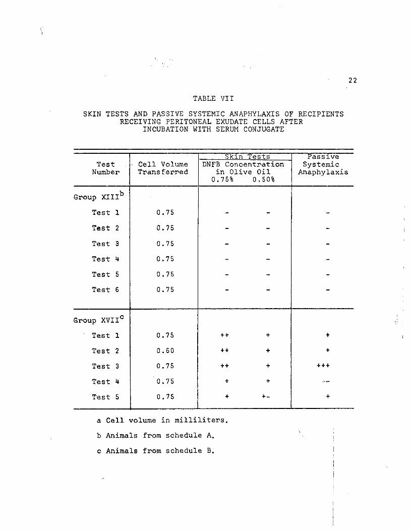
When peritoneal exudate cells were incubated with
homologous conjugated serum, the recipients of such cells
gave the reactions in Table VII. Cells from XIII groups
gave no reactions. However, cells from XVII groups gave
positive passive transfers of the delayed state and passive
systemic anaphylaxis. When the supernatants from these
incubations frere injected, the recipients from the XVII
groups were completely negative. However, the XIII groups
exhibited both the delayed response and passive systemic
anaphylaxis. This is shown in Table VIII.
Two experiments, XIII-6 and XVII-5, were run to study
all procedures which were described in the preceding chapter
for passive transfers. Table IX shows the differences and
similarities of the passive transfer capabilities of the two
time periods studied.
The control animals, which received homologous serum !
conjugate equal in volume to that used in the incubation j
part of the experiments, were all negative. |
Passive Hemagglutination Test
Using the passive hemagglutination technique, all
supernatants from incubation experiments in groups XIII-5;,

21
TABLE VI
SKIN TESTS AND PASSIVE SYSTEMIC ANAPHYLAXIS OF RECIPIENTS RECEIVING SUPERNATANT AFTER INCUBATION IN HANKS OF
PERITONEAL EXUDATE CELLS
Skin Tests Passive Test Number
DNFB Concentration in Olive Oil 0.75% 1 0.50%
Systemic Anaphylaxis
Group XIIIa
Test 4 +++ -
Test 5 ++ -
Test 6ab + + -
Test 6b \
+ + -
Group XVIIc
Test 3 + + + -
Test 4 + + + -
Test 5a^ + -
Test 5b + +-
—L
-
aAnimals from schedule A.
^XIII-6 Ifcun in duplicate,
c Animals from schedule B.
XVII-5 rvm in duplicate.
22
TABLE VII
SKIN TESTS AND PASSIVE SYSTEMIC ANAPHYLAXIS OF RECIPIENTS RECEIVING PERITONEAL EXUDATE CELLS AFTER
INCUBATION WITH SERUM CONJUGATE
Skin Tests Passive Test Number
- Cell Volume Transferred
DNFB Concentration in Olive Oil 0.75% 0.50%
Systemic Anaphylaxis
Group XIIIb
Test 1 0.75
Test 2 0.75 _ -
Test 3 0.75 - -
Test 4 0.75 - -
Test 5 0.75 - -
Test 6 0.75 - -
Group XVIIC
Test 1 0.75 ++ + +
Test 2 0.60 ++ + +
Test 3 0.75 ++ + +++
Test 4 0.75 + +
Test 5 0.75 + +- +
a Cell volume in milliliters,
b Animals from schedule A.
c Animals from schedule B.

23
TABLE VIII
SKIN TESTS AND PASSIVE SYSTEMIC ANAPHYLAXIS OF RECIPIENTS RECEIVING SUPERNATANT AFTER INCUBATION OF PERITONEAL
EXUDATE CELLS WITH SERUM CONJUGATE
Skin Tests Passive Test Number
DNFB Concentration in Olive Oil 0.75% 0.50%
Systemic Anaphylaxis
Group XIII*
Test 1 + + + 4-
Test 2 + +
Test 3 + - -
Test 4 + - +
Test 5 + - -
Test 6 + - ++
Group XVII**
Test. 1 + - + - -
Test 2 - - -
Test 3 - - -
Test k + - + - -
Test 5 — -| |
*Animals from schedule A.
**Animals from schedule B,

24
TABLE IX
COMPARISON OF TRANSFERS FOR SCHEDULES A AND B
Procedure Represented Skin Tests
DNFB Concentration in Olive Oil 0.75% 0.50%
Passive Systemic
Anaphylaxis
Experiment XIII-6a
(A) Whole Celjlsc
(D) Sonic Supernatant (D) Sonic Sediment (B) Whole Cells (B) Incubation Supernatant (E) Sonic Supernatant (E) Sonic Sediment (C) Whole Cellse
(C) Incubation Supernatant (F) Sonic Supernatant (F) Sonic Sediment
+ +
+-+
++
Experiment XVII-5^3
(A) Whole Cells0
(D) Sonic Supernatant (D) Sonic Sediment (B) Whole Cellsd
(B) Incubation Supernatant (E) Sonic Supernatant (E) Sonic Sediment (C) Whole Cellse
(C) Incubation Supernatant (F) Sonic Supernatant (F) Sonic Sediment
+ +
+
+
+
+
+ -
^From schedule A.
^From schedule B. j i
cPeritoneal exudate cells untreated* ; !
^Peritoneal exudate cells incubated in Hanks. |
ePeritoneal exudate cells incubated with serum conjugate.

25
XIII-6, XVII-1*, and XVII-5 and all sonicated cell supernatants
from groups XIII-6 and XVII-5 were tested for presence
of antibody. In no test was a positive hemagglutination
observed.

CHAPTER IV
DISCUSSION
These experiments have shown that there are differences
in the reactivity of peritoneal exudate cells collected at
different time periods. As reported earlier (5), it was
found that sonic extracts from peritoneal exudate cells
collected thirteen days after donors' sensitization would
not passively transfer the delayed state, although whole
cells did. If the cells were collected four days later,
both sonic extracts and whole cells gave positive passive
transfers. Many workers have reported positive transfers
using sonic extracts (1,3,5,6,7,8,9,12,11,18,19,20), while
others have been unsuccessful (2,10,11,15,17,21). These
discrepancies may have been in the animal species used,
the allergens used, the cell types used, time schedules, or
other peculiarities of each experiment.
As reported in recent reviews (4,13,16),it was found
that lymph node cells from sensitive donors will passively i
transfer delayed sensitivity. j
In the experiments where the cells were incubated with 1
homologous serum conjugate there is more evidence that the
cells of the two time periods are different. These results,
Tables VII and VIII, show that the XIII cells, from the time !
period which does not yield a sonic extract that will passively |
26 !

27
transfer, are desensitized by contact with homologous serum
conjugate. Some of the transfer activity did appear in the
supernatant after incubation. The XVII cells, collected
four days later and which yield a positive passive trans-
ferring sonic extract, seem to have the transfer activity
"fixed" with them. No transfer activity appeared in the
supernatant after incubation. Incubation experiments
have been devised by others whereby collected cells were
allowed to come in contact with the specific allergen
in vitro (12,13). It is not possible at this time to
directly compare these results with others for, according
to Chase (16), this incubation for contact sensitivity
in the guinea pig has not been reported.
A technique designed at first to be a control over
the preceeding experiments developed into still another
experimental procedure which demonstrated the afore-
stated time differences in collected cells. When cells
were incubated in Hanks solution, it was found that both
groups release transfer activity into the supernatant as
shown in Table VI. However, these results show that the
cells which did not yield sonic extract transfers would
still yield whole cell transfers. And the cells which did
yield sonic extract transfers no longer gave whole cell.-1 I
transfers. This type of cell incubation has not beenrre-I
ported previously. In working with human leucocytes, j
Lawrence (12) found that incubation of cells in serum for a
short period would yield a supernatant which gave passive
transfers-. I

28
In the time periods studied, it was found that passive
systemic anaphylaxis did not become evident in recipients
unless the cells were first incubated in the presence of
homologous serum conjugate. After such incubation, in the
group which gave no sonic extract transfers, only the super-
natant gave positive passive systemic anaphylaxis. It
appears that, like the delayed properties, the anaphylactic
property of these cells is released in the presence of the
conjugate. In the group which gave sonic extract transfers
the cells retained their anaphylactic property after con-
tact or were stimulated to produce it by the contact. It
may be that these reactions are not due to classical anti-
body. This is indicated by the negative hemagglutination
tests for the samples yielding positive passive systemic
anaphylaxis. Perhaps these positive reactions were due to an
antibody other than the classical type.
Unexplained by these experiments is the apparent in-
compatibility in the failure of sonic extracts of cells
collected thirteen days after donor sensitization to give
passive transfers»while incubation of these cells yields a
supernatant which will transfer. This might be explained i
by the experiments of Tsuji et al. (18), in which they found
that an "inhibitor" was indicated and could be removed from
cellular extracts by dialysis. If this is the case, then
the "inhibitor" is either not present in the XVII cells or
is in low enough concentration in proportion to the "trans-
fer factor" that it cannot express itself in these sonic!

29
extracts. Perhaps the XVII cells, Table V, which have re-
leased "transfer factor", do not give positive transfers
after Hanks incubation because the "inhibitor" is now in
sufficient quantity to mask it.
The experiments which have been discussed support the
idea (5) of differences in cellular reactivities at differ-
ent time periods in guinea pigs hypersensitive to 1-fluoro-
2, if-dinitrobenzene.

CHAPTER BIBLIOGRAPHY
1. Baram, P. and Mosko, N. M., "A Dialysable Fraction from Tuberculin Sensitive Human White Blood Cells Capable of Inducing Tuberculin-Delayed Hypersensi-tivity in Negative Recipients," Immunology, LXXXI (May, 1965), 461-474.
2. Chase, M. W., "The Cellular Transfer of Cutaneous Hypersensitivity to Tuberculin," Proceedings: Society of Experimental Biology and Medicine, LIX (June, 1945), 134-133".
3. Ehrei^kranz, N. J. and Waksman, B. H., "Failure to Transfer "Tuberculin Sensitivity Passively with Plasma Fractions Containing Alpha Globulin," Journal of Experimental Medicine, CIV (December, 1956), 9 35-946.
4. Gowans, J. L. and Mc Gregor, D. D., "The Immunological Activities of Lymphocytes," Progress in Allergy, IX (June, 1965), 1-63.
5. Guthrie, R. K., Lowke, G. E., Ferguson, J. K., and Ellis, W. L., "Contact Hypersensitivity to Simple Chemicals. Time After Donor Sensitization as a Factor in Passive Transfer," Journal of Investigative Dermatology, XLVI, (February, 1966), 224-229.
6. Jansen, L. H. and Delden, J. V., "Investigation into the Possibility of Passive Transfer of Hypersensitivity to Tuberculin (PPD) in Pigs, and from Man to Pig," Dermatologica , CXXVIII (February, 1964), 202-222.
7. Jeter, W. S., Lawrence, K. A. and Seebohm, P. M., "Analysis of Leukocytic Extracts from Guinea Pigs Hypersensitive to Tuberculin and 2,4-Dinitrochloro-benzene," Journal of Bacteriology, LXXIV (November, 1957), 680-683.
8. Kind, P. D., Bocobo, F. C., Curtis, A. C., and Bulala, P., "Cellular Passive Transfer of Contact Hypersensi-tivity to Paraplenylenediamine and to 2,4-Dinitro-chlorobenzene in Guinea Pigs," Journal of Investi-gative Dermatology, XLIV (January, 196577 7-11.
30

31
9. Kirchheimer, W. F. and Weiser, R. S., "The Tuberculin Reaction. I. Passive Transfer of Tuberculin Sensitivity with Cells of Tuberculous Guinea Pigs," Proceedings; Society of Experimental Biology and Medicine, LXVl (["October, 1947), 166-170.
10. Landsteiner, K., The Specificity of Serological Reactions, Rev. ed., Cambridge, Massachusetts, Harvard University Press, 1947.
11. Lawrence, H. S., "The Cellular Transfer of Cutaneous Hypersensitivity to Tuberculin in Man," Proceedings: Society of Experimental Biology and Medicine, LXXI, (August ,~~T949), 516-522.
12. Lawrence, H. S. and Pappenheimer, A. M., "Transfer of Delayed Hypersensitivity to Diptheria Toxin in Man," Journal of Experimental Medicine, CIV (September, 1956T,321-335 .
13. Lawrence, H. S., Cellular and Humoral Aspects of the Hypersensitive States, New York, New York, Paul B. Hoeber, Inc.7 19 59.
14. Metaxas, M. N. and Metaxas-Buhler, M., "Passive Transfer of Local Cutaneous Hypersensitivity to Tuberculin," Proceedings: Society of Experimentali-Biology and Medicine, EXIX (October, 1948) , 163-165.
15. Metaxas, M. N. and Metaxas-Buhler, M., "Studies on the Cellular Transfer of Tuberculin Sensitivity in the Guinea Pig," Journal of Immunology, LXXV (November, 1955), 333-347::
16. Raffel, S., ed., The Medical Clinics of North America, Philadelphia, Pennsylvania, W. B. Saunders Company, 19 65.
17. Tremaine, M. M. and Jeter, W. S., "Passive Cellular Transfer of Hypersensitivity to Serum Antigens in Rabbits," Journal of Immunology, LXXIV (February, 1955), 96-100. i
18. Tsuji, S., Oshima, S., Oshiro, M. and Izumi, T., "Studies on the'Transfer Factor' of Tuberculin Hypersensi-tivity in Animals," Journal of Immunology, XCIII (November, 1964), 838-849.

32
19. Turk, J. L., "Passive Transfer of Contact Sensitivity to Picryl Chloride in Guinea Pigs, with Sub-cellular Material, " Nature, CXCI (August, 1961), 915-916.
20. Turk, J. L. and Asherson, G. L., "Attempts to Transfer Contact Sensitivity Passively with Subcellular Fractions in the Guinea Pig," International Archives of Allergy and Applied Immunology, XXI (June, 1962), 321-325.
21. Zinsser, H. and Mueller, J. H., "On the Nature of Bac-terial Allergies," Journal of Experimental Medicine, XLI (January," 19 3"5T7 159-177.

CHAPTER V
SUMMARY
Guinea pigs were sensitized with l-fluoro-2,4-dinitro-
benzene. Peritoneal exudate cells were studied in two
time periods in relation to time lapse after donor sensiti-
zation. Cells collected thirteen days after sensitization
differed from those collected seventeen days after sensiti-
zation in terms of transfer reactivities. Experiments
showed the following:
1. Sonic extracts of thirteen day cells failed to
transfer while seventeen day sonic extracts did transfer.
2. Incubation in Hanks gave rise to a release into the
mediumsa transfer reactivity. The thirteen day cells
were still capable of positive transfers, but the seven-
teen day cells no longer had this capability.
3. Incubation with homologous serum conjugate desensi-
tized the thirteen day cells, leaving only the supernatant
with transfer activity. Seventeen day cells retained
transfer activity, with none appearing in the supernatant.
Passive systemic anaphylaxis was transferred by: the
supernatant from the incubation of thirteen day cells
with homologous serum conjugate. Such incubation of
seventeen day cells left them, but not the supernatant,
with the transfer capability. | i
33 - I

3H
Only after incubation of cells with homologous serum
conjugate was passive systemic anaphylaxis observed in re-
cipient guinea pigs. Passive hemagglutination tests of all
incubation and sonication supernatants failed to demonstrate
classical antibody.
It remains to be determined whether an "inhibitor" of
"transfer factor" is really present in the time periods
studied and, if so, the mechanism of its action.

BIBLIOGRAPHY
BOOKS
Burnet, F. M. and Fenner, F., The Production of Antibodies, 2nd ed., New York, New York, Macmillan,-T949.
Campbell, D. H., Garey, J. S., Cremer, N. E., and Sussdorf, D. H., Methods in Immunology, New York, New York,
Landsteiner, K., The Specificity of Serological Reactions. Rev. ed. Cambridge, Massachusetts, Harvard University Press, 1947.
Lawrence, D. H., Cellular and Humoral Aspects of the Hyper-sensitive States, New York, New York, Paul B. Hoeber, Inc., 19591
Raffel, S., The Medical Clinics of North America, Philadelphia, Pennsylvania, W. B~ Saunders Company, iabt>.
ARTICLES
Baram, P. and Mosko, N. M., "A Dialysable Fraction from Tuberculin Sensitive Human White Blood Cells Capable of Inducing Tuberculin-Delayed Hypersensitivity in Negative Recipients," Immunology LXXXI (May, 1965), 461-474.
Blazkovec, A. A., Sorkin, E. and Turk, J. L., "A Study of the Passive Cellular Transfer of Local Cutaneous Hyper-sensitivity," International Archives of Allergy and Applied Immunology, XXVlI (October, I9F5")̂ 2 8 a - 3 0"3T~
Chase, M. W., "The Cellular Transfer of Cutaneous Hypersen-sitivity," Journal of Bacteriology, LI (May, 1946), 643.
Chase, M. W., "The Cellular Transfer of Cutaneous Hyper-sensitivity to Tuberculin," Proceedings; Society of Experimental Biology and Medicine, LIX (June, 1945), 134-135.
cha.'-se ; Chase, M. W., "Experimental Sensitization with Particular
Reference to Picryl Chloride," International Archives of Allergy and Applied Immunology, V (April, li)b4), 163-191.
35
36
Chase, M. W. , Dameshek, W. , Haverman, S., Samter, M. and Sqiver, T. L., "The Role of the Formed Elements of the Blood in Allergy and Hypersensitivity," Journal of Allergy, XXVI (June, 1955), 219-252.
Cole, L. R. and Favour, C. B., "Correlations Between Plasma Protein Fractions, Antibody Titers, and the Passive Transfer of Delayed and Immediate Cutaneous Reactivity to Tuberculin PPD and Tuberculopolysaccharides," Journal of Experimental Medicine, CI (April, 1955), 391-420.
Crepea, S. B. and Cooke, R. A., "Study on the Mechanism of Dermatitis Venenataiin Guinea Pigs with a Demonstration of Skin-sensitizing Antibody by Passive Transfer," Journal of Allergy, XIX (June, 1948), 353-370.
Cummings, M. M., Hoyt, M. and Gottshall, R. Y., "Passive. Transfer of Tuberculin Sensitivity in the Guinea Pig," Public Health Reports, LXII (December, 1947), 994—998.
Cummings, M. M., Patnode, R. A., and Hudgins, P. C., "Passive Transfer of Tuberculin Hypersensitivity in Guinea figs Using Cells Disrupted by Sonic Vibration,V The American Review of Tuberculosis and Pulmonary Diseases, LXSTXT (February, 195677 246-250.
Ehrenkranz, N. J.5and Waksman, B. H., "Failure to Transfer Tuberculin Sensitivity Passively with Plasma Fractions Containing Alpha Globulin," Journal of Experimental Medicine, CIV (December-* 1956Tj 935-941".
Gowans, J. L. and Mc Gregor, P. P., "The Immunological Activities of Lymphocytes," Progress in Allergy, IX (June, 1965), 1-63.
Guthrie, R. K., Lowke, G. E., Ferguson, J. K., and Ellis, W. L. , "Contact Hypersensitivity to Simple Chemicals. Time After Donor Sensitization as a Factor in Passive Transfer," Journal of Investigative Dermatology, XLVI (February, 19 6677 224-229.
Hanks, J. H., "The Longevity of Chick Tissue Cultures Without Renewal of Medium," Journal of Cellular and Comparative Physiology, XXXI (April, 1941*7, 235-260. i
Jansen, L. H. and Delden, J. V., "Investigation into the ! Possibility of Passive Transfer of Hypersensitivity to Tuberculin (PPD) in Pigs, and From Man to Pig," ! Dermatologica, CXXVII (February, 1964), 202-222. :

37
Jeter, W. S., Tremaine, M. H., and Seebohm, P. M., "Passive Transfer of Delayed Hypersensitivity to 2,4-Dinitro-chlorobenzene in Guinea Pigs with Leucocytic Extracts," Proceedings: Society of Experimental Biology and Medicine,LXXXVI (June," 1954), 251-253.
Jeter, W. S. and Laurence,K. A. and Seebohm, P. M., "Analysis of Leukocytic Extracts from Guinea Pigs Hypersensitive to Tuberculin and 2,4-Dinitrochlorobenzen," Journal of Bacteriology, LXXVI (November, 1957), 680-683.
Kind, P. D. , Bocobo, F. C., Curtis, A. C., and Bulala, P., "Cellular Passive Transfer of Contact Hypersensitivity to Paraphenylenediamine and to 2,4-Dinitrochlorobenzene in Guinea Pigs," Journal of Investigative Dermatology, XLIV (January, 1965)", 7-11.
Kirchheimer, W. F. and Weiser, R. S., "The Tuberculin Reaction. I. Passive Transfer of Tuberculin Sensi-tivity with cells of Tuberculous Guinea Pigs," Proceedings: Society of Experimental Biology and Medicine, LXVI (October, 1947), 166-170.
Landsteiner, K. and Chase, M. W., "Experiments on Transfer of Cutaneous Sensitivity to Simple Compounds," Proceedings: Society of Experimental Biology and Medicine, XLIX (April, "^9 4 2), 688-690 .
Landsteiner, K. and Chase, M. W., "Studies on the Sensitization of Animals with Simple Chemical Compounds. XII. Skin Sensitization by Intraperitoneal Injections," Journal of Experimental Medicine, LXXI (February, 1940), 237-245.
Lawrence, H. S., "The Cellular Transfer in .Humans of Delayed Cutaneous Reactivity to Hemolytic Streptococci," Journal of Immunology, LXVIII (February, 1952), 159-178.
Lawrence, H. S., "The Cellular Transfer of Cutanous Hyper-sensitivity to Tuberculin in Man," Proceedings: Society of Experimental Biology and Medicine, LXXI
~~*"(August, 1949; , 516-522.
Lawrence, H. S., "The Transfer in Humans of Delayed Skin j Sensitivity to Streptococcol M Substance and to Tuberculin with Disrupted Leucocytes," Journal of Clinical Investigation, XXXIV (February, 1955), 7T9-230.
Lawrence, H. S. and Pappenheimer, A. M., "Transfer of Delayed Hypersensitivity to Diptheria Toxin in Man," Journal of Experimental Medicine, CIV (September, 1956), 321-335.
38
Metaxas, M. N. and Metaxas-Buhler, H., "Passive Transfer of Local Cutaneous Hypersensitivity to Tuberculin," Proceedings: Society of Experimental Biology and Medicine, EXIX (October, 194-8), 163-1651
Metaxas, M. N. and Metaxas-Buehler, M., "Studies on the Cellular Transfer of Tuberculin Sensitivity in the Guinea Pig," Journal of Immunology, LXXV (November, 1955), 333-347:
Najarian, J. S. and Feldman, J. D., "Specificity of Passively Transferred Delayed Hypersensitivity," Journal of Experimental Medicine, CXVIII (September^ 1963), m-3b2.
Raffel, S., "Delayed Hypersensitivities," Progress in Allergy. IV (June, 1951-0 , 173-178.
Segre, D. and Sharp, J. B., "Quantitation of Delayed Hypersensitivity in Guinea Pigs by a Local Passive Transfer Reaction," International Archives of Allergy and Applied Immunology, XXVII (March, 1965), 82-101.
Stavitsky, A. B., "Passive Cellular Transfer of the Tuber-culin Type of Hypersensitivity," Proceedings: Society of Experimental Biology and Medicine, LXVII (February, m s ) ,
Tremaine, M. M. and Jeter, W. S., "Passive Cellular Transfer of Hypersensitivity to Serum Antigens in Rabbits," Journal of Immunology, LXXXIV (February, 1955), 96-100.
Tsuji, S., Oshima, S., Oshiro, M., and Izumi, T., "Studies on the 'Transfer Factor1 of Tuberculin Hypersensitivity in Animals," Journal of Immunology, XCIII (November, 1964), 83 8-8491
Turk, J. L., "Passive Transfer of Contact Sensitivity to Picryl Chloride in Guinea Pigs, with Subcellular Material," Nature, CXCI (August, 1961), 915-916.
Turk, J. L., "Attempts to Transfer Contact Sensitivity Passively with Subcellular Fractions in the Guinea Pig," International Archives of Allergy and Applied Immuno-logy, XXlTjune, 1962), 321-325 . ~~
Zinsser, H. and Mueller, J. H., "On the Nature of Bacterial Allergies," Journal of Experimental Medicine, XLI (January, 1925)159-T77.