faculty of resource science and technology molecular ... characterization of listeria...molecular...
TRANSCRIPT

MOLECULAR CHARACTERIZATION OF Listeria monocytogenes FROM RAW
MILK
WONG GWO RONG
Bachelor of Science with Honours
(Resource Biotechnology)
2013
Faculty of Resource Science and Technology

Molecular Characterization of Listeria monocytogenes from Raw Milk
Wong Gwo Rong
(28645)
This project is submitted in partial fulfillment of the requirements for the degree of
Bachelor of Science with Honours
(Resource Biotechnology)
Supervisor: Dr. Samuel Lihan
Co-supervisor: Dr. Lesley Maurice Bilung
Programme Resource Biotechnology
Department of Molecular Biology
Faculty of Resource Science and Technology
University Malaysia Sarawak
2013
I
ACKNOWLEDGEMENTS
A thousand thanks to my final year project supervisor, Dr. Samuel Lihan which
gave valuable guidance and advices. I am truly appreciating the supports, encouragements
and motivation which gave me the strength and enable me to accomplish my final year
project in time and in a proper manner.
I would like to thank to my co-supervisor Dr. Lesley Maurice Bilung and all
Seniors, Kathleen Michelle Mikal, Felecia anak Collick, Mildred Pemela anak Jalin
(Master Students) as well as Leong Sui Sien (PhD student) for the guidance and technical
advices while I conducting the experiment for my final year project. Additional thanks also
given to Abang Iskandar as our lab assistants for the materials supplied as well as to my
course mates (Chai Siong Kiat, Ng Kok Hua, Tang Ping Sia and Foo Tun Xian) who had
helped me along the project.
Besides, I also appreciate the support from my parents in term of motivation and
positive advices.
II
DECLARATION
I hereby declare that the thesis submitted for the Degree Program at the University
Malaysia Sarawak is my own work except for quotation and citation which have been
acknowledged. I also declare that no portion of the work referred in this project has been
submitted in support of an application for another degree qualification of this or any other
university or institution of higher learning.
Wong Gwo Rong
Resource Biotechnology Programme
Department of Molecular Biology
Faculty of Resource Science and Technology
University Malaysia Sarawak

III
Table of Contents
Acknowledgements .......................................................................................................... I
Declaration ...................................................................................................................... II
Table of Contents ........................................................................................................... III
List of Abbreviations ...................................................................................................... V
List of Tables ................................................................................................................ VII
List of Figures ............................................................................................................. VIII
Abstract ........................................................................................................................... X
1.0 Introduction ...........................................................................................................1
2.0 Literature Review ..................................................................................................3
2.1 Listeria species ....................................................................................................3
2.2 L. monocytogenes ................................................................................................4
2.3 Pathogenecity of L. monocytogenes......................................................................5
2.4 Virulence genes of L. monocytogenes ..................................................................5
2.5 Outbreak of L. monocytogenes .............................................................................7
2.6 Isolation of L. monocytogenes ..............................................................................8
2.7 Molecular Confirmation of L. monocytogenes using Species-specific Polymerase
Chain Reaction ....................................................................................................8
2.8 Molecular Typing Methods of L. monocytogenes .................................................9
2.9 Agarose Gel Electrophoresis .............................................................................. 10
2.10 RAPDistance ..................................................................................................... 11
3.0 Materials and Methods ....................................................................................... 12
3.1 Raw Milk Samples Collection ............................................................................ 12
3.2 Enrichment of Listeria species from Raw Milk .................................................. 12
3.3 Isolation of L. monocytogenes among Listeria species from Raw Milk............... 12
3.4 DNA Extraction using Boiled Cell Method ........................................................ 13
3.5 Molecular Confirmation of L. monocytogenes using Species-specific PCR ........ 13
3.6 Screening of RAPD primers and (GTC)5 primers ............................................... 14
3.7 Molecular Typing using (GTC)5-PCR ................................................................ 16
3.8 Agarose Gel Electrophoresis .............................................................................. 17
3.9 (GTC)5-PCR Fingerprinting Analysis................................................................. 17
4.0 Results .................................................................................................................. 18
4.1 Enumeration of samples ..................................................................................... 18
IV
4.2 Sample processing ............................................................................................. 20
4.2.1 Serial Dilution ............................................................................................ 20
4.2.2 Isolation of L. monocytogenes from Raw Milk ............................................ 20
4.2.2.1 The First Batch of Sample Isolation:........................................................ 21
4.2.2.2 The Second Batch of Sample Isolation .................................................... 22
4.2.2.3 The Third Batch of Sample Isolation ....................................................... 23
4.3 Species-specific PCR ......................................................................................... 25
4.3.1 Bau Isolates ................................................................................................ 25
4.3.2 Kampung Haji Baki Isolates ....................................................................... 26
4.3.3 Ladang Lapan Isolates ................................................................................ 28
4.3.4 Petra Jaya Isolates ....................................................................................... 29
4.4 Screening of RAPD Primers............................................................................... 31
4.5 Molecular Characterization of L. monocytogenes using (GTG)5-PCR ................. 32
4.5.1 Scoring ....................................................................................................... 33
4.5.2 Dendrogram ................................................................................................ 35
5.0 Discussion ............................................................................................................ 36
6.0 Conclusion and Recommendations ..................................................................... 43
6.1 Conclusion ......................................................................................................... 43
6.2 Recommendations .............................................................................................. 44
7.0 References ............................................................................................................ 45
Appendix 1 ..................................................................................................................... 50
Appendix 2 ..................................................................................................................... 51
Appendix 3 ..................................................................................................................... 52
Appendix 4 ..................................................................................................................... 53
V
List of Abbreviations
°C Degree Celcius
µl Microlitre
actA Actin-polymerizing factor
ATCC American Type Culture Collection
bp base pairs
cm Centimetre
CTAB Cetyltrimethylammonium Bromide
DNA Deoxyribonucleic acid
dNTPs Deoxynucleotide Triphosphate
GC Content Guanine and Cytosine Content
(GTG)5-PCR (GTG)5 Polymerase Chain Reaction
hlyA Hemolysin A
inlA, inlB, inlC Internalin A, Internalin B, Internalin C
KOH Potassium Hydroxide
LB Luria-Bertani
LIPI-1 Listeria Pathogenecity Island 1
LLO Listeriosin O
min Minutes
mL Millilitre
mpl Metallooprotease
NT Nucleotide
PALCAM Polymyxin-acriflavine-LiCl-ceftazidime-aesculin-mannitol
VI
PCR Polymerase Chain Reaction
plcA Phosphatidylinositol-specific Phospholipase C
plcB Encodes for lecithinase
prfA Positive Regulatory Factor A
pVGC prfA Virulence Gene Cluster
RAPD Random Amplified Polymorphic DNA
RNA Ribonucleic Acid
SDS Sodium Dodecyl Sulphate
TBE Tris-boric EDTA
TE Buffer Tris-EDTA
VII
List of Tables
Table Page
Table 1: L. monocytogenes Genomic 4
Table 2: Function of Virulence genes in prfA Virulence Gene Cluster (pVGC) 6
Table 3: Species-specific PCR master mix cocktail 14
Table 4: Species-specific PCR thermal condition, temperature and cycles. 14
Table 5: Table 5: Master mix cocktail for Gen1_50_09 and Gen1_50_10 15
Table 6: Thermal condition, temperature and cycles for the PCR using Primers
Gen1_50_09 and Gen1_50_10 15
Table 7: Master mix cocktail for OPA 14 and OPA 15 16
Table 8: Thermal condition, temperature and cycles of OPA 14 and OPA 15 16
Table 9: Master mix cocktail of (GTG)5-PCR 17
Table 10: (GTG)5-PCR thermal condition, temperature and cycles 17
Table 11: Enumeration of milk samples on nutrient agar 18
Table 12: Enumeration of milk samples on nutrient agar 18
Table 13: Enumeration of milk samples on nutrient agar 19
Table 14: Isolation for the first batch milk samples 21
Table 15: Enumeration of first batch milk sample on PALCAM 21
Table 16: Isolation of second batch milk sample 22
Table 17: Enumeration of second batch milk sample on PALCAM 23
Table 18: Isolation for the third batch sampling 24
Table 19: Enumeration of third batch milk sample on PALCAM 24
Table 20: Matrix generated from (GTG)5-PCR for RAPDistance 34
Table 21: Summary of Isolates 53
VIII
List of Figures
Figure Page
Figure 1: prfA Virulence Gene Cluster (pVGC) of L. monocytogenes………………5
Figure 2: Isolation of Listeria Sp. on PALCAM…………………………………….20
Figure 3: PCR detection of listeriolysin O gene of L. monocytogenes of first batch isolates
(Bau isolates)………………………………………………………………25
Figure 4: PCR detection of listeriolysin O gene of L. monocytogenes of second batch
isolates (Bau isolates)……………………………………………………...25
Figure 5: PCR detection of listeriolysin O gene of L. monocytogenes of third batch isolates
(Bau isolates)………………………………………………………………26
Figure 6: PCR detection of listeriolysin O gene of L. monocytogenes of first batch isolates
(Kampung Haji Baki)………………………………………………………26
Figure 7: PCR detection of listeriolysin O gene of L. monocytogenes of second batch
isolates (Kampung Haji Baki)……………………………………………...27
Figure 8: PCR detection of listeriolysin O gene of L. monocytogenes of third batch isolates
(Kampung Haji Baki)………………………………………………………27
Figure 9: PCR detection of listeriolysin O gene of L. monocytogenes of first batch isolates
(Ladang Lapan)…………………………………………………………….28
Figure 10: PCR detection of listeriolysin O gene of L. monocytogenes of second batch
isolates (Ladang Lapan)……………………………………………………28
Figure 11: PCR detection of listeriolysin O gene of L. monocytogenes of third batch
isolates (Ladang Lapan)……………………………………………………29
Figure 12: PCR detection of listeriolysin O gene of L. monocytogenes of first batch isolates
(Petra Jaya)…………………………………………………………………29
Figure 13: PCR detection of listeriolysin O gene of L. monocytogenes of second batch
isolates (Petra Jaya)………………………………………………………...30

IX
Figure 14: PCR detection of listeriolysin O gene of L. monocytogenes of third batch
isolates (Petra Jaya)………………………………………………………..30
Figure 15: Results gel electrophoresis using primers Gen1_50_09 and Gen1_50_10 for
molecular typing…………………………………………………………...31
Figure 16: Result of gel electrophoresis of RAPD-PCR using primer OPA 14 and OPA
15 …………………………………………………………………………..32
Figure 17: Molecular typing of L. monocytogenes by (GTG)5-PCR………………...33
Figure 18: Indication of bands on gel electrophoresis for molecular typing………...33
Figure 19: Dendrogram showing relatedness among the L. monocytogenes isolates..35
X
Molecular Characterization of Listeria monocytogenes from Raw Milk
Wong Gwo Rong
Resource Biotechnology Programme
Faculty of Resource Science and Technology
University Malaysia Sarawak
ABSTRACT
Listeria monocytogenes has been reported as causative agent of foodborne disease which had gain public
health concern as it can cause listeriosis in human especially in pregnant women, immunocompromised
adults and infants. Various studies on molecular characterization of L. monocytogenes in foods had been
done in Malaysia such as chicken, beef and ready-to-eat foods. However, the study of L. monocytogenes in raw milk is still not well established. The objectives of this study were to detect, isolate and characterize the
presence of L. monocytogenes from raw milk by using (GTG)5-PCR. Raw milk was collected from Kota
Samarahan and Kuching, Sarawak, and it was transported to laboratory for analysis. Samples were enriched
using Tryptone Soy Broth, then plated on PALCAM agar for isolation purposes. Next, the colonies formed
on PALCAM agar were confirmed by species-specific PCR using LM1 and LM2 specific primer for
detection of L. monocytogenes. The 234 bp hly gene was successfully amplified by PCR. Confirmed isolates
were characterized molecularly using (GTG)5-PCR method with 15-mer primers, GTG. (GTG)5-PCR results
were analysed using RAPDistance bioinformatics software. A dendrogram was successfully formed from
(GTG)5-PCR binding pattern and genetic distribution of L. monocytogenes was obtained. Based on the study,
14 positives L. monocytogenes were successfully isolated and characterized using (GTG)5-PCR from the total
of 120 isolates. Three clusters A, B and C showing the genetic distribution were formed from RAPDistance
based on (GTG)5-PCR binding patterns. In conclusion, 14 L. monocytogenes isolates were detected, isolated and characterized into three clusters.
Keywords: Listeria monocytogenes, Raw milk, Species-specific PCR, (GTG)5-PCR, Genetic distribution.
ABSTRAK
Listeria monocytogenes telah dilaporkan sebagai agen penyebab penyakit bawaan makanan yang telah
mendapat perhatian daripada kesihatan awam kerana boleh menyebabkan listeriosis di kalangan wanita
hamil, immunokompromi orang dewasa dan bayi. Pelbagai kajian mengenai pencirian molekul L.
monocytogenes dalam makanan telah dijalankan di Malaysia seperti ayam, daging lembu, dan sedia
dimakan makanan. Walau bagaimanapun, pengajian L. monocytogenes dalam susu mentah masih tidak
mantap. Objektif kajian ini adalah untuk menganalisis, mengesan dan mengasingkan L. monocytogenes
daripada susu mentah dengan kaedah melekul. Susu mentah dikumpulkan dari Kota Samarahan dan
Kuching, Sarawak, dan telah dihantar ke makmal untuk analisis. Sampel telah diperkayakan dengan “Trytic
Soy Broth” dan sebaran atas agar PALCAM untuk pengasingan. Seterusnya, coloni terbentuk atas agar
PALCAM telah disahkan oleh PCR dengan menggunakan LM1 dan LM2 sebagai primer untuk mengesan L.
monocytogenes. Size 234 bp hly gen telah berjaya diamplifikasikan oleh PCR. (GTG)5-PCR dilakukan untuk
mengasingkan cirri-ciri L. monocytogenes yang telah berjaya diamplifikasikan dengan menggunakan 15-mer
primer, GTG. Keputusan (GTG)5-PCR telah dianalisi oleh RAPDistance. Dendrogram telah berjaya
dibentukan daripada (GTG)5-PCR dan pengedaran genetic L. monocytogenes telah diperolehi. Berdasarkan
kajian, 14 positif L.monocytogenes telah berjaya diasingkan dan menghasilkan pengedaran genetik. Tiga
kumpulan iaitu A, B dan C telah dibentuk dengan menggunakan RAPDistance berdasarkan data daripada
(GTG)5-PCR. Secara kesimpulanya, 14 L. monocytogenes pencilan telah diasingkan dan dibahagikan kepada
tiga kumplulan.
Kata Kunci: Listeria monocytogenes, Susu mentah, Spesies-spesifik PCR, (GTG)5-PCR, Pengedaran genetik.
1
1.0 Introduction
Listeria monocytogenes has been reported as causative agent of foodborne disease which
had gain public health concern as it can cause listeriosis in human. L. monocytogenes was
known and described completely when Murray et al. in the year of 1926 isolated and
reported a Gram-positive, short, non-sporing, rod-shaped bacterium from rabbits and
guinea-pigs that caused diseases. Murray et al. (1996) claimed that it was given name as
monocytogenes because it infects white blood cells (monocytes) in the blood.
In late 19th century, Gray and Killinger (1996) explained that L. monocytogenes was able
to cause infection in humans and animal which include cattle, sheep, birds, rodents and fish.
Existence of listeriosis on human was not much appreciated until the huge outbreak in
Germany in 1949 and scientists have proven that it was caused by consumption of raw
milk (Seeliger, 1961; Gray & Killinger, 1966). In addition, the first to be concerned is the
dairy industry as it can easily contaminated by L. monocytogenes and most of the outbreak
is related to it (Griffiths, 1989; Harvey & Gilmour, 1992; Jacquet et al, 1993).
L. monocytogenes is considered as one of the major problem in food safety as it can cause
listeriosis to human (Hamdi et al., 2006) and food can be contaminated by L.
monocytogenes along food chain through farm-to-fork (Farber & Peterkin, 2006).
According to Shen et al. (2006) fatality rate of L. monocytogenes is high and this makes it
as an important food-borne pathogen. L. monocytogenes produce listeriosin O (LLO)
which will cause listeriosis in human mostly in pregnant women, immunocompromised
individual and infants (Shen et al., 2006). Besides, Shen et al. (2006) has also stated that in
non-pregnant, the mortality rate is about 20 to 25%, this range of percentage can be
2
considered as high and L. monocytogenes primarily causes meningoencephalitis and
septicemia.
L. monocytogenes has been studied worldwide on various type of food like raw milk in
Algiers (Algeria) and Mashhad (Iran) (Hamdi et al., 2006; Jami et al. 2010), ready-to-eat
foods in Florida (Shen et al., 2006), fresh and cold-smoked fish in Portugal and England
(Vaz-Velho et al., 2001). In Malaysia, L. monocytogenes occurred in 75% of the frozen
beef, 30.4% of the local meat and 12% from fermented fish (Hassan et al., 2001). Besides,
study of L. monocytogenes has also been done on various type of food in Malaysia such as
chicken and beef (Samuel, 2007), raw salad vegetables (Jeyaletchumi et al., 2010), raw and
ready-to-eat foods (Marian et al., 2012). However, study and research on L.
monocytogenes in raw milk in Malaysia is still not well established.
Hence, this study was carried out to determine the occurrence of L. monocytogenes in raw
milk. The ojectives of this study were to:
i. analyse raw milk samples for the presence of L. monocytogenes,
ii. isolate L. monocytogenes from raw milk
iii. characterize L. monocytogenes in raw milk samples by molecular method.

3
2.0 Literature Review
2.1 Listeria species
Based on Rocourt and Cossart (1997) the genus Listeria is part of Clostridium sub-branch
together with Staphylacoccus, Streptococcus, Lactobacillus and Brochothrix. The
advancement on the basis of DNA-DNA hybridization, 16S rRNA sequencing and
multilocus enzyme analysis techniques have classified genus Listeria comprises of six
species namely L. monocytogenes, L. innocua, L. ivanovii, L. welshimeri, L. seeligeri and L.
grayi (Rocourt & Cossart, 1997). Among Listeria species, L. monocytogenes and L.
ivanovii have been identified as pathogens which L.monocytogenes is a pathogen for
human and L. ivanovii is a pathogen for animal (Montville & Matthews, 2008). In addition,
Rocourt and Cossart (1997) supported that L. monocytogenes and L. ivanovii are
pathogenic with respect to both the 50% lethal dose in mice. The pathogenicity of L.
monocytogenes had gain public health concern as it can infect human which lyse
monocytes in human blood (Rocourt & Cossart, 1997). Montiville and Matthews (2008)
stated that a few biochemical traits can differentiate Listeria species. Acid production is the
main component to characterize the Listeria species for example D-xylose, L-rhamnose,
alpha-methyl-D-mannoside and D-mannitol (Montiville & Matthews, 2008). L.
monocytogenes having the ability to lyse red blood cells and this capability differentiated L.
monocytogenes from other Listeria species (Montiville & Matthews, 2008). Based on the
work of researchers which had been done previously, hemolysis was found as a
biochemical markers used to differentiate L. monocytogenes from other Listeria species
(Rocourt & Cossart, 1997).
4
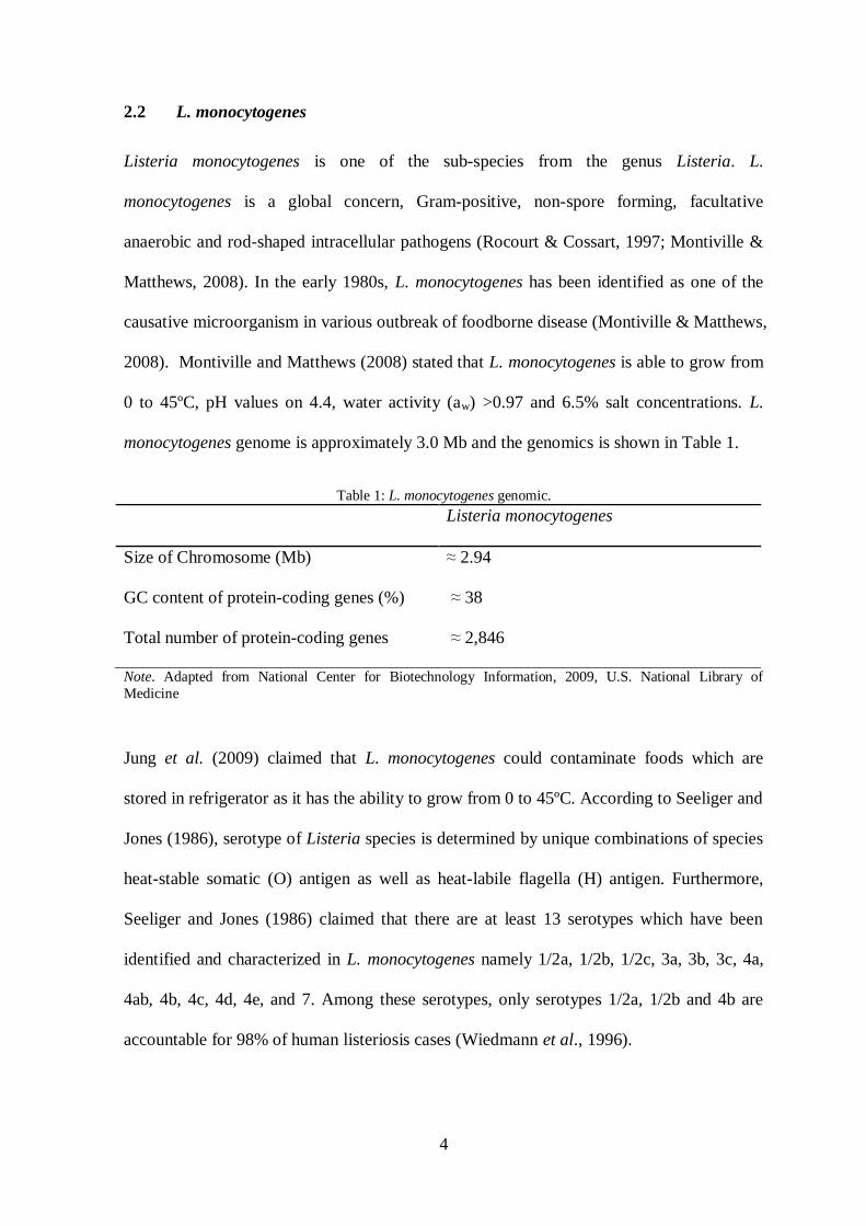
2.2 L. monocytogenes
Listeria monocytogenes is one of the sub-species from the genus Listeria. L.
monocytogenes is a global concern, Gram-positive, non-spore forming, facultative
anaerobic and rod-shaped intracellular pathogens (Rocourt & Cossart, 1997; Montiville &
Matthews, 2008). In the early 1980s, L. monocytogenes has been identified as one of the
causative microorganism in various outbreak of foodborne disease (Montiville & Matthews,
2008). Montiville and Matthews (2008) stated that L. monocytogenes is able to grow from
0 to 45ºC, pH values on 4.4, water activity (aw) >0.97 and 6.5% salt concentrations. L.
monocytogenes genome is approximately 3.0 Mb and the genomics is shown in Table 1.
Table 1: L. monocytogenes genomic.
Listeria monocytogenes
Size of Chromosome (Mb) ≈ 2.94
GC content of protein-coding genes (%) ≈ 38
Total number of protein-coding genes ≈ 2,846
Note. Adapted from National Center for Biotechnology Information, 2009, U.S. National Library of
Medicine
Jung et al. (2009) claimed that L. monocytogenes could contaminate foods which are
stored in refrigerator as it has the ability to grow from 0 to 45ºC. According to Seeliger and
Jones (1986), serotype of Listeria species is determined by unique combinations of species
heat-stable somatic (O) antigen as well as heat-labile flagella (H) antigen. Furthermore,
Seeliger and Jones (1986) claimed that there are at least 13 serotypes which have been
identified and characterized in L. monocytogenes namely 1/2a, 1/2b, 1/2c, 3a, 3b, 3c, 4a,
4ab, 4b, 4c, 4d, 4e, and 7. Among these serotypes, only serotypes 1/2a, 1/2b and 4b are
accountable for 98% of human listeriosis cases (Wiedmann et al., 1996).
5
2.3 Pathogenecity of L. monocytogenes
Listeria monocytogenes is capable to produce hemolysis (hlyA) or listeriolysin O (LLO)
that lyse monocytes of animal and human (Montiville & Matthews, 2008) and this
capability has classified L. monocytogenes as pathogenic microorganism. L.
monocytogenes has gained global public health concern and it has been associated as a
foodborne diseases which cause diseases such as septicemia, meningoencephalitis,
miscarriages, meningitis among infants, pregnant women and immunocompromised
individuals (Choi & Hong, 2003; Kayser, 2001; Rossmanith et al., 2006). Swaminathan
(2007) claimed that mortality rate caused by L. monocytogenes is 20 to 25%.
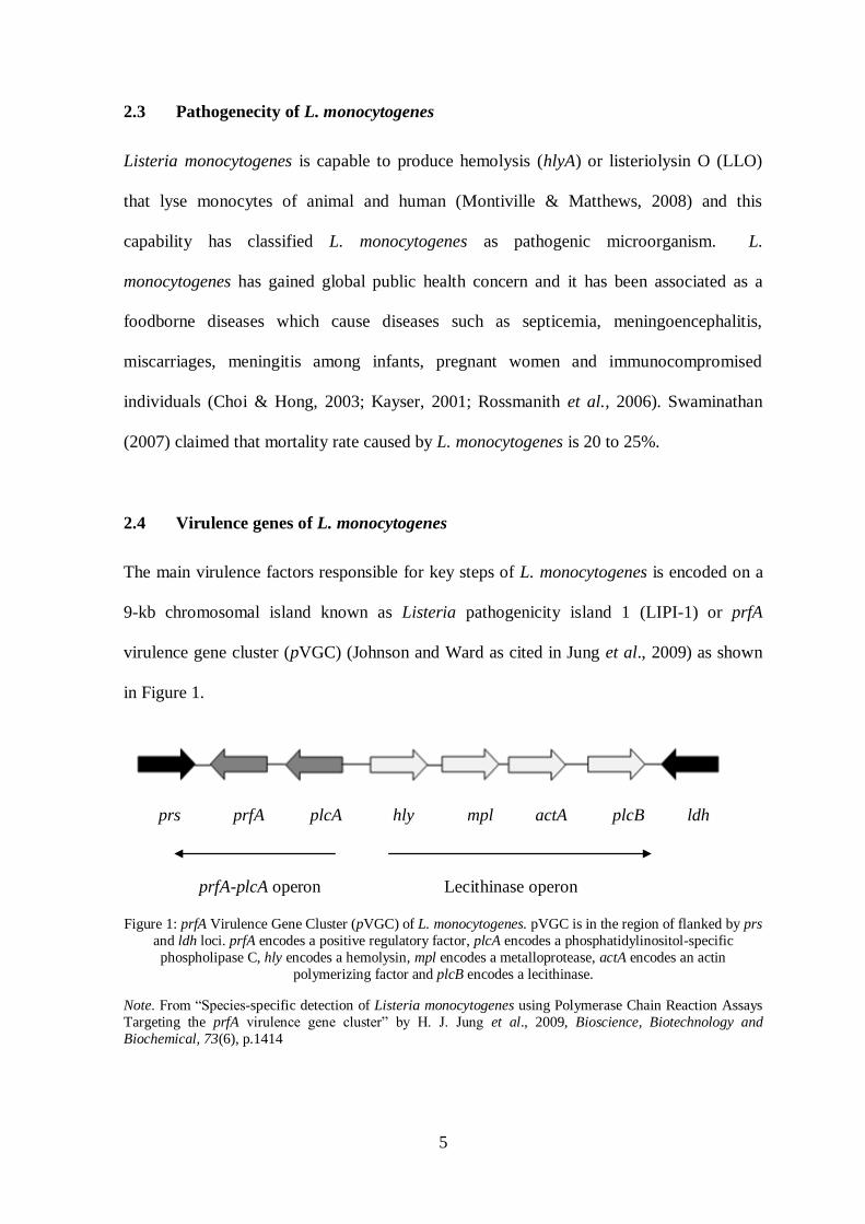
2.4 Virulence genes of L. monocytogenes
The main virulence factors responsible for key steps of L. monocytogenes is encoded on a
9-kb chromosomal island known as Listeria pathogenicity island 1 (LIPI-1) or prfA
virulence gene cluster (pVGC) (Johnson and Ward as cited in Jung et al., 2009) as shown
in Figure 1.
Figure 1: prfA Virulence Gene Cluster (pVGC) of L. monocytogenes. pVGC is in the region of flanked by prs
and ldh loci. prfA encodes a positive regulatory factor, plcA encodes a phosphatidylinositol-specific
phospholipase C, hly encodes a hemolysin, mpl encodes a metalloprotease, actA encodes an actin
polymerizing factor and plcB encodes a lecithinase.
Note. From “Species-specific detection of Listeria monocytogenes using Polymerase Chain Reaction Assays
Targeting the prfA virulence gene cluster” by H. J. Jung et al., 2009, Bioscience, Biotechnology and
Biochemical, 73(6), p.1414
prs plcA hly mpl actA plcB ldh prfA
prfA-plcA operon Lecithinase operon

6
In the region of pVGC, it has six virulence genes which are positive regulatory regulator
prfA, hemolysin (hly), two phospholipases (plcA and plcB), a metalloprotease (mpl) that
function as to activates plcB and actA that works as actin-based motibility (Johnson and
Ward, 2009). This pVGC cluster is the only one known to date that is involved in the
virulence of Listeria. Based on Charkraborty et al. (2000), prfA gene is the key regulator
for the virulence of L. monocytogenes and it acts as a master regulator in the pVGC.
Virulence gene of L. monocytogenes does not necessary locates in the pVGC and there are
several other virulence genes that located outside of the cluster (Kuhn & Goebel, 1999).
These virulence genes are involved in the production of surface proteins that necessary for
internalization of the L. monocytogenes to the host cells which include inlA, inlB and inlC
genes that codes for internalin A, B and C (Kuhn & Goebel, 1999). In addition, Slutsker
and Schuchat (1999) also claimed that iap virulence like genes also located outside the
cluster and it can be dependent or independent to prfA gene. The function of the virulence
genes are shown in Table 2.
Table 2: Function of Virulence genes in prfA Virulence Gene Cluster (pVGC)
Virulence Genes Name Functions
prfA Positive Regulatory Factor A Transcriptional activator
plcA Phosphatidylinositol-specific Phospholipase C Aids in escape from vacuole
plcB Encodes for lecithinase Aids in escape from vacuole
hlyA Hemolysin A or Listeriolysin O (LLO) Hemolyse monocytes
mpl Metallooprotease Maturation of plcB
actA Actin-polymerizing factor Actin-based motibiliy and
cell-to-cell spread
inlA, inlB, inlC Internalin A, Internalin B, Internalin C
respectively
Internalization of Listeria
monocytogenes to host cells Note. Adapted from “Pathogenesis of Listeria monocytogenes”, p. 97-130 by M. Kuhn and W. Goebel, 1999,
New York: Marcel Dekker, Inc and “Listeriosis in humans”, p. 75-96, by L. Slutsker and A. Schuchat (1999),
New York: Marcel Dekker, Inc.
7
Among these virulence genes, Jung et al. (2009) revealed that hlyA encodes hemolysin,
called as listeriolysin O that is vital for the invasion of the pathogen to the host cells which
lyse phagosomes of the host cells and resulting in spreading. Furthermore, Jung et al.
(2009) also stated that hlyA, plcA and plcB virulence genes and their translational product
can be virulence markers to differentiate pathogenic species from non-pathogenic species.
2.5 Outbreak of L. monocytogenes
Centers for Disease Control and Prevention (2012) has reported that in September 11, 2012,
L. monocytogenes outbreak causes a total of 14 persons infected with the listeriosis from
11 states and the District of Columbia. Furthermore, CDC (2012) claimed that the states
include California, Colorado, District of Columbia, Maryland, Minnesota, Nebraska, New
Jersey, New Mexico, New York, Ohio, Pennsylvania and Virginia. CDC (2012) reported
that among the 3 out of 14 person were dead. CDC (2012) explained that the outbreak of
listeriosis was linked to imported Frescolina Brand Ricotta Salata Cheese. Besides, Centers
for Disease Control and Prevention (2012) also reported that in year 2011, a total of 147
persons infected with any of the 5 outbreak-associated subtypes of L. monocytogenes from
28 states which include Alabama, Arkansas, California, Colorado, Idaho, Illinois, Indiana,
Iowa, Kansas, Louisiana, Maryland, Missouri, Montana, Nebraska, Nevada, New Mexico,
New York, North Dakota, Oklahoma, Oregon, Pennsylvania, South Dakota, Texas, Utah,
Virginia, West Virginia, Wisconsin and Wyoming. Tang et al., (1994) stated that in
Malaysia there has been no report of foodborne listeriosis and Lim (2002) supported that
true incidence of foodborne infections in Malaysia is unknown.
8
2.6 Isolation of L. monocytogenes
Listeria enrichment broth is to enrich L. monocytogenes present in raw milk samples. After
enrichment process, raw milk samples are spreaded on Polymyxin-acriflavine-LiCl-
ceftazidime-aesculin-mannitol, PALCAM agar (Oxoid). PALCAM agar is based on the
formulation described by Van Netten et al. (1989) and is used for isolation of L.
monocytogenes from foods. Based on Thermo Scientific (2012), PALCAM is highly
selective because of lithium chloride, ceftazidime, polymyxin B and acriflavine
hydrochloride presents in the agar. L. monocytogenes will hydrolyse aesculin which is a
chemical composition of PALCAM (Oxoid) and this hydrolysis will result in the formation
of a black halo around colonies on PALCAM (Oxoid) (Thermo Scientific, 2012)
2.7 Molecular Confirmation of L. monocytogenes using Species-specific
Polymerase Chain Reaction
Polymerase Chain Reaction (PCR) is a technique to amplify a small region of DNA to
make unlimited amount of copies of the DNA (Russell, 2010).This techniques was
discovered by Kary Mullis as a method to amplify strand of DNA using dNTPs, DNA
polymerase and primers and this technique involve heating and cooling process which is
useful for analysis and manipulation of DNA (McPherson & Muller, 2006). There are 4
components needed in order to perform PCR which is DNA template, DNA polymerase,
primers (designated) and dNTPs. The purpose of using Species-specific PCR is to identify
L. monocytogenes molecularly. For Species-specific PCR, the DNA template used is DNA
strain from L. monocytogenes and the primers used in this PCR has to be designated based
on the molecular maker of L. monocytogenes such as listeriolysin O (LLO) virulence gene.
Based on Border et al. (1990), the primers used for detection of L. monocytogenes was

9
LM1 (5’ CGG AGG TTC CGC AAA AGA TG – 3’) and LM2 (5’ CCT CCA GAG TGA
TCG ATG TT -3’) which was specifically design based on the listerioysin O (LLO)
virulence gene with expected amplicon of 234 bp. L. monocytogenes ATCC (American
Type Culture Collection) 15313 as well as 19155 will be the positive control of Species-
specific PCR.
2.8 Molecular Typing Methods of L. monocytogenes
DNA fingerprinting also can be known as DNA typing or DNA profiling (Russell, 2010).
The use of restriction enzyme has enable researches to compare the base sequences of
different organism (Tortora et al., 2007). Enzyme is specific as well as restriction enzyme;
it will cut at specific and recognize sequence (Tortora et al., 2007). For example like
EcoRI and BamHI having totally different restriction among each other. As a result,
different fragment will be restricted by restriction enzyme and this different fragment will
be used as DNA fingerprinting which is benefit to forensic microbiology. Restriction
Fragment Length Polymorphic (RFLP) is a type of DNA fingerprinting and Amplified
Fragment Length Polymorphic is the PCR-based RFLP. Besides, RAPD is also a type of
DNA fingerprinting as it uses 10-mer primer to randomly bind to the DNA of organism
and will produce different size of fragment and this fragment will be profiled. Random
Amplified Polymorphic DNA (RAPD) markers are short DNA fragments nearly from 8 to
12 base pair and used as primer in PCR (Hodek et al., 2012). Based on Quinn et al., (2011),
RAPD does not require any specific knowledge on the DNA sequence of the target
organism. 10-mer primers (RAPD) will or will not amplify a segment of DNA randomly
along the target DNA sequence through PCR. Furthermore, Quinn et al., (2011) claimed
that RAPD is cost effective and having discriminatory power for many bacterial species. In
molecular characterization, Pulsed Field Gel Electrophoresis (PFGE) and RAPD-PCR are
10
techniques that usually used by the scientist (Cocolin et al., 2005). PFGE is not chosen for
molecular characterization is due to time consuming and require expensive apparatus
although this technique has the most discriminatory power (Franciosa et al., 1998). As for
RAPD technique it is widely used and it can monitor bacterial strains on a wide scale and
able to determine whole genome diversity (Wagner et al., 1996). This technique will be
performed as described by Lee et al., (2011). The primers proposed by Lee et al., (2011)
are 5’-TGT GTG CTG G-3’ and 5’-TTC CGA ACC C-3’ with the name given as OPA 14
and OPA 15 respectively. OPA 14 and OPA 15 were designed and this primers work best
on L. monocytogenes ATCC 15313 and 19155 as a result this strain is used as positive
control in RAPD-PCR. Molecular typing methods rely solely on PCR include RAPD
(Dellaglio et al., 2005) and repetitive genomic element PCR (Gevers et al., 2001).
Repetitive PCR primers amplify repetitive bacterial DNA elements which includes ERIC,
BOX or (GTG)5 (Versalovic et al., 1991). In many studies, (GTG)5-PCR was proven to be
useful for the differentiate of species, subspecies and potentially strain levels (Fernanda et
al., 2010).
2.9 Agarose Gel Electrophoresis
Slish (1998) claimed that agarose gel electrophoresis is a technique used to separate DNA
fragments by their molecular weight. The technique of electrophoresis is based on the fact
that DNA are negatively charged due to its phosphate backbone (Slish, 1998). As a result,
a negative pole and positive pole is needed in the technique of electrophoresis. The
negative pole will be put near to the sample so that it could push the sample along the
agarose gel and for the positive pole is to pull the DNA sample along the agarose gel. Thus,
the biggest size of DNA fragment will move slowest and the smallest size fragment will
move fastest in the agarose gel. In order to view the banding pattern of the DNA fragments
11
(sample) it has to be stained with Ethidium Bromide (EtBr) so that the sample will be
visible under UV light.
2.10 RAPDistance
RAPDistance is a bioinformatic tool that used to analyse RAPD-PCR or (GTG)5-PCR
result. In this study, cluster analysis was done using RAPDistance.

12
3.0 Materials and Methods
3.1 Raw Milk Samples Collection
Raw milk samples were collected from different areas within Kota Samarahan and
Kuching namely Bau, Kampung Haji Baki (Seven miles), Ladang Lapan (Eight miles) and
Petra Jaya. The samples were labelled (area and date), stored in the ice box and transported
to laboratory in UNIMAS for analysis within 2 to 3 hours. The samples were collected
every 2 weeks or 1 month interval. At sampling site, one sample was taken because the raw
milk is quite hard to obtain thus the sampling period was prolonged to one month. Five
hundred milli-liter of raw milk were collected from each sampling site. Bau and Kampung
Haji Baki (Seven miles) raw milk samples were collected on 3rd
of November, 2012
whereas Ladang Lapan (Eight miles) and Petra Jaya were collected on 1st of December,
2012. The second batch of sampling was carried out on the 2nd
of March 2013 and the third
batch sampling was carried out on the 16th of March 2013.
3.2 Enrichment of Listeria species from Raw Milk
The samples were enriched using Tryptone Soy Broth (TSB) which was incubated at 30ºC
for 48 hours with shaking at 120 rpm. Twenty-five mL of the sample were poured into 225
mL of Tryptone Soy Broth.
3.3 Isolation of L. monocytogenes among Listeria species from Raw Milk
After enrichment using TSB, serial dilution was performed and each dilution was spread
on two PALCAM to create replication. The plates were incubated at 37ºC for 48 hours
under micro-aerophilic conditions. This micro-aerophilic condition can be done by sealing
PALCAM plates with Parafilm. The PALCAM plates were examined after 48 hours of