fast regulation of ap-1 activity through interaction of lamin a/c
TRANSCRIPT

TH
EJ
OU
RN
AL
OF
CE
LL
BIO
LO
GY
JCB: ARTICLE
The Rockefeller University Press $30.00J. Cell Biol. Vol. 183 No. 4 653–666www.jcb.org/cgi/doi/10.1083/jcb.200805049 JCB 653
Correspondence to Vicente Andr é s: [email protected]
Abbreviations used in this paper: AP-1, activator protein 1; c-Fos, cellular homologue of the Finkel-Biskis-Jinkins osteosarcoma virus gene; EMSA, electrophoretic mobility shift assay; ERK, extracellular signal-regulated kinase; ERNF, extraction-resistant nuclear fraction; FRET, fluorescence resonance energy transfer; MEF, mouse embryonic fi broblast; NE, nuclear envelope; pERK1/2, phosphorylated ERK1/2; pRb, retinoblastoma protein; SNF, soluble nuclear fraction.
Introduction c-Fos is a member of the dimeric activator protein 1 (AP-1) tran-
scription factor family that regulates key cellular processes,
including cell proliferation, death, survival differentiation, and
oncogenic transformation ( Shaulian and Karin, 2002 ; Eferl and
Wagner, 2003 ). c-fos is an “ early response ” gene that undergoes
rapid transcriptional activation in response to multiple patho-
physiological stimuli (e.g., growth factors, chemical and physi-
cal stress, etc.) ( Shaulian and Karin, 2002 ). Fine regulation of
c-Fos activity is achieved via its interaction with regulatory pro-
teins that can either enhance or inhibit AP-1 activity, and through
posttranslational processing of preexisting or newly synthesized
c-Fos protein ( Piechaczyk and Blanchard, 1994 ; Shaulian and
Karin, 2002 ). For example, c-Fos can be phosphorylated by pro-
tein kinases C and A, cdc2 ( Abate et al., 1991 ), FRK ( Deng and
Karin, 1994 ), extracellular signal-regulated kinase 7 (ERK7) ( Abe
et al., 1999 ), and the mitogen-activated protein kinases (MAPK)
ERK1/2 ( Monje et al., 2003 ) and p38 ( Tanos et al., 2005 ).
Importantly, c-Fos transcriptional activity is modulated through
reversible phosphorylation by some of these kinases, including
ERK1/2 ( Hunter and Karin, 1992 ; Hill and Treisman, 1995 ; Monje
et al., 2003 ). Phosphorylation of c-Fos by ERK1 and RSK oc-
curs soon after serum stimulation ( Chen et al., 1993 , 1996 ).
Moreover, a docking site for ERK in c-Fos facilitates its sequen-
tial phosphorylation in multiple C-terminal sites upon prolonged
ERK activation ( Murphy et al., 2002 ). The binding of growth
factors, differentiation stimuli, and cytokines to cell surface re-
ceptors promotes the activation of the small GTPase Ras and
dual phosphorylation (activation) of ERKs. Active ERK1 and
ERK2 translocate into the nucleus, where they phosphorylate sev-
eral transcriptional regulators (e.g., c-Fos) that cause a rapid in-
duction of immediate early genes ( Sharrocks, 2001 ).
Sequestration of c-Fos at the nuclear envelope (NE)
through interaction with A-type lamins suppresses
AP-1 – dependent transcription. We show here that
c-Fos accumulation within the extraction-resistant nu-
clear fraction (ERNF) and its interaction with lamin A are
reduced and enhanced by gain-of and loss-of ERK1/2
activity, respectively. Moreover, hindering ERK1/2-
dependent phosphorylation of c-Fos attenuates its re-
lease from the ERNF induced by serum and promotes its
interaction with lamin A. Accordingly, serum stimula-
tion rapidly releases preexisting c-Fos from the NE via
ERK1/2-dependent phosphorylation, leading to a fast
activation of AP-1 before de novo c-Fos synthesis. More-
over, lamin A – null cells exhibit increased AP-1 activity
and reduced levels of c-Fos phosphorylation. We also
fi nd that active ERK1/2 interacts with lamin A and co-
localizes with c-Fos and A-type lamins at the NE. Thus,
NE-bound ERK1/2 functions as a molecular switch for
rapid mitogen-dependent AP-1 activation through phos-
phorylation-induced release of preexisting c-Fos from its
inhibitory interaction with lamin A/C.
Fast regulation of AP-1 activity through interaction of lamin A/C, ERK1/2, and c-Fos at the nuclear envelope
Jos é Mar í a Gonz á lez , 1 Ana Navarro-Puche , 1 Berta Casar , 2 Piero Crespo , 2 and Vicente Andr é s 1
1 Laboratory of Vascular Biology, Department of Molecular and Cellular Pathology and Therapy, Instituto de Biomedicina de Valencia, Consejo Superior de Investigaciones Cientifi cas (CSIC), 46010 Valencia, Spain
2 Instituto de Biomedicina y Biotecnolog í a de Cantabria (IBBTEC), CSIC-IDICAN-Universidad de Cantabria, Department of Molecular Biology, Faculty of Medicine, 39011 Santander, Spain
© 2008 González et al. This article is distributed under the terms of an Attribution–Noncommercial–Share Alike–No Mirror Sites license for the fi rst six months after the publica-tion date (see http://www.jcb.org/misc/terms.shtml). After six months it is available under a Creative Commons License (Attribution–Noncommercial–Share Alike 3.0 Unported license, as described at http://creativecommons.org/licenses/by-nc-sa/3.0/).
on April 7, 2018jcb.rupress.org Downloaded from http://doi.org/10.1083/jcb.200805049Published Online: 17 November, 2008 | Supp Info:
JCB • VOLUME 183 • NUMBER 4 • 2008 654
Results
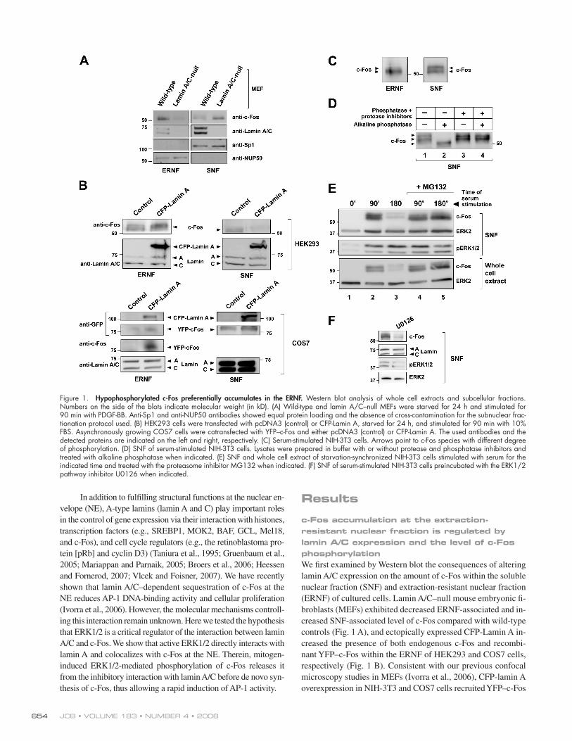
c-Fos accumulation at the extraction-resistant nuclear fraction is regulated by lamin A/C expression and the level of c-Fos phosphorylation We fi rst examined by Western blot the consequences of altering
lamin A/C expression on the amount of c-Fos within the soluble
nuclear fraction (SNF) and extraction-resistant nuclear fraction
(ERNF) of cultured cells. Lamin A/C – null mouse embryonic fi -
broblasts (MEFs) exhibited decreased ERNF-associated and in-
creased SNF-associated level of c-Fos compared with wild-type
controls ( Fig. 1 A ), and ectopically expressed CFP-Lamin A in-
creased the presence of both endogenous c-Fos and recombi-
nant YFP – c-Fos within the ERNF of HEK293 and COS7 cells,
respectively ( Fig. 1 B ). [ID]FIG1[/ID] Consistent with our previous confocal
microscopy studies in MEFs ( Ivorra et al., 2006 ), CFP-lamin A
overexpression in NIH-3T3 and COS7 cells recruited YFP – c-Fos
In addition to fulfi lling structural functions at the nuclear en-
velope (NE), A-type lamins (lamin A and C) play important roles
in the control of gene expression via their interaction with histones,
transcription factors (e.g., SREBP1, MOK2, BAF, GCL, Mel18,
and c-Fos), and cell cycle regulators (e.g., the retinoblastoma pro-
tein [pRb] and cyclin D3) ( Taniura et al., 1995 ; Gruenbaum et al.,
2005 ; Mariappan and Parnaik, 2005 ; Broers et al., 2006 ; Heessen
and Fornerod, 2007 ; Vlcek and Foisner, 2007 ). We have recently
shown that lamin A/C – dependent sequestration of c-Fos at the
NE reduces AP-1 DNA-binding activity and cellular proliferation
( Ivorra et al., 2006 ). However, the molecular mechanisms controll-
ing this interaction remain unknown. Here we tested the hypothesis
that ERK1/2 is a critical regulator of the interaction between lamin
A/C and c-Fos. We show that active ERK1/2 directly interacts with
lamin A and colocalizes with c-Fos at the NE. Therein, mitogen-
induced ERK1/2-mediated phosphorylation of c-Fos releases it
from the inhibitory interaction with lamin A/C before de novo syn-
thesis of c-Fos, thus allowing a rapid induction of AP-1 activity.
Figure 1. Hypophosphorylated c-Fos preferentially accumulates in the ERNF. Western blot analysis of whole cell extracts and subcellular fractions. Numbers on the side of the blots indicate molecular weight (in kD). (A) Wild-type and lamin A/C – null MEFs were starved for 24 h and stimulated for 90 min with PDGF-BB. Anti-Sp1 and anti-NUP50 antibodies showed equal protein loading and the absence of cross-contamination for the subnuclear frac-tionation protocol used. (B) HEK293 cells were transfected with pcDNA3 (control) or CFP-Lamin A, starved for 24 h, and stimulated for 90 min with 10% FBS. Asynchronously growing COS7 cells were cotransfected with YFP – c-Fos and either pcDNA3 (control) or CFP-Lamin A. The used antibodies and the detected proteins are indicated on the left and right, respectively. (C) Serum-stimulated NIH-3T3 cells. Arrows point to c-Fos species with different degree of phosphorylation. (D) SNF of serum-stimulated NIH-3T3 cells. Lysates were prepared in buffer with or without protease and phosphatase inhibitors and treated with alkaline phosphatase when indicated. (E) SNF and whole cell extract of starvation-synchronized NIH-3T3 cells stimulated with serum for the indicated time and treated with the proteasome inhibitor MG132 when indicated. (F) SNF of serum-stimulated NIH-3T3 cells preincubated with the ERK1/2 pathway inhibitor U0126 when indicated.
655AP-1 REGULATION BY C -FOS – ERK1/2 – LAMIN A/C INTERACTIONS • Gonz á lez et al.
Compared with serum-starved cells, the level of c-Fos protein
was up-regulated in cultures stimulated for 90 min, which was
markedly reduced after 180 min of serum stimulation ( Fig. 1 E ,
lanes 1 – 3). Moreover, treatment with the proteasome inhibitor
MG132 abrogated c-Fos down-regulation at 180 min after serum
stimulation ( Fig. 1 E , lane 5 vs. 3). These fi ndings are in agree-
ment with the concept that mitogen stimulation leads to de novo
c-Fos expression, which then undergoes proteasome-dependent
degradation ( Bossis et al., 2003 ; Ito et al., 2005 ). In line with
previous studies ( Monje et al., 2003 ), we found that preincuba-
tion of serum-starved cells with the ERK1/2 inhibitor U0126
greatly reduces c-Fos level in cells stimulated with serum for 90
min ( Fig. 1 F ). Therefore, to circumvent the possible infl uence
of ERK1/2 inhibitors on c-Fos synthesis and/or degradation, the
effects of U0126 and PD98059 on c-Fos expression and sub-
nuclear localization was investigated using the protocol schema-
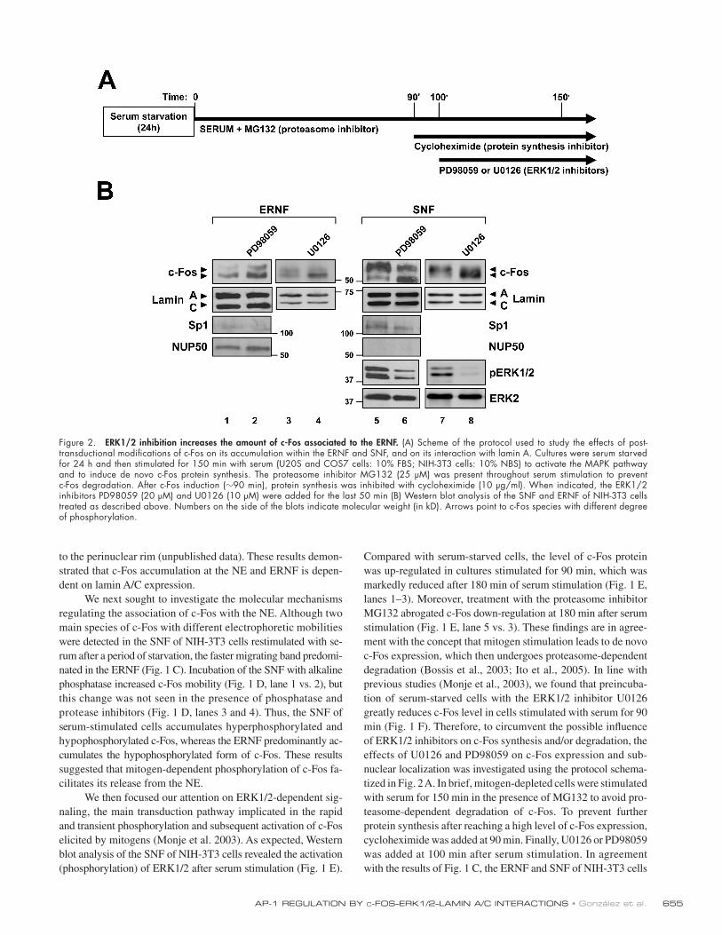
tized in Fig. 2 A . In brief, mitogen-depleted cells were stimulated
with serum for 150 min in the presence of MG132 to avoid pro-
teasome-dependent degradation of c-Fos. To prevent further
protein synthesis after reaching a high level of c-Fos expression,
cycloheximide was added at 90 min. Finally, U0126 or PD98059
was added at 100 min after serum stimulation. In agreement
with the results of Fig. 1 C , the ERNF and SNF of NIH-3T3 cells
to the perinuclear rim (unpublished data). These results demon-
strated that c-Fos accumulation at the NE and ERNF is depen-
dent on lamin A/C expression.
We next sought to investigate the molecular mechanisms
regulating the association of c-Fos with the NE. Although two
main species of c-Fos with different electrophoretic mobilities
were detected in the SNF of NIH-3T3 cells restimulated with se-
rum after a period of starvation, the faster migrating band predomi-
nated in the ERNF ( Fig. 1 C ). Incubation of the SNF with alkaline
phosphatase increased c-Fos mobility ( Fig. 1 D , lane 1 vs. 2), but
this change was not seen in the presence of phosphatase and
protease inhibitors ( Fig. 1 D , lanes 3 and 4). Thus, the SNF of
serum-stimulated cells accumulates hyperphosphorylated and
hypophosphorylated c-Fos, whereas the ERNF predominantly ac-
cumulates the hypophosphorylated form of c-Fos. These results
suggested that mitogen-dependent phosphorylation of c-Fos fa-
cilitates its release from the NE.
We then focused our attention on ERK1/2-dependent sig-
naling, the main transduction pathway implicated in the rapid
and transient phosphorylation and subsequent activation of c-Fos
elicited by mitogens ( Monje et al. 2003 ). As expected, Western
blot analysis of the SNF of NIH-3T3 cells revealed the activation
(phosphorylation) of ERK1/2 after serum stimulation ( Fig. 1 E ).
Figure 2. ERK1/2 inhibition increases the amount of c-Fos associated to the ERNF . (A) Scheme of the protocol used to study the effects of post-transductional modifi cations of c-Fos on its accumulation within the ERNF and SNF, and on its interaction with lamin A. Cultures were serum starved for 24 h and then stimulated for 150 min with serum (U20S and COS7 cells: 10% FBS; NIH-3T3 cells: 10% NBS) to activate the MAPK pathway and to induce de novo c-Fos protein synthesis. The proteasome inhibitor MG132 (25 μ M) was present throughout serum stimulation to prevent c-Fos degradation. After c-Fos induction ( � 90 min), protein synthesis was inhibited with cycloheximide (10 μ g/ml). When indicated, the ERK1/2 inhibitors PD98059 (20 μ M) and U0126 (10 μ M) were added for the last 50 min (B) Western blot analysis of the SNF and ERNF of NIH-3T3 cells treated as described above. Numbers on the side of the blots indicate molecular weight (in kD). Arrows point to c-Fos species with different degree of phosphorylation.

JCB • VOLUME 183 • NUMBER 4 • 2008 656
c-Fos or YFP (as negative control). Fig. 3, A – C show represen-
tative images and intensity line profi les. In agreement with our
previous FRET studies ( Ivorra et al., 2006 ), cells cotransfected
with CFP-lamin A/YFP – c-Fos exhibited a signifi cant increase
in FRET effi ciency in the perinuclear rim compared with nega-
tive controls ( Fig. 3 D ). Addition of U0126 inhibited ERK1/2,
as revealed by reduced ERK1/2 phosphorylation in the SNF
( Fig. 3 E ), and signifi cantly increased FRET effi ciency in cells
cotransfected with CFP-lamin A/YFP but not with CFP-lamin
A/YFP ( Fig. 3 D ). [ID]FIG3[/ID] These results indicated that ERK1/2 inhibi-
tion increases the level of c-Fos/lamin A interaction in vivo.
Constitutive ERK1/2 activation reduces the presence of c-Fos in the ERNF Consistent with the results of Fig. 2 B , treatment of U2OS cells
with U0126 reduced the amount of hyperphosphorylated c-Fos
in the SNF and ERNF fractions, and increased the amount of
hypophosphorylated c-Fos within the ERNF ( Fig. 4 A , lane
1 vs. 2). Overexpression of MEKE, a constitutively active form
of MEK1, markedly augmented ERK1/2 phosphorylation in
the SNF compared with controls transfected with empty vector
( Fig. 4 A , pERK1/2, lane 2 vs. 3). This effect coincided with di-
minished retention of c-Fos in the ERNF, especially of the hy-
perphosphorylated species, which appeared to accumulate in
the SNF ( Fig. 4 A , lane 2 vs. 3). [ID]FIG4[/ID] Thus, constitutive activation of
ERK1/2 reduces the amount of ERNF-associated c-Fos.
manipulated as described above predominantly contained the
fastest migrating hypophosphorylated and the two migrating
forms of c-Fos, respectively ( Fig. 2 B , ERNF: lanes 1 and 3; SNF:
lanes 5 and 7). [ID]FIG2[/ID] Treatment with either PD98059 or U0126 inhib-
ited ERK1/2 activation, as revealed by the reduced amount
of phosphorylated ERK1/2 (pERK1/2) in the SNF ( Fig. 2 B ,
PD98059: lane 5 vs. 6; U0126: lane 7 vs. 8), and caused the accu-
mulation of hypophosphorylated c-Fos both in the ERNF ( Fig. 2 B ,
PD98059: lane 1 vs. 2; U0126: lane 3 vs. 4) and SNF ( Fig. 2 B ,
PD98059: lane 5 vs. 6; U0126: lane 7 vs. 8). The pattern of ex-
pression of Sp1 and nucleoporin-50 + Npap60 (NUP50) suggested
the absence of cross-contamination in these studies ( Fig. 2 B ).
The results presented thus far indicated that the amount of c-Fos
in the ERNF and perinuclear rim is regulated by the level of lamin
A/C expression. They also suggested that inhibition of ERK1/2
activity reduces the extent of c-Fos phosphorylation and promotes
its accumulation within the ERNF.
ERK1/2 inhibition increases the interaction of c-Fos with lamin A in vivo We next performed fl uorescence resonance energy transfer (FRET)
confocal microscopy studies to examine the in vivo interaction
between c-Fos and lamin A. FRET effi ciency determined as
the increment in CFP fl uorescence after YFP photobleaching
( Kenworthy, 2001 ) was quantifi ed in asynchronously growing
COS7 cells cotransfected with CFP-lamin A and either YPF –
Figure 3. ERK1/2 inhibition increases the interaction between c-Fos and lamin A in vivo. FRET was measured in NIH-3T3 cells using the acceptor photo-bleaching method. Cells were transiently cotransfected with CFP-lamin A and either YFP (negative control) or YFP – c-Fos. Transfected cells were maintained in 10% NBS for 24 h and U0126 was added for the last 50 min when indicated. (A) Representative images of one YPF – c-Fos/CFP-lamin A – transfected U0126-treated cell before and after YFP photobleaching. The two perinuclear regions marked with white squares enlarged in B illustrate increased CFP fl uorescence after photobleaching. (C) Fluorescence line profi le analysis of CFP-lamin A intensity throughout the entire perinuclear rim shown in A before (green) and after (red) photobleaching of YFP. (D) Quantifi cation of protein – protein interactions calculated as the percentage of CFP fl uorescence increment after photobleaching in 20 – 30 cells from three independent experiments. (E) Western blot analysis of the SNF of cells maintained in 10% NBS treated or not with U0126. Numbers on the side of the blots indicate molecular weight (in kD).

657AP-1 REGULATION BY C -FOS – ERK1/2 – LAMIN A/C INTERACTIONS • Gonz á lez et al.
ERK1/2 activation is a physiological mechanism for releasing
c-Fos from the ERNF.
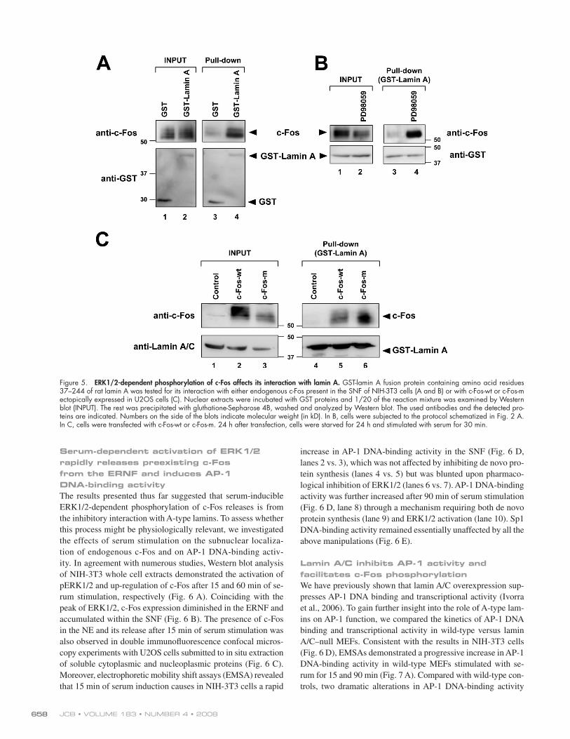
To directly examine whether ERK1/2-dependent phos-
phorylation of c-Fos affects its affi nity for lamin A, we per-
formed in vitro pull-down assays using a GST-lamin A fusion
protein containing amino acids 37 – 244 of rat lamin A, which
interacts with recombinant c-Fos ( Ivorra et al., 2006 ). GST-
lamin A specifi cally interacted with the endogenous c-Fos pro-
tein present in extracts from serum-stimulated NIH-3T3 cells
( Fig. 5 A , lane 3 vs. 4). [ID]FIG5[/ID] Moreover, the amount of GST-lamin A –
bound c-Fos was greatly increased upon treatment of cells with
PD98059 ( Fig. 5 B , lane 3 vs. 4). We also compared the binding
of GST-lamin A to c-Fos-wt and c-Fos-m, which were ectopi-
cally expressed in serum-stimulated U2OS cells. Examination
of input samples suggested the presence of phosphorylation
activity in these cultures because c-Fos-wt exhibited slower
electrophoretic mobility than c-Fos-m ( Fig. 5 C , lane 2 vs. 3).
Moreover, GST-lamin A interacted more effi ciently with c-Fos-m
compared with c-Fos-wt ( Fig. 5 C , lane 5 vs. 6). These re-
sults suggested that the interaction of c-Fos with lamin A is
facilitated by inhibiting ERK1/2-mediated phosphorylation of
c-Fos, either by mutational blockade of ERK1/2 phosphory-
lation sites in c-Fos or by pharmacological inhibition of the
ERK1/2 pathway.
Mutations that hinder ERK1/2-dependent phosphorylation of c-Fos impair its release from the ERNF and enhance its interaction with lamin A We next sought to assess whether physiological levels of
ERK1/2 activation can release c-Fos from the ERNF. To this
end, we investigated in U2OS cells the subnuclear localiza-
tion of ectopically expressed wild-type c-Fos (c-Fos-wt) and
c-Fos-m, a mutant in which four residues that undergo ERK1/2-
dependent phosphorylation are rendered unphosphorylatable
(T232A, T325A, T331A, S374A) ( Monje et al., 2003 ) (see
scheme in Fig. 4 B ). The level of serum-dependent ERK1/2
activation was similar in cells transfected with c-Fos-wt and
c-Fos-m, as revealed by comparable increases in the amount
of pERK1/2 in the SNF ( Fig. 4 B , c-Fos-wt: lane 2 vs. 3;
c-Fos-m: lane 4 vs. 5). Serum-inducible ERK1/2 activation
coincided with reduced amount of ERNF-associated c-Fos-wt
( Fig. 4 B , lane 2 vs. 3), but this effect was weaker for c-Fos-m
( Fig. 4 B , lane 4 vs. 5). The mobility shift of mutated c-Fos-m
upon stimulation is likely due to ERK1/2-dependent phos-
phorylation at site(s) that is(are) not important for lamin A/C
association and possibly for c-Fos activation, per se (e.g., S21,
S32, S70, S133; see scheme in Fig. 4 B ). These fi ndings sug-
gested that serum-inducible phosphorylation of c-Fos through
Figure 4. ERK1/2 constitutive activation reduces c-Fos accumulation in the ERNF and mutations that hinder ERK1/2-dependent phosphorylation of c-Fos impair its release from the ERNF. The SNF and ERNF of U2OS cells were analyzed by Western blot. Numbers on the side of the blots indicate molecular weight (in kD). (A) Cells were transfected with pcDNA3 or with a plasmid encoding MEKE, a constitutively active form of MEK1. 1 d after transfection, cells were subjected to the protocol shown in Fig. 2 A . (B) Scheme of the potentially phosphorylatable residues of wild-type c-Fos (c-Fos-wt) and c-Fos-m, which contains the following amino acid substitutions in residues phosphorylated by ERK1/2: T232A, T325A, T331A, S374A. Cells were transfected with a plasmid encoding c-Fos-wt or c-Fos-m. 1 d after transfection, cells were starved for 24 h and serum stimulated for 30 min when indicated. Of note, four putative sites for ERK1/2-dependent phosphorylation are intact in c-Fos-m (S21, S32, S70, S133).
JCB • VOLUME 183 • NUMBER 4 • 2008 658
increase in AP-1 DNA-binding activity in the SNF ( Fig. 6 D ,
lanes 2 vs. 3), which was not affected by inhibiting de novo pro-
tein synthesis (lanes 4 vs. 5) but was blunted upon pharmaco-
logical inhibition of ERK1/2 (lanes 6 vs. 7). AP-1 DNA-binding
activity was further increased after 90 min of serum stimulation
( Fig. 6 D , lane 8) through a mechanism requiring both de novo
protein synthesis (lane 9) and ERK1/2 activation (lane 10). Sp1
DNA-binding activity remained essentially unaffected by all the
above manipulations ( Fig. 6 E ).
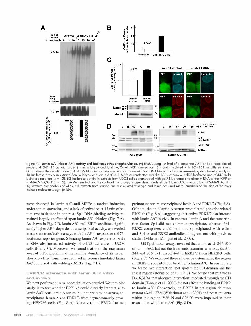
Lamin A/C inhibits AP-1 activity and facilitates c-Fos phosphorylation We have previously shown that lamin A/C overexpression sup-
presses AP-1 DNA binding and transcriptional activity ( Ivorra
et al., 2006 ). To gain further insight into the role of A-type lam-
ins on AP-1 function, we compared the kinetics of AP-1 DNA
binding and transcriptional activity in wild-type versus lamin
A/C – null MEFs. Consistent with the results in NIH-3T3 cells
( Fig. 6 D ), EMSAs demonstrated a progressive increase in AP-1
DNA-binding activity in wild-type MEFs stimulated with se-
rum for 15 and 90 min ( Fig. 7 A ). [ID]FIG7[/ID] Compared with wild-type con-
trols, two dramatic alterations in AP-1 DNA-binding activity
Serum-dependent activation of ERK1/2 rapidly releases preexisting c-Fos from the ERNF and induces AP-1 DNA-binding activity The results presented thus far suggested that serum-inducible
ERK1/2-dependent phosphorylation of c-Fos releases is from
the inhibitory interaction with A-type lamins. To assess whether
this process might be physiologically relevant, we investigated
the effects of serum stimulation on the subnuclear localiza-
tion of endogenous c-Fos and on AP-1 DNA-binding activ-
ity. In agreement with numerous studies, Western blot analysis
of NIH-3T3 whole cell extracts demonstrated the activation of
pERK1/2 and up-regulation of c-Fos after 15 and 60 min of se-
rum stimulation, respectively ( Fig. 6 A ). Coinciding with the
peak of ERK1/2, c-Fos expression diminished in the ERNF and
accumulated within the SNF ( Fig. 6 B ). [ID]FIG6[/ID] The presence of c-Fos
in the NE and its release after 15 min of serum stimulation was
also observed in double immunofl uorescence confocal micros-
copy experiments with U2OS cells submitted to in situ extraction
of soluble cytoplasmic and nucleoplasmic proteins ( Fig. 6 C ).
Moreover, electrophoretic mobility shift assays (EMSA) revealed
that 15 min of serum induction causes in NIH-3T3 cells a rapid
Figure 5. ERK1/2-dependent phosphorylation of c-Fos affects its interaction with lamin A. GST-lamin A fusion protein containing amino acid residues 37 – 244 of rat lamin A was tested for its interaction with either endogenous c-Fos present in the SNF of NIH-3T3 cells (A and B) or with c-Fos-wt or c-Fos-m ectopically expressed in U2OS cells (C). Nuclear extracts were incubated with GST proteins and 1/20 of the reaction mixture was examined by Western blot (INPUT). The rest was precipitated with gluthatione-Sepharose 4B, washed and analyzed by Western blot. The used antibodies and the detected pro-teins are indicated. Numbers on the side of the blots indicate molecular weight (in kD). In B, cells were subjected to the protocol schematized in Fig. 2 A . In C, cells were transfected with c-Fos-wt or c-Fos-m. 24 h after transfection, cells were starved for 24 h and stimulated with serum for 30 min.

659AP-1 REGULATION BY C -FOS – ERK1/2 – LAMIN A/C INTERACTIONS • Gonz á lez et al.
Figure 6. Serum-inducible ERK1/2 activation correlates with the release of preexisting c-Fos from the ERNF and causes a rapid induction of AP-1 DNA-binding activity. (A and B) NIH-3T3 cells were starved for 24 h and stimulated with 10% NBS for the indicated times. Whole cell extracts (A), or SNF and ERNF (B) were analyzed by Western blot. Of note, the amount of protein analyzed in the blots of B was � 14 times higher than in A. Numbers on the side of the blots indicate molecular weight (in kD). (C) Double immunofl uorescence of c-Fos and lamin A/C in U2OS cells. Top images: Asynchronously growing control cells. Middle and bottom images: Starved and serum-stimulated cells in situ extracted and treated with DNase. Images in the right column show 3D representation of c-Fos signal intensity in lamin A/C – containing perinuclear rim. Graph shows the total intensity of c-Fos signal per total lamin A/C – containing perinuclear area of in situ extracted cells ( n = 30 cells of each condition). (D and E) NIH 3T3 were starved for 24 h and stimulated with 10% NBS for the indicated times. When indicated, cycloheximide (CHX, 10 μ g/ml) or U0126 (10 μ M) was added 1 h before serum stimulation. EMSA was performed using 10 fmol of a consensus AP-1 (D) or Sp1 (E) radiolabeled probe and SNF (5 μ g total protein). The graphs show the quantifi cation of DNA-binding activity as assessed by densitometric analysis. Reactions in lanes 11 and 22 contained a 50-fold molar excess of competitor (unlabeled AP-1 or Sp1 consensus oligonucleotide, respectively).
JCB • VOLUME 183 • NUMBER 4 • 2008 660
Figure 7. Lamin A/C inhibits AP-1 activity and facilitates c-Fos phosphorylation. (A) EMSA using 10 fmol of a consensus AP-1 or Sp1 radiolabeled probe and SNF (15 μ g total protein) from wild-type and lamin A/C – null MEFs starved for 48 h and stimulated with 10% FBS for different times. Graph shows the quantifi cation of AP-1 DNA-binding activity after normalization with Sp1 DNA-binding activity as assessed by densitometric analysis. (B) Luciferase activity in extracts from wild-type and lamin A/C – null MEFs cotransfected with the AP-1 – responsive coll73-luciferase and pGL4-Renilla luciferase reporters ( n = 12). (C) Luciferase activity in extracts from U2OS cells cotransfected with coll73-luciferase and either miRNA-control/GFP or miRNA-LMNA/GFP ( n = 12). The Western blot and the confocal microscopy images demonstrate effi cient lamin A/C silencing by miRNA-LMNA/GFP. (D) Western blot analysis of whole cell extracts from starved and restimulated wild-type and lamin A/C – null MEFs. Numbers on the side of the blots indicate molecular weight (in kD).
were observed in lamin A/C – null MEFs: a marked induction
under serum starvation, and a lack of activation at 15 min of se-
rum restimulation; in contrast, Sp1 DNA-binding activity re-
mained largely unaffected upon lamin A/C ablation ( Fig. 7 A ).
As shown in Fig. 7 B , lamin A/C – null MEFs exhibited signifi -
cantly higher AP-1-dependent transcriptional activity, as revealed
in transient transfection assays with the AP-1 – responsive coll73-
luciferase reporter gene. Silencing lamin A/C expression with
miRNA also increased activity of coll73-luciferase in U2OS
cells ( Fig. 7 C ). Moreover, we found that both the maximum
level of c-Fos protein and the relative abundance of its hyper-
phosphorylated form were reduced in serum-stimulated lamin
A/C compared with wild-type MEFs ( Fig. 7 D ).
ERK1/2 interacts with lamin A in vitro and in vivo We next performed immunoprecipitation-coupled Western blot
analysis to test whether ERK1/2 could directly interact with
lamin A/C. Anti-lamin A serum, but not preimmune serum, co-
precipitated lamin A and ERK1/2 from asynchronously grow-
ing HEK293 cells ( Fig. 8 A ). Moreover, anti-ERK2, but not
preimmune serum, coprecipitated lamin A and ERK1/2 ( Fig. 8 A ).
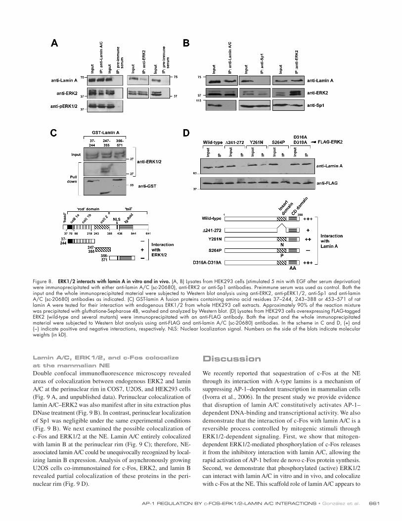
[ID]FIG8 [/ID] Of note, the anti-lamin A serum precipitated phosphorylated
ERK1/2 ( Fig. 8 A ), suggesting that active ERK1/2 can interact
with lamin A/C in vivo. In contrast, lamin A and the transcrip-
tion factor Sp1 did not coimmunoprecipitate, whereas Sp1-
ERK2 complexes could be immunoprecipitated with either
anti-Sp1 or anti-ERK2 antibodies, in agreement with previous
studies ( Milanini-Mongiat et al., 2002 ).
GST pull-down assays revealed that amino acids 247 – 355
of lamin A/C, but not the fragments spanning amino acids 37 –
244 and 356 – 571, associated to ERK1/2 from HEK293 cells
( Fig. 8 C ). We extended these studies by determining the region
in ERK2 responsible for binding to lamin A/C. In particular,
we tested two interaction “ hot spots ” : the CD domain and the
Insert region ( Robinson et al., 1998 ). We found that mutations
D316,319A that abrogate interactions mediated through the CD
domain ( Tanoue et al., 2000 ) did not affect the binding of ERK2
to lamin A/C. Conversely, an ERK2 Insert region deletion
mutant ( � 241-272) ( Whitehurst et al., 2004 ) and point mutants
within this region, Y261N and S264Y, were impaired in their
association with lamin A/C ( Fig. 8 D ).
661AP-1 REGULATION BY C -FOS – ERK1/2 – LAMIN A/C INTERACTIONS • Gonz á lez et al.
Discussion
We recently reported that sequestration of c-Fos at the NE
through its interaction with A-type lamins is a mechanism of
suppressing AP-1 – dependent transcription in mammalian cells
( Ivorra et al., 2006 ). In the present study we provide evidence
that disruption of lamin A/C constitutively activates AP-1 –
dependent DNA-binding and transcriptional activity. We also
demonstrate that the interaction of c-Fos with lamin A/C is a
reversible process controlled by mitogenic stimuli through
ERK1/2-dependent signaling. First, we show that mitogen-
dependent ERK1/2-mediated phosphorylation of c-Fos releases
it from the inhibitory interaction with lamin A/C, allowing the
rapid activation of AP-1 before de novo c-Fos protein synthesis.
Second, we demonstrate that phosphorylated (active) ERK1/2
can interact with lamin A/C in vitro and in vivo, and colocalize
with c-Fos at the NE. This scaffold role of lamin A/C appears to
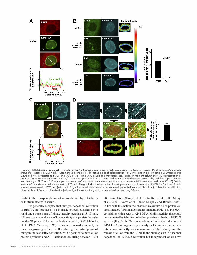
Lamin A/C, ERK1/2, and c-Fos colocalize at the mammalian NE Double confocal immunofl uorescence microscopy revealed
areas of colocalization between endogenous ERK2 and lamin
A/C at the perinuclear rim in COS7, U2OS, and HEK293 cells
( Fig. 9 A , and unpublished data). Perinuclear colocalization of
lamin A/C – ERK2 was also manifest after in situ extraction plus
DNase treatment ( Fig. 9 B ). In contrast, perinuclear localization
of Sp1 was negligible under the same experimental conditions
( Fig. 9 B ). [ID]FIG9[/ID] We next examined the possible colocalization of
c-Fos and ERK1/2 at the NE. Lamin A/C entirely colocalized
with lamin B at the perinuclear rim ( Fig. 9 C ); therefore, NE-
associated lamin A/C could be unequivocally recognized by local-
izing lamin B expression. Analysis of asynchronously growing
U2OS cells co-immunostained for c-Fos, ERK2, and lamin B
revealed partial colocalization of these proteins in the peri-
nuclear rim ( Fig. 9 D ).
Figure 8. ERK1/2 interacts with lamin A in vitro and in vivo. (A, B) Lysates from HEK293 cells (stimulated 5 min with EGF after serum deprivation) were immunoprecipitated with either anti-lamin A/C (sc-20680), anti-ERK2 or anti-Sp1 antibodies. Preimmune serum was used as control. Both the input and the whole immunoprecipitated material were subjected to Western blot analysis using anti-ERK2, anti-pERK1/2, anti-Sp1 and anti-lamin A/C (sc-20680) antibodies as indicated. (C) GST-lamin A fusion proteins containing amino acid residues 37 – 244, 243 – 388 or 453 – 571 of rat lamin A were tested for their interaction with endogenous ERK1/2 from whole HEK293 cell extracts. Approximately 90% of the reaction mixture was precipitated with gluthatione-Sepharose 4B, washed and analyzed by Western blot. (D) Lysates from HEK293 cells overexpressing FLAG-tagged ERK2 (wild-type and several mutants) were immunoprecipitated with an anti-FLAG antibody. Both the input and the whole immunoprecipitated material were subjected to Western blot analysis using anti-FLAG and anti-lamin A/C (sc-20680) antibodies. In the scheme in C and D, (+) and (−) indicate positive and negative interactions, respectively. NLS: Nuclear localization signal. Numbers on the side of the blots indicate molecular weights (in kD).
JCB • VOLUME 183 • NUMBER 4 • 2008 662
after stimulation ( Kruijer et al., 1984 ; Kerr et al., 1988 ; Monje
et al., 2003 ; Ivorra et al., 2006 ; Murphy and Blenis, 2006 ).
In line with this notion, we observed maximum c-Fos protein ex-
pression at 60 – 90 min after serum stimulation ( Fig. 1 E , Fig. 6 A ),
coinciding with a peak of AP-1 DNA-binding activity that could
be attenuated by inhibitors of either protein synthesis or ERK1/2
activity ( Fig. 6 D ). Our novel observation is the induction of
AP-1 DNA-binding activity as early as 15 min after serum ad-
dition concomitantly with maximum ERK1/2 activity and the
release of c-Fos from the ERNF to the nucleoplasm in a manner
dependent on ERK1/2 activation but independent of de novo
facilitate the phosphorylation of c-Fos elicited by ERK1/2 in
cells stimulated with serum.
It is generally accepted that mitogen-dependent activation
of ERK1/2 in fi broblasts is a biphasic process consisting of a
rapid and strong burst of kinase activity peaking at 5 – 15 min,
followed by a second wave of lower activity that persists through-
out the G1 phase of the cell cycle ( Kahan et al., 1992 ; Meloche
et al., 1992 ; Meloche, 1995 ). c-Fos is expressed minimally in
most nongrowing cells as well as during the initial phase of
mitogen-induced ERK activation, with a peak of de novo c-Fos
protein synthesis and AP-1 activation occurring between 1 – 2 h
Figure 9. ERK1/2 and c-Fos partially colocalize at the NE. Representative images of cells examined by confocal microscopy. (A) ERK2-lamin A/C double immunofl uorescence in COS7 cells. Graph shows a line profi le illustrating areas of colocalization. (B) Control and in situ – extracted plus DNase-treated U2OS cells were subjected to ERK2 – lamin A/C or Sp1 – lamin A/C double immunofl uorescence. Images in the right column show 3D representation of ERK2 or Sp1 signal intensity in the lamin A/C – containing perinuclear rim of control and in situ – extracted/DNase-treated cells, and the graph shows the total intensity of ERK2 and Sp1 signal per total lamin A/C – containing perinuclear area in the in situ – extracted/DNase-treated cells ( n = 30). (C) Double lamin A/C – lamin B immunofl uorescence in U2OS cells. The graph shows a line profi le illustrating nearly total colocalization. (D) ERK2 – c-Fos – lamin B triple immunofl uorescence in U2OS cells (left). Lamin B signal was used to delineate the nuclear envelope (white lines in middle column) to allow the quantifi cation of perinuclear ERK2-c-Fos colocalization (yellow signal) shown in the graph, as determined by analyzing 50 cells.

663AP-1 REGULATION BY C -FOS – ERK1/2 – LAMIN A/C INTERACTIONS • Gonz á lez et al.
from the interaction with A-type lamins (see above). Indeed,
AP-1 – dependent DNA-binding and transcriptional activity were
signifi cantly higher in lamin A/C – null versus wild-type MEFs
and in U2OS cells in which lamin A/C expression was silenced
with miRNA ( Fig. 7, A – C ). Our results also suggest that the ef-
fi ciency of serum-inducible activation (phosphorylation) of the
small preinduction levels of c-Fos elicited by ERK1/2 is aided
by their colocalization at the NE ( Fig. 7 D ). We are currently
investigating whether ERK1/2 – lamin A/C complex formation
may help prolong the activation state of ERK1/2 by maintaining
these signal transducers in a reservoir out of the reach of solu-
ble nuclear phosphatases. It will be also of interest to examine
whether A-type lamins serve a scaffolding function only for
ERK1/2-dependent activation of c-Fos, or whether induction
of other transcription factors also requires the activity of NE-
associated signal transducers.
The Insert and the CD domains of ERK1/2 are involved in
protein – protein interactions. ERK1/2 substrate docking can be se-
lectively dissociated in vitro by single point mutations without per-
turbing ERK activation or its intrinsic catalytic activity ( Dimitri
et al., 2005 ). Indeed, mutations affecting the Insert region ( � 241-
272, Y261N, and S264P) or the CD domain (D316,319A) alter
the interaction of ERK2 with some substrates ( Tanoue et al., 2000 ;
Whitehurst et al., 2004 ; Casar et al., 2007 ). We have shown here
that the ERK2 – lamin A/C interaction requires the Insert domain of
ERK2 and the coil 2 of lamin A/C. Other proteins such as PEA15
( Whitehurst et al., 2004 ) and Mxi2 ( Casar et al., 2007 ) also inter-
act with ERK through the Insert region. The determinant mediat-
ing in this interaction is a “ reverse D domain ” , with a consensus
sequence R/K- � a -X 3/4 - � b (where � are Leu, Ile, or Val) ( Callaway
et al., 2005 ). Notably, the R/K- � a -X 3/4 - � b motif is present in the
region of A-type lamins, which we found to intervene in their in-
teraction with ERK1/2 (RIRISL at residues 296 – 301).
protein synthesis ( Fig. 6 ). Based on these fi ndings and the ob-
servations that gain-of and loss-of ERK1/2 activity reduces and
enhances, respectively, c-Fos accumulation within the ERNF
and its interaction with lamin A, and that regulation of these
processes is impaired by hindering ERK1/2-dependent phos-
phorylation of c-Fos, we propose the model depicted in Fig. 10 .
[ID]FIG10[/ID] In serum-deprived cells, sequestration of c-Fos at the NE via its
interaction with lamin A/C limits transcription of AP-1 target
genes. Subsequent to mitogen stimulation, lamin A – bound ac-
tive ERK1/2 phosphorylates c-Fos and releases it from the
NE, thus allowing the rapid transcriptional activation of AP-1 –
responsive genes before de novo c-Fos protein synthesis. This
mechanism permits the continuous existence in the cell of a cer-
tain level of inactive c-Fos (e.g., in quiescent cells) that can be
rapidly activated by mitogen stimulation via ERK1/2-dependent
phosphorylation without requiring de novo protein synthesis.
Intracellular compartmentalization through interactions
among MAPKs and scaffold proteins plays an important role in
the regulation of signal transduction pathways ( Morrison and
Davis, 2003 ; Kolch, 2005 ). For example, active MEK – ERK com-
plexes are retained in the cytoplasm and in sites of focal adhe-
sion through interaction with the transmembrane protein Sef
( Torii et al., 2004 ) and paxilin ( Ishibe et al., 2004 ), respectively.
Moreover, kinase suppressor of Ras 1 ( Muller et al., 2001 ) and
the complex p14-MEK-partner 1 ( Teis et al., 2002 ; Pullikuth
et al., 2005 ) are recruited to the plasma membrane and the en-
dosome, respectively, where they enhance MEK and ERK activity.
On the other hand, the association between ERKs and structural
nuclear proteins, such as kinetochores, may serve anchoring pur-
poses ( Shapiro et al., 1998 ). To the best of our knowledge, we
show here for the fi rst time that A-type lamins function as a nu-
clear docking platform for EKR1/2, and that NE-bound ERK1/2
contributes to the rapid activation of AP-1 via releasing c-Fos
Figure 10. Model for the rapid activation of AP1 upon mitogen stimulation. In serum-deprived cells, the c-Fos protein is expressed at low level predomi-nantly in a hypophosphorylated state and associated to the NE through its interaction with A-type lamins. Under these conditions, transcription of AP-1 target genes is off. Upon mitogen stimulation, phosphorylated (active) ERK1/2 bound to lamin A/C phosphorylates c-Fos, causing its release from the NE. Hyperphosphorylated nucleoplasmic c-Fos can then heterodimerize with AP-1 family members (e.g., c-Jun), thus allowing the transcriptional activation of AP-1 – responsive genes before de novo c-Fos synthesis.
JCB • VOLUME 183 • NUMBER 4 • 2008 664
2003 ); pCMV-FLAG-ERK2; pCMV-FLAG-ERK2-Y261N; pCMV-FLAG-ERK2- � 241-272; pCMV-FLAG-ERK2-S264P and pCMV-FLAG-ERK2-D316A-D319A ( Robinson et al., 2002 ); pCEFL-AU5-c-Fos (c-Fos-wt); and pCEFL-AU5-c-Fos-mut (c-Fos-m, which contains the following mutations: T232A, T325A, T331A, and S374A; gift from S. Gutkind, National Institutes of Health, MD; see Fig. 4 B ) ( Monje et al., 2003 ).
Cell culture NIH-3T3, COS-7, HEK293, U20S, and HeLa cells were obtained from the American Type Culture Collection. Lmna -null and littermate wild-type MEFs are described elsewhere ( Sullivan et al., 1999 ). Cells maintained in DME supplemented with 100 U/ml penicillin, 0.1 mg/ml streptomycin, and 2 mmol/L L- glutamine (Invitrogen) and 10% FBS (or 10% NBS for NIH-3T3 cells) were incubated at 37 ° C in a 5% CO 2 /95% O 2 atmosphere. Cultures were serum starved for 24 h and then stimulated with either 10% FBS, 10% NBS, PDGF-BB (20 ng/ml; Sigma-Aldrich), or EGF (100 ng/ml; Sigma-Aldrich). For ERK1/2 inhibition, PD98059 (20 μ M; Tocris) or U0126 (10 μ M; Promega) were added before serum stimulation. MG132 and cyclohexi-mide were from Sigma-Aldrich. When indicated, cells were treated as schematized in Fig. 2 A .
In vitro pull-down assays GST proteins were purifi ed using glutathione-Sepharose 4B (GE Health-care) and eluted with 50 mM Tris-Cl (pH 8.0). For the experiments of Fig. 5 , GST proteins and cell lysates from NIH-3T3 cells were incubated in 20 mM Hepes (pH 7.9), 0.4 M NaCl, 1 mM EDTA, 1 mM EGTA, 1 mM DTT, and 1 mM PMSF supplemented with complete protease inhibitor cocktail (Roche). After 16 h of incubation at 4 ° C, glutathione-Sepharose 4B was added to a fi nal concentration of 10% and agitated at 4 ° C for 45 min. The beads were collected by centrifugation and washed three times with 20 mM Hepes (pH 7.9), 0.4 M NaCl, 1 mM EDTA, 1 mM EGTA, 1 mM DTT, and 1 mM PMSF. For the experiments of Fig. 8 C , whole cell lysates from HEK293 cells were incubated with GST-lamin A bound to glutathione- Sepharose 4B beads. After 4 h of incubation at 4 ° C, the beads were col-lected by centrifugation and washed twice with 1% NP-40/PBS. In all cases, pellets were air-dried, resuspended in 2x Laemmli buffer, boiled for 5 min, and separated onto 12% SDS – polyacrylamide gels (SDS-PAGE).
Subcellular fractionation, immunoprecipitation, and immunoblot experiments Immunoprecipitation and Western blot analysis were performed as previ-ously described ( Ivorra et al., 2006 ; Casar et al., 2007 ). Subcellular frac-tionation was performed as described by Schreiber et al. ( 1989 ) with minor modifi cations ( Ivorra et al., 2006 ). In brief, cells were washed with PBS and scraped into TEN buffer (150 mM NaCl, 1 mM EDTA, and 40 mM TrisCl at pH 7.4). Cells were collected by brief centrifugation in microfuge tubes and resuspended in 10 mM Hepes (pH 7.9), 10 mM KCl, 0.1 mM EDTA, 0.1 mM EGTA, 1 mM DTT, and 0.5 mM PMSF. After 15 min on ice, Nonidet NP-40 (Fluka) was added to a fi nal concentration of 10%, and tubes were vortexed. Lysates were centrifuged at 4 ° C in a microfuge set at maximum speed to ob-tain the soluble cytoplasmic fraction (supernatant) and the nuclear pellet, which was resuspended in ice-cold 20 mM Hepes (pH 7.9), 0.4 M NaCl, 1 mM EDTA, 1 mM EGTA, 1 mM DTT, and 1 mM PMSF, and agitated at 4 ° C for 15 min. The nuclear lysate was centrifuged for 45 min at 4 ° C to obtain the SNF and the pellet containing the ERNF.
In situ nuclear matrix isolation and indirect immunofl uorescence analysis In situ nuclear matrix isolation was performed as described previously ( Fey et al., 1984 ). In brief, cells grown on coverslips were washed in PBS and extracted twice in cytoskeleton buffer (CSK: 100 mM NaCl, 300 mM sucrose, 10 mM PIPES (pH 6.8), 3 mM MgCl 2 , 0.5% Triton X-100, and 1.2 mM PMSF) for 10 min at 0 ° C. The resulting soluble fraction was removed. Extraction buffer (250 mM ammonium sulfate, 300 mM sucrose, 10 mM PIPES (pH 6.8), 3 mM MgCl 2 , 1.2 mM PMSF, and 0.5% Triton X-100) was added to the Triton X-100 insoluble structures for 10 min at 0 ° C and the cytoskeleton fraction was removed. DNase digestion was performed twice in digestion buffer (CSK buffer containing 100 μ g/ml DNase I and 50 mM NaCl) followed by extraction in digestion buffer containing 0.25 M (NH 4 ) 2 SO 4 . In situ – extracted and control cells were fi xed in 4% formalde-hyde/PBS and permeabilized with 0.5% Triton X-100. All samples were blocked for 5 min with 10 mM glycine (pH 8.5) and 1 h with 5% dry milk in 10% FBS, 0.5% BSA, 0.1% Triton X-100, and PBS, followed by an over-night incubation at 4 ° C with anti-c-Fos (1:100), anti-ERK2 (1:100), or anti-Sp1 (1:100) antibodies. Samples were then incubated with species-appropriate FITC-conjugated secondary antibody. After washes and incubation with anti-lamin A/C (1:100; sc-7292) for 1 h at room temperature, specimens
In this study, we have focused our attention on how mito-
genic signals regulate the interaction between c-Fos and lamin
A/C. Further research is required to assess whether additional
pathophysiological conditions regulate this interaction and the
underlying molecular mechanisms. Of notable interest in this
regard are the inherited diseases termed laminopathies, which
are caused by either mutations in the LMNA gene (which en-
codes for A-type lamins) or defective posttranslational process-
ing of prelamin A ( Worman and Bonne, 2007 ). Notably, lamin
A/C and lamin-associated polypeptides can physically interact
with histones, chromatin, and transcription factors (e.g., c-Fos,
SREBP1, MOK2, BAF, GCL, Mel18), suggesting that altered
gene expression contributes to the pathogenesis of laminopa-
thies ( Taniura et al., 1995 ; Gruenbaum et al., 2005 ; Broers et al.,
2006 ; Heessen and Fornerod, 2007 ; Vlcek and Foisner, 2007 ).
Indeed, microarray studies using fi broblast from Hutchinson-
Gilford progeria syndrome patients revealed differential tran-
scription factor expression ( Ly et al., 2000 ; Csoka et al., 2004 ).
Moreover, certain pathogenic lamin A mutations cause altera-
tions in the transcription factors MOK2 ( Dreuillet et al., 2008 )
and SREBP1 ( Lloyd et al., 2002 ; Hubner et al., 2006 ) and in the
cell cycle regulator pRb ( Hubner et al., 2006 ). Interestingly,
transgenic mice overexpressing either wild-type MEK5 or con-
stitutively active MEK1 exhibit excessive ERK1/2-dependent
signaling and dilated cardiomyopathy ( Bueno et al., 2000 ;
Nicol et al., 2001 ), a clinical manifestation of several laminopa-
thies. Moreover, expression of the Emery-Dreifuss muscular
dystrophy – causing H222P – lamin A mutant protein in homozy-
gous lmna H222P/H222P knock-in mice causes in the heart aberrant
ERK1/2 activity and myopathy, and leads to activation of several
MAPKs — including ERK1/2 — and downstream target genes in
cultured cells ( Muchir et al., 2007 ). It is also noteworthy that
MEK/ERK pathway inhibition improves defective myogenic
factor expression and differentiation of C2C12 myoblasts
expressing the Emery-Dreifuss muscular dystrophy – causing
R453W – lamin A mutant ( Favreau et al., 2004 , 2008 ). In the
light of the aforementioned studies and the fi ndings reported
herein, it will be of interest to assess whether pathogenic lamin
A/C mutants alter the interaction of c-Fos and/or ERK1/2 at the
NE, and if so, whether they affect the regulation and function of
AP-1 target genes.
Materials and methods Antibodies Primary antibodies directed against c-Fos (sc-52), lamin A/C (sc-7292 in Figs. 1 B, 4, 6 C, 7 C, and 9 ; sc-7293 in Figs.1 A, C – F, 2, 5, 6 A and B, and 7 D ; and sc-20680 in Fig. 8 ), lamin B (sc-6217), GST (sc-138), ERK2 (sc-154 and sc-1647 only in Fig. 9 D ), pERK1/2 (sc-7383), Sp1 (sc-59-G), � -tubulin (sc-8035), anti-FLAG, and HRP-coupled secondary antibodies were purchased from Santa Cruz Biotechnology, Inc. Anti-NUP50 and anti-GFP were from Abcam (ab4005) and Invitrogen (A6455), respectively.
Plasmids pECFP-YFP was constructed by inserting YFP into pECFP-C1 (gift from J. L ó pez-Gim é nez, University of Glasgow, Scotland). The following plasmids are described elsewhere: pGEX-lamin A (37 – 244), pGEX-lamin A (247 – 355), and pGEX-lamin A (356 – 571) (gift from T. Ozaki, Chiba Cancer Center Research Institute, Japan) ( Ozaki et al., 1994 ); pEYFP-c-Fos and pECFP-lamin A ( Ivorra et al., 2006 ); pmycCMV-ERK2-MEK1 ( Robinson et al., 1998 ); pCDNAI-HA-ERK2 ( Crespo et al., 1994 ); pCEFL-MEKE ( Sanz-Moreno et al.,
665AP-1 REGULATION BY C -FOS – ERK1/2 – LAMIN A/C INTERACTIONS • Gonz á lez et al.
We thank M.J. Andr é s-Manzano for help with the preparation of fi gures, and D. Barettino for critical reading of the manuscript. We are also grateful to col-leagues who generously provided reagents (S. Gutkind: c-Fos-wt, c-Fos-m; T. Ozaki: pGEX-lamin A; J. L ó pez-Gim é nez: pECFP-YFP; R. Foisner, and C.L. Stewart: wt and lamin A – null MEFs). V. Andr é s ’ laboratory is supported by grants SAF2004-03057 and SAF2007-6211 (Spanish Ministry of Science and Innovation – MICINN, and European Regional Development Fund -FEDER), and RD06/0014/0021 (Red Tem á tica de Investigaci ó n Cooperativa en En-fermedades Cardiovasculares, Instituto de Salud Carlos III – ISCIII).
P. Crespo ’ s laboratory is supported by grants BFU2005-00777 and GEN2003-20239-C06-03 (MICINN), the EU Sixth Framework Program under the GROWTHSTOP (LSHC CT-2006-037731) and SIMAP (IST-2004-027265) projects, and RD06/0020/0105 (Red Tem á tica de Investigaci ó n Cooperativa en C á ncer, ISCIII). J.M. Gonz á lez received salary support from ISCIII and a re-search grant from Generalitat Valenciana (GVPRE/2008/163).
Submitted: 9 May 2008 Accepted: 20 October 2008
References Abate , C. , D.R. Marshak , and T. Curran . 1991 . Fos is phosphorylated by p34cdc2,
cAMP-dependent protein kinase and protein kinase C at multiple sites clustered within regulatory regions. Oncogene . 6 : 2179 – 2185 .
Abe , M.K. , W.L. Kuo , M.B. Hershenson , and M.R. Rosner . 1999 . Extracellular signal-regulated kinase 7 (ERK7), a novel ERK with a C-terminal domain that regulates its activity, its cellular localization, and cell growth. Mol. Cell. Biol. 19 : 1301 – 1312 .
Bossis , G. , P. Ferrara , C. Acquaviva , I. Jariel-Encontre , and M. Piechaczyk . 2003 . c-Fos proto-oncoprotein is degraded by the proteasome independently of its own ubiquitinylation in vivo. Mol. Cell. Biol. 23 : 7425 – 7436 .
Broers , J.L. , F.C. Ramaekers , G. Bonne , R.B. Yaou , and C.J. Hutchison . 2006 . Nuclear lamins: laminopathies and their role in premature ageing. Physiol. Rev. 86 : 967 – 1008 .
Bueno , O.F. , L.J. De Windt , K.M. Tymitz , S.A. Witt , T.R. Kimball , R. Klevitsky , T.E. Hewett , S.P. Jones , D.J. Lefer , C.F. Peng , et al . 2000 . The MEK1-ERK1/2 signaling pathway promotes compensated cardiac hypertrophy in transgenic mice. EMBO J. 19 : 6341 – 6350 .
Callaway , K. , M.A. Rainey , and K.N. Dalby . 2005 . Quantifying ERK2-protein in-teractions by fl uorescence anisotropy: PEA-15 inhibits ERK2 by blocking the binding of DEJL domains. Biochim. Biophys. Acta . 1754 : 316 – 323 .
Casar , B. , V. Sanz-Moreno , M.N. Yazicioglu , J. Rodriguez , M.T. Berciano , M. Lafarga , M.H. Cobb , and P. Crespo . 2007 . Mxi2 promotes stimulus- independent ERK nuclear translocation. EMBO J. 26 : 635 – 646 .
Chen , R.H. , C. Abate , and J. Blenis . 1993 . Phosphorylation of the c-Fos trans-repression domain by mitogen-activated protein kinase and 90-kDa ribo-somal S6 kinase. Proc. Natl. Acad. Sci. USA . 90 : 10952 – 10956 .
Chen , R.H. , P.C. Juo , T. Curran , and J. Blenis . 1996 . Phosphorylation of c-Fos at the C-terminus enhances its transforming activity. Oncogene . 12 : 1493 – 1502 .
Crespo , P. , N. Xu , W.F. Simonds , and J.S. Gutkind . 1994 . Ras-dependent activa-tion of MAP kinase pathway mediated by G-protein beta gamma sub-units. Nature . 369 : 418 – 420 .
Csoka , A.B. , S.B. English , C.P. Simkevich , D.G. Ginzinger , A.J. Butte , G.P. Schatten , F.G. Rothman , and J.M. Sedivy . 2004 . Genome-scale expres-sion profi ling of Hutchinson-Gilford progeria syndrome reveals wide-spread transcriptional misregulation leading to mesodermal/mesenchymal defects and accelerated atherosclerosis. Aging Cell . 3 : 235 – 243 .
Deng , T. , and M. Karin . 1994 . c-Fos transcriptional activity stimulated by H-Ras-activated protein kinase distinct from JNK and ERK. Nature . 371 : 171 – 175 .
Dimitri , C.A. , W. Dowdle , J.P. MacKeigan , J. Blenis , and L.O. Murphy . 2005 . Spatially separate docking sites on ERK2 regulate distinct signaling events in vivo. Curr. Biol. 15 : 1319 – 1324 .
Dreuillet , C. , M. Harper , J. Tillit , M. Kress , and M. Ernoult-Lange . 2008 . Mislocalization of human transcription factor MOK2 in the presence of pathogenic mutations of lamin A/C. Biol. Cell . 100 : 51 – 61 .
Eferl , R. , and E.F. Wagner . 2003 . AP-1: a double-edged sword in tumorigenesis. Nat. Rev. Cancer . 3 : 859 – 868 .
Favreau , C. , D. Higuet , J.C. Courvalin , and B. Buendia . 2004 . Expression of a mutant lamin A that causes Emery-Dreifuss muscular dystrophy inhibits in vitro differentiation of C2C12 myoblasts. Mol. Cell. Biol. 24 : 1481 – 1492 .
Favreau , C. , E. Delbarre , J.C. Courvalin , and B. Buendia . 2008 . Differentiation of C2C12 myoblasts expressing lamin A mutated at a site responsible for Emery-Dreifuss muscular dystrophy is improved by inhibition of the MEK-ERK pathway and stimulation of the PI3-kinase pathway. Exp. Cell Res. 314 : 1392 – 1405 .
were washed and incubated with an anti – mouse secondary antibody con-jugated to Alexa 633 (1:300).
Phosphatase treatment NIH-3T3 cells were rapidly washed with cold PBS and collected for subcel-lular fractionation as described above. When indicated, fractionation was performed in the absence of phosphatase inhibitors, DTT and PMSF. Ali-quots of the SNF were incubated in the absence or presence of 1 U of al-kaline phosphatase (Roche) for 1 h at 37 ° C. Reactions were stopped by adding SDS sample buffer and processed for immunoblotting.
EMSA Double-stranded oligonucleotides containing the AP-1 (5 � -CGCTTGA T-GAGTCA G-3 � ; AP-1 site underlined) and the Sp1 (5 � -ATTCGATCG GGGC-GG GGCGAGC-3 � ; Sp1 site underlined) consensus sites were labeled with � [ 32 P]dATP using polynucleotide kinase (New England Biolabs, Inc.) and pu-rifi ed on a Sephadex G-50 column. EMSA was performed using the SNF of NIH-3T3 cells (5 μ g total protein) and wild-type and lamin A/C – null mice MEFs (15 μ g total protein) as previously described ( Ivorra et al., 2006 ).
Confocal microscopy Images were acquired on a laser confocal microscope (TCS/SP2; Leica) with a 63x oil immersion objective (NA 1.4). For single lamin A/C ( Fig. 7 C ) and lamin B ( Fig. 7 C ), double ERK2 – lamin A/C ( Fig. 9 A ) and lamin B – lamin A/C ( Fig. 9 C ), and triple ERK2 – c-Fos – lamin B immunofl uores-cence ( Fig. 9 D ): cells were fi xed with 4% PFA/PBS at room temperature (RT) on glass coverslips and permeabilized using 0.5% Triton X-100. Sam-ples were blocked for 5 min with 10 mM glycine (pH 8.5) and 1 h with 5% dry milk in 10% FBS, 0.5% BSA, 0.1% Triton X-100, and PBS before an overnight incubation at 4 ° C with anti-lamin A/C (1:100; sc-7292) or anti-lamin B antibody (1:100). In double immunofl uorescence experiments, samples were incubated with anti-ERK2 (1:100) or anti-lamin B (1:100) antibodies for 1 h at room temperature after anti-lamin A/C antibody incu-bation. For triple immunofl uorescence, samples were incubated simultane-ously with anti-c-Fos (1:100) and anti-ERK2 (1:100; sc-1647) antibodies after anti-lamin B antibody incubation. Finally, secondary antibodies con-jugated to Alexa 488, Texas red, and Alexa 633 (Molecular Probes, Inc.) were used (1:300). Image quantifi cation was done using MetaMorph soft-ware (MDS Analytical Technologies).
FRET NIH-3T3 cells were cotransfected with pECFP-lamin A + pEYFP-c-Fos or pECFP-Lamin A + EYFP as a negative control (1 μ g each plasmid) using Li-pofectamine (Invitrogen). Cotransfection of pECFP-YFP + pcDNA3 (0.5 μ g each) was used as positive control to calibrate the system. Images were ac-quired on a confocal microscope (TCS/SP2; Leica) with a 63x oil immer-sion objective (NA 1.4). An argon laser line of 458 nm was used to excite CFP (PMT window 465 – 505 nm) and a 514-nm line (20% laser intensity for acquisition and 65% for photobleaching) to excite YFP (PMT window: 525 – 600 nm). Studies were performed in 4% paraformaldehyde-fi xed cells using the acceptor-photobleaching method ( Kenworthy, 2001 ) as pre-viously described ( Ivorra et al., 2006 ), in which FRET is calculated as the relative increase in donor fl uorescence as a result of the reduction or elimi-nation of energy transfer when the acceptor is photobleached. Specifi cally, we used the following equation: FRET = (C after � C before )/C after x 100, where C before and C after are the total fl uorescence intensity of the CFP channel be-fore and after photobleaching, respectively. For negative values, this pa-rameter was considered 0.
Luciferase gene reporter assays Wild-type and lamin A/C – null MEFs were transiently transfected with 5 μ g of AP-1 – dependent coll 73-luciferase reporter plasmid and pGL4-Renilla lu-ciferase using the calcium phosphate method. U2OS were transfected with 5 μ g of coll73-luciferase plus either a plasmid encoding for a miRNA- control/GFP or miRNA-LMNA/GFP (BLOCK-IT; Invitrogen). After 48 h in 10% FBS, cells were harvested and luciferase activity was measured following the manufacturer ’ s instructions (dual luciferase reporter assay system; Promega). Luciferase activity and GFP expression were measured in a luminometer (Vic-tor). miRNA-transfected cells were also fi xed in 4% PFA and studied by immunofl uorescence confocal microscopy, or lysed for Western blot analysis.
Statistical analysis Results are reported as mean ± SE. In experiments with two groups, differ-ences were evaluated using a two-tail, unpaired Student ’ s t test. One-way ANOVA and Bonferroni ’ s post hoc test was used for experiments involving more than two groups.
JCB • VOLUME 183 • NUMBER 4 • 2008 666
Murphy , L.O. , S. Smith , R.H. Chen , D.C. Fingar , and J. Blenis . 2002 . Molecular interpretation of ERK signal duration by immediate early gene products. Nat. Cell Biol. 4 : 556 – 564 .
Nicol , R.L. , N. Frey , G. Pearson , M. Cobb , J. Richardson , and E.N. Olson . 2001 . Activated MEK5 induces serial assembly of sarcomeres and eccentric cardiac hypertrophy. EMBO J. 20 : 2757 – 2767 .
Ozaki , T. , M. Saijo , K. Murakami , H. Enomoto , Y. Taya , and S. Sakiyama . 1994 . Complex formation between lamin A and the retinoblastoma gene prod-uct: identifi cation of the domain on lamin A required for its interaction. Oncogene . 9 : 2649 – 2653 .
Piechaczyk , M. , and J.M. Blanchard . 1994 . c-fos proto-oncogene regulation and function. Crit. Rev. Oncol. Hematol. 17 : 93 – 131 .
Pullikuth , A. , E. McKinnon , H.J. Schaeffer , and A.D. Catling . 2005 . The MEK1 scaffolding protein MP1 regulates cell spreading by integrating PAK1 and Rho signals. Mol. Cell. Biol. 25 : 5119 – 5133 .
Robinson , F.L. , A.W. Whitehurst , M. Raman , and M.H. Cobb . 2002 . Identifi cation of novel point mutations in ERK2 that selectively disrupt binding to MEK1. J. Biol. Chem. 277 : 14844 – 14852 .
Robinson , M.J. , S.A. Stippec , E. Goldsmith , M.A. White , and M.H. Cobb . 1998 . A constitutively active and nuclear form of the MAP kinase ERK2 is suffi cient for neurite outgrowth and cell transformation. Curr. Biol. 8 : 1141 – 1150 .
Sanz-Moreno , V. , B. Casar , and P. Crespo . 2003 . p38alpha isoform Mxi2 binds to extracellular signal-regulated kinase 1 and 2 mitogen-activated protein kinase and regulates its nuclear activity by sustaining its phosphorylation levels. Mol. Cell. Biol. 23 : 3079 – 3090 .
Schreiber , E. , P. Matthias , M.M. Muller , and W. Schaffner . 1989 . Rapid detection of octamer binding proteins with ‘ mini-extracts ’ , prepared from a small number of cells. Nucleic Acids Res. 17 : 6419 .
Shapiro , P.S. , E. Vaisberg , A.J. Hunt , N.S. Tolwinski , A.M. Whalen , J.R. McIntosh , and N.G. Ahn . 1998 . Activation of the MKK/ERK pathway during somatic cell mitosis: direct interactions of active ERK with ki-netochores and regulation of the mitotic 3F3/2 phosphoantigen. J. Cell Biol. 142 : 1533 – 1545 .
Sharrocks , A.D. 2001 . The ETS-domain transcription factor family. Nat. Rev. Mol. Cell Biol. 2 : 827 – 837 .
Shaulian , E. , and M. Karin . 2002 . AP-1 as a regulator of cell life and death. Nat. Cell Biol. 4 : E131 – E136 .
Sullivan , T. , D. Escalante-Alcalde , H. Bhatt , M. Anver , N. Bhat , K. Nagashima , C.L. Stewart , and B. Burke . 1999 . Loss of A-type lamin expression compromises nuclear envelope integrity leading to muscular dystrophy. J. Cell Biol. 147 : 913 – 920 .
Taniura , H. , C. Glass , and L. Gerace . 1995 . A chromatin binding site in the tail domain of nuclear lamins that interacts with core histones. J. Cell Biol. 131 : 33 – 44 .
Tanos , T. , M.J. Marinissen , F.C. Leskow , D. Hochbaum , H. Martinetto , J.S. Gutkind , and O.A. Coso . 2005 . Phosphorylation of c-Fos by members of the p38 MAPK family. Role in the AP-1 response to UV light. J. Biol. Chem. 280 : 18842 – 18852 .
Tanoue , T. , M. Adachi , T. Moriguchi , and E. Nishida . 2000 . A conserved docking motif in MAP kinases common to substrates, activators and regulators. Nat. Cell Biol. 2 : 110 – 116 .
Teis , D. , W. Wunderlich , and L.A. Huber . 2002 . Localization of the MP1-MAPK scaffold complex to endosomes is mediated by p14 and required for sig-nal transduction. Dev. Cell . 3 : 803 – 814 .
Torii , S. , M. Kusakabe , T. Yamamoto , M. Maekawa , and E. Nishida . 2004 . Sef is a spatial regulator for Ras/MAP kinase signaling. Dev. Cell . 7 : 33 – 44 .
Vlcek , S. , and R. Foisner . 2007 . A-type lamin networks in light of laminopathic diseases. Biochim. Biophys. Acta . 1773 : 661 – 674 .
Whitehurst , A.W. , F.L. Robinson , M.S. Moore , and M.H. Cobb . 2004 . The death effector domain protein PEA-15 prevents nuclear entry of ERK2 by in-hibiting required interactions. J. Biol. Chem. 279 : 12840 – 12847 .
Worman , H.J. , and G. Bonne . 2007 . “ Laminopathies ” : a wide spectrum of human diseases. Exp. Cell Res. 313 : 2121 – 2133 .
Fey , E.G. , K.M. Wan , and S. Penman . 1984 . Epithelial cytoskeletal framework and nuclear matrix-intermediate fi lament scaffold: three-dimensional or-ganization and protein composition. J. Cell Biol. 98 : 1973 – 1984 .
Gruenbaum , Y. , A. Margalit , R.D. Goldman , D.K. Shumaker , and K.L. Wilson . 2005 . The nuclear lamina comes of age. Nat. Rev. Mol. Cell Biol. 6 : 21 – 31 .
Heessen , S. , and M. Fornerod . 2007 . The inner nuclear envelope as a transcrip-tion factor resting place. EMBO Rep. 8 : 914 – 919 .
Hill , C.S. , and R. Treisman . 1995 . Transcriptional regulation by extracellular signals: mechanisms and specifi city. Cell . 80 : 199 – 211 .
Hubner , S. , J.E. Eam , A. Hubner , and D.A. Jans . 2006 . Laminopathy-inducing lamin A mutants can induce redistribution of lamin binding proteins into nuclear aggregates. Exp. Cell Res. 312 : 171 – 183 .
Hunter , T. , and M. Karin . 1992 . The regulation of transcription by phosphoryla-tion. Cell . 70 : 375 – 387 .
Ishibe , S. , D. Joly , Z.X. Liu , and L.G. Cantley . 2004 . Paxillin serves as an ERK-regulated scaffold for coordinating FAK and Rac activation in epithelial morphogenesis. Mol. Cell . 16 : 257 – 267 .
Ito , Y. , D. Inoue , S. Kido , and T. Matsumoto . 2005 . c-Fos degradation by the ubiquitin-proteasome proteolytic pathway in osteoclast progenitors. Bone . 37 : 842 – 849 .
Ivorra , C. , M. Kubicek , J.M. Gonzalez , S.M. Sanz-Gonzalez , A. Alvarez-Barrientos , J.E. O ’ Connor , B. Burke , and V. Andres . 2006 . A mechanism of AP-1 suppression through interaction of c-Fos with lamin A/C. Genes Dev. 20 : 307 – 320 .
Kahan , C. , K. Seuwen , S. Meloche , and J. Pouyssegur . 1992 . Coordinate, bi-phasic activation of p44 mitogen-activated protein kinase and S6 kinase by growth factors in hamster fi broblasts. Evidence for thrombin-induced signals different from phosphoinositide turnover and adenylylcyclase inhibition. J. Biol. Chem. 267 : 13369 – 13375 .
Kenworthy , A.K. 2001 . Imaging protein-protein interactions using fl uorescence resonance energy transfer microscopy. Methods . 24 : 289 – 296 .
Kerr , L.D. , J.T. Holt , and L.M. Matrisian . 1988 . Growth factors regulate transin gene expression by c-fos-dependent and c-fos-independent pathways. Science . 242 : 1424 – 1427 .
Kolch , W. 2005 . Coordinating ERK/MAPK signalling through scaffolds and in-hibitors. Nat. Rev. Mol. Cell Biol. 6 : 827 – 837 .
Kruijer , W. , J.A. Cooper , T. Hunter , and I.M. Verma . 1984 . Platelet-derived growth factor induces rapid but transient expression of the c-fos gene and protein. Nature . 312 : 711 – 716 .
Lloyd , D.J. , R.C. Trembath , and S. Shackleton . 2002 . A novel interaction be-tween lamin A and SREBP1: implications for partial lipodystrophy and other laminopathies. Hum. Mol. Genet. 11 : 769 – 777 .
Ly , D.H. , D.J. Lockhart , R.A. Lerner , and P.G. Schultz . 2000 . Mitotic misregula-tion and human aging. Science . 287 : 2486 – 2492 .
Mariappan , I. , and V.K. Parnaik . 2005 . Sequestration of pRb by cyclin D3 causes intranuclear reorganization of lamin A/C during muscle cell differentia-tion. Mol. Biol. Cell . 16 : 1948 – 1960 .
Meloche , S. 1995 . Cell cycle reentry of mammalian fi broblasts is accompanied by the sustained activation of p44mapk and p42mapk isoforms in the G1 phase and their inactivation at the G1/S transition. J. Cell. Physiol. 163 : 577 – 588 .
Meloche , S. , K. Seuwen , G. Pages , and J. Pouyssegur . 1992 . Biphasic and synergistic activation of p44mapk (ERK1) by growth factors: correla-tion between late phase activation and mitogenicity. Mol. Endocrinol. 6 : 845 – 854 .
Milanini-Mongiat , J. , J. Pouyssegur , and G. Pages . 2002 . Identifi cation of two Sp1 phosphorylation sites for p42/p44 mitogen-activated protein kinases: their implication in vascular endothelial growth factor gene transcription. J. Biol. Chem. 277 : 20631 – 20639 .
Monje , P. , M.J. Marinissen , and J.S. Gutkind . 2003 . Phosphorylation of the carboxyl-terminal transactivation domain of c-Fos by extracellular signal-regulated kinase mediates the transcriptional activation of AP-1 and cel-lular transformation induced by platelet-derived growth factor. Mol. Cell. Biol. 23 : 7030 – 7043 .
Morrison , D.K. , and R.J. Davis . 2003 . Regulation of MAP kinase signaling modules by scaffold proteins in mammals. Annu. Rev. Cell Dev. Biol. 19 : 91 – 118 .
Muchir , A. , P. Pavlidis , V. Decostre , A.J. Herron , T. Arimura , G. Bonne , and H.J. Worman . 2007 . Activation of MAPK pathways links LMNA mutations to cardiomyopathy in Emery-Dreifuss muscular dystrophy. J. Clin. Invest. 117 : 1282 – 1293 .
Muller , J. , S. Ory , T. Copeland , H. Piwnica-Worms , and D.K. Morrison . 2001 . C-TAK1 regulates Ras signaling by phosphorylating the MAPK scaffold, KSR1. Mol. Cell . 8 : 983 – 993 .
Murphy , L.O. , and J. Blenis . 2006 . MAPK signal specifi city: the right place at the right time. Trends Biochem. Sci. 31 : 268 – 275 .