fatal accelerated cirrhosis after imported hev 10 genotype
TRANSCRIPT

of healing of suppurative nodes and is the only evidence-based effective treatment (9). Surgical excision remains controversial because of potentially high rates of signifi-cant scarring (10). For nonsuppurative lymphadenitis, a watch-and-wait approach is recommended because most resolve rapidly (8).
Given our findings, the National TB Program in Geor-gia subsequently created a management protocol. This protocol recommends no intervention for nonsuppurative lymphadenitis and needle aspiration for suppurative local lymphadenitis.
In summary, we found an increasing rate of BCG-as-sociated lymphadenitis after a shift to exclusive BCG SSI vaccine use in Georgia. Countries with a BCG vaccina-tion policy should have a clear protocol on management of BCG vaccine–related adverse events to avoid inappropriate treatment in children.
References 1. Mangtani P, Abubakar I, Ariti C, Beynon R, Pimpin L, Fine PE,
et al. Protection by BCG vaccine against tuberculosis: a systematic review of randomized controlled trials. Clin Infect Dis. 2014;58:470–80. http://dx.doi.org/10.1093/cid/cit790
2. Brosch R, Gordon SV, Garnier T, Eiglmeier K, Frigui W, Valenti P, et al. Genome plasticity of BCG and impact on vaccine efficacy. Proc Natl Acad Sci U S A. 2007;104:5596–601. http://dx.doi.org/10.1073/pnas.0700869104
3. Rabin AS, Kuchukhidze G, Sanikidze E, Kempker RR, Blumberg HM. Prescribed and self-medication use increase delays in diagnosis of tuberculosis in the country of Georgia. Int J Tuberc Lung Dis. 2013;17:214–20. http://dx.doi.org/10.5588/ijtld.12.0395
4. Statens Serum Institute. Description of BCG VACCINE SSI [cited 2015 Jan 20]. http://www.ssi.dk/English/Vaccines/BCG%20Vaccine%20Danish%20Strain%201331/Discription%20of%20BCG%20Vaccine%20SSI.aspx
5. Alrabiaah AA, Alsubaie SS, Bukhari EI, Gad A, Alzamel FA. Outbreak of Bacille Calmette-Guérin–related lymphadenitis in Saudi children at a university hospital after a change in the strain of vaccine. Ann Saudi Med. 2012;32:4–8.
6. Hengster P, Schnapka J, Fille M, Menardi G. Occurrence of suppurative lymphadenitis after a change of BCG vaccine. Arch Dis Child. 1992;67:952–5. http://dx.doi.org/10.1136/adc.67.7.952
7. Bukhari E, Alzahrani M, Alsubaie S, Alrabiaah A, Alzamil F. Bacillus Calmette-Guérin lymphadenitis: a 6-year experience in two Saudi hospitals. Indian J Pathol Microbiol. 2012;55:202–5. http://dx.doi.org/10.4103/0377-4929.97869
8. Cuello-García CA, Pérez-Gaxiola G, Jiménez Gutiérrez C. Treating BCG-induced disease in children. Cochrane Database Syst Rev. 2013;1:CD008300.
9. Banani SA, Alborzi A. Needle aspiration for suppurative post-BCG adenitis. Arch Dis Child. 1994;71:446–7. http://dx.doi.org/10.1136/adc.71.5.446
10. Chan WM,. Kwan YW, Leung CW. Management of Bacillus Calmette-Guérin lymphadenitis. Hong Kong Journal of Paediatrics. 2011;16:85–94.
Address for correspondence: Giorgi Kuchukhidze, National Center for Disease Control and Public Health, 9 Asatiani St, 0177 Tbilisi, Georgia; email: [email protected], [email protected]
Fatal Accelerated Cirrhosis after Imported HEV Genotype 4 Infection
Ryan B. Perumpail, Aijaz Ahmed, John P. Higgins, Samuel K. So, J. Lynn Cochran, Jan Drobeniuc, Tonya R. Mixson-Hayden, Chong-Gee TeoAuthoraffiliations:StanfordUniversitySchoolofMedicine, PaloAlto,California,USA(R.B.Perumpail,A.Ahmed, J.P.Higgins,S.K.So);BirminghamGastroenterologyAssociates,Birmingham,Alabama,USA(J.L.Cochran);TrinityMedical Center,Birmingham(J.L.Cochran);CentersforDiseaseControlandPrevention,Atlanta,Georgia,USA(J.Drobeniuc, T.R.Mixson-Hayden,C.-G.Teo)
DOI:http://dx.doi.org/10.3201/eid2109.150300
To the Editor: Hepatitis E is a viral hepatitide that is endemic in many developing countries. In its classic form, it results from ingesting fecally contaminated water that carries hepatitis E virus (HEV), and it frequently resolves without treatment. When hepatitis E is imported to the United States, it originates mainly from persons who have acquired HEV genotype 1 infection from South Asia (1). We report imported HEV genotype 4 infection (Technical Appendix Figure, panel A) in a patient during which cir-rhosis and fatal hepatic decompensation ensued.
The patient was a 68-year-old man of Chinese ethnicity who had been a California resident since 1985. He sought treatment for mild jaundice in April 2013 in Hong Kong, where he had been staying for 7 weeks. Sixteen years be-fore, he had undergone orthotopic liver transplantation at Stanford University Medical Center (Palo Alto, California, USA) for hepatitis B cirrhosis. Since then, he had received entecavir and tacrolimus for maintenance and had been vaccinated against hepatitis A virus. Until his current ill-ness, routine liver function tests had not indicated hepatic dysfunction (values in November 2012: alanine amino-transferase 2 IU/L, aspartate aminotransferase 24 IU/L, alkaline phosphatase 67 IU/L, total bilirubin 0.5 mg/dL).
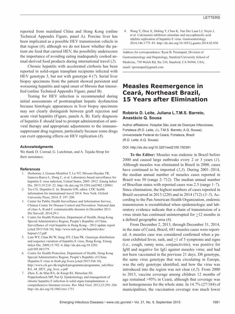
When the patient returned to the United States, 3 weeks after onset of jaundice, the initial work-up showed the fol-lowing values: alanine aminotransferase 149 IU/L, aspar-tate aminotransferase 59 IU/L, alkaline phosphatase 193 IU/L, total bilirubin 2.8 mg/dL (online Technical Appendix Figure, panel B, http://wwwnc.cdc.gov/EID/article/21/9/ 15-0300-Techapp1.pdf). Hepatitis B virus DNA and anti-nuclear antibodies were not detected, and the tacrolimus level was stable. Ultrasound revealed a normal transplanted liver. A liver biopsy specimen showed mild portal, biliary, and lobular inflammation and early biliary injury (Figure,
EmergingInfectiousDiseases•www.cdc.gov/eid•Vol.21,No.9,September2015 1679
LETTERS

panels A, B; a color version of this figure is available online [http://wwwnc.cdc.gov/EID/article/21/9/15-0300-F.htm]). The prednisone dosage was escalated, and mycophenolate mofetil was added. Liver enzyme activity showed some im-provement, but the bilirubin level continued to rise (online Technical Appendix Figure, panel B).
A biopsy specimen taken 3 months later showed grade 3 hepatitis with bile ductular reaction, bridging hepatocytic necrosis and fibrosis, and regenerative nodule formation (Fig-ure, panels C, D). A blood sample taken about this time tested positive for HEV RNA. The patient was then given ribavirin (1,000 mg/d). Before hepatitis E was diagnosed, tacrolimus was given (1 mg 2×/d); when the diagnosis was confirmed, the tacrolimus dose was reduced to 0.5 mg every other day. Four months after the patient sought treatment, ascites was noted. Ribavirin was stopped because of pancytopenia. Blood samples subsequently tested negative for HEV RNA, but HEV IgM and IgG were found. Hepatic function did not improve.
Eight months after onset of the patient’s condition, marked hepatic decompensation occurred (online Techni-cal Appendix Figure), culminating in esophageal variceal hemorrhage. The patient was placed on a waiting list and
then underwent liver transplantation, but he died during the operation from complications of hemorrhage. Biopsy of the liver explant revealed intense lobular inflammation with the hepatocellular reactive changes persisting and stage IV fibrosis (Figure, panels E, F).
The patient had lived and worked in Hong Kong be-fore he became a resident of the United States. He had not visited Hong Kong in the 3 years preceding his most recent trip, nor had he traveled to Europe. Review of his medi-cal records revealed no evidence of hepatic dysfunction after his previous travels. Considering that his most recent visit to Hong Kong coincided with the incubation period of hepatitis E (2), he most likely acquired HEV genotype 4 infection during that visit.
In China over the past decade, national notifications of HEV infection have risen, with 28,232 cases reported in 2013 (3). In Hong Kong, where a rising trend in hepatitis E notifications also has been observed (150 cases reported in 2012 [4]), HEV infections are almost all associated with HEV genotype 4 (5).
This patient’s HEV subgenomic sequence was closely related to human and porcine HEV genotype 4 sequences
1680 EmergingInfectiousDiseases•www.cdc.gov/eid•Vol.21,No.9,September2015
LETTERS
Figure.SerialhistologicchangesinliverofthepatientwhoreceivedadiagnosisofhepatitisEafteravisittoHongKongin2013 (AandB:atfirstbiopsy;CandD:secondbiopsy;EandF:thirdbiopsy.A)Mildmixedportalinfiltration;minimallobularinflammation;acidophilbodypresentatupperright;andbileductshowinginjurywithlymphocyticinfiltration(originalmagnification×400).b)Mildportalinflammation;someinterfaceactivity;andportaltractsnotshowingincreasedfibrosity(originalmagnification×200).C)Mononuclearinfiltrationofportaltractatupperrightwithbileduct/ductularinfiltrationandinjury;lobularchangesmoresevere,showingmoreinflammation,acidophilbodiesandreactivenuclearchangeinhepatocyteswithballooningofsomehepatocytes(originalmagnification×400).d)Portalandlobularinflammation;andmarkedincreaseinfibrosiswithbridgingandregenerativenoduleformation(originalmagnification×100).E)Extensivelobularinflammationandreactivehepatocyticchangeswithnuclearenlargement,prominentnucleoli,andballooning(originalmagnification×400).F)Well-developedcirrhosis(originalmagnification×40).Hematoxylinandeosinstaining(A,C,E);Massontrichromestaining.(B,D,F).
reported from mainland China and Hong Kong (online Technical Appendix Figure, panel A). Porcine liver has been implicated as a possible HEV transmission vehicle in that region (6); although we do not know whether the pa-tient ate food that carried HEV, the possibility underscores the importance of avoiding eating inadequately cooked an-imal-derived food products during international travel (2).
Chronic hepatitis with accelerated cirrhosis has been reported in solid-organ transplant recipients infected with HEV genotype 3, but not with genotype 4 (7). Serial liver biopsy specimens from the patient showed persistent and worsening hepatitis and rapid onset of fibrosis that intensi-fied (online Technical Appendix Figure, panel B).
Testing for HEV infection is recommended during initial assessments of posttransplant hepatic dysfunction because histologic appearances in liver biopsy specimens may not clearly distinguish between graft rejection and acute viral hepatitis (Figure, panels A, B). Early diagnosis of hepatitis E should lead to prompt administration of anti-viral therapy and appropriate adjustments to the immuno-suppressant drug regimen, particularly because some drugs can exert opposing effects on HEV replication (8).
AcknowledgmentsWe thank D. Conrad, G. Lutchman, and A. Tejada-Strop for their assistance.
References 1. Drobeniuc J, Greene-Montfort T, Le NT, Mixson-Hayden TR,
Ganova-Raeva L, Dong C, et al. Laboratory-based surveillance for hepatitis E virus infection, United States, 2005–2012. Emerg Infect Dis. 2013;19:218–22. http://dx.doi.org/10.3201/eid1902.120961
2. Teo CG. Hepatitis E. In: Brunette GW, editor. CDC health information for international travel 2014. New York: Oxford University Press; 2014. P. 197–200.
3. Center for Public Health Surveillance and Information Service, Chinese Center for Disease Control and Prevention. National data of class A, B and C communicable diseases in December 2013. Dis Surveill. 2014;29:1.
4. Centre for Health Protection, Department of Health, Hong Kong Special Administrative Region, People’s Republic of China. Surveillance of viral hepatitis in Hong Kong—2012 update report [cited 2015 Feb 10]. http://www.info.gov.hk/hepatitis/doc/ hepsurv12.pdf
5. Lam WY, Chan RCW, Sung JJY, Chan PK. Genotype distribution and sequence variation of hepatitis E virus, Hong Kong. Emerg Infect Dis. 2009;15:792–4. http://dx.doi.org/10.3201/eid1505.081579
6. Centre for Health Protection, Department of Health, Hong Kong Special Administrative Region, People’s Republic of China. Hepatitis E virus in fresh pig livers [cited 2015 Feb 10]. http://www.cfs.gov.hk/english/programme/programme_rafs/files/RA_44_HEV_pig_liver_e.pdf
7. Zhou X, de Man RA, de Knegt RJ, Metselaar HJ, Peppelenbosch MP, Pan Q. Epidemiology and management of chronic hepatitis E infection in solid organ transplantation: a comprehensive literature review. Rev Med Virol. 2013;23:295–304. http://dx.doi.org/10.1002/rmv.1751
8. Wang Y, Zhou X, Debing Y, Chen K, Van Der Laan LJ, Neyts J, et al. Calcineurin inhibitors stimulate and mycophenolic acid inhibits replication of hepatitis E virus. Gastroenterology. 2014;146:1775–83. http://dx.doi.org/10.1053/j.gastro.2014.02.036
Address for correspondence: Ryan B. Perumpail, Division of Gastroenterology and Hepatology, Stanford University School of Medicine, 750 Welch Rd, Ste 210, Stanford, CA 94304, USA; email: [email protected]
Measles Reemergence in Ceará, Northeast Brazil, 15 Years after Elimination
Robério D. Leite, Juliana L.T.M.S. Barreto, Anastácio Q. SousaAuthoraffiliations:HospitalSãoJosédeDoençasInfecciosas,Fortaleza(R.D.Leite,J.L.T.M.S.Barreto,A.Q.Sousa); UniversidadeFederaldoCeará,Fortaleza,Brazil (R.D.Leite,A.Q.Sousa)
DOI:http://dx.doi.org/10.3201/eid2109.150391
To the Editor: Measles was endemic in Brazil before 2000 and caused large outbreaks every 2 or 3 years (1). Although measles was eliminated in Brazil in 2000, cases have continued to be imported (2,3). During 2001–2014, the median annual number of measles cases reported in Brazil was 50 (range 2–712). The median annual number of Brazilian states with reported cases was 2.5 (range 1–7). Since elimination, the highest numbers of cases reported in Brazil occurred in 2013 (220) and in 2014 (712) (3–5). Ac-cording to the Pan American Health Organization, endemic transmission is reestablished when epidemiologic and lab-oratory evidence indicate that a chain of transmission of a virus strain has continued uninterrupted for >12 months in a defined geographic area (6).
From December 2, 2013, through December 31, 2014, in the state of Ceará, Brazil, 681 measles cases were report-ed. A measles case was considered confirmed when a pa-tient exhibited fever, rash, and >1 of 3 symptoms and signs (i.e., cough, runny nose, conjunctivitis); was positive for IgM and negative for IgG against measles virus; and had not been vaccinated in the previous 21 days. D8 genotype, the same virus genotype that was circulating in Europe, was the only genotype identified, and how the virus was introduced into the region was not clear (4,5). From 2000 to 2013, vaccine coverage among children 12 months of age remained >95% in Ceará, although that coverage was not homogeneous for the whole state. In 14.7% (27/184) of municipalities, the vaccination coverage was much lower
EmergingInfectiousDiseases•www.cdc.gov/eid•Vol.21,No.9,September2015 1681
LETTERS