fatty acids in energy metabolism of the central nervous system.pdf
TRANSCRIPT

Review ArticleFatty Acids in Energy Metabolism of the Central Nervous System
Alexander Panov,1 Zulfiya Orynbayeva,2 Valentin Vavilin,1 and Vyacheslav Lyakhovich1
1 Institute of Molecular Biology and Biophysics, Siberian Division of the Russian Academy of Medical Sciences (SB RAMS),2 Timakova Street, Novosibirsk 630117, Russia
2 Department of Surgery, Drexel University College of Medicine, Philadelphia, PA, USA
Correspondence should be addressed to Alexander Panov; [email protected]
Received 2 February 2014; Revised 29 March 2014; Accepted 29 March 2014; Published 4 May 2014
Academic Editor: Ancha Baranova
Copyright © 2014 Alexander Panov et al. This is an open access article distributed under the Creative Commons AttributionLicense, which permits unrestricted use, distribution, and reproduction in any medium, provided the original work is properlycited.
In this review, we analyze the current hypotheses regarding energy metabolism in the neurons and astroglia. Recently, it wasshown that up to 20% of the total brain’s energy is provided by mitochondrial oxidation of fatty acids. However, the existinghypotheses consider glucose, or its derivative lactate, as the only main energy substrate for the brain. Astroglia metabolicallysupports the neurons by providing lactate as a substrate for neuronal mitochondria. In addition, a significant amount ofneuromediators, glutamate and GABA, is transported into neurons and also serves as substrates for mitochondria. Thus, neuronalmitochondria may simultaneously oxidize several substrates. Astrocytes have to replenish the pool of neuromediators by synthesisde novo, which requires large amounts of energy. In this review, we made an attempt to reconcile 𝛽-oxidation of fatty acids byastrocytic mitochondria with the existing hypothesis on regulation of aerobic glycolysis. We suggest that, under condition ofneuronal excitation, both metabolic pathways may exist simultaneously. We provide experimental evidence that isolated neuronalmitochondria may oxidize palmitoyl carnitine in the presence of other mitochondrial substrates. We also suggest that variations inthe brain mitochondrial metabolic phenotype may be associated with different mtDNA haplogroups.
1. Introduction
The architecture of the body and the structure of enzymes,which determine the body’s functions, are encoded in thenuclear (nDNA) and mitochondrial (mtDNA) DNA. Allfunctions, including replication of DNA and synthesis ofenzymes, require energy provided in higher organisms bymitochondria.Thus, life involves the interplay between struc-ture and energy [1].
Until recent centuries, the indigenous populations of theEarth lived sedentary for thousands of years in differentclimate zones and thus were forced to adapt to different typesof food and temperatures. Mitochondria play the leadingrole in adaptation of animals and humans to environmentalconditions to a large degree due to high mutation rate ofmtDNA [1]. Rapid segregation of variant mtDNAs withinthe female germline results in maternal lineages close tohomoplasmic mtDNAs [2]. Wallace stressed that individualsharboring variant mtDNA genotypes differ in mitochondrialphysiologies and thus will respond differently to changing
environmental and pathological conditions [3]. This ledto significant variations of mtDNA in indigenous popula-tions from different parts of the world. As a result, thehuman mtDNA tree has discrete branches with each branchincluding a group of related mtDNA sequences (haplotypes)called a haplogroup [4]. The haplogroups correlate withthe geographic distribution of indigenous populations andconsequently with their environmental niche, and, moreover,they have different predisposition to various pathologicalconditions [4–8]. The mutation rate of mtDNA is high notonly in the female germline but also in somatic tissues ofthe body. Consequently, mtDNA rearrangement and basesubstitution have been found to accumulate with age inmultiple tissues [1, 3].
In contrast to the impressive achievements of molec-ular biologists in the study of mtDNA and association ofvariations and mutations of mtDNA with pathology, whichwere depicted in several fundamental reviews by one of the“fathers” of Mitochondrial Medicine, Wallace, investigationsof mitochondrial physiology and biochemistry failed to give
Hindawi Publishing CorporationBioMed Research InternationalVolume 2014, Article ID 472459, 22 pageshttp://dx.doi.org/10.1155/2014/472459

2 BioMed Research International
clear answers on the mechanisms that underlie connectionsbetween mtDNA haplogroups and pathology [1, 3, 4]. Also,an important fundamental question is why, in patients withsystemic distribution of mutated mtDNA, clinical manifesta-tions are usually organ-specific and different mutations havedifferent clinical pictures.
In a number of recent publications, we have stated that themost common or traditional methods of studying mitochon-drial functions are outdated and even obsolete, if the goals ofthe studies are mitochondrial physiology and pathophysiol-ogy [9–11]. It is clear, that in situ mitochondria oxidize not asingle substrate but a mixture of substrates depending on themetabolic situation, hormonal status, and type of the host cell.However, in most publications on mitochondrial functions,the authors used only a single substrate or substrate mixtureglutamate + malate and pyruvate + malate. In too manypapers, succinate + rotenone was used as the only substrate,which is completely unacceptable. There is also a popularbelief that glutamate and pyruvate are “classical” substratesfor complex I, while succinate is substrate for complex II.However, we have shown that, in brain and spinal cordmitochondria, up to 50% of added glutamate or pyruvate wasmetabolized via transamination to 𝛼-ketoglutarate and thenconverted to succinate [10]. Some researchers still believethat there is no major difference between mitochondria fromvarious organs and consider liver mitochondria as a “stand-ard” for mitochondrial functions.
In addition, a sharp drop in publications describingmitochondrial functions, respiration in particular, duringthe last decade was probably associated with the ubiquitoususage of Milli-Q water for preparation of buffers.TheMilli-Qwater contains very little amount of cations and anions butis highly contaminated with peroxides [11]. Plastic containersand filter sterilization of buffers also contribute to significantcontamination of experimental solutions with peroxides [11].Thus, a complex mixture of circumstances and causes wasresponsible for inability of researchers studying mitochon-drial physiology and biochemistry to match the advances inthe mitochondrial molecular biology. The technical issueswere discussed recently in [11, 12].
In this review we discuss facts and ideas available inthe literature regarding the energy metabolism of maturebrain in order to better understand mitochondrial functionsin CNS. We formulate a new methodological approach tothe brain’s energy metabolism and neuronal mitochondria:both astroglial and neuronal mitochondria oxidize not asingle substrate but a mixture of substrates, which include𝛽-oxidation of fatty acids, pyruvate, lactate, and neurome-diators glutamate and GABA. This will lead us to a betterunderstanding on the roles of metabolic phenotypes, whichare physiological expressions of the variant haplogroups ofmtDNA and variant nDNA, in the development of metabolicand neurological diseases.
2. General Considerations RegardingMitochondrial Metabolism
All cellular functions in higher animals require energy in theform of ATP, NADPH, ΔpH, and ΔΨ across the membranes
and concentration gradients of metabolites, all of whichare generated directly or indirectly by mitochondria. Mito-chondria are organelles, which produce “useful” energy byutilizing part of the free energy released during the burning ofhydrogen derived from different food sources: carbohydrates(glucose), fats (fatty acids), and proteins (amino acids). Thispresumes that the basic principles ofmitochondrial functionshave to be the same in all organs and tissues, but as we willsee in the examples of neurons and astrocytes, the “details”and “variations” are very important for adaptation of mito-chondrial metabolism to the specific functions of organs andcell specialization.
Since mitochondria produce energy by burninghydrogen, it is useful to appraise the hydrogen storesin the body taking into consideration functional andmetabolic specialties of different organs. Let us first reckoncarbohydrates. The content of glucose in the blood is closeto 1000mg per 1 L, that is, 5 grams in an average person with5 liters of blood. This amount of glucose will be consumedby erythrocytes alone just in 20–30 minutes, and, in a muchlesser time, if glucose is also consumed by the brain, spinalcord, heart, skeletal muscles, kidneys, and liver. The largeststorage of glucose in the liver as glycogen is about 100–120gram. Only glucose from the liver’s glycogen can be sharedwith other organs [13]. We should also keep in mind thatfat tissue also consumes large amount of glucose. This isbecause, in the fat cells, there is a constant cycling of fattyacids: first, triglycerides split into free fatty acids and glycerol,and then the same fatty acids become reconnected to a newglycerol forming a “new” triglyceride, while the “old” glycerolis catabolized. As a result, after about 2-3 hours of mild work,all reserves of carbohydrates (glucose) will be depleted. Theconstant level of glucose in the blood must be maintained forthe sake of normal functioning of cells, which have absoluterequirement for glucose: erythrocytes, nerve tissue, and fattissue. Therefore, in order to maintain the physiologicallyconstant level of glucose in the blood, the liver metabolismmust switch early from glycolysis to gluconeogen-esis.
The total storage of amino acids is very small, and, duringgluconeogenesis and neuronal activity, the pool of aminoacids must be constantly replenished by transamination of 𝛼-ketoglutarate, pyruvate, or proteolysis of proteins. Fat is thelargest and most energetically rich and long lasting storage ofhydrogen.
Thus, the constantly hard working organs, such as heartand kidneys, must utilize fatty acids as the major source ofhydrogen.There is a saying which states that “fats are burnedin the flame of carbohydrates.” In small animals and in somehuman phenotypes (northern indigenous populations), livermust produce glucose almost constantly. It is known thatfunctions of the isolated liver mitochondria from experimen-tal animals are highly dependent on themetabolic state of theliver [11, 14, 15].
From the common sense, it seems logical that mito-chondria in situ never oxidize a single substrate: electronsenter the respiratory chain at different sites and from dif-ferent metabolic pathways. Thus, the scientifically soundmethod to study mitochondrial functions suggests that a

BioMed Research International 3
researcher must utilize the physiologically relevant mixturesof substrates, which may be different for different tissues.However, this issue has not yet been thoroughly studied.Although physiologists had known for a long time that heart,for example, utilizes for energy production a mixture offatty acids and glucose, researchers, when studying isolatedheart mitochondria, continue utilizing succinate + rotenoneor glutamate, which is not a physiological substrate forthe heart. We have stressed recently on the significance ofphysiologically relevant substrate mixtures to study the brainand spinal cord mitochondria functions [10, 16].
3. The Role of the Blood Brain Barrier inRegulation of the Brain Energy Metabolism
Unlike liver mitochondria, the respiratory activities of theisolated brain and spinal cord mitochondria do not dependmuch on the metabolic state of experimental animals due tothe presence of the blood brain barrier (BBB) [17]. BBB isformed by the endothelial cells of the blood capillaries [18–20] and astroglial cells [21] that line cerebralmicrovessels.Thecombined surface area of these microvessels constitutes thelargest interface for the blood-brain exchange. This surfacearea, depending on the anatomical region, is between 150and 200 cm2 × g−1 tissue giving a total area for exchangein the brain between 12–18m2 for the average human adult[22]. The processes of astrocytes form a virtually continuoussheath around the vascular walls; only about 11% of the vesselperimeter lack this astrocytic glia covering [21]. Neurons,glia, and microvessels are organized into well-structuredneurovascular units, which regulate cerebral blood flowand maintain a precisely regulated microenvironment forreliable neuronal signalling [17, 18, 20]. BBB forms a barrierbecause tight junctions between adjacent endothelial cellsforce most molecular traffic to occur across the BBB throughthe cells [17]. Gases, O
2, CO2,and NH
3, can freely diffuse
through the lipid membranes, and the unbound long-chainfatty acids also diffuse through the membranes [23]. Formost other compounds, including ions, there are specifictransport systems. Finally, a combination of intracellular andextracellular enzymes provides a “metabolic barrier,” whichcan metabolize and inactivate many neuroactive and toxiccompounds [17, 24]. However, the independence of the brainfrom the whole body metabolism is relative. In fact, hypotha-lamus has a rather easy access for many compounds in theblood, for example, carnitine and long-chain fatty acids,through numerous “fenestrations” in the BBB [23–25]. Thehypothalamus has regions where the capillary endotheliumis channeled to allow free passage of even large proteins andother molecules such as peptide hormones, whereas othersites serve as sensors of nutrients in the blood [23, 26]. Thereis a hypothesis that fatty acids metabolism within discreteregions of hypothalamus functions as a sensor of nutrientavailability that participate in the integration of the energybalance via controlling multiple nutritional and hormonalsignals [23]. Other brain sites did not show variance forglucose and fatty acid metabolism relative to feeding status[17].
4. The Problem of the Energy Substrate inthe Brain
Brain functions are absolutely dependent on the aerobicmitochondrial energy metabolism. Individual neurons arerarely more than 8–20𝜇m away from a brain capillary [27].Oxygen is better soluble in lipids than in water; therefore,the membranous structures of the BBB are not a barrier forO2.The short distance from the blood capillaries ensures that
neurons obtain enoughoxygen formitochondrial respiration.To this day, most researchers accept that “glucose is the onlyblood-borne substrate used by the normal, adult brain thatcan support its metabolic demands” [28]. The dissensionsconsider only what form neurons use glucose as a source ofenergy: as glucose [29, 30] or as lactate [31–39]. Pellerin andMagistretti have discovered the glutamate-stimulated aerobicglycolysis in astrocytes and that lactate, after conversion topyruvate, serves as the substrate for mitochondrial respira-tion in activated neurons [32–39]. The concept is knownas the astrocyte-neuron lactate shuttle hypothesis (ANLSH)[38, 40, 41].
In a recent comprehensive review, Bouzier-Sore andPellerin [42] summarized the latest publications and analyzedthe results of different approaches regarding utilization ofglucose and lactate by neurons and astrocytes. The resultsof genetic and biochemical studies, both in vivo and in vitro,using nuclear magnetic resonance (NMR) spectroscopyof the 1H- and 13C-labeled glucose, lactate, and othermetabolites, have unequivocally led to conclusion that mostof the glucose is utilized by astrocytes providing neuronswith lactate as a source of energy (see [42] for references anddiscussion). However, other researchers expressed objectionsand asked important questions that called for alternativehypotheses on the brain’s energymetabolism [28, 43, 44].Thedissensions considered mostly the metabolism of astrocytes.Dienel and Hertz, despite admitting that lactate can be agood fuel for the brain cells under certain conditions, arguedthat the available evidence suggests that, when large amountsof lactate were formed, lactate was quickly cleared both fromthe cells and from the region of activated tissue, where it wasgenerated to be consumed elsewhere [28]. Based on a seriesof studies, Dienel and Hertz suggested that (i) the activity ofoxidative pathways increases in working astrocytes in vivoand in vitro, (ii) oxidative pathways produce two to threetimes more ATP than glycolytic lactate production duringexposure of cultured astrocytes to 100mM glutamate, and(iii) glutamate accumulated in the astrocyte can enter theTCA cycle and serve as a substrate for mitochondrial ATPproduction [28].The latter hypothesiswas quickly abandonedbecause it was discovered that, in astrocytic mitochondria,the content of the glutamate-aspartate transporter (GAT) isvery low [45, 46]. Dienel and Cruz have noted that the ener-getic demands of activated astrocytes were higher and morecomplex than recognized [43]. In addition, in vivo activationstudies revealed that the rise in consumption of the blood-borne glucose usually exceeded that of oxygen [43]. Thismismatch between glucose and oxygen utilization becomesparticularly large if, to consider the contribution of glycogen,the brain’s major glucose reserve is located in astrocytes.

4 BioMed Research International
The failure of local oxygen consumption to become equalto that of glucose plus glycogen in vivo was consideredas strong evidence against stoichiometric transfer of lac-tate from astrocytes to neighboring neurons for oxidation[43]. Dienel and Cruz suggested that astrocytes, not nearbyneurons, use the glycogen and glycolysis for energy duringphysiological activation in normal brain [43]. They havealso admitted that tissue culture studies do not consistentlysupport the lactate shuttle hypothesis because key elementsof the model, glutamate-induced increases in glucose utiliza-tion, and lactate release are not observed in many astrocytepreparations [43].This brushed awaymany objections againstthe astrocyte-neuron lactate shuttle (ANLS) model. Here, wehave to add that in many published experiments, the cellswere cultured in the presence of penicillin + streptomycin(see, e.g., [47–49]). Mitochondria isolated from cultured cellsgrown in the presence of streptomycin have no or very lowrespiratory activity [50]. Therefore, the results obtained withcultured cells, grown in the presence of streptomycin, mustbe treated with caution.
5. Alternative to Glucose Substrates forthe Brain Energy Metabolism
It has long been known that fatty acids can enter the brain[51, 52] and be oxidized [53–55]. It was estimated recentlythat about 20% of the total energy expenses of the adultbrain are satisfied by oxidation of fatty acids [56]. It isbelieved that fatty acid oxidation occurs almost exclusivelyin astrocytes [56], and BBB is capable to transport carnitineand fatty acids from the blood to astrocytes [17, 57]. However,even in the latest discussions of the brain’s metabolism [41,42, 44, 58, 59], the role of fatty acids as important energysubstrates for the brain energymetabolismwas not taken intoconsideration. Although some researchers accept that theneuromediators glutamate [60] and GABA [61] may undergooxidative degradation both in neurons and astroglia, therewas little evidence obtained at the mitochondrial level. Wehave recently presented the evidence that isolated brain andspinal cord mitochondria require amino acid glutamate forthe maximal rates of oxidative phosphorylation [10, 62].Thusthe energy metabolism of the central nervous system is muchmore complex and compartmentalized than it was thought.
6. Mitochondrial Metabolism and FunctionsDepend on Localization within the Cells
In order to understand mitochondrial metabolism in thebrain and spinal cord, we have to take into considerationthe anatomical structure of these organs and localizationof the neuronal mitochondria. Cortical tissue is composedmainly of two types of cells: the highly heterogeneouspopulation of neurons and the neuroglial cells (or glia). Gliacells outnumber the neurons by far [63–66]. Some authorssuggested that the ratio of astrocyte/neuron is close to 10 [65],although this ratio may vary significantly between the brainregions. Of particular importance for the neuronal func-tions are macroglial cells or astroglia. Although astrocytesdo not participate in the interactions between neurons on
the millisecond scale, they are fundamental for supportingneurons with energy substrates and neuromediators [41,63, 67, 68]. The importance of astroglia for the neuronalenergy metabolism becomes evident from the distribution ofmitochondria in the neurons.
Using a combination of immunocytochemistry, histo-chemistry, cytochemistry, and optical densitometrymethods,Wong-Riley showed that cytochrome c oxidase (complexIV), an endogenous mitochondrial marker, correlated pos-itively with the neuronal functional level of activity [64].Somatic enzyme activity was found to be related to bothspontaneous and synaptically evoked activities. Accordingto the physiological effects of the cortical cells on eachother, there are two categories of neurons: excitatory andinhibitory cells [63]. Excitatory cells release transmitters(mostly glutamate) at their synaptic ends that, on contactwiththe postsynaptic membrane, create currents that depolarizethe postsynaptic cell. Inhibitory cells release transmitters(GABA) that tend to hyperpolarize the postsynaptic cell,thereby diminishing the effects of the depolarizing currentsgenerated by the excitatory synapses [63]. Cell bodies arecommonly sites of the inhibitory input, and the levels ofcomplex IV tend to be low because repolarization subsequentto hyperpolarization is largely passive [64]. When cell bodiesreceive both excitatory and inhibitory synapses, as certaincortical interneurons do, the energy demand for ion pumpingfollowing depolarizing potentials increases the level of theircytochrome c oxidase activity [64].
In general, it was found that the depolarizing activitiesimposed a greater energy demand on postsynaptic neuronsthan did the hyperpolarizing activities. Therefore, dendriticmetabolism makes the largest single contribution to themetabolic activity of the brain [63, 69]. As exemplified by thequantitative analysis of mitochondria in the neuropil of theprimate visual cortex, 85% of mitochondria were localized indendrites and axon terminals, whereas glial cells accountedfor less than 5% of the total count of mitochondria [64].
Sokoloff et al., using the autoradiographic [14C]deoxy-glucose method, have demonstrated that energy metabolismincreased almost linearly with the degree of functionalactivation, that is, spike frequency, in the terminal projectionzones of activated pathways [67]. The increased metabolismwas found in neuropil and was minimal or undetectable inneuronal cell bodies andmyelinated axons. During increasedneuronal activity, increased oxygen consumption occurslocally, in the small volumes of synaptic junctions, whilefurther propagation of the signals is supported by anaerobicglycolysis in the much larger volumes of the axons and cellbody [70, 71]. This explains why energetic cost in terms ofglucose of the brain functions is a linear function of thenumber of neurons and the estimated glucose use per neuronis remarkably constant, varying only by 40% across the sixspecies of rodents and primates (including humans) [66].
In the cortex, approximately 85–90% of neurons are exci-tatory, and only 10–15% are inhibitory [63, 69]. Abeles [63]estimated a synaptic density of 8 × 10⁸ per 1mm3 in allcortices. This density varies not much between the species.Then, in a mouse (with 100,000 neurons per 1mm3), eachneuron receives 8,000 synapses, whereas, in a human (with

BioMed Research International 5
Astrocyte processesOligodendrocyte
Giantboutons
Dendrites
Axon
Boutons
Myelin sheath 15
Figure 1:Motoneuronal perikaryon and its synaptic coveringMoto-neuronal perikaryon and its synaptic covering. Parent fibers are notshown. Dendrites are covered with boutons at all distances from cellbody. Notice astrocytic processes cover some of oligodendrocyticsurface as well as motoneuronal. (Adapted from Poritsky [72]).
20,000 neurons per 1mm3), each neuron receives 40,000synapses [63]. How the neuron receiving this amount ofsynaptic inputs might look is illustrated in Figure 1. Eachneuron, depending on the size, receives synaptic inputs fromhundreds or thousands of neurons.
Figure 2 illustrates the look of a relatively small cerebellarGolgi cell: one can see the cell body, principal dendrites,and a diffuse cloud of axon terminals where mitochondriaare located. From these calculations and visual illustra-tions we can conclude that in the central nervous systemmost mitochondria are located in very small volumes. Thisexcludes colocalization of any metabolic enzyme that couldsupply mitochondria with sufficient amount of respiratorysubstrates, glycolytic enzymes for example.
In full accord with this suggestion, we have found thatisolated nonsynaptic brain mitochondria (BM) and spinalcord mitochondria (SCM) do not contain endogenous sub-strates: upon addition of freshly isolated BM or SCM tothe polarographic chamber without substrates, the oxygenconsumption and reduction of mitochondrial NAD(P) wereclose to zero. This is in striking contrast to fresh liver mito-chondria, which can respire for manyminutes without addedsubstrates, and, after addition of uncoupler (CCCP), it takesabout 15 minutes for mitochondrial NAD(P)H to becomefully oxidized (Figure 3). Freshly isolated mitochondria fromthe heart, kidney, and skeletal muscle also demonstraterelatively long respiration on endogenous substrates (notshown).
Estimations of energy distribution between various brainfunctions have shown that about 90% of energy is spent onpresynaptic fluxes of Na+, K+, Cl−, and Ca2+ and less than10% for transmitter recycling and metabotropic responsesof spines [63, 69]. Thus, most of the energy is spent onsynaptic activities leaving very little energy for housekeepingof neurons.
Figure 2: Photomicrograph of a cerebellar Golgi cell. The cell wasjuxtacellularly filled with neurobiotin and viewed in dark field atmagnification of 240x. The soma, principal dendrites, and a diffusecloud of axon terminals are visible, each terminal correspondingto a contact with one of thousands of granule cells (adapted fromHoltzman et al. [73]).
0 60 120 180 240 300 3600
250
500
750
1000
1250
1500
CCCP
Fluo
resc
ence
(a.u
.)
Time (s)
CCCP
No substrate
Rat liver mitochondria
Rat brain mitochondria
Figure 3: Mitochondrial pyridine nucleotides in freshly isolated ratliver and brainmitochondria incubated in the absence of added sub-strates. Incubation medium contained 120mM KCl, 10mM NaCl,2mM MgCl
2, 2mM KH
2PO4, 20mM MOPS, pH 7.2, 1mM EGTA,
and 0.7mMCaCl2. Mitochondrial protein 0.5mg/mL. Fluorescence
of mitochondrial NAD(P)H was measured as described in [74].
All events at synaptic junctions occur in a very shorttime scale (msec). Therefore, diffusion of pyruvate or lac-tate, produced by glycolysis, along the neuron’s axons anddendrites would not be fast enough to supply substrates formitochondria located at synaptic terminals. Under these con-ditions, the only energy for synaptic activitiesmay come fromsynaptic and postsynaptic mitochondria oxidizing substratesprovided by astroglia that embrace the synapses with itsprocesses [75].
It should also be kept in mind that the radioactivelylabelled glucose and deoxyglucose methods do not allowidentification of cellular elements in neuropil participatingin the metabolic activation, for example, axonal terminals,

6 BioMed Research International
dendrites, or astrocytic processes enveloping the synapses[67]. To our opinion, the results obtained with the radioac-tively labelled glucose and glucose metabolites, formed invivo or in vitro, to study the brain’s energy metabolism weremisleading because they did not take into account the roleof fatty acids oxidation both in astrocytes and probably inthe neurons as well. In addition, in earlier works on thestoichiometry between glucose consumption and cycling ofneurotransmitters (glutamate and GABA), it was assumedthat almost all neurotransmitters released into synaptic cleftparticipated in the neuronal-astrocytic cycle [76, 77], and nopre- or postsynaptic uptake of GABA occurred [78].
7. Neuromediators Glutamate andGABA Serve as Substrates for Neuronal andAstroglial Mitochondria
Not so long ago, it was established that a significant fractionof glutamate is rapidly bound and transported postsynapti-cally by the glutamate transporter isoform, EAAT4, locatedjuxtasynaptically in the membranes of spines and dendrites[79, 80]. At the climbing fiber to Purkinje cell synapses incerebellum about 17% [80] or more than 50% [81] of synap-tically released glutamate may be removed by postsynaptictransporters. Besides the cerebellum, EAAT4 transporter wasfound to be omnipresent throughout the fore- and midbrain[82]. Moreover, it was shown that although most of theEAAT2 protein is astroglial, in hippocampal slices around15% is distributed in nerve terminals and axons and maybe responsible for more than half of the total uptake ofglutamate from synaptic clefts [77]. These data suggest thatpostsynaptic transport of glutamate into nerve terminals,where mitochondria are located [83], may occur in all brainregions. According to calculations of Brasnjo and Otis [80],in a single synapse EAAT4 glutamate transporters bind andtransport postsynaptically about 1.3 ± 0.1 × 10⁶ glutamatemolecules. In the brain, on average, 1mm3 of tissue contains1 × 108 synapses [63, 84]. Because of the high density ofsynaptic contacts, the neuronal cells may be exposed tomediators released from hundreds of firing synapses. Thus,in a narrow space of spines and dendrites, several mil-lion glutamate molecules postsynaptically transported fromsynaptic boutons may create local cytosolic concentration ofglutamate in a low mM range [10]. Consequently, neuronalmitochondria, particularly those located at the axonal ordendritic synaptic junctions, may temporarily metabolize, inaddition to pyruvate, significant amounts of glutamate [10].The GABAergic neurons reuptake a larger portion of thereleased neurotransmitter GABA compared to their gluta-matergic counterparts [85]. GABA’s degradation followingrelease from the synapse takes place in both neuronal andastrocytic cells [61, 85, 86]. The GABA degradation formssuccinate [61], which may stimulate oxidative stress [10].
8. The ‘‘Metabolic Cauldron’’ of Astroglia
Until recently, the functions of astroglia were consideredas secondary, supporting the neuronal functions. However,
more and more evidence is accumulating, which leads us tounderstanding that, without astroglia, the neuronal functionswould, probably, never go evolutionary higher than shellfishor worms. Therefore, we have to consider functions of thecentral nervous system, as a result of concerted work ofastroglia and neurons. The main functions of the astrogliaare metabolic support of neurons with nutrients such aslactate; storing glycogen, which is the glucose reserve buffer;transport glucose and other nutrients from blood flow intothe brain; neurotransmitter uptake and release and neuro-transmitter synthesis de novo; and regulation of ion concen-tration in the extracellular space, as well as other functionswemay not yet know. A simple enumeration of the astrocyte’sfunctions forces us to admit that astroglial mitochondriamust constantly produce large amount of energy to supportthese functions [43]. Taking into consideration that up to20% of the total brain’s energy expenditures are satisfiedby oxidation of fatty acids [56, 87], it is clear that, withoutconsidering fatty acids as a source of energy and the carbonskeleton for the de novo synthesis of neuromediators, it isimpossible to understand how astrocytes function.
9. Current Views on Energy Metabolismin Astroglia
According to estimation of Wong-Riley, astrocytes containabout 5% of the total brain mitochondria [64]. Lovatt etal. using the microarray analysis showed that astrocytesand neurons each express transcripts predicting individualself-sufficiency in both glycolysis and oxidative metabolism[75]. Importantly, most enzymes in the tricarboxylic acid(TCA) cycle were expressed at higher relative levels inastrocytes than in neurons. Mass spectrometric analysis ofthe TCA cycle intermediates confirmed that freshly isolatedadult astrocytes maintained an active TCA cycle, whereasimmunoelectron microscopy revealed that fine astrocyticprocesses encompassing synapses contained a higher densityof mitochondria than the surrounding cells [75]. Genoudet al. have shown that synaptic release of glutamate leadsto an increased astrocytic coverage of the bouton-spineinterface and an increase in glutamate transporter expressionin astrocytic processes [88]. These observations indicate thatastrocytes exhibit a vigorous oxidative metabolism in theintact adult brain and do not rely on just glycolysis as a majorsource of ATP as was suggested earlier [75].
The recognition of the large scale postsynaptic transportand utilization of glutamate and GABA as mitochondrialsubstrates suggest that a significant amount of amino acidsare removed from the pool of neurotransmitters. The keyenzymes involved in the de novo synthesis of glutamate andglutamine, which is also a precursor of GABA in neurons,are located in astroglia [85, 89–92].Therefore, a large portionof mitochondrial energy and substrates must be spent inastrocytes for the anaplerotic replenishment of neurotrans-mitters in addition to the simultaneous provision of lactatefor neuronal mitochondria [28, 42, 43].
Neurons contribute at most 50% of cerebral corticalvolume, and high astrocyte/neuron ratio is a feature ofmost brain regions [65]. The cytological and metabolic

BioMed Research International 7
Glutamatergic synapse Astrocyte Capillary
Glutamate
GG
Glutamate receptorsNa+ Ca2+ 3Na+
Glutamine
2 lactate
Glutamate
EAAT1 and 2
Glycolysis
PGK
ADP
ADP
ATP
ATP
Na+/K+
ATPase
2K+
GlucoseGlucose
Vm
Figure 4: Schematic representation of the mechanism for glutamate-induced glycolysis in astrocytes during physiological activation (from[93]). At glutamatergic synapses, presynaptically released glutamate depolarizes postsynaptic neurons by acting at specific receptor subtypes.The action of glutamate is terminated by an efficient glutamate uptake system located primarily in astrocytes. Glutamate is cotransportedwith Na+, resulting in an increase in the astrocytic sodium concentration leading to activation of the astrocyte Na+/K+-ATPase. Activationof the Na+/K+-ATPase stimulates glycolysis, that is, glucose consumption and lactate production. Lactate, once released by astrocytes, can betaken up by neurons and serves them as an adequate energy substrate. (Note: from further reading of the paper, it becomes clear that “PGK”should, in fact, be “PDH,” pyruvate dehydrogenase, which provides acetyl-CoA to the TCA cycle and further electrons to respiratory chainof mitochondria.)
relationships of neurons with astrocytes and blood capillarieswere well described by Magistretti and Pellerin and schemat-ically shown in Figure 4 [93]. The legend to Figure 4 (citedfrom [93]) describes the essence of the original concept of thecoupling mechanism between neuronal activity and glucoseutilization. The concept involves activation of the aerobicglycolysis in astrocytes and lactate consumption by neurons,known as the astrocyte-neuron lactate shuttle hypothesis(ANLSH) [38, 40, 41, 93]. Glutamate uptake into astrocytesis driven by the electrochemical gradient of Na+; it is aNa+-dependent mechanism with the stoichiometry of threeNa+ ions cotransported with one glutamate molecule [93].Much of the glutamate taken up by astrocytes is convertedto glutamine and released into the synaptic cleft for uptakeby neurons to be used for resynthesis of neurotransmittersglutamate and GABA or utilized for energy [94].
In the astrocyte, glutamate is predominantly converted toglutamine by the ATP-dependent glutamine synthase, whichis located almost exclusively in astrocytes [90–92, 95, 96].Another consequence of the transporter-mediated glutamateuptake and conversion to glutamine is stimulation of aerobicglycolysis in astrocytes [38, 93]. This metabolic effect of glu-tamate is expressed with EC
50of about 80𝜇Mglutamate [97].
During neuronal activation, reuptake of glutamate byastrocyte induced a rapid (within a few seconds) and
reversible increase of glucose transport from the bloodinto astrocytes [98] that parallels the increase in glucoseutilization [31]. In vivo activation studies in normal subjectsrevealed that the rise in consumption of blood-borne glucoseusually exceeded the increase in oxygen consumption [28,98]. The two processes perform on a timescale consistentwith physiological processes occurring in vivo upon brainactivation [38]. About 60% of the glucose consumed isconverted in astrocytes to lactate [42]. Over several decades,dozens of studies performed in vitro, ex vivo, and in vivo haverepeatedly demonstrated that lactate constitutes an excellentoxidative substrate for neurons (reviewed in [34, 42]). Lactatewas found to be the preferable oxidative substrate overglucose in cultured neurons [47, 99].
The astrocyte-neuron lactate shuttle hypothesis was agreat breakthrough in understanding how neuronal synap-tic activity is supplied with substrates for mitochondrialrespiration. However, Dienel in the recent comprehensivereview described alternative views on the formation andmetabolic fates of lactate in the brain [44]. Anyway, neitherthe ANLSH hypothesis [40, 41] nor alternative hypotheses[31, 44] did incorporate the fact that up to 20% of thebrain’s energy metabolism is supplied by the 𝛽-oxidationof fatty acids in astrocytic mitochondria [56, 87]. Withoutthis, it is very difficult to explain rationally the sources
8 BioMed Research International
GlucoseGlycolysis
Pyruvate Pyruvate
LDH
Lactate
To neurons
Cytosol
NADH + H+
MgATP + CO2MgADP+ Pi
NADH + H+ + CO2
NAD+ NAD+ NAD+
H2O
FADH2
FAD Succinate
GTP GDP + Pi
ADP
APC
ATP
Succinyl-CoA
NADH + H+ + CO2
NAD+
CoA
NADH + H+ + CO2
𝛽-Oxidation
ADP + Pi
GS To neurons
OxaloacetateCitrate
Isocitrate
Mitochondria
GLDH
Fatty acids
Astrocyte
Glutamate Glutamine
Acetyl-CoACoA
Malate
Fumarate
H+ + NAD(P)H + NH4+
ATP + NH4+
𝛼-Ketoglutarate
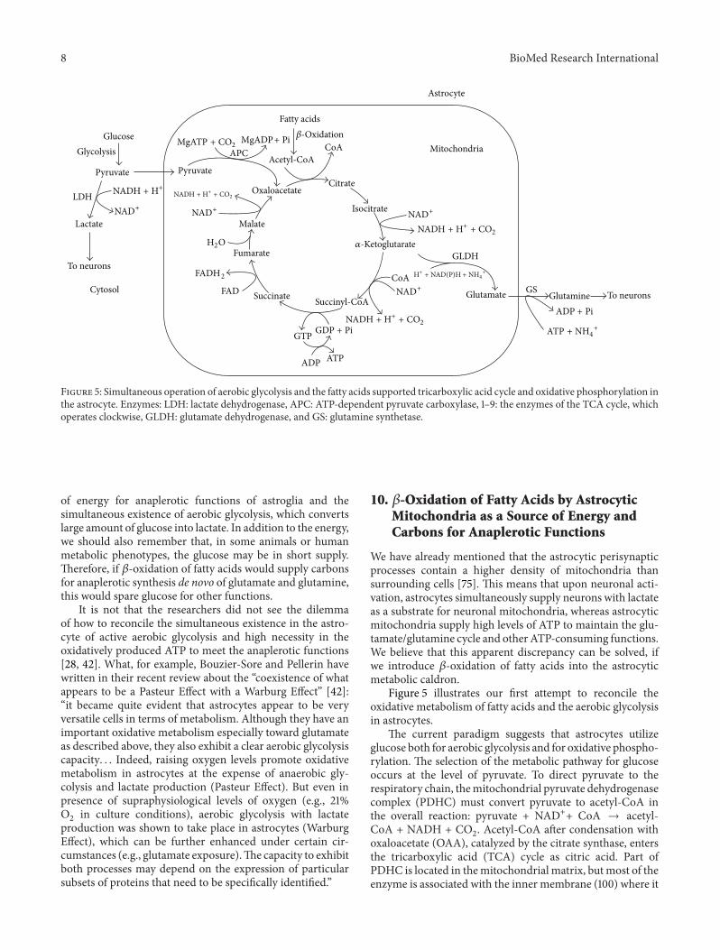
Figure 5: Simultaneous operation of aerobic glycolysis and the fatty acids supported tricarboxylic acid cycle and oxidative phosphorylation inthe astrocyte. Enzymes: LDH: lactate dehydrogenase, APC: ATP-dependent pyruvate carboxylase, 1–9: the enzymes of the TCA cycle, whichoperates clockwise, GLDH: glutamate dehydrogenase, and GS: glutamine synthetase.
of energy for anaplerotic functions of astroglia and thesimultaneous existence of aerobic glycolysis, which convertslarge amount of glucose into lactate. In addition to the energy,we should also remember that, in some animals or humanmetabolic phenotypes, the glucose may be in short supply.Therefore, if 𝛽-oxidation of fatty acids would supply carbonsfor anaplerotic synthesis de novo of glutamate and glutamine,this would spare glucose for other functions.
It is not that the researchers did not see the dilemmaof how to reconcile the simultaneous existence in the astro-cyte of active aerobic glycolysis and high necessity in theoxidatively produced ATP to meet the anaplerotic functions[28, 42]. What, for example, Bouzier-Sore and Pellerin havewritten in their recent review about the “coexistence of whatappears to be a Pasteur Effect with a Warburg Effect” [42]:“it became quite evident that astrocytes appear to be veryversatile cells in terms of metabolism. Although they have animportant oxidative metabolism especially toward glutamateas described above, they also exhibit a clear aerobic glycolysiscapacity. . . Indeed, raising oxygen levels promote oxidativemetabolism in astrocytes at the expense of anaerobic gly-colysis and lactate production (Pasteur Effect). But even inpresence of supraphysiological levels of oxygen (e.g., 21%O2in culture conditions), aerobic glycolysis with lactate
production was shown to take place in astrocytes (WarburgEffect), which can be further enhanced under certain cir-cumstances (e.g., glutamate exposure).The capacity to exhibitboth processes may depend on the expression of particularsubsets of proteins that need to be specifically identified.”
10. 𝛽-Oxidation of Fatty Acids by AstrocyticMitochondria as a Source of Energy andCarbons for Anaplerotic Functions
We have already mentioned that the astrocytic perisynapticprocesses contain a higher density of mitochondria thansurrounding cells [75]. This means that upon neuronal acti-vation, astrocytes simultaneously supply neurons with lactateas a substrate for neuronal mitochondria, whereas astrocyticmitochondria supply high levels of ATP to maintain the glu-tamate/glutamine cycle and other ATP-consuming functions.We believe that this apparent discrepancy can be solved, ifwe introduce 𝛽-oxidation of fatty acids into the astrocyticmetabolic caldron.
Figure 5 illustrates our first attempt to reconcile theoxidative metabolism of fatty acids and the aerobic glycolysisin astrocytes.
The current paradigm suggests that astrocytes utilizeglucose both for aerobic glycolysis and for oxidative phospho-rylation. The selection of the metabolic pathway for glucoseoccurs at the level of pyruvate. To direct pyruvate to therespiratory chain, themitochondrial pyruvate dehydrogenasecomplex (PDHC) must convert pyruvate to acetyl-CoA inthe overall reaction: pyruvate + NAD++ CoA → acetyl-CoA + NADH + CO
2. Acetyl-CoA after condensation with
oxaloacetate (OAA), catalyzed by the citrate synthase, entersthe tricarboxylic acid (TCA) cycle as citric acid. Part ofPDHC is located in themitochondrial matrix, butmost of theenzyme is associated with the inner membrane (100) where it

BioMed Research International 9
may form functional complexes with other enzymes of theTCA cycle. According to our hypothesis, presented here (seeFigure 5), 𝛽-oxidation of fatty acids is the source of acetyl-CoA for the TCA cycle. Oxaloacetate for the citrate synthasereaction is provided by the pyruvate carboxylase, which isexclusively the astrocytic enzyme [100–102]. Waagepetersenet al. concluded that neuronal pyruvate carboxylation isunlikely to be of quantitative significance [102]. Pyruvatecarboxylase catalyzes the ATP-dependent irreversible car-boxylation of pyruvate to form oxaloacetate: pyruvate +HCO3
− + ATP → oxaloacetate + ADP + Pi.The idea that 𝛽-oxidation of fatty acids may be the prefer-
able substrate for production of acetyl-CoA in astrocytesis supported by the properties of the PDHC in astrocyticmitochondria. Halim et al. have shown that all componentsof the PDHC were expressed in both neurons and astrocytesin culture [103]. However, in astrocytes, the PDHC activitywas kept strongly inhibited through phosphorylation of thepyruvate dehydrogenase alpha subunit. In contrast, neuronalPDHC operated close to maximumwithmuch lower levels ofphosphorylated PDH alpha. Dephosphorylation of astrocyticPDH alpha restored the PDHC activity and lowered lactateproduction [103]. This intrinsic property of the astrocyticmitochondrial PDHC will favor glucose entering the gly-colytic pathway and production of OAA, rather than enteringthe TCA cycle. This also suggests that, in astrocytes, fattyacids may be the major source for acetyl-CoA.
Upon neuronal activation, aspartate increased glutamatesynthesis in both control and the aralar-deficient astrocytes,mainly by serving as amino donor [96], but also by producingadditional OAA and thus saving more glucose for lactateproduction. Under these conditions, 𝛼-ketoglutarate willbe constantly produced in the TCA cycle and convertedby the mitochondrial aspartate aminotransferase (AAT) toglutamate [104]. It has been shown that mitochondrial glu-tamate dehydrogenase (GLDH) is important for glutamatedegradation, whereas glutamate biosynthesis occurs mainlyas a transamination via AAT [104]. It is important that bothin neurons and astrocytes AAT and GDLH may function asmultienzyme complexes [104].The complex regulation of glu-tamate formation or disposal by the multienzyme complexesis discussed in the comprehensive review by McKenna et al.[104].
The newly produced glutamate will be actively removedfrom mitochondria by the cytosolic ATP-dependent glu-tamine synthase, and at high cytosolic ATP, the phosphory-lated glutamine transporters will pump out glutamine fromthe astrocyte [105].
11. In Astrocytes, OxidativePhosphorylation Proceeds Simultaneouslywith Aerobic Glycolysis
Upon neuronal activation, astrocytes remove significant partof glutamate from the synaptic cleft by the Na+-dependentuptake system, which requires energy, and thus stimulatesglycolysis [31, 38]. Glutamate induces also a rapid (withina few seconds) and reversible increase of glucose transport
into astrocytes [106] that parallels the increase in glucoseutilization [31, 38]. Moreover, glutamatemay cause inhibitionof neuronal glucose transport, which is even stronger in thepresence of lactate [107]. The primary isoforms of glucosetransporter in brain are GLUT1, detected at high concen-trations in blood-brain barrier and glia; GLUT3 in neurons;and GLUT5 in microglia [108]. After glucose entry, glucoseis phosphorylated by type I hexokinase (HK1) [109, 110]. Inastrocytes, most of HK1 is associated with mitochondria andthe activity of HK1 bound to mitochondria is greater thanthe cytosolic hexokinase [111, 112]. Moreover, this associationof HK1 is modulated in coordination with changes in thecell’s metabolic state [111]. Thus, in the astrocytes, formationof glucose-6-phosphate occurs not for the expense of thecytosolic ATP but for the expense of mitochondrial ATP andthis will stimulate formation of pyruvate.
Neurons lack pyruvate carboxylase; instead, all activitiesof this enzyme were found in astroglia [113, 114]. In addition,PDHC in astrocytic mitochondria is inhibited [75], verysimilar to liver mitochondria when the liver metabolismswitches from glucose oxidation to gluconeogenesis andoxidation of fatty acids [115]. Together, these properties of thekey enzymes in astrocytes direct more pyruvate to formationof lactate and OAA. This will also promote 𝛽-oxidationof fatty acids that will supply the TCA cycle with acetyl-CoA, which is allosteric activator of pyruvate carboxylase[116]. Upon neuronal activation, OAA produced by pyruvatecarboxylase will be directed toward the TCA cycle and 𝛼-ketoglutarate will be removed from the cycle for glutamatesynthesis; in resting synapses, when the necessity in newlyformed glutamate and lactate is diminished, the OAA will bedirected towards gluconeogenesis, which also requires ATP.Again, 𝛽-oxidation of fatty acids will provide ATP for thisenergy-dependent metabolic pathway and more glucose willbe stored as glycogen.
Active oxidative phosphorylation will maintain in thecytosol high ATP/ADP and NADH/NAD+ ratios. This isbecause 𝛽-oxidation enzymes, the membrane-bound TCAcycle enzymes, and the respiratory chainmay be organized inthe functional complexes and work relatively independentlyfrom the cytosolic pool of ATP and NADH. Therefore,a large part of pyruvate formed during glycolysis will bereduced to lactate, which will be rapidly removed fromthe astrocytes [38]. In this way, both the aerobic glycolysisand oxidative phosphorylation will proceed simultaneouslyin the astrocyte as quasi-irreversible pathways, which weremetabolically activated by transported glutamate from theactivated synapses.
When the neuronal synaptic terminals become “quiet,”astrocytes will also becomemetabolically inactivated becauseaccumulation of lactatewill inhibit glycolysis,𝛼-ketoglutaratewill not be converted to glutamate and thus will be metab-olized in the TCA cycle towards OAA. Altogether, this willstimulate gluconeogenesis and accumulation of glycogen.Glycogen is the glucose reserve buffer during periods ofhigh rate of glucose consumption and glucose shortage. Toour opinion, glucose from the glycogen during periods ofactivation will not be released to neurons but, rather, willbe converted to lactate inside the astrocyte. Thus oxidation

10 BioMed Research International
of fatty acids by astroglial mitochondria provides energyand part of the carbon skeleton for the anaplerotic synthesisof neuromediators and saves glucose, which is convertedeither into lactate to fuel neurons, or stored for emergencyas glycogen.
12. Properties of Neuronal Mitochondria
In experiments with isolated forebrain or spinal cord mito-chondria, we, as well as many other researchers, used thepopular method by Sims [117] with slight modifications[10, 11]. The method uses a very mild homogenizationtechnique and a purification step with the discontinuousPercoll gradient. The resulting mitochondria are consideredto originate predominantly from postsynaptic elements ofthe synapses, partly from the blown presynaptic vesicles,neuronal cell bodies, and, possibly, astroglia [45, 84]. The“contamination” with the astroglial mitochondria is, morelikely, negligible because they showedhigh rates of respirationwith glutamate and pyruvate. As we have mentioned above,astrocytic mitochondria have low expression of glutamate-aspartate transporter [46, 118] and low activity of PDHC[103].Electron microscopic study has shown that, purified in thePercoll gradient, brain and spinal cord mitochondria werenot contaminated by other organelles [16]. Panov et al. [10]have shown that nonsynaptic mitochondria have respiratoryqualities similar to those found for synaptic mitochondria[60]. In this review, we will designate these, the so callednonsynaptic mitochondria, as brain mitochondria (BM).
13. Brain but Not Astroglial MitochondriaPossesses the ElectrogenicGlutamate/Aspartate Transporter (GAT)
Brain mitochondria have some principal properties thatstrongly distinguish them from the astrocytic mitochondria.First, astroglial mitochondria have low or no expression ofthe glutamate/aspartate transporter (GAT), but, instead, GATis mainly neuronal [45, 118–120]. The glutamate-aspartatetransporter is importantmetabolically because it is a requiredcomponent of the malate/aspartate shuttle (MAS) [120, 121].The malate-aspartate shuttle is considered the most impor-tant shuttle in brain. It is particularly important in neuronsand may be extremely low or even nonexistent in brainastrocytes [59, 120]. Hertz suggested that aralar/AGC1 inbrain astrocytes, even at very low levels, could also playa role in a modified aspartate-malate shuttle to oxidizereducing equivalents in mitochondria [59]. MAS is the majorpathway by which cytosolic electrons from NADH can enterthe mitochondria and be oxidized [120, 121]. Lactate, as asubstrate, is energetically richer than pyruvate, but withoutMAS; neuronal mitochondria cannot effectively utilize thisextra hydrogen and convert lactate to pyruvate.
The absence of GAT from astrocytes in the brain impliesa compartmentation of the intermediary metabolism ofglucose, with glycolysis taking place in astrocytes and lactateexported to the extracellular fluid and oxidized to CO
2
and H2O in neurons [43]. Glycolysis starting from glucose
can, of course, proceed actively in neurons as well as inastrocytes, but the very fact that most neuronal mitochondriaare localized in the narrow space of synapses, suggests thatglycolysis cannot be the major source of mitochondrialsubstrates for synaptic activities. However, propagation ofthe electrical signal, along dendrites, axons, and cell body, ismaintained with participation of the glycolytic ATP.
GAT is expressed in mammals in two isoforms, aralarand citrin [122]. Aralar is expressed in the liver and bothisoforms are expressed in the heart. Although both isoformsare detected in the brain during the first weeks of life, onlyaralar is detected in the mature brain [118]. Both aralar andcitrin are electrogenic and unidirectional. They transport aprotonated glutamate into the mitochondria in exchange foraspartate anion utilizing the energy of the mitochondrialtransmembrane electrochemical gradient (ΔΨ) [121].
Berkich et al. stressed that aralar requires the presenceof mitochondrial aspartate aminotransferase (AST) to gen-erate aspartate and cannot provide glutamate for glutamatedehydrogenase (GLDH) because GLDH does not produceaspartate for exchange with glutamate [45]. However, bothneurons and astroglial cells have two glutamate/hydroxylcarriers, GC1 and GC2 [122, 123]. These carriers operatereversibly providing substrate for GLDH.
In the energized cells, active transport of glutamate intoneuronal mitochondria by the electrogenic aralar, which isstructurally and functionally coupled to AAT, will producethe reciprocal amount of aspartate. Aspartate may be trans-ported into astrocytic cells, where it will be converted by thecytosolic AAT into glutamate and oxaloacetate (OAA) [68].
14. Isolated Brain Mitochondria Have StronglyInhibited Succinate Dehydrogenase
A large scale utilization of glutamate by BM, as the energy-delivering substrate, is strongly suggested not only by the doc-umented large scale transport of the mediator into neurons[80, 124] but also by the properties of isolated BM and SCM[9, 10, 16, 62, 125]. In a number of recent works, we haveshown that in the brain and spinal cord mitochondria, themajor source of reactive oxygen species (ROS) productionwas associated with the reverse electron transport (RET)[10, 125].Therefore, one of themost interesting and importantfeatures of the neuronal mitochondria was that mitochon-drial succinate dehydrogenase (SDH) was usually stronglyinhibited (see Figure 7) by endogenous oxaloacetate (OAA)[10, 105]. The degree of this intrinsic SDH inhibition variesstrongly between the species. In other words, it depends onthe metabolic phenotype of the BM [62, 126].
The significance of this inhibition of SDH becomesevident from the fact that most neuronal mitochondria arelocated in the narrow spaces of synaptic junctions [63, 64].Upon activation, the synaptic mitochondria have no otherfunction except restoration of the ionic homeostasis acrossthemembranes of the synapses.Therefore, when synapses arenot activated, the inactive mitochondria become hyperpolar-ized. In other words, the synaptic mitochondria will respirein the metabolic State 4, when the reverse electron transport

BioMed Research International 11
(RET) activates production of reactive oxygen species (ROS)on complex I [9, 10].This is in strict contrast to mitochondriain other organs, such as liver, kidney, and heart, wheremitochondria are constantly producing ATP and the RET isusually at minimum.
Brain (BM) and spinal cord (SCM)mitochondria isolatedin the absence of bovine serum albumin (non-BSA-BM)usually display strong inhibitions of succinate oxidation (seeFigure 7(a)). Therefore, most researchers traditionally isolateBM and SCM in the presence of defatted BSA [126], whichsignificantly improved oxidation of succinate (Figure 7(b)).Figure 7(b) shows, however, that even with the BSA-BMoxidizing succinate, it required more than a minute for themembrane potential to reach the maximum.
It should be noted that, with many species, the BMisolated in the presence of BSA and initially oxidizingsuccinate develop strong inhibition of SDH after 40–60minutes after isolation. For comparison, in the non-BSA-BM oxidizing pyruvate + malate (Figure 8), energization ofBM, as measured by a TPP+-sensitive electrode, reached themaximum within few seconds.
It must be mentioned that too many researchers usedsuccinate as a substrate in the presence of rotenone, whichprevented formation of OAA, but also abolished otherphysiologically important events, such as reverse electrontransport and the associated ROS production.We believe thatintrinsic inhibition of SDH is an evolutionary mechanismagainst oxidative damage of the most vulnerable synapticmitochondria. This is of particular importance because wehave shown that in the BM and SCM up to 50% of pyru-vate and glutamate are oxidized via alanine and aspartateaminotransferases, which produce 𝛼-ketoglutarate and thensuccinate [10, 16]. Another adaptive mechanism in BM andSCM against excessive production of ROS in the synapticmitochondria of quiescent neurons is probably the lowcontents of the intramitochondrial substrates.
15. Metabolic Phenotypes of Mitochondriadue to Different Affinities of SDH toOxaloacetate
The intrinsic inhibition of succinate oxidation in BM andSCM varies significantly between the species [62, 126]. SCMin general showed much lower inhibition of SDH as com-pared to the BM together with the lower rates of respirationwith all substrates. This resulted in the lower rates of ATPprovision and increased rate of the RET-dependent ROSproduction in resting SCM [16, 62, 125]. BM and SCMhave very high activities of the matrix superoxide dismutase(SOD2) that very rapidly converts superoxide radicals toH2O2[10, 62]. Therefore, it is very likely that most of the
damaging effects of increased ROS production are associatedwith the increased H
2O2[62, 127].We have recently provided
evidence that the haplotype-specific differences in produc-tion of ROS (H
2O2) may be responsible for development of
sporadic and familial cases of amyotrophic lateral sclerosis(ALS) [62]. Evidently, the degree of the intrinsic inhibition ofSDH may be important phenotypic feature that determines
the susceptibility of a particular organism to diseases, such asALS, Alzheimer’s disease, and Parkinson’s disease, where theincreased oxidative stress plays important pathogenic roles.
There may be several mechanisms responsible for vari-ations in the OAA-dependent intrinsic inhibition of SDH.OAA is themost powerful inhibitor of SDH [128].The affinityof SDH for OAA changes with reduction of the enzyme’ssulfhydryl group depending on mitochondrial energization.Upon deenergization of mitochondria, the affinity of SDHto OAA may increase tenfold [129]. Another importantproperty of SDH is that, besides succinate, the enzyme canalso oxidize malate with the similar affinity [130]. Whileexternally addedOAAcompeteswith succinate for binding tothe enzyme,OAA formed during oxidation ofmalate remainstightly bound to the enzyme, which makes malate a powerfulinhibitor of SDH [130]. Of interest, the metabolic phenotype(nontransgene) of the Sprague Dawley rats (from TaconicFarms Inc. Germantown, NY 12526) in 2007 displayed astrong inhibition of ROSproduction in the presence ofmalate(Figure 9).
This ability of malate to inhibit ROS production waslost in the nontransgene Sprague Dawley rats in 2010, whenthe animals failed to develop ALS symptoms upon receivingthe mutated human SOD1 gene [62]. In this metabolicphenotype, the presence of malate in the substrate mixturesstimulated production of the RET-dependent ROS (data notshown).
16. Activation of Oxidative Phosphorylationduring Simultaneous Oxidation ofPyruvate, Glutamate, and Malate
In BM, there are close functional and structural relationshipsbetween enzymes that metabolize the tricarboxylic acid(TCA) cycle intermediates and amino acids [131]. Thereare three enzymes, ALT, AAT, and GLDH, which, in BM,convert alanine and glutamate to 𝛼-ketoglutarate (𝛼-KG)[94, 132]. The enzymes are found structurally colocalizedand may form functional complexes without releasing theintermediary metabolites [68, 94]. This type of enzymesinteraction is important for the neuronal mitochondria,which have a very high content of proteins in the matrix.For this reason, the matrix of mitochondria from the heart,brain, and skeletal muscle is a hard gel, which excludesor greatly hampers the diffusion of small molecules [133,134]. Therefore, many enzymes of the TCA cycle are tightlyassociated with the inner mitochondrial membrane. Forexample, the membrane-bound pools of pyruvate dehydro-genase complex (PDHC) and 𝛼-ketoglutarate dehydrogenasecomplex (KGDHC) about 200 times exceed the “soluble”pool of these enzymes in the matrix [133]. The enzymes areorganized into stable functional complexes, which tunnelmetabolites along the complexes forming efficient metabolicchannels. Such multienzyme complexes have been shownfor 𝛽-oxidation of fatty acids [135], malate-aspartate shuttle(aralar) with AAT and the TCA cycle enzymes [136, 137],AAT and GLDH [68], and respiratory chain complexes [138,139]. It was shown that, for the heart mitochondria, the

12 BioMed Research International
ratio for oxidative phosphorylation (OXPHOS) complexesI : II : III : IV : V was 1 : 2 : 3 : 6-7 : 3–5 [140] or more recentlydetermined as 1 : 1.5 : 3 : 6 : 3 [141]. The respiratory complexesinteract with each other to form a supercomplex namedthe respirasome [138]. Based on the above ratios of theOXPHOS complexes, Schagger and Pfeiffer [139] suggestedthat the respirasome exists as a mixture of two large super-complexes and one smaller supercomplex. Each of the twolarge supercomplexes is comprised of a complex I monomer,a complex III dimer, and four copies of complex IV. Thesmaller supercomplex contains two complexes III and fourcomplexes IV [16, 118]. The major advantages of the super-complex structure of the mitochondrial respiratory chain aresubstrate channeling, catalytic enhancement, sequestrationof reactive intermediates, and structural stabilization [138,139]. Evidently, similar considerations consider the neuronaland astroglial mitochondria, which also have high respira-tory activities and complex organization of the metabolicenzymes.
We suggest that, in addition to the benefits listed above,the respirasome is also an evolutionary adaptive mechanismdesigned to prevent excessive production of ROS. Several-fold excess of complex IV in the clusters of respiratoryenzymes promotes oxidation of the potential sites of superox-ide radical production in complexes I and III [9]. Evidently,this mechanism was developed early during the evolutionof the aerobic organisms because it is available in aerobicbacteria and yeast [138, 139]. In addition, mitochondria invivo oxidize several different substrates simultaneously com-ing from different metabolic pathways, which are stronglyorgan and species specific [10, 16], and thusmay have differentimpacts on oxidative stress.
The three glutamate-transforming enzymes have highMichaelis constants for glutamate: for GLDH 8–17mM [142]and for ALT and AST in the range of 7–20mM [132].However, in view of the electrogenic nature of the aralar andremoval of 𝛼-KG by the TCA cycle [45], in energized BM, thethree glutamate-transforming reactions operate irreversiblywith the net loss of glutamate. Therefore, even at concentra-tions much lower than the Km’s of the enzymes, glutamatewill be rapidly oxidized.
During neuronal activation, astrocytes provide additionalamounts of lactate for postsynaptic neuronal mitochondria[34, 143, 144]. For rapid conversion of lactate to pyruvatepostsynaptic mitochondria must receive a certain amountof glutamate to fuel MAS in order to recycle the cytosolicNAD+ [10, 45]. Importantly, several research groups [145–148] have shown that upon neuronal activation, nanomolarconcentrations of external Ca2+ specifically activate oxidativephosphorylation via the aralar-dependent transport of gluta-mate intomitochondria.Therefore, increased neuronal activ-ity through small changes in the extramitochondrial Ca2+activates MAS and OXPHOS, whereas increased mitochon-drial Ca2+ may further activate mitochondrial respirationvia specific dehydrogenases [149]. As a result, the glutamate,which is present in the neurons, may be rapidly oxidized bothpresynaptically and postsynaptically [10, 60].
We have suggested [10] that in the presence of pyruvate+ glutamate, particularly when malate was also present (with
Cytosol
Glutamate
Aspartate
AST
AST
𝛼-kg
𝛼-kg
OAANADH
NADH
NAD+
NAD+
Malate
MDH
GAT
MKgT
Matrix
Glutamate
Aspartate
Malate
MDH
OAA
OAA
Inner mitochondrialmembrane
+ −
𝛼-Ketoglutarate (𝛼-kg)
Figure 6: The malate-aspartate shuttle (MAS). The mitochondrialinner membrane is impermeable for NADH. In order to effec-tively utilize lactate for mitochondrial respiration, lactate mustbe converted to pyruvate in the reaction lactate + NAD+ →pyruvate + NADH. NADH is reoxidized by the MAS.The process isunidirectional because GAT (aralar) is electrogenic and the matrixNADH is rapidly oxidized by the respiratory chain.
the exception shown above), the respiration overcomes somelimiting steps in the respiratory chain. We considered at leasttwo such limiting steps [10]. First, the activity of succinatedehydrogenase (SDH/Complex II) may be inhibited by OAAand thus limit the rate of the TCA cycle operation during thestate 3 and state 3U. The ability of pyruvate and glutamateto overcome inhibition of SDH was associated with themetabolic removal of OAA in the citrate synthase, aspartate,and alanine transaminase reactions, respectively.
The second limitation point is the 𝛼-KGDHC reaction.It has been shown [150] that activity of 𝛼-KGDHC is thelowest among the TCA cycle enzymes and is controlled by theavailability of 𝛼-KG [151] and the enzyme’s affinity for 𝛼-KG,which is controlled by Ca2+ and Mg2+ [152, 153]. In addition,a decreased matrix ATP/ADP ratio due to increased energyconsumption in activated neurons would also increase theavailability of GDP for the substrate level phosphorylationand thus the overall activity of 𝛼-KGDHC [94]. The key roleof 𝛼-KG for the TCA-related hydrogen supply was shownin experiments with labelled metabolites [154]. Balazs (1965)has shown that the amount of 𝛼-KG increased 30-fold inBM oxidizing glutamate + pyruvate + malate [154]. Underthese conditions, GLDH does not participate in productionof 𝛼-KG [155]. Since both AAT and ALT are present in agreat excess, as compared with the respiration rate, the OAAis continuously removed by the transamination reactions.Balazs concluded that a competition takes place betweenthe 𝛼-KGDHC and GLDH, probably for NAD+, resulting inpreferential oxidation of 𝛼-ketoglutarate [155].
Yudkoff et al. suggested that in the presence of glutamate+ pyruvate, the TCA cycle in brain mitochondria operates astwo coupled cycles (see Figure 10): cycle A is leading from 𝛼-KG to OAA and cycle B from OAA to 𝛼-KG which includesthe citrate synthase reaction (see Figure 10) [60]. Accordingto Yudkoff et al., the flux of substrates through cycle A is

BioMed Research International 13
1
39
RBM
RBM
TPP+
ADP
ADP
79
155
DT
CCCP
RCR = 2.0
2
0 120 240 360 480 600 720 840
Time (s)
(a)
1
RBM
RBM
TPP+
ADP
ADP
CCCP
RCR = 4.3
2
60
257
63
264
0 120 240 360 480 600 720
Time (s)
(b)
Figure 7: Respiratory activity and membrane potential of the rat (Lewis) brain mitochondria isolated in the absence (a) and in the presence(b) of 0.1% BSA. Incubation conditions: 125mM KCl, 10mM NaCl, 10mM MOPS, pH 7.2, 2mM MgCl
2, 2mM KH
2PO4, 1mM EGTA, and
0.7mMCaCl2. At Ca2+/EGTA = 0.7, the [Ca2+]Free was 1 𝜇M. Chamber volume = 0.65 mL. Substrate: succinate 5mM, no rotenone was added.
Additions: BM 0.3mg, ADP 150 𝜇M, and CCCP 0.5 𝜇M. Numbers at the traces are respiratory activities in nmol/min/mg mitochondrialprotein. Respiratory activity ratio (RCR) is 𝑉State 3/𝑉State 4.
0 120 240 360 480 600
Membrane potential
Time (s)
21
121
24
138
120
24
RBM
ADP
ADP CCCP
RBM
Respiration
State 3U
State 3State 4 0
TPP+
TPP+
−180mV
−140mV
O2 = 0
Figure 8: Respiratory activity and membrane potential of the rat(Sprague Dawley) brain mitochondria isolated in the absence of0.1% BSA. Incubation conditions as in Figure 7. Substrates: pyruvate2.5mM, malate 2mM. Additions: ADP 150 𝜇M and CCCP 0.5 𝜇M.Numbers at the traces are respiratory activities in nmol/min/mgmitochondrial protein.
3–5-fold faster than through the cycle B [60]. Thus, withpyruvate + glutamate + malate, activation of 𝛼-KGDHC andSDHmay significantly increase the rates of the TCA cycle andrespiratory chain in state 3 and state 3U (see Figure 9). A highturnover of cycle A, with activated SDH, would increase RETand the associated ROS production in resting mitochondria.As a result, the rate of oxygen consumption in the metabolicState 4 also increases [10].
700
650
600
550
500
450
400
350
300
250
200
150
100
50
0
P+
M
G+
M
P+
G+
M S
S+
P
S+
P+
G+
M
S+
G
S+
G+
M
S+
M
NSNS
Gen
erat
ion
ofH
2O2
as %
of p
yruv
ate+
mal
ate
∗∗∗
∗∗∗
∗∗∗
∗∗∗
∗∗∗
∗∗∗
Figure 9: Generation of H2O2by non-BSA rat brain mitochondria
oxidizing various substrates and substrate mixtures. Incubationconditions as in Figure 7. Substrates: pyruvate 2.5mM, glutamate5mM, succinate 5mM, and malate 2mM. The method of ROSmeasurements was described in [10]. Statistics: ∗∗∗𝑃 < 0.001; NS:not significant.
17. Neuronal Mitochondria DoOxidize Fatty Acids in the Mixtures withOther Substrates
It has long been recognized that fatty acids can enter the brainand be metabolized to CO
2and H
2O [51, 52, 156, 157]. Much

14 BioMed Research International
Malate
Fumarate
SDHMalonate
Succinate
Succinyl-CoA
Oxaloacetate
GlutamateAST
Aspartate
PyruvateALTAlanine
Citrate
Acetyl-CoA
Pyruvate
cis-Aconitate
Isocitrate
(a) (b)
𝛼-Ketoglutarate
Figure 10: A schematic presentation of operation of the tricarboxylic acid cycle in brain mitochondria oxidizing glutamate and pyruvate.The figure was adapted from [95]. Abbreviations: AST: aspartate aminotransferase, ALT: alanine aminotransferase, and SDH: succinatedehydrogenase.The symbol of the closed lock means the step catalyzed by SDH is inhibited.The dashed arrows indicate inhibitory influencesof malate and oxaloacetate on SDH.
of the evidence was obtained in the in vitro studies usingprimary cultures of various cells from the developing brain[67, 156] and experiments with brain perfusion [53, 54]. It wasconcluded that the brain’s capacity to oxidize fatty acids andthe levels of the enzymes of fatty acid oxidation in the brainwere much higher in the suckling rat than in the adult rat [55,156]. In primary cultures, only astrocytes were able to utilizefatty acids for 14CO
2production, and the rate of utilization
was greater than that of the ketone bodies. The metabolicpatterns of the brain cells derived from the developing braincomplemented the nature of the diet of the suckling animals,which was rich in fat and low in carbohydrate [156]. Andthere was evidence that the brain of adult animals (at leastfrom dogs and cats) did not utilize free fatty acids in vivo[53, 54]. Thus, currently it is widely accepted that neuronalmitochondria in adult brain do not oxidize fatty acids [158].
There is very little evidence regarding the fatty acidsmetabolism in the isolated brain mitochondria from adultanimals with the use of the new paradigm, that is, with theuse of the physiologically relevant substrate mixtures. Weaddressed this issue using rat brain mitochondria and ournewmethodology based on the assumption that in situmito-chondria oxidize not a single but several substrates. There-fore, we tested palmitoyl carnitine as a substrate in combina-tionwith the “classical” substrates for the brainmitochondria.Figure 11 presents direct polarographic recordings of respi-ratory activities of the rat brain mitochondria with differentsubstrates and their mixtures. Figure 12 presents the sum-mary of three separate isolations and also shows the rates ofROS production with each substrate and a substrate mixture.
The panels (a), (b), and (c) of Figure 11 illustrate respira-tory activities of BMwith the “classical” substrates: glutamate+ malate, pyruvate + malate and succinate. Notice that withsuccinate (Figure 11(c)), the State 4 respiration was several-fold higher than with glutamate (Figure 11(a)) or pyruvate(Figure 11(b)). The rates of the State 4 respiration to a large
degree are determined by the reverse electron transport(RET) and the associated production of superoxide radical.RET is an energy-dependent function and, since formationof the superoxide radical, serves as a sink for electrons;mitochondria increase the State 4 respiration. Thus, the State4 respiration may serve as a qualitative indicator of changesin ROS production by energized mitochondria.
Figure 11(c) shows that due to intrinsic inhibition ofSDH, the response of BM oxidizing succinate to additionof ADP was negligible. The inhibition was released uponaddition of 5mMglutamate (Figure 11(c)). Figure 11(d) showsthat, during oxidation of the substrates mixture succinate +glutamate + pyruvate, the rate of oxidative phosphorylationwas significantly higher than with glutamate or pyruvate (seealso Figure 12).
When palmitoyl carnitine was used as a substrate (Figure11(e)), the BM responded toADP by a 3-fold increase in respi-ration, whichwas followed by significant inhibition of oxygenconsumption. Addition of uncoupler (CCCP) also failed tosignificantly increase respiration. This experiment indicatesthat BM are capable, albeit not efficiently, of oxidizing fattyacids.We found that addition ofmalate to palmitoyl carnitinehad no effect on the rates of respiration. Only 𝐿-palmitoylcarnitine must be used as a substrate because 𝐷, 𝐿-palmitoylcarnitine is inhibitory.
In situations when palmitoyl carnitine was oxidized bythe BM in the presence of either pyruvate (Figure 11(f)),glutamate (Figure 11(g)), or succinate (Figure 11(h)), the ratesof respiration were significantly increased in all metabolicstates (see also Figure 12). The stimulation of respiration wasparticularly large during simultaneous oxidation of succinate+ palmitoyl carnitine (see Figure 11(h) and Figure 12).
At this time, we have no explanations on the mechanismor mechanisms that were involved in the stimulatory effectof respiration with the mixtures of palmitoyl carnitine withother substrates. This issue requires further investigation.

BioMed Research International 15
0 120 240 360 480 600 720Time (s)
CCCP
15419
145
13 ADP
DT
BM 0.3mg
RCR1 = 11
Glutamate + malate O2 = 0
(a)
0 120 240 360 480 600 720Time (s)
ADP
36
158 CCCP
185
31
12717 ADP
BM 0.3mg
O2 = 0
RCR1 = 7.5
RCR2 = 5.1
Pyruvate + malate
(b)
0 120 240 360 480 600Time (s)
54
Spontaneous ADP
191
Glutamate
60
158
CCCP
52 ADP
Succinate
BM 0.3mg
O2 = 0
inhibition
(c)
0 120 240 360 480 600 720Time (s)
ADP
38
CCCPADP
202
19840
180
44 ADP
O2 = 0
RCR1 = 4.1
RCR2 = 5.0
BM 0.2mg
Succinate + glutamate + pyruvate
(d)
0 120 240 360 480 600 720 840Time (s)
Dithionite
CCCP
7930
6718 ADP
BM 0.3mg
O2 = 0
RCR = 3.7
Palmitoylcarnitine
(e)
0 120 240 360 480 600 720Time (s)
22
127
ADP
25
158
CCCP
122
20 ADP
BM 0.3mg
O2 = 0
RCR1 = 7.9
RCR2 = 5.1
Pyruv. + palm.-carn. + malate
(f)
0 120 240 360 480 600Time (s)
38
167
CCCP
167
30 ADP
BM 0.3mg
O2 = 0Glut. + palm.-carn. + malate
RCR = 5.6
(g)
0 60 120 180 240 300 360 420 480 540Time (s)
Spontaneous inhibition
175 Glutamate60
223
CCCP
55 ADPBM 0.3mg
O2 = 0Succinate + palm.-carn.
RCR = 4.1
(h)
Figure 11:Oxygen consumption by rat (SpragueDawley, 2010) brainmitochondria isolatedwithout BSAoxidizing various substrates and theirmixtures in different metabolic states. Experimental conditions as described in Figure 6. Metabolic states are shown in Figure 7. Substrates:glutamate 5mM, malate 2mM, pyruvate 2.5mM, succinate 5mM, and palmitoyl carnitine 25 𝜇M. Numbers at the traces are respiratoryactivities in nmol/min/mgmitochondrial protein. Respiratory activity ratio (RCR) is𝑉State 3/preceeding𝑉State 4. Additions: brainmitochondria0.3mg, ADP 150 𝜇M, CCCP 0.5 𝜇M, and glutamate 5mM.

16 BioMed Research International
70
60
50
40
30
20
10
0
State 4
Glu
tam
ate
Pyru
vate
Palm
-car
n.
Glu
+P-
C
Pyr+
P-C
Succ
inat
e
S+
P-C
Nan
omol
O2
(min
/mg
prot
ein)
(a)
250
225
200
175
150
125
100
75
50
25
0
State 3
Glu
tam
ate
Pyru
vate
Palm
-car
n.
Glu
+P-
C
Pyr+
P-C
Succ
inat
e
S+
P-C
Nan
omol
O2
(min
/mg
prot
ein)
(b)
250
225
200
175
150
125
100
75
50
25
0
State 3U
Glu
tam
ate
Pyru
vate
Palm
-car
n.
Glu
+P-
C
Pyr+
P-C
Succ
inat
e
S+
P-C
Nan
omol
O2
(min
/mg
prot
ein)
(c)
600
550
500
450
400
350
300
250
200
150
100
50
0
ROS productionPe
rcen
t of g
luta
mat
e + m
alat
e (10
0%)
G+
M
P+
M
Palm
+ca
rn.
G+
M+
P-C
P+
M+
P-C
Succ
inat
e
S+
P-C
100%
(d)
Figure 12: Respiratory activity and ROS production by rat brain mitochondria oxidizing palmitoyl carnitine in various substrate mixtures.Incubation conditions as in Figure 7 and substrate mixtures as described in Figure 11. The data are average of three different isolations (M ±standard error). ROS production was measured with the Amplex red method as described in [10]. The minimal rate of ROS production wasobserved with glutamate + malate, which was taken as 100%.
However, the results presented in Figures 11 and 12 unequiv-ocally demonstrate the ability of BM to efficiently utilize fattyacids as a source of energy not only in astrocytes but in theneurons as well.
Of particular importance was the fact that simultaneousoxidation of palmitoyl carnitinewith the “classical” substratesresulted in the increased State 3 respiration and several-foldincrease in the generation of ROS in BM (see Figure 12). Thismay have implications for understanding the pathogeneses ofneurodegenerative diseases and pathologies associated withaging and the metabolic syndrome.
18. Conclusions
During the last decade, a large body of evidence has beenaccumulated on participation of mitochondrial dysfunctionsin pathogeneses of practically all major pathologies in human[1, 8, 159, 160]. The roles of mitochondrial dysfunctionswere particularly evident in a number of neurodegenerativediseases (NDD): Parkinson’s disease, Alzheimer’s disease,and amyotrophic lateral sclerosis (ALS) [159, 160]. Differentgenetic and toxic animal models have been developed tostudy the roles of mitochondria more closely [10, 161–163].There is one important feature common for the NDDs and

BioMed Research International 17
animal models of these diseases: most cases of NDDs aresporadic with only few percentage of cases having cleargenetic cause [62]; regarding the animal models, for example,the rotenone model of Parkinson’s disease or transgenicmodel of ALS, can be reproduced on only one or two species,while other species, rats in particular, did not reproduce thesymptoms of a pathology [164].
According to Wallace, variations in the haplotypes ofmtDNA may determine to a large degree the differencesin susceptibility of human individuals and experimentalanimal species to a particular pathology [1, 4]. At the levelof physiology, variations in the haplotypes of mtDNA maycome into view as variations in the mitochondrial metabolicphenotypes. That is, mitochondria may have differencesin affinity to various substrates, allosteric regulators, andso forth. These variations would be further manifested asdifferent rates of ATP synthesis or ATP/ADP ratios anddifferent rates of ROS production.Therefore, in order to studymore closely the relationships between variant haplogroupsof mtDNA and pathologies, we have to study variances inmitochondrial metabolic phenotypes. The old methodologyto study mitochondrial functions using only one substrate,succinate + rotenone, in particular, is outdated. We now haveseveral examples of advantages by using substratemixtures instudying mitochondrial functions [10, 165]. In this review, weshow that isolated brain mitochondria do oxidize palmitoylcarnitine in the presence of “classical” substrates glutamate,pyruvate, and succinate; and inclusion of fatty acids oxidationinto the scheme of energy metabolism of astroglia removedmany questions to astrocyte’s functioning. It is time to changethe current view on the neuronal energymetabolism as basedalmost exclusively on glucose.
19. Comment
Soon after we have submitted the paper of this review tothe Biomed Research International, Schonfeld and Reiserhave published a review, which was entitled: “Why doesbrain metabolism not favor burning of fatty acids to provideenergy? Reflections on disadvantages of the use of free fattyacids as fuel for brain” [158]. The conclusions of the authors,which are reflected in the title, were quite opposite to the con-clusions we present in the current review. Of course, it wouldbe interesting and instructive to write a critical appraisal ofthe arguments presented by Schonfeld and Reiser and in thecurrent review. But we think that, at the present, it is firstnecessary to provide the readers with our alternative pointof view and evidences in support. As we mentioned in thisreview, some of the discrepancies stem from the differences inconditions andmethods. For example, in one critical paper bySchonfeld et al., the authors did not observe reverse electrontransport and ROS production during oxidation by rat heartmitochondria of the carnitine derivatives of fatty acids,which was observed (though also not high) with succinateas a substrate [157]. We have found that the authors usedincubation medium, which contained 0.5mM EDTA andrather high concentration of palmitoyl carnitine (0.5mM).It is known that EDTA removes Mg2+ from the high affinitysites on mitochondria, and this causes an increase in the
proton conductance of the inner membrane [11, 166]. As aresult, the membrane potential drops by about 30–40mV,which is enough to blockRET [11]. In addition, in the rat heartmitochondria, oxidation of succinate is significantly inhibitedby the endogenous oxaloacetate. The degree of inhibitiondoes not depend on the presence of bovine serum albuminand varies between the animal species [11]. We hope that inthe near future the discrepancies between the two groups willbe resolved in the fruitful discussions for the benefit of theresearchers working with mitochondria.
Conflict of Interests
The authors declare that there is no conflict of interestsregarding the publication of this paper.
References
[1] D. C. Wallace, “A mitochondrial paradigm of metabolic anddegenerative diseases, aging, and cancer: a dawn for evolution-ary medicine,” Annual Review of Genetics, vol. 39, pp. 359–407,2005.
[2] J. P. Jenuth, A. C. Peterson, K. Fu, and E. A. Shoubridge, “Rand-om genetic drift in the female germline explains the rapid seg-regation of mammalian mitochondrial DNA,” Nature Genetics,vol. 14, no. 2, pp. 146–151, 1996.
[3] D. C. Wallace, “Why do we still have a maternally inheritedmitochondrial DNA? insights from evolutionary medicine,”Annual Review of Biochemistry, vol. 76, pp. 781–821, 2007.
[4] D. C. Wallace, “Mitochondria as Chi,” Genetics, vol. 179, no. 2,pp. 727–735, 2008.
[5] K. Darvishi, S. Sharma, A. K. Bhat, E. Rai, and R. N. K. Bamezai,“Mitochondrial DNA G10398A polymorphism imparts mater-nal Haplogroup N a risk for breast and esophageal cancer,”Cancer Letters, vol. 249, no. 2, pp. 249–255, 2007.
[6] R.-K. Bai, S. M. Leal, D. Covarrubias, A. Liu, and L.-J. C. Wong,“Mitochondrial genetic background modifies breast cancerrisk,” Cancer Research, vol. 67, no. 10, pp. 4687–4694, 2007.
[7] L. M. Booker, G. M. Habermacher, B. C. Jessie et al., “NorthAmerican white mitochondrial haplogroups in prostate andrenal cancer,” Journal of Urology, vol. 175, no. 2, pp. 468–472,2006.
[8] M. Brandon, P. Baldi, and D. C. Wallace, “Mitochondrial muta-tions in cancer,”Oncogene, vol. 25, no. 34, pp. 4647–4662, 2006.
[9] A. Panov, S. Dikalov, N. Shalbuyeva, R. Hemendinger, J. T.Greenamyre, and J. Rosenfeld, “Species- and tissue-specificrelationships between mitochondrial permeability transitionand generation of ROS in brain and liver mitochondria of ratsandmice,”American Journal of Physiology—Cell Physiology, vol.292, no. 2, pp. C708–C718, 2007.
[10] A. Panov, P. Schonfeld, S. Dikalov, R. Hemendinger, H. L.Bonkovsky, and B. R. Brooks, “The neuromediator glutamate,through specific substrate interactions, enhancesmitochondrialATP production and reactive oxygen species generation innonsynaptic brain mitochondria,” The Journal of BiologicalChemistry, vol. 284, no. 21, pp. 14448–14456, 2009.
[11] A. Panov, Practical Mitochondriology. Pitfalls and Problems inStudies of Mitochondria, Amazon, Lexington, Ky, USA, 2013.
[12] A. Panov and S. Dikalov, “Structural and metabolic determi-nants of mitochondrial superoxide and detection methods ofmitochondrial superoxide and hydrogen peroxide,” in Mito-chondria in Health and Death, chapter 5, Elsevier, 2014.

18 BioMed Research International
[13] N. A. Campbell, B. Williamson, and R. J. Hyden, Biology:Exploring Life, Pearson Prentice Hall, Boston,Mass, USA, 2006.
[14] A. V. Panov, Y. M. Konstantinov, and V. V. Lyakhovich, “Thepossible role of palmitoyl CoA in the regulation of the adeninenucleotides transport in mitochondria under different meta-bolic states. I. Comparison of liver mitochondria from starvedand fed rats,” Journal of Bioenergetics, vol. 7, no. 2, pp. 75–85,1975.
[15] A. V. Panov, V. A. Vavilin, V. N. Solovyov, and V. V. Lyakhovich,“Relationships between the adenine nucleotide system and oxi-dative phosphorylation in rat liver in the dynamics of starva-tion,” Biokhimiya, vol. 48, no. 2, pp. 235–243, 1983.
[16] A. V. Panov, N. Kubalik, N. Zinchenko et al., “Metabolic andfunctional differences between brain and spinal cordmitochon-dria underlie different predisposition to pathology,” AmericanJournal of Physiology—Regulatory Integrative and ComparativePhysiology, vol. 300, no. 4, pp. R844–R854, 2011.
[17] T. R. Kasser, A.Deutch, andR. J.Martin, “Uptake and utilizationof metabolites in specific brain sites relative to feeding status,”Physiology and Behavior, vol. 36, no. 6, pp. 1161–1165, 1986.
[18] N. J. Abbott, L. Ronnback, and E. Hansson, “Astrocyte-endo-thelial interactions at the blood-brain barrier,” Nature ReviewsNeuroscience, vol. 7, pp. 41–53, 2006.
[19] N. J. Abbott, A. A. K. Patabendige, D. E.M. Dolman, S. R. Yusof,and D. J. Begley, “Structure and function of the blood-brainbarrier,” Neurobiology of Disease, vol. 37, no. 1, pp. 13–25, 2010.
[20] P. Ballabh, A. Braun, and M. Nedergaard, “The blood-brainbarrier: an overview: structure, regulation, and clinical impli-cations,” Neurobiology of Disease, vol. 16, no. 1, pp. 1–13, 2004.
[21] D. Virgintino, P. Monaghan, D. Robertson et al., “An immuno-histochemical and morphometric study on astrocytes andmicrovasculature in the human cerebral cortex,”TheHistochem-ical Journal, vol. 29, no. 9, pp. 655–660, 1997.
[22] S. Nag and D. J. Begley, “Blood-brain barrier, exchange of meta-bolites and gases,” in Pathology and Genetics. CerebrovascularDiseases, H. Kalimo, Ed., pp. 22–29, Neuropath Press, Basel,Switzerland, 2005.
[23] T. K. T. Lam, G. J. Schwartz, and L. Rossetti, “Hypothalamicsensing of fatty acids,”NatureNeuroscience, vol. 8, no. 5, pp. 579–584, 2005.
[24] R. S. el-Bacha and A. Minn, “Drug metabolizing enzymes incerebrovascular endothelial cells afford a metabolic protectionto the brain,” Cellular and Molecular Biology, vol. 45, no. 1, pp.15–23, 1999.
[25] N. Bresolin, L. Freddo, L. Vergani, and C. Angelini, “Carnitine,carnitine acyltransferases, and rat brain function,”ExperimentalNeurology, vol. 78, no. 2, pp. 285–292, 1982.
[26] P. Ciofi, M. Garret, O. Lapirot et al., “Brain-endocrine interac-tions: a microvascular route in the mediobasal hypothalamus,”Endocrinology, vol. 150, no. 12, pp. 5509–5519, 2009.
[27] K. E. Schlageter, P. Molnar, G. D. Lapin, and D. R. Groothuis,“Microvessel organization and structure in experimental braintumors: microvessel populations with distinctive structural andfunctional properties,”Microvascular Research, vol. 58, no. 3, pp.312–328, 1999.
[28] G.A.Dienel and L.Hertz, “Glucose and lactatemetabolismdur-ing brain activation,” Journal of Neuroscience Research, vol. 66,no. 5, pp. 824–838, 2001.
[29] C.-P. Chih, P. Lipton, and E. L. Roberts Jr., “Do active cerebralneurons really use lactate rather than glucose?” Trends inNeurosciences, vol. 24, no. 10, pp. 573–578, 2001.
[30] C.-P. Chi and E. L. Roberts Jr., “Energy substrates for neuronsduring neural activity: a critical review of the astrocyte-neuronlactate shuttle hypothesis,” Journal of Cerebral Blood Flow andMetabolism, vol. 23, no. 11, pp. 1263–1281, 2003.
[31] L. Hertz, “The astrocyte-neuron lactate shuttle: a challenge of achallenge,” Journal of Cerebral Blood Flow and Metabolism, vol.24, no. 11, pp. 1241–1248, 2004.
[32] L. Pellerin and P. J. Magistretti, “Glutamate uptake into astro-cytes stimulates aerobic glycolysis: a mechanism coupling neu-ronal activity to glucose utilization,” Proceedings of the NationalAcademy of Sciences of the United States of America, vol. 91, no.22, pp. 10625–10629, 1994.
[33] L. Pellerin and P. J. Magistretti, “Excitatory amino acids stim-ulate aerobic glycolysis in astrocytes via an activation of theNa+/K+ ATPase,” Developmental Neuroscience, vol. 18, no. 5-6,pp. 336–342, 1996.
[34] P. J.Magistretti and L. Pellerin, “Metabolic coupling during acti-vation: a cellular view,” Advances in Experimental Medicine andBiology, vol. 413, pp. 161–166, 1997.
[35] L. Pellerin, “Lactate as a pivotal element in neuron-glia meta-bolic cooperation,”Neurochemistry International, vol. 43, no. 4-5, pp. 331–338, 2003.
[36] L. Pellerin and P. J. Magistretti, “Empiricism and rationalism:two paths toward the same goal,” Journal of Cerebral Blood Flowand Metabolism, vol. 24, no. 11, pp. 1240–1241, 2004.
[37] L. Pellerin and P. J. Magistretti, “Neuroenergetics: calling uponastrocytes to satisfy hungry neurons,”Neuroscientist, vol. 10, no.1, pp. 53–62, 2004.
[38] L. Pellerin, A. P. Halestrap, and K. Pierre, “Cellular and subcel-lular distribution of monocarboxylate transporters in culturedbrain cells and in the adult brain,” Journal of NeuroscienceResearch, vol. 79, no. 1-2, pp. 55–64, 2005.
[39] L. Pellerin, A.-K. Bouzier-Sore, A. Aubert et al., “Activity-dependent regulation of energy metabolism by astrocytes: anupdate,” Glia, vol. 55, no. 12, pp. 1251–1262, 2007.
[40] A. Schousboe, L. K. Bak, and H. S. Waagepetersen, “Astrocyticcontrol of biosynthesis and turnover of the neurotransmittersglutamate and GABA,” Frontiers in Endocrinology, vol. 4, no.102, pp. 1–11, 2013.
[41] L. Pellerin and P. J. Magistretti, “Sweet sixteen for ANLS,” Jour-nal Cerebral Blood FlowMetabolism, vol. 32, pp. 1152–1166, 2012.
[42] A. -K. Bouzier-Sore and L. Pellerin, “Unraveling the complex,metabolic nature of astrocytes,” Frontiers in Cellular Neuro-science, vol. 7, article 179, pp. 1–13, 2013.
[43] G. A. Dienel and N. F. Cruz, “Nutrition during brain activation:does cell-to-cell lactate shuttling contribute significantly tosweet and sour food for thought?” Neurochemistry Interna-tional, vol. 45, no. 2-3, pp. 321–351, 2004.
[44] G. A. Dienel, “Brain lactate metabolism: the discoveries and thecontroversies,” Journal of Cerebral Blood Flow and Metabolism,vol. 32, pp. 1107–1138, 2012.
[45] D. A. Berkich, M. S. Ola, J. Cole, A. J. Sweatt, S. M. Hutson, andK. F. LaNoue, “Mitochondrial transport proteins of the brain,”Journal of Neuroscience Research, vol. 85, no. 15, pp. 3367–3377,2007.
[46] L. Contreras, A. Urbieta, K. Kobayashi, T. Saheki, and J.Satrustegui, “Low levels of citrin (SLC25A13) expression inadult mouse brain restricted to neuronal clusters,” Journal ofNeuroscience Research, vol. 88, no. 5, pp. 1009–1016, 2010.
[47] Y. Itoh, T. Esaki, K. Shimoji et al., “Dichloroacetate effects onglucose and lactate oxidation by neurons and astroglia in vitro

BioMed Research International 19
and on glucose utilization by brain in vivo,” Proceedings of theNational Academy of Sciences of the United States of America,vol. 100, no. 8, pp. 4879–4884, 2003.
[48] L. Sokoloff, S. Takahashi, J. Gotoh, B. F. Driscoll, and M. J.Law, “Contribution of astroglia to functionally activated energymetabolism,” Developmental Neuroscience, vol. 18, no. 5-6, pp.343–352, 1996.
[49] A. Tarozzi, F. Morroni, C. Bolondi et al., “Neuroprotectiveeffects of erucin against 6-hydroxydopamine-induced oxidativedamage in a dopaminergic-like neuroblastoma cell line,” Inter-national Journal of Molecular Sciences, vol. 13, no. 9, pp. 10899–10910, 2012.
[50] A. Panov and Z. Orynbayeva, “Bioenergetic and antiapoptoticproperties of mitochondria from cultured human prostatecancer cell lines PC-3, DU145 and LNCaP,” PloS ONE, vol. 8,no. 8, Article ID e72078, 2013.
[51] G. A. Dhopeshwarkar and J. F. Mead, “Fatty acid uptake by thebrain. III. Incorporation of [I-14C]oleic acid into the adult rat-brain,” Biochimica et Biophysica Acta, vol. 210, no. 2, pp. 250–256, 1970.
[52] G. A. Dhopeshwarkar, C. Subramanian, and J. F. Mead, “Fattyacid uptake by the brainV. incorporation of [1-14c]linolenic acidinto adult rat brain,” Biochimica et Biophysica Acta, vol. 239, no.2, pp. 162–167, 1971.
[53] C. Allweis, T. Landau, M. Abeles, and J. Magnes, “The oxidationof uniformly labelled albumin-bound palmitic acid to CO2 bythe perfused cat brain,” The Journal of Neurochemistry, vol. 13,no. 9, pp. 795–804, 1966.
[54] J. J. Spitzer, “CNS and fatty acid metabolism,” Physiologist, vol.16, no. 1, pp. 55–68, 1973.
[55] J. B.Warshaw andM. L. Terry, “Cellular energymetabolismdur-ing fetal development. VI. Fatty acid oxidation by developingbrain,” Developmental Biology, vol. 52, no. 1, pp. 161–166, 1976.
[56] D. Ebert, R. G. Haller, and M. E. Walton, “Energy contributionof octanoate to intact rat brain metabolism measured by13Cnuclear magnetic resonance spectroscopy,”The Journal of Neu-roscience, vol. 23, no. 13, pp. 5928–5935, 2003.
[57] K. A. Nałecz, D.Miecz, V. Berezowski, and R. Cecchelli, “Carni-tine: transport and physiological functions in the brain,”Molec-ular Aspects of Medicine, vol. 25, no. 5-6, pp. 551–567, 2004.
[58] B. Pardo B, L. Contreras, and J. Satrustegui, “De novo synthesisof glial glutamate and glutamine in young mice requiresaspartate provided by the neuronal mitochondrial aspartate-glutamate carrier aralar/AGC1,” Frontiers in Endocrinology, vol.4, article 149, pp. 1–4, 2013.
[59] L. Hertz, “The glutamate-glutamine (GABA) cycle: importanceof late postnatal development and potential reciprocal inter-actions between biosynthesis and degradation,” Frontiers inEndocrinology, vol. 4, p. 59, 2013.
[60] M. Yudkoff, D. Nelson, Y. Daikhin, and M. Erecinska, “Tricar-boxylic acid cycle in rat brain synaptosomes. Fluxes and inter-actions with aspartate aminotransferase and malate/aspartateshuttle,”The Journal of Biological Chemistry, vol. 269, no. 44, pp.27414–27420, 1994.
[61] N. J. K. Tillakaratne, L. Medina-Kauwe, and K. M. Gibson,“Gamma-aminobutyric acid (GABA) metabolism in mam-malian neural and nonneural tissues,” Comparative Biochem-istry and Physiology A: Physiology, vol. 112, no. 2, pp. 247–263,1995.
[62] A. Panov, N. Steuerwald, V. Vavilin et al., “Role of neuronalmitochondrial metabolic phenotype in pathogenesis of ALS,”
in Amyotrophic Lateral Sclerosis, M. H. Maurer, Ed., chapter 6,INTECH Open Access Publisher, 2012.
[63] M. Abeles, Corticonics: Neural Circuits of the Cerebral Cortex,Cambridge University Press, New York, NY, USA, 1991.
[64] M. T. T. Wong-Riley, “Cytochrome oxidase: an endogenousmetabolic marker for neuronal activity,” Trends in Neuro-sciences, vol. 12, no. 3, pp. 94–101, 1989.
[65] A. Bignami, “Glial cells in thew central nerve system,” in Dis-cussions in Neuroscience, P. J. Magistretti, Ed., vol. 8, pp. 1–45,Elsevier, Amsterdam, 1991.
[66] S. Herculano-Houzel, “Scaling of brain metabolism with a fixedenergy budget per neuron: implications for neuronal activity,plasticity and evolution,” PLoS ONE, vol. 6, no. 3, Article IDe17514, 2011.
[67] L. Sokoloff, S. Takahashi, J. Gotoh, B. F. Driscoll, and M. J.Law, “Contribution of astroglia to functionally activated energymetabolism,” Developmental Neuroscience, vol. 18, no. 5-6, pp.343–352, 1996.
[68] M. C. McKenna, I. B. Hopkins, S. L. Lindauer, and P. Bamford,“Aspartate aminotransferase in synaptic and nonsynaptic mito-chondria: differential effect of compounds that influence tran-sient hetero-enzyme complex (metabolon) formation,” Neuro-chemistry International, vol. 48, no. 6-7, pp. 629–636, 2006.
[69] D. Attwell and S. B. Laughlin, “An energy budget for signalingin the grey matter of the brain,” Journal of Cerebral Blood Flowand Metabolism, vol. 21, no. 10, pp. 1133–1145, 2001.
[70] C. Morland, S. Henjum, E. G. Iversen, K. K. Skrede, and B.Hassel, “Evidence for a higher glycolytic than oxidative meta-bolic activity in white matter of rat brain,” NeurochemistryInternational, vol. 50, no. 5, pp. 703–709, 2007.
[71] L. Sokoloff, “Energetics of functional activation in neuraltissues,” Neurochemical Research, vol. 24, no. 2, pp. 321–329,1999.
[72] R. Poritsky, “Two and three dimenstional ultrastructure ofboutons and glial cells on the motoneuronal surface in the catspinal cord,”The Journal of Comparative Neurology, vol. 135, no.4, pp. 423–452, 1969.
[73] T. Holtzman, T. Rajapaksa, A. Mostofi, and S. A. Edgley, “Dif-ferent responses of rat cerebellar Purkinje cells and Golgi cellsevoked by widespread convergent sensory inputs,” The Journalof Physiology, vol. 574, part 2, pp. 491–507, 2006.
[74] A. V. Panov and R. C. Scaduto Jr., “Substrate specific effects ofcalcium on metabolism of rat heart mitochondria,” AmericanJournal of Physiology—Heart and Circulatory Physiology, vol.270, no. 4, pp. H1398–H1406, 1996.
[75] D. Lovatt, U. Sonnewald, H. S. Waagepetersen et al., “Thetranscriptome and metabolic gene signature of protoplasmicastrocytes in the adult murine cortex,” The Journal of Neuro-science, vol. 27, no. 45, pp. 12255–12266, 2007.
[76] N. C. Danbolt, “Glutamate uptake,” Progress in Neurobiology,vol. 65, no. 1, pp. 1–105, 2001.
[77] D. N. Furness, Y. Dehnes, A. Q. Akhtar et al., “A quantitativeassessment of glutamate uptake into hippocampal synapticterminals and astrocytes: new insights into a neuronal role forexcitatory amino acid transporter 2 (EAAT2),” Neuroscience,vol. 157, no. 1, pp. 80–94, 2008.
[78] N. R. Sibson, A. Dhankhar, G. F. Mason, D. L. Rothman, K. L.Behar, and R. G. Shulman, “Stoichiometric coupling of brainglucose metabolism and glutamatergic neuronal activity,” Pro-ceedings of the National Academy of Sciences of the United Statesof America, vol. 95, no. 1, pp. 316–321, 1998.

20 BioMed Research International
[79] Y. Dehnes, F. A. Chaudhry, K. Ullensvang, K. P. Lehre, J. Storm-Mathisen, and N. C. Danbolt, “The glutamate transporterEAAT4 in rat cerebellar Purkinje cells: a glutamate-gated chlo-ride channel concentrated near the synapse in parts of the den-dritic membrane facing astroglia,” The Journal of Neuroscience,vol. 18, no. 10, pp. 3606–3619, 1998.
[80] G. Brasnjo and T. S. Otis, “Isolation of glutamate transport-coupled charge flux and estimation of glutamate uptake atthe climbing fiber-Purkinje cell synapse,” Proceedings of theNational Academy of Sciences of the United States of America,vol. 101, no. 16, pp. 6273–6278, 2004.
[81] C. Auger and D. Attwell, “Fast removal of synaptic glutamate bypostsynaptic transporters,” Neuron, vol. 28, no. 2, pp. 547–558,2000.
[82] A. Massie, L. Cnops, I. Smolders et al., “High-affinity Na+/K+-dependent glutamate transporter EAAT4 is expressed through-out the rat fore- and midbrain,” The Journal of ComparativeNeurology, vol. 511, no. 2, pp. 155–172, 2008.
[83] Z. Li, K.-I. Okamoto, Y. Hayashi, and M. Sheng, “The impor-tance of dendritic mitochondria in the morphogenesis andplasticity of spines and synapses,” Cell, vol. 119, no. 6, pp. 873–887, 2004.
[84] D. G. Nicholls, “The glutamatergic nerve terminal,” EuropeanJournal of Biochemistry, vol. 212, no. 3, pp. 613–631, 1993.
[85] L. K. Bak, A. Schousboe, and H. S. Waagepetersen, “The gluta-mate/GABA-glutamine cycle: aspects of transport, neurotrans-mitter homeostasis and ammonia transfer,” The Journal ofNeurochemistry, vol. 98, no. 3, pp. 641–653, 2006.
[86] F. Conti, A. Minelli, and M. Melone, “GABA transporters inthe mammalian cerebral cortex: localization, development andpathological implications,” Brain Research Reviews, vol. 45, no.3, pp. 196–212, 2004.
[87] M. He, Z. Pei, A.-W. Mohsen et al., “Identification and char-acterization of new long chain Acyl-CoA dehydrogenases,”Molecular Genetics and Metabolism, vol. 102, no. 4, pp. 418–429,2011.
[88] C. Genoud, C. Quairiaux, P. Steiner, H. Hirling, E. Welker, andG. W. Knott, “Plasticity of astrocytic coverage and glutamatetransporter expression in adult mouse cortex,” PLoS Biology,vol. 4, no. 11, p. e343, 2006.
[89] N.M. Rowley, K. K.Madsen, A. Schousboe, andH. SteveWhite,“Glutamate and GABA synthesis, release, transport and meta-bolism as targets for seizure control,” Neurochemistry Interna-tional, vol. 61, pp. 546–558, 2012.
[90] A.Martinez Hernandez, K. P. Bell, andM. D. Norenberg, “Glut-amine synthetase: glial localization in brain,” Science, vol. 195,no. 4284, pp. 1356–1358, 1977.
[91] M. D. Norenberg and A. Martinez-Hernandez, “Fine structurallocalization of glutamine synthetase in astrocytes of rat brain,”Brain Research, vol. 161, no. 2, pp. 303–310, 1979.
[92] M. D. Norenberg, “The distribution of glutamine synthetase inthe rat central nervous system,” Journal of Histochemistry andCytochemistry, vol. 27, no. 3, pp. 756–762, 1979.
[93] P. J. Magistretti and L. Pellerin, “Cellular mechanisms of brainenergy metabolism and their relevance to functional brainimaging,” Philosophical Transactions of the Royal Society B:Biological Sciences, vol. 354, no. 1387, pp. 1155–1163, 1999.
[94] M. C. McKenna, J. H. Stevenson, X. Huang, and I. B. Hopkins,“Differential distribution of the enzymes glutamate dehydro-genase and aspartate aminotransferase in cortical synapticmitochondria contributes to metabolic compartmentation in
cortical synaptic terminals,” Neurochemistry International, vol.37, no. 2-3, pp. 229–241, 2000.
[95] M. C. McKenna, “The glutamate-glutamine cycle is not stoi-chiometric: fates of glutamate in brain,” Journal of NeuroscienceResearch, vol. 85, no. 15, pp. 3347–3358, 2007.
[96] B. Pardo, T. B. Rodrigues, L. Contreras et al., “Brain glutaminesynthesis requires neuronal-born aspartate as amino donor forglial glutamate formation,” Journal of Cerebral Blood Flow andMetabolism, vol. 31, no. 1, pp. 90–101, 2011.
[97] R. Dringen, R. Gebhardt, and B. Hamprecht, “Glycogen inastrocytes: possible function as lactate supply for neighboringcells,” Brain Research, vol. 623, no. 2, pp. 208–214, 1993.
[98] G. A. Dienel, R. Y. Wang, and N. F. Cruz, “Generalized sensorystimulation of conscious rats increases labeling of oxidativepathways of glucose metabolism when the brain glucose-oxygen uptake ratio rises,” Journal of Cerebral Blood Flow andMetabolism, vol. 22, no. 12, pp. 1490–1502, 2002.
[99] A.-K. Bouzier-Sore, P. Voisin, P. Canioni, P. J. Magistretti, andL. Pellerin, “Lactate is a preferential oxidative energy substrateover glucose for neurons in culture,” Journal of Cerebral BloodFlow and Metabolism, vol. 23, no. 11, pp. 1298–1306, 2003.
[100] R. P. Shank, G. S. Bennett, S. O. Freytag, and G. Campbell,“Pyruvate carboxylase: an astrocyte-specific enzyme implicatedin the replenishment of amino acid neurotransmitter pools,”Brain Research, vol. 329, no. 1-2, pp. 364–367, 1985.
[101] A. C. H. Yu, J. Drejer, L. Hertz, and A. Schousboe, “Pyruvatecarboxylase activity in primary cultures of astrocytes andneurons,”The Journal of Neurochemistry, vol. 41, no. 5, pp. 1484–1487, 1983.
[102] H. S. Waagepetersen, H. Qu, A. Schousboe, and U. Son-newald, “Elucidation of the quantitative significance of pyruvatecarboxylation in cultured cerebellar neurons and astrocytes,”Journal of Neuroscience Research, vol. 66, no. 5, pp. 763–770,2001.
[103] N. D. Halim, T. Mcfate, A. Mohyeldin et al., “Phosphorylationstatus of pyruvate dehydrogenase distinguishes metabolic phe-notypes of cultured rat brain astrocytes and neurons,” Glia, vol.58, no. 10, pp. 1168–1176, 2010.
[104] M. C. McKenna, J. H. Stevenson, X. Huang, and I. B. Hopkins,“Differential distribution of the enzymes glutamate dehydroge-nase and aspartate aminotransferase in cortical synaptic mito-chondria contributes to metabolic compartmentation in cor-tical synaptic terminals,” Neurochemistry International, vol. 37,no. 2-3, pp. 229–241, 2000.
[105] A. Broer, J. W. Deitmer, and S. Broer, “Astroglial glutaminetransport by system N is upregulated by glutamate,” Glia, vol.48, no. 4, pp. 298–310, 2004.
[106] A. Loaiza, O. H. Porras, and L. F. Barros, “Glutamate triggersrapid glucose transport stimulation in astrocytes as evidencedby real-time confocal microscopy,”The Journal of Neuroscience,vol. 23, no. 19, pp. 7337–7342, 2003.
[107] O. H. Porras, A. Loaiza, and L. Felipe Barros, “Glutamate medi-ates acute glucose transport inhibition in hippocampal neu-rons,” The Journal of Neuroscience, vol. 24, no. 43, pp. 9669–9673, 2004.
[108] S. J. Vannucci, F.Maher, and I. A. Simpson, “Glucose transporterproteins in brain: delivery of glucose to neurons and glia,” Glia,vol. 21, no. 1, pp. 2–21, 1997.
[109] D. L. Needels and J. E. Wilson, “The identity of hexokinaseactivities from mitochondrial and cytoplasmic fractions of ratbrain homogenates,”The Journal of Neurochemistry, vol. 40, no.4, pp. 1134–1143, 1983.

BioMed Research International 21
[110] L. D. Griffin, B. D. Gelb, V. Adams, and E. R. B.McCabe, “Devel-opmental expression of hexokinase 1 in the rat,” Biochimica etBiophysica Acta—Gene Structure and Expression, vol. 1129, no.3, pp. 309–317, 1992.
[111] R. M. Lynch, K. E. Fogarty, and F. S. Fay, “Modulation of hexok-inase association with mitochondria analyzed with quantitativethree-dimensional confocal microscopy,” The Journal of CellBiology, vol. 112, no. 3, pp. 385–395, 1991.
[112] S. Nagamatsu, Y. Nakamichi, N. Inoue, M. Inoue, H. Nishino,and H. Sawa, “Rat C6 glioma cell growth is related to glucosetransport and metabolism,” Biochemical Journal, vol. 319, no. 2,pp. 477–482, 1996.
[113] L. Hertz and H. R. Zielke, “Astrocytic control of glutamatergicactivity: astrocytes as stars of the show,”Trends in Neurosciences,vol. 27, no. 12, pp. 735–743, 2004.
[114] N. C. Danbolt, “Glutamate uptake,” Progress in Neurobiology,vol. 65, no. 1, pp. 1–105, 2001.
[115] P. J. Randle, “Fuel selection in animals,” Biochemical SocietyTransactions, vol. 14, no. 5, pp. 799–806, 1986.
[116] A. Adina-Zada, T. N. Zeczycki, and P. V. Attwood, “Regulationof the structure and activity of pyruvate carboxylase by acetylCoA,” Archives of Biochemistry and Biophysics, vol. 519, no. 2,pp. 118–130, 2012.
[117] N. R. Sims, “Rapid isolation of metabolically active mitochon-dria from rat brain and subregions using percoll density grad-ient centrifugation,” The Journal of Neurochemistry, vol. 55, no.2, pp. 698–707, 1990.
[118] M. Ramos, A. del Arco, B. Pardo et al., “Developmental changesin the Ca2+-regulated mitochondrial aspartate-glutamate car-rier aralar1 in brain and prominent expression in the spinalcord,” Developmental Brain Research, vol. 143, no. 1, pp. 33–46,2003.
[119] Y. Xu, M. S. Ola, D. A. Berkich et al., “Energy sources for glut-amate neurotransmission in the retina: absence of the aspar-tate/glutamate carrier produces reliance on glycolysis in glia,”The Journal of Neurochemistry, vol. 101, no. 1, pp. 120–131, 2007.
[120] M. C. McKenna, H. S. Waagepetersen, A. Schousboe, and U.Sonnewald, “Neuronal and astrocytic shuttle mechanisms forcytosolic-mitochondrial transfer of reducing equivalents: cur-rent evidence and pharmacological tools,” Biochemical Pharma-cology, vol. 71, no. 4, pp. 399–407, 2006.
[121] K. F. LaNoue and A. C. Schoolwerth, “Metabolite transport inmitochondria,” Annual Review of Biochemistry, vol. 48, pp. 871–922, 1979.
[122] F. Palmieri, “The mitochondrial transporter family (SLC25):physiological and pathological implications,” Pflugers ArchivEuropean Journal of Physiology, vol. 447, no. 5, pp. 689–709,2004.
[123] G. Fiermonte, L. Palmieri, S. Todisco, G. Agrimi, F. Palmieri,and J. E. Walker, “Identification of the mitochondrial glutamatetransporter. Bacterial expression, reconstitution, functionalcharacterization, and tissue distribution of two human iso-forms,”The Journal of Biological Chemistry, vol. 277, no. 22, pp.19289–19294, 2002.
[124] T. S. Otis, M. P. Kavanaugh, and C. E. Jahr, “Postsynaptic glut-amate transport at the climbing fiber-Purkinje cell synapse,” Sci-ence, vol. 277, no. 5331, pp. 1515–1518, 1997.
[125] A. Panov, N. Kubalik, N. Zinchenko, R. Hemendinger, S.Dikalov, and H. L. Bonkovsky, “Respiration and ROS produc-tion in brain and spinal cord mitochondria of transgenic ratswith mutant G93a Cu/Zn-superoxide dismutase gene,” Neu-robiology of Disease, vol. 44, no. 1, pp. 53–62, 2011.
[126] A. V. Panov, V. A. Vavilin, V. V. Lyakhovich, B. R. Brooks, andH. L. Bonkovsky, “Effect of bovine serum albumin on mito-chondrial respiration in the brain and liver of mice and rats,”Bulletin of Experimental Biology and Medicine, vol. 149, no. 2,pp. 187–190, 2010.
[127] S. I. Dikalov, M. P. Vitek, and R. P. Mason, “Cupric-amyloid 𝛽peptide complex stimulates oxidation of ascorbate and genera-tion of hydroxyl radical,” Free Radical Biology andMedicine, vol.36, no. 3, pp. 340–347, 2004.
[128] B. A. C. Ackrell, E. B. Kearney, andD. Edmondson, “Mechanismof the reductive activation of succinate dehydrogenase,” TheJournal of Biological Chemistry, vol. 250, no. 18, pp. 7114–7119,1975.
[129] A. D. Vinogradov, D. Winter, and T. E. King, “The binding sitefor oxaloacetate on succinate dehydrogenase,” Biochemical andBiophysical Research Communications, vol. 49, no. 2, pp. 441–444, 1972.
[130] B. A. C.Ackrell, E. B. Kearney, andM.Mayr, “Role of oxalacetatein the regulation of mammalian succinate dehydrogenase,”TheJournal of Biological Chemistry, vol. 249, no. 7, pp. 2021–2027,1974.
[131] D. E. Bergles, J. S. Diamond, and C. E. Jahr, “Clearance of glut-amate inside the synapse and beyond,” Current Opinion in Neu-robiology, vol. 9, no. 3, pp. 293–298, 1999.
[132] M. Ruscak, J. Orlicky, V. Zubor, and H. Hager, “Alanineaminotransferase in bovine brain: purification and properties,”The Journal of Neurochemistry, vol. 39, no. 1, pp. 210–216, 1982.
[133] C. J. Stanley and R. N. Perham, “Purification of 2-oxo aciddehydrogenase multienzyme complexes from ox heart by a newmethod,” Biochemical Journal, vol. 191, no. 1, pp. 147–154, 1980.
[134] E. A. Lopez-Beltran, M. J. Mate, and S. Cerdan, “Dynamics andenvironment of mitochondrial water as detected by 1H NMR,”Journal Biological Chemistry, vol. 271, pp. 10648–10653, 1996.
[135] J. Angdisen, V. D. G. Moore, J. M. Cline, R. M. Payne, and J. A.Ibdah, “Mitochondrial trifunctional protein defects: molecularbasis and novel therapeutic approaches,” Current Drug Targets:Immune, Endocrine and Metabolic Disorders, vol. 5, no. 1, pp.27–40, 2005.
[136] K. F. LaNoue, J. Bryla, and J. R. Williamson, “Feedback inter-actions in the control of citric acid cycle activity in rat heartmitochondria,”The Journal of Biological Chemistry, vol. 247, no.3, pp. 667–679, 1972.
[137] K. F. LaNoue and J. R. Williamson, “Interrelationships betweenmalate-aspartate shuttle and citric acid cycle in rat heartmitochondria,”Metabolism, vol. 20, no. 2, pp. 119–140, 1971.
[138] H. Schagger, “Respiratory chain supercomplexes,” IUBMB Life,vol. 52, no. 3–5, pp. 119–128, 2002.
[139] H. Schagger and K. Pfeiffer, “Supercomplexes in the respiratorychains of yeast and mammalian mitochondria,” The EMBOJournal, vol. 19, no. 8, pp. 1777–1783, 2000.
[140] Y. Hatefi, “The mitochondrial electron transport and oxidativephosphorylation system,” Annual Review of Biochemistry, vol.54, pp. 1015–1069, 1985.
[141] S. T. Ohnishi, T. Ohnishi, S. Muranaka et al., “A possible siteof superoxide generation in the complex I segment of rat heartmitochondria,” Journal of Bioenergetics and Biomembranes, vol.37, no. 1, pp. 1–15, 2005.
[142] S.-W. Cho, J. Lee, and S. Y. Choi, “Two soluble forms of gluta-mate dehydrogenase isoproteins from bovine brain,” EuropeanJournal of Biochemistry, vol. 233, no. 1, pp. 340–346, 1995.
22 BioMed Research International
[143] P. J. Shaw, “Molecular and cellular pathways of neurodegen-eration in motor neurone disease,” Journal of Neurology, Neu-rosurgery and Psychiatry, vol. 76, no. 8, pp. 1046–1057, 2005.
[144] L. Hertz, L. Peng, and G. A. Dienel, “Energy metabolism inastrocytes: high rate of oxidative metabolism and spatiote-mporal dependence on glycolysis/glycogenolysis,” Journal ofCerebral Blood Flow and Metabolism, vol. 27, no. 2, pp. 219–249,2007.
[145] F. N. Gellerich, Z. Gizatullina, O. Arandarcikaite et al., “Extra-mitochondrial Ca2+ in the nanomolar range regulates gluta-mate-dependent oxidative phosphorylation on demand,” PLoSONE, vol. 4, no. 12, Article ID e8181, 2009.
[146] F. N. Gellerich, Z. Gizatullina, S. Trumbeckaite et al., “The regu-lation of OXPHOS by extramitochondrial calcium,” Biochimicaet Biophysica Acta—Bioenergetics, vol. 1797, no. 6-7, pp. 1018–1027, 2010.
[147] L. Contreras and J. Satrustegui, “Calcium signaling in brainmitochondria: interplay of Malate aspartate NADH shuttle andcalcium uniporter/mitochondrial dehydrogenase pathways,”The Journal of Biological Chemistry, vol. 284, no. 11, pp. 7091–7099, 2009.
[148] B. Pardo, L. Contreras, A. Serrano et al., “Essential role of aralarin the transduction of small Ca2+ signals to neuronal mito-chondria,” The Journal of Biological Chemistry, vol. 281, no. 2,pp. 1039–1047, 2006.
[149] L. Hertz, “Intercellular metabolic compartmentation in thebrain: past, present and future,” Neurochemistry International,vol. 45, no. 2-3, pp. 285–296, 2004.
[150] J. C. K. Lai, J. M. Walsh, S. C. Dennis, and J. B. Clark, “Synapticand non synaptic mitochondria from rat brain: isolation andcharacterization,” The Journal of Neurochemistry, vol. 28, no. 3,pp. 625–631, 1977.
[151] A. J. L. Cooper and F. Plum, “Biochemistry and physiology ofbrain ammonia,” Physiological Reviews, vol. 67, no. 2, pp. 440–519, 1987.
[152] J. G. McCormack and R. M. Denton, “The effects of calciumions and adenine nucleotides on the activity of pig heart 2-oxoglutarate dehydrogenase complex,” Biochemical Journal, vol.180, no. 3, pp. 533–544, 1979.
[153] A. Panov and A. Scarpa, “Independent modulation of the acti-vity of 𝛼-ketoglutarate dehydrogenase complex by Ca2+ andMg2+,” Biochemistry, vol. 35, no. 2, pp. 427–432, 1996.
[154] R. Balazs, “Control of glutamatemetabolism.The effect of pyru-vate,”The Journal of Neurochemistry, vol. 12, pp. 63–76, 1965.
[155] R. Balazs, “Control of glutamate oxidation in brain and livermitochondrial systems,” The Biochemical Journal, vol. 95, pp.497–508, 1965.
[156] J. Edmond, R. A. Robbins, J. D. Bergstrom, R. A. Cole, and J.De Vellis, “Capacity for substrate utilization in oxidative meta-bolism by neurons, astrocytes, and oligodendrocytes fromdeveloping brain in primary culture,” Journal of NeuroscienceResearch, vol. 18, no. 4, pp. 551–561, 1987.
[157] P. Schonfeld, M. R. Wieckowski, M. Lebiedzinska, and L.Wojtczak, “Mitochondrial fatty acid oxidation and oxidativestress: lack of reverse electron transfer-associated productionof reactive oxygen species,” Biochimica et Biophysica Acta—Bio-energetics, vol. 1797, no. 6-7, pp. 929–938, 2010.
[158] P. Schonfeld and G. Reiser, “Why does brain metabolism notfavor burning of fatty acids to provide energy? Reflections ondisadvantages of the use of free fatty acids as fuel for brain.,”Journal Cerebral Blood Flow Metabolism, vol. 33, pp. 1493–1499,2013.
[159] M. F. Beal, “Energetics in the pathogenesis of neurodegenerativediseases,” Trends in Neurosciences, vol. 23, no. 7, pp. 298–304,2000.
[160] M. F. Beal, “Mitochondria take center stage in aging and neu-rodegeneration,” Annals of Neurology, vol. 58, no. 4, pp. 495–505, 2005.
[161] E. Brouillet, F. Conde, M. F. Beal, and P. Hantraye, “Replicat-ing Huntington’s disease phenotype in experimental animals,”Progress in Neurobiology, vol. 59, no. 5, pp. 427–468, 1999.
[162] M. F. Beal, “Experimentalmodels of Parkinson’s disease,”NatureReviews Neuroscience, vol. 2, no. 5, pp. 325–332, 2001.
[163] R. Betarbet, T. B. Sherer, G. MacKenzie, M. Garcia-Osuna, A.V. Panov, and J. T. Greenamyre, “Chronic systemic pesticideexposure reproduces features of Parkinson’s disease,” NatureNeuroscience, vol. 3, no. 12, pp. 1301–1306, 2000.
[164] A. Panov, S.Dikalov,N. Shalbuyeva,G. Taylor, T. Sherer, and J. T.Greenamyre, “Rotenone model of Parkinson disease: multiplebrain mitochondria dysfunctions after short term systemicrotenone intoxication,”The Journal of Biological Chemistry, vol.280, no. 51, pp. 42026–42035, 2005.
[165] A. Panov, S. Dikalov, and S. Dambinova, “Tissue-specific meta-bolic regulations of respiration and ROS production of theheart, brain and spinal cordmitochondria,” in Proceedings of theFASEB Meeting, Washington, DC, USA, April 2011.
[166] S. Cadenas andM.D. Brand, “Effects ofmagnesium and nucleo-tides on the proton conductance of rat skeletal-muscle mito-chondria,” Biochemical Journal, vol. 348, part 1, pp. 209–213,2000.