fe a. bartolome, md, fpasmap department of...
TRANSCRIPT

NEOPLASIA 3Fe A. Bartolome, MD, FPASMAP
Department of Pathology
Our Lady of Fatima University

Fundamental Changes in Cell Physiology
That Determine Malignant Phenotype
1. Self-sufficiency in growth signals
2. Insensitivity to growth-inhibiting
signals
3. Evasion of apoptosis
4. Limitless replicative potential
5. Sustained angiogenesis
6. Ability to invade and metastasize
7. Defects in DNA repair
M
O
L
E
C
U
L
A
R
B
A
S
I
S

Evasion of apoptosisM
O
L
E
C
U
L
A
R
B
A
S
I
S
Review of Apoptosis
• Programmed cell death
• Triggers: range from DNA damage
to loss of adhesion to the basement
membrane (“anoikis”)
• Two pathways: extrinsic (via death
receptor CD95/Fas) and intrinsic
(mitochondrial)

Evasion of apoptosisM
O
L
E
C
U
L
A
R
B
A
S
I
S
Extrinsic Pathway
1. Binding of CD95/Fas to CD95L/FasL
2. Trimerization of receptor and its
cytoplasmic death domain attract
FADD
3. Recruitment of procaspase 8 by
FADD formation of death-
inducing signalling complex
4. Generation of caspase 8
5. Activation of caspase 3
(executioner caspase) cell death

Evasion of apoptosisM
O
L
E
C
U
L
A
R
B
A
S
I
S
Intrinsic Pathway
1. Cleavage and activation of BH3-
only protein BID by caspase 8
2. Permeabilization of mitochondrial
membrane
3. Release of cytochrome c
4. Binding of cytochrome c to APAF-1
activation of caspase 9
cleavage and activation of
executioner caspases

Evasion of apoptosisM
O
L
E
C
U
L
A
R
B
A
S
I
S
• Integrity of mitochondrial outer
membrane regulated by anti-
apoptotic proteins BCL2 & BCL-XL
• Pro-apoptotic proteins: BAX & BAK
• BH3-only proteins
(BAD, BID, PUMA) regulate the
balance between pro- and anti-
apoptotic proteins

Evasion of apoptosisM
O
L
E
C
U
L
A
R
B
A
S
I
S
CD95/Fa
s
FADD

Evasion of apoptosisM
O
L
E
C
U
L
A
R
B
A
S
I
S
CD95 receptor-induced and
DNA damage-triggered
pathways of apoptosis and
mechanisms used by tumor
cells to evade cell death.
(1) Reduced CD95 level. (2)
Inactivation of death-induced
signaling complex by FLICE
protein (caspase 8; apoptosis-
related cysteine peptidase).
(3) Reduced egress of
cytochrome c from
mitochondrion as a result of
up-regulation of BCL2. (4)
Reduced levels of pro-
apoptotic BAX resulting from
loss of p53. (5) Loss of
apoptotic peptidase activating
factor 1 (APAF1). (6) Up-
regulation of inhibitors of
apoptosis (IAP). FADD, Fas-
associated via death domain

Evasion of apoptosisM
O
L
E
C
U
L
A
R
B
A
S
I
S
1. Reduced levels of CD95/Fas
decreased susceptibility of tumor
cells to apoptosis
2. Some tumors with high FLIP levels
prevent activation of caspase 8
3. Overexpression of BCL2 inhibit
apoptosis

M
O
L
E
C
U
L
A
R
B
A
S
I
S
Fundamental Changes in Cell Physiology
That Determine Malignant Phenotype
1. Self-sufficiency in growth signals
2. Insensitivity to growth-inhibiting
signals
3. Evasion of apoptosis
4. Limitless replicative potential
5. Sustained angiogenesis
6. Ability to invade and metastasize
7. Defects in DNA repair

Limitless replicative potentialM
O
L
E
C
U
L
A
R
B
A
S
I
S
• Most normal human cells with
capacity of 60 – 70 doublings after
doublings, cell lose ability to divide
become senescent due to
shortening of telomeres at ends of
chromosomes
• Short telomeres recognized by
DNA-repair machinery cell cycle
arrest mediated by p53 and RB

Limitless replicative potentialM
O
L
E
C
U
L
A
R
B
A
S
I
S
1. Cells with disabled checkpoints
nonhomologous end-joining pathway
activated fusion of shortened ends
of two chromosomes dicentric
chromosomes (+) genomic
instability
2. Reactivation of telomerase bridge-
fusion-breakage cycle cease cell
survives despite genomic instability
accumulation of numerous mutations
malignancy

M
O
L
E
C
U
L
A
R
B
A
S
I
S
Inactive
telomerase
expression
Shortened
telomeres after
multiple
doublings
Competent
checkpoints
Cell cycle
arrest and
senescenc
e
Absent
checkpoints
Inappropriate
activation of
DNA-repair
pathwaysDicentric
chromosomes
Metaphase
Anaphas
e
New double-
stranded
breaks
Bridge-fusion-
breakage cycle
No re-
expression of
telomerase
Mitotic
catastrophe
and cell
death
With re-
expression of
telomerase
Escape from
bridge-fusion
cycleTumorigenesi
s

Limitless replicative potentialM
O
L
E
C
U
L
A
R
B
A
S
I
S
• Telomerase active in normal stem cells
but normally absent, or expressed at
very low levels in most somatic cells
• 85% - 95% of cancers with up-
regulation of enzyme telomerase
lengthening of telomeres no cell
cycle arrest or senescence
M
O
L
E
C
U
L
A
R
B
A
S
I
S
Fundamental Changes in Cell Physiology
That Determine Malignant Phenotype
1. Self-sufficiency in growth signals
2. Insensitivity to growth-inhibiting
signals
3. Evasion of apoptosis
4. Limitless replicative potential
5. Sustained angiogenesis
6. Ability to invade and metastasize
7. Defects in DNA repair

M
O
L
E
C
U
L
A
R
B
A
S
I
S
Sustained angiogenesis
• Solid tumors cannot enlarge beyond
1 to 2 mm in diameter unless they
are vascularized
• Cancer cells can stimulate:
1. Neoangiogenesis – new vessels
from previously existing
capillaries
2. Vasculogenesis – endothelial
cells recruited from bone marrow

M
O
L
E
C
U
L
A
R
B
A
S
I
S
Sustained angiogenesis
• Tumor vasculature abnormal
Leaky and dilated
Haphazard pattern of connection
• Effects of neovascularization on
tumor growth:
1. Supply of nutrients and oxygen
2. (+) secretion of growth factors
(e.g. IGFs, PDGF, granulocyte-
macrophage colony stimulating
factor) stimulation of growth of
adjacent tumor cells

M
O
L
E
C
U
L
A
R
B
A
S
I
S
Sustained angiogenesis
• Angiogenesis is required for:
1. Continued tumor growth
2. Access to the vasculature
metastasis
• Angiogenesis is a necessary
biologic correlate of malignancy

Anti-angiogenesis factors
1. Thrombospondin
• Platelet factor 4; regulated by p53
2. Angiostatin
• Cleavage product of plasminogen
3. Endostatin
• Cleavage product of collagen type
XVIII
4. Vasostatin
• Cleavage product of transthyretin
M
O
L
E
C
U
L
A
R
B
A
S
I
S
Sustained angiogenesis

M
O
L
E
C
U
L
A
R
B
A
S
I
S
Sustained angiogenesis
• Tumor angiogenesis is controlled by
a balance between angiogenesis
promoters and inhibitors involves
proteases secreted by tumor cells or
inflammatory cells
Increased production of
angiogenic factors and/or loss of
angiogenic inhibitors
• Angiogenic switch controlled by
physiologic stimuli such as hypoxia

M
O
L
E
C
U
L
A
R
B
A
S
I
S
Sustained angiogenesis
Hypoxia Stimulate
HIF 1α
Transcription
of VEGF
Transcription
of bFGF
Activation of
Notch signalling
pathway
Regulate
branching &
density of new
vessels
Mutation of
RAS or MYC
Proliferation of
endothelial cells & growth
of new vessels toward the
tumor
M
O
L
E
C
U
L
A
R
B
A
S
I
S


• Angiogenesis factors produced by
tumor cells include:
1. Vascular endothelial growth factor
(VEGF)
2. Basic fibroblast growth factor (bFGF)
3. Angiopoietin (Ang)
M
O
L
E
C
U
L
A
R
B
A
S
I
S
Sustained angiogenesis

Angiopoietins (Ang)
a) Ang 1
• Promotes stabilization and growth of
vessels from capillary types to larger
types by recruiting peri-endothelial
cells
b) Ang 2
• Promotes remodelling and
maturation of developing vascular
networks
M
O
L
E
C
U
L
A
R
B
A
S
I
S
Sustained angiogenesis

M
O
L
E
C
U
L
A
R
B
A
S
I
S

M
O
L
E
C
U
L
A
R
B
A
S
I
S
1. Self-sufficiency in growth signals
2. Insensitivity to growth-inhibiting
signals
3. Evasion of apoptosis
4. Limitless replicative potential
5. Sustained angiogenesis
6. Ability to invade and metastasize
7. Defects in DNA repair
Fundamental Changes in Cell Physiology
That Determine Malignant Phenotype

M
O
L
E
C
U
L
A
R
B
A
S
I
S
Ability to invade and metastasize
• Biologic hallmarks of malignant
tumors
• Metastatic cascade divided into two
phases:
1. Invasion of extracellular matrix
(basement membrane & interstitial
connective tissue)
2. Vascular dissemination, homing of
tumor cells, and colonization

Invasion of Extracellular Matrix: Steps
1. Dissociation of cells from one another
• Down regulation of E-cadherin due
to mutation in the gene for E-
cadherin or gene for catenins
reduced ability of cells to adhere to
each other facilitate detachment
from primary tumor
Local InvasionM
O
L
E
C
U
L
A
R
B
A
S
I
S

Invasion of Extracellular Matrix: Steps
1. Dissociation of cells from one another
Local InvasionM
O
L
E
C
U
L
A
R
B
A
S
I
S
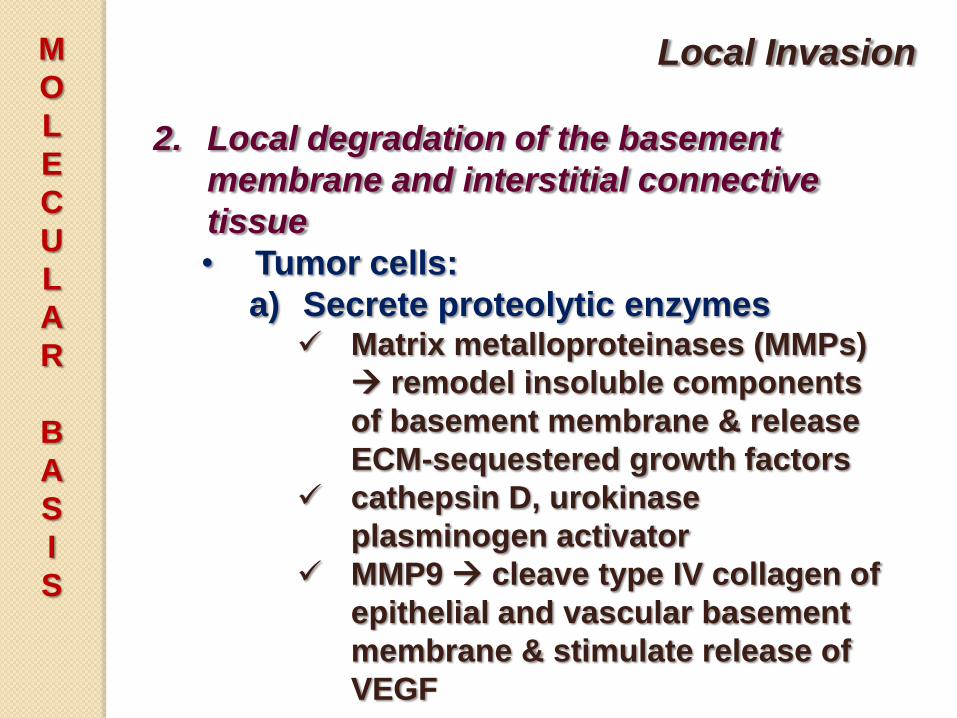
2. Local degradation of the basement
membrane and interstitial connective
tissue
• Tumor cells:
a) Secrete proteolytic enzymes Matrix metalloproteinases (MMPs)
remodel insoluble components
of basement membrane & release
ECM-sequestered growth factors
cathepsin D, urokinase
plasminogen activator
MMP9 cleave type IV collagen of
epithelial and vascular basement
membrane & stimulate release of
VEGF
Local InvasionM
O
L
E
C
U
L
A
R
B
A
S
I
S

2. Local degradation of the basement
membrane and interstitial connective
tissue
• Tumor cells:
a) Secrete proteolytic enzymes
b) Induce stromal cells (e.g.
Fibroblasts, inflammatory cells) to
elaborate proteases
Local InvasionM
O
L
E
C
U
L
A
R
B
A
S
I
S

2. Local degradation of basement
membrane and interstitial connective
tissue
• Tumor cells can adopt a second
mode of invasion called amoeboid
migration
Tumor cells squeeze through
spaces in the matrix utilize
collagen fibers as high-speed
“railways”
Quicker
Local InvasionM
O
L
E
C
U
L
A
R
B
A
S
I
S

2. Local degradation of basement
membrane and interstitial connective
tissue
Local InvasionM
O
L
E
C
U
L
A
R
B
A
S
I
S

3. Changes in attachment of tumor cells to
ECM proteins
• Normal epithelial cells with receptors
such as integrins for basement
membrane laminin and collagen
help maintain cells in a
resting, differentiated state
• Loss of adhesion in normal cells
induction of apoptosis but tumor cells
resistant
Local InvasionM
O
L
E
C
U
L
A
R
B
A
S
I
S

3. Changes in attachment of tumor cells to
ECM proteins
• Cleavage of basement membrane
proteins collagen IV and laminin by
MMP2 or MMP9 generate novel
sites bind to receptors on tumor
cells stimulate migration
Local InvasionM
O
L
E
C
U
L
A
R
B
A
S
I
S

3. Changes in attachment of tumor cells to
ECM proteins
Local InvasionM
O
L
E
C
U
L
A
R
B
A
S
I
S

4. Locomotion
• Final step in invasion
• Tumor cells attach to the matrix at the
leading edge detach from matrix at
trailing edge contract the actin
cytoskeleton to ratchet forward
• Movement potentiated and directed by
tumor-derived cytokines
Autocrine motility factors
Local InvasionM
O
L
E
C
U
L
A
R
B
A
S
I
S
4. Locomotion
• Cleavage products of matrix
components (collagen, laminin) and
some growth factors (IGFs I and II)
with chemotactic activity for tumor
cells
• Stromal cells produce paracrine
effectors of cell motility (e.g.
Hepatocyte growth factor-scatter
factor) bind to receptors on tumor
cells
Local InvasionM
O
L
E
C
U
L
A
R
B
A
S
I
S

4. Locomotion
Local InvasionM
O
L
E
C
U
L
A
R
B
A
S
I
S


Vascular Dissemination & Homing of Tumor
Cells
• In the circulation, tumor cells are
vulnerable to destruction by the
following mechanisms:
1. Mechanical shear stress
2. Apoptosis stimulated by loss of
adhesion
3. Innate and adaptive immune
defenses
• Within the circulation, tumor cells
tend to aggregate in clumps
Local InvasionM
O
L
E
C
U
L
A
R
B
A
S
I
S

Vascular Dissemination & Homing of Tumor
Cells
M
O
L
E
C
U
L
A
R
B
A
S
I
S
Clumps of tumor
cells in circulation
Homotypic
adhesion among
tumor cells
Heterotypic
adhesion between
tumor cells and
blood
cells, particularly
platelets
Platelet-tumor
aggregates
Enhanced
tumor cell
survival and
implantability
Bind and activate
coagulation factors
Formation
of emboli
Adhere to
endothelium
Extravasatio
n across BM
Distant
sites

Vascular Dissemination & Homing of Tumor
Cells
• Involvement of adhesion molecules
(integrins, laminin receptors) &
proteolytic enzymes
CD44 adhesion molecule
Expressed on normal T cells
Bind to hyaluronate on high
endothelial venules
Overexpression favor
metastatic spread
• Most metastases occur in the first
capillary bed available to the tumor
M
O
L
E
C
U
L
A
R
B
A
S
I
S

Vascular Dissemination & Homing of Tumor
Cells
• Certain tumors with organ tropism for
spread not explained by natural
pathways of drainage, which may be
related to the following mechanisms:
1. Preferential expression of ligands
for tumor cell-adhesion molecules
on the endothelial cells of the
target organ.
M
O
L
E
C
U
L
A
R
B
A
S
I
S

Vascular Dissemination & Homing of Tumor
Cells
2. Chemokines have an important role in
determining the target tissues for
metastasis.
• Breast cancer cells express the
chemokine receptors CXCR4 and
CCR7 chemokines highly
expressed in tissues to which
breast cancers commonly
metastasize
• Some target organs liberate
chemo- attractants that recruit
tumor cells to the site (e.g. IGFs I
and II)
M
O
L
E
C
U
L
A
R
B
A
S
I
S

Vascular Dissemination & Homing of Tumor
Cells
3. In some cases, the target tissue may
be a non-permissive environment for
the growth of tumor seedlings
• Skeletal muscles are rarely the site
of metastases
M
O
L
E
C
U
L
A
R
B
A
S
I
S

Vascular Dissemination & Homing of Tumor
Cells
• Tumor cells secrete cytokines, growth
factors, and ECM molecules that act
on resident stromal cells make the
metastatic site habitable for the
cancer cells
Breast cancer cells secrete para-
thyroid hormone-related protein
(PTHRP) stimulate osteoblasts
to make RANK ligand activate
osteoclasts breast cancer
metastases to bone osteolytic
M
O
L
E
C
U
L
A
R
B
A
S
I
S

Tumour–bone paracrine interactions. Left-hand side, an osteolytic interaction in
which parathyroid-hormone-related protein (PTHrP), which is produced by tumour
cells, stimulates the activity of bone-eating osteoclasts (multinucleate cell).
Osteoclasts in turn produce transforming growth factor- (TGF-), which stimulates
the tumour cells. Right-hand side, osteoblastic interactions. Tumour cells produce
many factors, including endothelin 1 (ET1), PTHrP and platelet-derived growth
factor (PDGF)-BB, which stimulate the activity of bone-producing osteoblasts
(blue nuclei). Osteoblasts in turn produce factors that stimulate the growth of the
tumour cells.

M
O
L
E
C
U
L
A
R
B
A
S
I
S
The metastatic
cascade.
Sequential steps
involved in the
hematogenous
spread of a tumor.

M
O
L
E
C
U
L
A
R
B
A
S
I
S
Mechanisms of metastasis development
within a primary tumor:
1. Clonal evolution model
• Metastasis is caused by rare variant
clones that develop in the primary
tumor.

M
O
L
E
C
U
L
A
R
B
A
S
I
S
Mechanisms of metastasis development
within a primary tumor:
2. Expression of metastasis signature
• Metastasis is caused by multiple
abnormalities that occur in most cells
of the primary tumor early in the
development of the tumor

M
O
L
E
C
U
L
A
R
B
A
S
I
S
Mechanisms of metastasis development
within a primary tumor:
3. Appearance of metastatic variants in a
tumor with a metastatic gene signature

M
O
L
E
C
U
L
A
R
B
A
S
I
S
Mechanisms of metastasis development
within a primary tumor:
4. Corollary of tumor stem cell hypothesis
• Metastasis development is greatly
influenced by the tumor stroma, which
may regulate angiogenesis, local
invasiveness, and resistant to immune
elimination allow cells of the
primary tumor to become metastatic

M
O
L
E
C
U
L
A
R
B
A
S
I
S
Mechanisms of metastasis development
within a primary tumor:
4. Corollary of tumor stem cell hypothesis

M
O
L
E
C
U
L
A
R
B
A
S
I
S
Genes implicated in control of metastasis
Metastatic suppressor gene
• A gene whose loss promotes the
development of metastasis without an
effect on the primary tumor
• Examples: mir335 and mir126
suppress metastasis in breast cancer

M
O
L
E
C
U
L
A
R
B
A
S
I
S
Genes implicated in control of metastasis
Metastatic oncogene
• A gene that favors the development of
metastasis without effect upon the
primary tumor
• Examples:
Mir10b
SNAIL & TWIST promote
epithelial-to-mesenchymal
transistion (EMT) down-regulate
E-cadherin expression in breast
carcinoma

M
O
L
E
C
U
L
A
R
B
A
S
I
S
1. Self-sufficiency in growth signals
2. Insensitivity to growth-inhibiting
signals
3. Evasion of apoptosis
4. Limitless replicative potential
5. Sustained angiogenesis
6. Ability to invade and metastasize
7. Defects in DNA repair
Fundamental Changes in Cell Physiology
That Determine Malignant Phenotype
M
O
L
E
C
U
L
A
R
B
A
S
I
S
Defects in DNA repair
• Individuals born with inherited
defects in DNA-repair proteins are at
a higher risk of developing cancer.
• Defects in repair mechanisms are
present in sporadic human cancers.
• DNA-repair genes are not oncogenic
abnormalities allow mutations in
other genes during the process of
normal cell division.

M
O
L
E
C
U
L
A
R
B
A
S
I
S
Defects in DNA repair
• Three types of DNA-repair systems:
1. Mismatch repair
2. Nucleotide excision repair
3. Recombination repair
• Genomic instability occurs when
both copies of the DNA-repair genes
are lost.

M
O
L
E
C
U
L
A
R
B
A
S
I
S
Defects in DNA repair
Defects in genes involved in mismatch
repairHereditary Nonpolyposis Colon Cancer
Synd.
• Familial carcinomas of the colon
affecting predominantly the cecum
and proximal colon
• Result of error in “proofreading”
• Hallmark is microsatellite instability
Microsatellites – tandem repeats
of one to six nucleotides; length
constant in normal people
In HNPCC, length varies in
tumor cells

M
O
L
E
C
U
L
A
R
B
A
S
I
S
Defects in DNA repair
Defects in genes involved in mismatch
repairHereditary Nonpolyposis Colon Cancer
Synd.
• Affected individuals inherit one
defective copy of a DNA mismatch-
repair gene “second hit” occurs
in colonic epithelial cells
• Involves mutations in the growth
regulating genes encoding TGF-ß
receptor II, TCF component of ß-
catenin pathway, BAX


M
O
L
E
C
U
L
A
R
B
A
S
I
S
Defects in DNA repair
Defects in nucleotide excision repair
Xeroderma pigmentosum
• Increased risk for cancers of the
skin, particularly after exposure to
UV radiation from sunlight
• UV radiation cross-linking of
pyrimidine residues inhibit DNA
replication


M
O
L
E
C
U
L
A
R
B
A
S
I
S
Defects in DNA repair
Defects in recombination repair
Bloom syndrome, Ataxia
telangiectasia, Fanconi anemia
• Characterized by hypersensitivity to
other DNA-damaging agents such as:
Ionizing radiation (Bloom synd.
and ataxia telangiectasia)
DNA cross-linking chemo-
therapeutic agens (Fanconi
anemia)
M
O
L
E
C
U
L
A
R
B
A
S
I
S
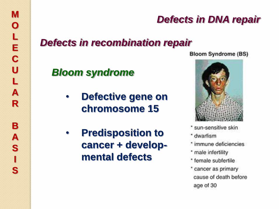
Defects in DNA repair
Defects in recombination repair
Bloom syndrome
• Defective gene on
chromosome 15
• Predisposition to
cancer + develop-
mental defects

M
O
L
E
C
U
L
A
R
B
A
S
I
S
Defects in DNA repair
Defects in recombination repair
Ataxia telangiectasia
• Predisposition to cancer + neural
symptoms
• Mutation of ATM gene gene
product important in recognizing
and responding to DNA damage
caused by ionizing radiation

M
O
L
E
C
U
L
A
R
B
A
S
I
S
Defects in DNA repair

M
O
L
E
C
U
L
A
R
B
A
S
I
S
Defects in DNA repair
Ataxia telangiectasia.
The hallmark of clinical
presentation is a
debilitating
progressive
neurodegeneration.
Other characteristics
are extreme
radiosensitivity, immun
odeficiency, a
predisposition to
cancer (haematopoietic
malignancy) and
sterility due to
defective meiotic
recombination. Ocular
and facial
telangiectasia are also
associated with AT.

M
O
L
E
C
U
L
A
R
B
A
S
I
S
Defects in DNA repair
Defects in recombination repair
Fanconi anemia
• Predisposition to cancer + bone
marrow aplasia
• Fanconi anemia proteins and the
BRCA proteins form a DNA-
damage response network
resolve and repair intrastrand and
interstrand cross-links induced by
chemical agents


M
O
L
E
C
U
L
A
R
B
A
S
I
S
Metabolic alterations in tumors
Warburg effect (aerobic glycolysis)
• Glycolysis that occurs in the face
of adequate oxygen for oxidative
phosphorylation
• Eighth hallmark of cancer

M
O
L
E
C
U
L
A
R
B
A
S
I
S
Metabolic alterations in tumors
Warburg effect: Hypotheses
1. Altered metabolism confers a growth
advantage in the hypoxic tumor micro-
environment.
• Hypoxia HIF 1α stimulate
angiogenesis and up-regulate
expression of enzymes for
glycolysis
2. Continuous rounds of hypoxia
followed by normoxia select for tumor
cells that constitutively upregulate
glycolysis.

M
O
L
E
C
U
L
A
R
B
A
S
I
S
Metabolic alterations in tumors
Warburg effect: Hypotheses
3. Mutations in oncogenes and tumor
suppressors that favor growth (e.g.
RAS, p53, and PTEN) also stimulate
metabolic changes in the cell.
• Alterations in signalling pathways
in cancer can also stimulate the
uptake of glucose and other
nutrients favor glycolysis

M
O
L
E
C
U
L
A
R
B
A
S
I
S
Metabolic alterations in tumors
Warburg effect: Hypotheses
4. Tumor cells are able to grow under
marginal environmental conditions
without triggering autophagy
• Mutation or epigenetic silencing of
genes involved in autophagy, most
notably PTEN

M
O
L
E
C
U
L
A
R
B
A
S
I
S
Dysregulation of cancer-associated genes
Chromosomal Changes
• Change in chromosome number
(aneuploidy) and chromosomal
instability may be the initiating events
in tumor growth
• Two types of chromosomal re-
arrangements that can activate proto-
oncogenes: translocations (more
common) and inversions

M
O
L
E
C
U
L
A
R
B
A
S
I
S
Dysregulation of cancer-associated genes
Chromosomal Changes
• Mechanisms of activation by
translocation:
1. Swapping of regulatory elements
with those of another gene over-
expression of proto-oncogene (e.g.
lymphoid tumors - Burkitt’s
lymphoma)

M
O
L
E
C
U
L
A
R
B
A
S
I
S
Dysregulation of cancer-associated genes
Chromosomal Changes
• Mechanisms of activation by
translocation:
2. Recombination of unrelated
sequences from two different
chromosomes form hybrid
fusion genes e.g. Philadelphia
chromosome of CML; hemato-
poietic tumors, sarcomas

M
O
L
E
C
U
L
A
R
B
A
S
I
S
Dysregulation of cancer-associated genes
Chromosomal Changes
• Deletions
Second most common structural
abnormality in tumor cells
More common in nonhemato-
poietic solid tumors
Associated with loss of particular
tumor suppressor genes
e.g. Retinoblastoma (deletion of
chr. 13q14)

M
O
L
E
C
U
L
A
R
B
A
S
I
S
Dysregulation of cancer-associated genes
Gene Amplification
• Reduplication and amplification of
DNA sequences of proto-oncogenes
over-expression of products
• Examples:
Amplification of N-MYC in
neuroblastoma
ERBB2 amplification in breast
cancers
C-MYC, L-MYC, N-MYC
amplification in small cell lung
CA

M
O
L
E
C
U
L
A
R
B
A
S
I
S
Dysregulation of cancer-associated genes
Epigenetic Changes
• Reversible, heritable changes in
gene expression that occur
without mutation
• Involve post-translational
modifications of histones and
DNA methylation

M
O
L
E
C
U
L
A
R
B
A
S
I
S
Dysregulation of cancer-associated genes
Epigenetic Changes
• Normal cells:
Majority of genes not
expressed silenced by DNA
methylation and histone
modification
heterochromatin formation
• Cancer cells:
Characterized by global hypo-
methylation and selective
promoter-localized hyperme-
thylation

How genetic and epigenetic alterations may cooperate in
the genesis of cancer. Potential pathways are shown
indicating how genetic change may precede epigenetic
change, and vice versa, as the cause of cancer.

M
O
L
E
C
U
L
A
R
B
A
S
I
S
Dysregulation of cancer-associated genes
Epigenetic Changes
• Tumor suppressor genes are
sometimes silenced by hyper-
methylation of promoter
sequences rather than mutation
Example: CDKN2A codes for
tumor suppressors p14/ARF
and p16/INK4a affect p53 and
Rb inhibit two checkpoints
BRCA 1 (breast cancer) and
VHL (renal cell CA)


M
O
L
E
C
U
L
A
R
B
A
S
I
S
miRNAs and Cancer
miRNAs
• Small, non-coding, single-stranded
RNAs (~22 nucleotides long) that are
incorporated into the RNA-induced
silencing complex
• Mediate sequence-specific
recognition of mRNAs and mediate
post-transcriptional gene silencing
• Control cell
growth, differentiation, and cell
survival

M
O
L
E
C
U
L
A
R
B
A
S
I
S
miRNAs and Cancer
miRNAs
• If a miRNA inhibits the translation
of an oncogene acts as a tumor
suppressor
• If a miRNA inhibits a tumor
suppressor gene acts as an
oncogene

M
O
L
E
C
U
L
A
R
B
A
S
I
S
miRNAs and Cancer
miRNAs
• Frequent amplifications and
deletions of miRNA loci identified in
many cancers
• Reduced activity of a miRNA that
inhibits translation of an oncogene
excess oncoprotein
• Overactivity of miRNA that targets a
tumor suppressor gene decreased
production of tumor suppressor
protein

M
O
L
E
C
U
L
A
R
B
A
S
I
S
miRNAs and Cancer
miRNAs
• Down-regulation or deletion of
certain miRNAs in some leukemias
and lymphomas increased
expression of BCL2 decreased
apoptosis
• miRNA-mediated up-regulation of
RAS (lung tumors) and MYC (B-cell
leukemias) oncogenes

M
O
L
E
C
U
L
A
R
B
A
S
I
S
Multistep Carcinogenesis
• Each cancer must result from the
accumulation of multistep mutations
Individual tumors accumulate an
average of 90 mutant genes
• No single oncogene can fully transform
non-immortalized cells in vitro
• Cells can generally be transformed by
combinations of oncogenes

M
O
L
E
C
U
L
A
R
B
A
S
I
S
Multistep Carcinogenesis
• “intrinsic tumor-suppressive
mechanisms” thwart the actions of
growth-promoting mutations
In cells with competent
checkpoints, oncogenic signalling
leads to senescence or apoptosis
rather than transformation
Emergence of malignant tumors
requires mutational loss of many
genes, including those that regulate
apoptosis and senescence


E
N
D
of
P
A
R
T
3