female gender attenuates cytokine and chemokine expression and leukocyte recruitment in experimental...
TRANSCRIPT

Female Gender Attenuates Cytokineand Chemokine Expression andLeukocyte Recruitment in ExperimentalRodent Abdominal Aortic Aneurysms
INDRANIL SINHA, BRENDA S. CHO, KAREN J. ROELOFS,JAMES C. STANLEY, PETER K. HENKE,AND GILBERT R. UPCHURCH, JR.
Department of Surgery, Jobst Vascular Research Laboratories, Section ofVascular Surgery, University of Michigan, Ann Arbor, MI 48109-0329
ABSTRACT: Female gender appears to be protective in the development ofabdominal aortic aneurysms (AAAs). This study sought to identify gen-der differences in cytokine and chemokine expression in an experimentalrodent AAA model. Male and female rodent aortas were perfused witheither saline (control) or elastase to induce AAA formation. Aortic di-ameter was determined and aortic tissue was harvested on postperfusiondays 4 and 7. Cytokine and chemokine gene expression was examinedusing focused gene arrays. Immunohistochemistry was used to quan-tify aortic leukocyte infiltration. Data were analyzed by Student’s t-testsand ANOVA. Elastase-perfused female rodents developed significantlysmaller aneurysms compared to males by day 7 (93±10% vs. 201±25%,P = 0.003). Elastase-perfused female aortas exhibited a fivefold decreasein expression of the BMP family and ligands of the TNF superfamily com-pared to males. In addition, the expression of members of the TGF � andVEGF families were three to fourfold lower in female elastase-perfusedaortas compared to males. Multiple members of the interleukin, CCchemokine receptor, and CC ligand families were detectable in only themale elastase-perfused aortas. Female elastase-perfused aortas demon-strated a corollary twofold lower neutrophil count (females: 17.5 ± 1.1PMN/HPF; males: 41 ± 5.2 neutrophils/HPF, P = 0.01) and a 1.5-foldlower macrophage count (females: 12 ± 1.1 macrophages/HPF; males:17.5 ± 1.1 macrophages/HPF, P = 0.003) compared to male elastase-perfused aortas. This study documents decreased expression of multiplecytokines and chemokines and diminished leukocyte trafficking in fe-male rat aortas compared to male aortas following elastase perfusion.
Address for correspondence: Gilbert R. Upchurch, Jr., M.D., University of Michigan Hospital, 2210THCC, 1500 East Medical Center Drive, Ann Arbor, MI 48109-0329. Voice: 734-936-5790; fax: 734-647-9867.
e-mail: [email protected]
Ann. N.Y. Acad. Sci. 1085: 367–379 (2006). C© 2006 New York Academy of Sciences.doi: 10.1196/annals.1383.027
367

368 ANNALS NEW YORK ACADEMY OF SCIENCES
These genes may contribute to the gender disparity seen in AAA forma-tion.
KEYWORDS: AAAs; gender; cytokine; chemokine; gene array
INTRODUCTION
Male gender is a well-established risk factor in the formation of abdominalaortic aneurysms (AAA). Clinically, men form AAAs at a 4:1 ratio comparedto women.1 However, AAA development becomes accelerated in women post-menopausal.2 These observations support the tenet that circulating estrogensmay play a protective role in limiting pathologic inflammation in the aorticwall associated with AAA formation.3 In this regard, it is known that estrogenattenuates the inflammatory response and decreases leukocyte infiltration inmany nonaneurysmal cardiovascular diseases.4
The mechanisms underlying the gender disparity in AAA formation are notwell understood. Few studies have examined the role of gender in experimen-tal AAA models. We reported the incidence and size of AAAs are signifi-cantly less in female rodents compared to males.5 In this regard, female aortasalso exhibit significantly decreased aortic wall macrophage secreting matrixmetalloproteinase (MMP)-9, an enzyme critical for AAA formation.5,6 In aseparate study, tamoxifen, a selective estrogen receptor modulator, also atten-uated experimental AAA formation and limited neutrophil infiltration.7 Thepresent study was undertaken to determine if gender disparities in cytokineand chemokine expression contribute to decreased leukocyte trafficking inexperimental female AAAs.
MATERIALS AND METHODS
All rats were obtained from Charles River Laboratories (Wilmington, MA,USA). All experiments were approved by the University of Michigan UniversalCommittee on the Use and Care of Animals (UCUCA No. 8566).
Aneurysm Induction
Male and female Sprague-Dawley rats, weighing 190–210 g, underwent elas-tase perfusion as previously described.8 Briefly, rats were anesthetized with1–2% isoflurane inhalation and the infrarenal aorta was isolated. Temporarycontrol of the aorta was obtained, lumbar branches were ligated, and an aorto-tomy was made near the bifurcation using a 30-gauge needle. The aorta wasthen perfused for 30 min with either 2 mL of normal saline or 12 units of

SINHA et al.: GENDER EFFECTS ON CYTOKINE AND CHEMOKINE EXPRESSION 369
porcine pancreatic elastase (Sigma, St. Louis, MO, USA) in 2 ml of normalsaline.
Infrarenal aortic diameters were measured prior to perfusion, immediatelypostperfusion, and prior to harvest. This was accomplished using a Spot InsightColor Optical Camera (Diagnostic Instruments, Inc., Sterling Heights, MI,USA) attached to an operating microscope (Nikon, Melville, NY, USA) utiliz-ing Image Pro Express software (Media Cybernetics, Inc., Silver Springs, MD,USA). Aortas harvested on 4 and 7 days postperfusion (n = 5 to 6 at each timepoint for both saline controls and elastase-perfused animals) were subjectedto RNA (seems like total RNA was used) extraction and paraformaldehydefixation.
Gene Arrays
Total RNA was isolated by treatment of aortic segments harvested on post-perfusion day 4 with TRIzol reagent (Life Technologies, Rockville, MD, USA).cDNA was produced by reverse transcription using oligo-(dT) primer andM-MLV reverse transcriptase (Life Technologies). Two micrograms of totalRNA from each animal (n = 4 per gender, per array, for both saline con-trols and elastase-perfused aortas) were obtained for microarray experiments.RNA labeling was accomplished using the SuperArray Amplolabeling Lin-ear Polymerase Reaction kit (SuperArray Bioscience Corp., Frederick, MD,USA) according to the manufacturer’s specifications. Hybridization was per-formed using Oligo-GE Q-series Rat Gene Arrays, ORN-021 (Rat Cytokine)and ORN-022 (Rat Chemokines and Receptors) per provided protocol. Geneexpression was standardized to all housekeeping genes included in the cor-responding array. Gene arrays were visualized using the provided SuperarrayChemiluminescent Detection Kit. Photographs were obtained using a CCDcamera. All data analysis was done using online GEArray Expression Analy-sis Suite software (http://geasuite.superarray.com).
Immunohistochemistry
Aortic tissue was fixed in 4% paraformaldehyde for 20 h, transferred to70% ethanol, and subsequently embedded in paraffin for immunohistochem-istry. Aortic tissue deparaffinization and rehydration were performed usingxylene and graded alcohols. The sections were stained for macrophages andneutrophils using the following procedures. The aortic sections were deparaf-finized in xylene and rehydrated in graded alcohols.
For neutrophil staining, blocking buffer was then applied to prevent nonspe-cific binding. Rabbit anti-rat neutrophil primary antibody (Accurate Chemicaland Scientific Corporation, Westbury, NY, USA) was used for staining,

370 ANNALS NEW YORK ACADEMY OF SCIENCES
followed by an antirabbit IgG biotinylated secondary antibody and avidin-biotin complex-AP reagent available in the Rabbit IgG Alkaline PhosphataseABC Kit (Vector Laboratories, Burlingame, CA, USA). The sections werethen visualized using the Vector Red Alkaline Phosphatase Substrate KitI (Vector Laboratories) and counterstained with Hematoxylin QS (VectorLaboratories).
Macrophage staining entailed heat-induced epitope retrieval using 10 mMsodium citrate, pH 6.0, in a microwave. The sections were subsequently in-cubated with 3% hydrogen peroxide in methanol to block endogenous perox-idase activity, followed by a blocking buffer to prevent nonspecific binding.Mouse antirat CD68 primary antibody (Serotec, Raleigh, NC, USA) was usedfor staining, followed by an antimouse IgG biotinylated secondary antibodyand an avidin-biotin-HRP complex, available in the Mouse IgG Elite Vectas-tain ABC Kit (Vector Laboratories). Sections were then visualized using aDAB Peroxidase Substrate Kit (Vector Laboratories) and counterstained withHematoxylin QS (Vector Laboratories). All cell counts were done by a trained,blinded observer in 10 separate 100× high-powered fields (HPF) of both theadventitia and media. A mean value was then calculated for positively stainedcells in each animal.
Statistics
Data analysis was performed using nonpaired Student’s t-tests and anal-ysis of variance (ANOVA) with significance set as P < 0.05. All data areexpressed as the mean ± standard error of the mean (SEM). Statistical anal-ysis was performed using SigmaStat (Version 2.03, Copyright 1992–1997,SPSS Inc.).
RESULTS
Female Aortas Form Smaller Experimental AAAs
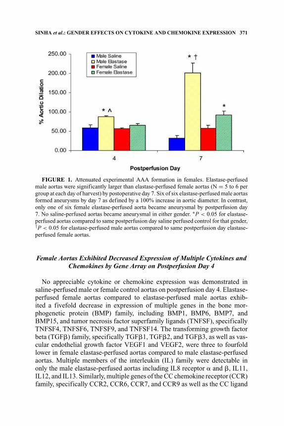
Saline-perfused aortas did not form aneurysms in either gender. Mean diam-eters of elastase-perfused male aortas increased 88 ± 2.7% and 201 ± 25.1%by postperfusion days 4 and 7, respectively. In contrast, the mean diameters offemale elastase-perfused aortas increased 65 ± 4.4% (P = 0.002) and 93 ±10% (P = 0.003) by postperfusion days 4 and 7, respectively (FIG. 1). Six ofsix elastase-perfused male aortas formed aneurysms as defined by a 100%increase in aortic diameter by postperfusion day 7 in contrast to one of sixelastase-perfused female aortas.
SINHA et al.: GENDER EFFECTS ON CYTOKINE AND CHEMOKINE EXPRESSION 371
FIGURE 1. Attenuated experimental AAA formation in females. Elastase-perfusedmale aortas were significantly larger than elastase-perfused female aortas (N = 5 to 6 pergroup at each day of harvest) by postoperative day 7. Six of six elastase-perfused male aortasformed aneurysms by day 7 as defined by a 100% increase in aortic diameter. In contrast,only one of six female elastase-perfused aorta became aneurysmal by postperfusion day7. No saline-perfused aortas became aneurysmal in either gender. ∗P < 0.05 for elastase-perfused aortas compared to same postperfusion day saline perfused control for that gender,†P < 0.05 for elastase-perfused male aortas compared to same postperfusion day elastase-perfused female aortas.
Female Aortas Exhibited Decreased Expression of Multiple Cytokines andChemokines by Gene Array on Postperfusion Day 4
No appreciable cytokine or chemokine expression was demonstrated insaline-perfused male or female control aortas on postperfusion day 4. Elastase-perfused female aortas compared to elastase-perfused male aortas exhib-ited a fivefold decrease in expression of multiple genes in the bone mor-phogenetic protein (BMP) family, including BMP1, BMP6, BMP7, andBMP15, and tumor necrosis factor superfamily ligands (TNFSF), specificallyTNFSF4, TNFSF6, TNFSF9, and TNFSF14. The transforming growth factorbeta (TGF�) family, specifically TGF�1, TGF�2, and TGF�3, as well as vas-cular endothelial growth factor VEGF1 and VEGF2, were three to fourfoldlower in female elastase-perfused aortas compared to male elastase-perfusedaortas. Multiple members of the interleukin (IL) family were detectable inonly the male elastase-perfused aortas including IL8 receptor � and �, IL11,IL12, and IL13. Similarly, multiple genes of the CC chemokine receptor (CCR)family, specifically CCR2, CCR6, CCR7, and CCR9 as well as the CC ligand

372 ANNALS NEW YORK ACADEMY OF SCIENCES
TABLE 1. Female gender and decreased cytokine and chemokine expression in exper-imental AAA formation. Note decreased cytokine and chemokine production in femaleelastase-perfused aortas on postperfusion day 4. ND (none detectable) denotes expressionwas present in males, but not detectable in female aortas
Cytokine/Chemokine Fold decrease in females
Bone morphogenetic protein (BMP) familyBMP1 6.1BMP6 6.3BMP7 5.6BMP15 6.3
C-C Chemokine ligand familyCCL24 NDCCL25 NDCCL28 ND
C-C Chemokine receptor familyCCR2 NDCCR7 NDCCR8 ND
Interleukin (IL) familyIL1 6.2IL2 6.1IL3 4.9IL5 4.9IL7 5.9IL8� NDIL8� NDIL11 6IL12 6.3
Transforming growth factor (TGF) familyTGF�1 4.1TGF�2 4.2
Tumor necrosis factor (TNF) familyTNFSF4 NDTNFSF6 NDTNFSF9 NDTNFSF15 ND
Vascular endothelial growth factor familyVEGF1 3.6VEGF2 3.6
(CCL) family, specifically CCL3, CCL5, CCL24, CCL25, and CCL28, weredetectable in male elastase-perfused aortas, but not in female elastase-perfusedaortas (TABLE 1).
Female Aortas Exhibit Decreased Neutrophil and Macrophage Infiltration
Minimal neutrophil and macrophage infiltration was detected in both maleand female saline perfumed aortas. Male elastase-perfused aortas demonstrated13.7 ± 5.9 positively staining cells/HPF in males compared to 5.0 ± 0.5

SINHA et al.: GENDER EFFECTS ON CYTOKINE AND CHEMOKINE EXPRESSION 373
neutrophils/HPF (P = 0.09) in females on postperfusion day 4. By postper-fusion day 7, male elastase-perfused aortas demonstrated a twofold greaterneutrophil count (40.75 ± 5.2 PMN/HPF) in comparison to female elastase-perfused aortas (17.5 ± 1.1 PMN/HPF, P = 0.01) (FIGS. 2A and B). Maleelastase-perfused aortas also demonstrated 12.2 ± 5.1 and 17.5 ± 1.1 CD68positive cells (macrophages)/HPF on postperfusion days 4 and 7, respec-tively. Number of macrophages was significantly fewer in females (4.7 ±1.1 macrophages/HPF on postperfusion day 4, P = 0.2, and 11.8 ± 1.1macrophages/HPF on postperfusion day 7, P = 0.003) (FIGS. 3A and B).
DISCUSSION
This study confirms that female rodents develop significantly smaller ex-perimental AAAs compared to males. Cytokine and chemokine gene arraysidentified several cytokine and chemokine families, which were differentiallyexpressed by gender including BMP, TNF, TGF�, IL, CCR, and CCL gene fam-ilies. Furthermore, female rat aortas exhibited significantly fewer infiltratingneutrophils and macrophages by postperfusion day 7.
It is possible that the known effect of estrogen in females on inflamma-tory processes in cardiovascular diseases may be associated with the observeddecreased leukocyte cell infiltration in females and their apparent resistanceto AAA formation.9 Multiple studies have shown estrogen plays a protectiverole by attenuating inflammatory cell mediated damage by downregulatingcytokine and chemokine expression. In two separate AAA models, femalegender has been shown to limit experimental AAA formation, at least in partby limiting neutrophil and macrophage infiltrate.4,10 In addition, Miller et al.observed that estrogen attenuation of the early inflammatory response follow-ing endoluminal vascular injury was associated with decreased expression ofseveral cytokines and chemokines, especially the CXC chemokine family.11
This was accompanied with decreased neutrophil and macrophage infiltrationand vasoprotection through an apparent anti-inflammatory pathway.11 Estro-gen is also known to decrease myocardial inflammation and damage follow-ing ischemia-reperfusion injury by downregulating proinflammatory cytokinessuch as TNF�, IL1�, and IL6.12
Estrogen also attenuates inflammation-mediated damage in nonvascular tis-sue. Santizo et al. demonstrated that chronic estrogen depletion enhancedleukocyte adhesion in the rat cerebral circulation and suggested this was amechanism underlying increased neural damage in ovariectomized femalesas compared to intact females.13 In addition, estrogen has been observed toenhance wound healing through an anti-inflammatory effect by limiting neu-trophil and macrophage infiltration and suppressing the production of numer-ous cytokines including TNF� and macrophage migration inhibitory factor(MIF).14

374 ANNALS NEW YORK ACADEMY OF SCIENCES
Estrogen is likely to decrease cytokine and chemokine expression throughmultiple mechanisms. First, estrogen decreases p38 mitogen activated pro-tein kinase (MAPK), a crucial upstream proinflammatory regulator of nu-merous cytokines, including TNF�, IL1, IL4, IL6, and IL8.15 Wang et al.demonstrated decreased p38 MAPK phosphorylation (activation) in controlfemales compared to oophorectomized females and control males followingischemia/reperfusion injury.12 Second, estrogen also decreases the activity ofnuclear factor �B (NF�B), a key proinflammatory transcription factor.16 Fi-nally, estrogen decreases oxidant stress, a stimuli for the production of multipleinflammatory cytokines in ischemia/reperfusion injury.15
FIGURE 2. Female gender and decreased aortic wall neutrophil infiltration. (A) Im-munohistochemistry (100×) by postoperative day 7 revealed significantly increased neu-trophil infiltration in the adventitia of male aortas compared to female aortas. Few positivelystained cells were identified in saline-perfused controls of either gender. ∗P < 0.05 forelastase-perfused aortas compared to same postperfusion day saline-perfused control forthat gender, †P < 0.05 for elastase-perfused male aortas compared to same postperfusionday elastase-perfused females, N = 4 per group at each day of harvest. (B,C) Representativeimages of positively staining cells (arrows) in the adventitia of elastase-perfused male (B)and fewer positively staining cells in the female aortas (C) (100×). [Saline-perfused aortasnot shown.]

SINHA et al.: GENDER EFFECTS ON CYTOKINE AND CHEMOKINE EXPRESSION 375
FIGURE 2. Continued.
The present intervention demonstrates multiple cytokine families that weredifferentially regulated by gender, although their role in AAA pathogenesishas not been thoroughly investigated. Others have provided some insight intothis subject. Dai et al. have previously shown that adenoviral delivery medi-ated exogenous overexpression of TGF-�1 stabilized preformed experimentalAAAs.17 However, no studies to date have examined gender differences inTGF� regulation in experimental AAAs. Similarly, the TNF superfamily isknown to have a role in AAA pathogenesis, but no studies have assessed

376 ANNALS NEW YORK ACADEMY OF SCIENCES
gender difference in AAA formation.6 Kobayashi et al. demonstrated VEGFto be upregulated in AAAs, but a role in AAA pathogenesis remains unclear.18
Certain ILs are known to be critical to AAA formation, but no evidence existsfor the roles of IL11, IL12, or IL13.6
A limited number of investigators have examined the role of chemokines inAAA formation. Zhao et al. demonstrated that macrophage inhibitory protein(MIP)1� (also known as CCL3), which was differentially expressed by genderin the present study, and MIP2 (CXCL2) were important in experimental AAAformation.19 Similarly, Yamagishi et al. observed an elevenfold increase inCXCR2 production in human AAAs compared to control healthy aortas.20
However, no studies to date have elucidated gender differences in the patternsof chemokine expression.
FIGURE 3. Female gender and decreased aortic wall macrophage infiltration. (A)Immunohistochemistry (100×) by postoperative day 7 revealed significantly increasedmacrophage infiltration in the adventitia of male aortas compared to female aortas. Mini-mally positively stained cells were identified in saline-perfused controls of either gender.∗P < 0.05 for elastase-perfused aortas compared to same postperfusion day saline-perfusedcontrol for that gender, †P < 0.05 for elastase-perfused male aortas compared to samepostperfusion day elastase-perfused females, N = 4 per group at each day of harvest.(B,C) Representative images of positively CD68 staining cells (arrows) in the adventitia ofelastase-perfused male (B) and minimal positively staining cells in the female aortas (C)(100×). (Saline-perfused aortas not shown.)

SINHA et al.: GENDER EFFECTS ON CYTOKINE AND CHEMOKINE EXPRESSION 377
FIGURE 3. Continued.
The present investigation supports the speculation that a number of cytokinesand chemokines may be differentially regulated by gender and may contributeto vessel wall injury. More reliable polymerase chain reaction or northern blot-ting data will be required to confirm these observations. This study also onlyexamined postperfusion day 4 and therefore does not take into account genesthat may be upregulated either earlier or later in experimental AAA formation.Additionally, it will be necessary to determine if gender differences in cytokineand chemokine differences are intrinsic to the wall or secondary to diminishedleukocyte trafficking into female aortas. Nevertheless, this study has identi-fied multiple gene families that deserve further consideration for a potentialrole in gender differences during AAA formation. Furthermore, continuingresearch is needed to more fully elucidate the mechanism by which estrogenacts to decrease cytokine and chemokine expression. A better understanding

378 ANNALS NEW YORK ACADEMY OF SCIENCES
of gender disparities may ultimately lead to new pharmacological targets forthe treatment of AAAs.
ACKNOWLEDGMENTS
This work was supported by NIH KO8 (HL67885-02) (GRU), Von LiebigAward-Lifeline Foundation (GRU), the Lifeline Medical Student ResearchAward (IS), the Griswold and Margery H. Ruth Alpha Omega Alpha MedicalStudent Research Fellowship (IS), and the Jobst Foundation.
REFERENCES
1. SINGH, K., K.H. BONAA, B.K. JOCOBSEN, et al. 2001. Prevalence and risk factorsfor abdominal aortic aneurysms in a population based study: the Tromso study.Am. J. Epidemiol. 154: 236–244.
2. BENGTSSON, H., B. SONESSON & D. BERGVIST. 1996. Incidence and prevalence ofAAA. N. Y. Acad. Sci. 800: 1–24.
3. LAVECCHIA, C., A. DECARLI, S. FRANCESCHI, et al. 1987. Menstrual and reproduc-tive factors and the risk of myocardial infarction in women under fifty-years ofage. Am. J. Obstet. Gynecol. 157: 1108–1112.
4. BAKER, L., K.K. MELDRUM, M. WANG, et al. 2003. The role of estrogen in cardio-vascular disease. J. Surg. Res. 115: 325–344.
5. AILAWADI, G., J.L. ELIASON, K.J. ROELOFS, et al. 2004. Gender differences inexperimental aneurysm formation. Arterioscler Thromb. Vasc. Biol. 24: 2116–2122.
6. AILAWADI, G., J.L. ELIASON & G.R. UPCHURCH. 2003. Current concepts in thepathogenesis of abdominal aortic aneurysms. J. Vasc. Surg. 38: 584–588.
7. GRIGORYANTS, V., K.K. HANNAWA, C.G. PEARCE, et al. 2005. Tamoxifen up-regulates catalase production, inhibits vessel wall neutrophil recruitment, andattenuates development of experimental abdominal aortic aneurysms. J. Vasc.Surg. 41: 108–114.
8. ANIDJAR, S., J.L. SALZMANN, D. GENTRIC, et al. 1990. Elastase induced experimen-tal aneurysms in rats. Circulation 82: 973–981.
9. LEINWAND, L.A. 2003. Sex is a potent modifier of the cardiovascular system.J. Clin. Invest. 112: 302–307.
10. MARTIN-MCNULTY, B., D.M. THAM, C. DA, et al. 2003. 17 �-estradiol atten-uates development of angiotensin II-induced aortic abdominal aneurysm inapolipoprotein E-deficient mice. Arterioscler Thromb. Vasc. Biol. 23: 1627–1632.
11. MILLER, A.P., W. FENG, D. XING, et al. 2004. Estrogen modulated inflammatorymediator expression and neutrophil chemotaxis in injured arteries. Circulation110: 1664–1669.
12. WANG, M., B.M. TSAI, K.M. REIGER, et al. 2006. 17-�-estradiol decreases p38MAPK-mediated myocardial inflammation and dysfunction following acute is-chemia. J. Mol. Cell. Cardiol. 40: 205–212.
13. SANTIZO, R. & D.A. PELLIGRINO. 1999. Estrogen reduces leukocyte adhesion in thecerebral circulation of female rats. J. Cereb Blood Flow Metab. 19: 1061–1065.

SINHA et al.: GENDER EFFECTS ON CYTOKINE AND CHEMOKINE EXPRESSION 379
14. ASHCROFT, G.S., S.J. MILLS, K. LEI, et al. 2003. Estrogen modulated cuta-neous wound healing by downregulating macrophage migration inhibitory factor.J. Clin. Invest. 111: 1309–1318.
15. KHER, A., M. WANG, B.M. TSAI, et al. 2005. Sex differences in the myocardialinflammatory response to acute injury. Shock 23: 1–10.
16. DESHPANDE, R., H. KHALILI, R.G. PERGOLIZZI, et al. 1997. Estradiol down-regulatesLPS-induced cytokine production and NF�B activation in murine macrophages.Am. J. Reprod. Immunol. 38: 46–54.
17. DAI, J., F. LOSY, A.M. GUINAULT, et al. 2005. Overexpression of transforminggrowth factor-beta1 stabilizes already formed aortic aneurysms: a first approachto induction of functional healing by endovascular gene therapy. Circulation 112:1008–1015.
18. KOBAYASHI, M., J. MATSUBARA, M. MATSHUSHITA, et al. 2002. Expression of an-giogenesis and angiogenic factors in human aortic vascular disease. J. Surg. Res.106: 239–245.
19. ZHAO, L., M.P. MOOS, R. GRABNER, et al. 2004. The 5-lipoxygenase pathwaypromotes pathogenesis of hyperlipidemia-dependent aortic aneurysm. Nat. Med.10: 966–973.
20. YAMAGISHI, M., T. HIGASHIKATA, H. ISHIBASHI-UEDA, et al. 2005. Sustained upreg-ulation of inflammatory chemokine and its receptor in aneurysmal and occlusiveatherosclerotic disease: results from tissue analysis with cDNA macroarray andreal-time reverse transcriptional polymerase chain reaction methods. Circ. J. 69:1490–1495.