fetal brain anomalies
TRANSCRIPT

Fetal brain anomalies(ventriculomegaly, ACC, Dandy-Walker malformations)
Kh. Batnasan
United Family Intermed Hospital, Mongolia
Korea University Hospital
31 Mar 2015

Fetal ventriculomegaly
• Ventriculomegaly – Enlargement of the lateral cerebral ventricles
• Nonspecific marker of abnormal brain development
• Measurement: Internal width of the atrium of the lateral ventricle at the level of glomus of the choroid plexus

Ventriculomegaly or Hydrocephalus?

Definition
Subtle difference
Ventriculomegaly – obstruction of normal CSF bulk flow due to global cerebral tissue loss after hypoxic-ischemic injury (eg., congenital anomalies, TORCH infections…)
Hydrocephalus – impaired, obstructed or altered CSF bulk flow

Fetal ventriculomegaly
• Normal range = 5-10 mm (from 15 weeks)
• Mild VM = 10-15 mm
• Severe VM = >15 mm
(dangling choroid plexus)

Mild ventriculomegaly
• ≥10 mm
• Bilateral or unilateral
(Distal and proximal ventricles are should be measured)
• Associated with:• Brain malformations • Genetic syndromes• Infections
• Prevalence: (extremely variable) 0.15 – 0.7%
• Hannon et al., (2012) - 7.9 per 10,000 singleton births

Mild ventriculomegaly
• Difficult task for counseling
• Parent’s anxiety
• We need an accurate diagnostic work-up

Mild VM – diagnostic work up
• ruling out for associated anomalies
• ruling out for congenital infections
• ruling out for feto-neonatal alloimmune thrombocytopenia
• ruling out for chromosomal abnormalities
• monitoring the development of mild VM in the progressing pregnancy.
Mild VM - ruling out for associated anomalies
• May be associated with neural and extraneural anomalies
• May be first sign of brain anomaly in 3rd trimester or after delivery
• Association range: 10-71% (average – 40%)
• Need an accurate and systematic evaluation of the fetal brain
• Fetal brain MRI – advocated in the case of mild VM (following neurosonogramm)
• Appropriate time – 3rd trimester
• Helpful in the case of cortical anomalies

Mild VM - ruling out for congenital infections
May be seen:
• small periventricular calcifications
• small subependimal cysts
• extraneural abnormal findings:• liver calcifications • ascites • hepatosplenomegaly • echogenic bowel• placentomegaly• growth restriction
• Recommended tests for TORCH infections (CMV, toxoplasmosis)

Mild VM - Ruling out for feto-neonatal alloimmunethrombocytopenia
• Very rare
• Hyperechogenicity of the ventricular walls
• Intraventricular echogenic blood clots
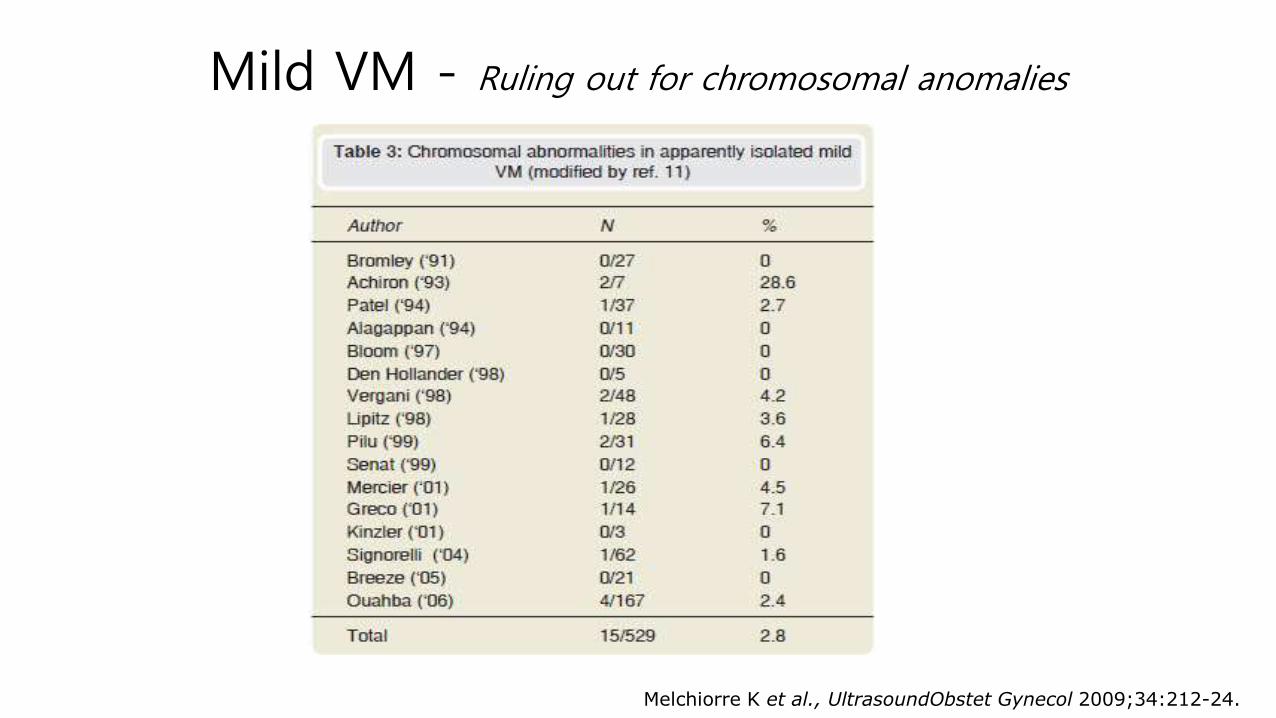
Mild VM - Ruling out for chromosomal anomalies
Melchiorre K et al., UltrasoundObstet Gynecol 2009;34:212-24.
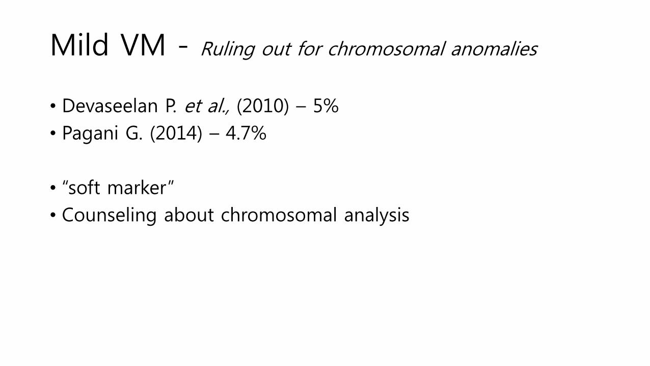
Mild VM - Ruling out for chromosomal anomalies
• Devaseelan P. et al., (2010) – 5%
• Pagani G. (2014) – 4.7%
• “soft marker”
• Counseling about chromosomal analysis
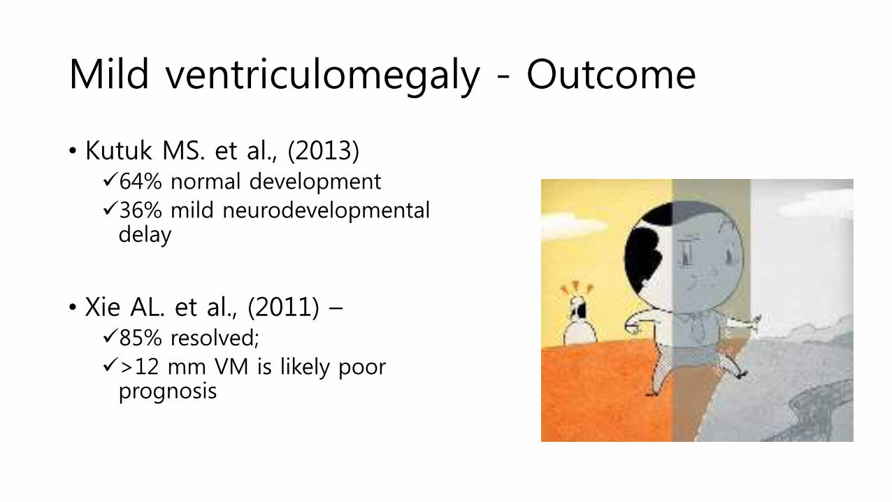
Mild ventriculomegaly - Outcome
• Kutuk MS. et al., (2013) 64% normal development
36% mild neurodevelopmental delay
• Xie AL. et al., (2011) –85% resolved;
>12 mm VM is likely poor prognosis


Severe VMPrognosis:
• Survival or neurodevelopmental outcome is poor
• Related to the underlying condition (hydrocephalus, cerebral atrophy or additional anomalies…)
Prevalence:
• 3.6 per 10,000 singleton births
(Hannon et al., 2012)

Severe VM
• Associated anomalies – 49.8%
• Chromosomal anomalies: 3.2 – 8.3%
• Structural anomalies – 42.7%
• Neonatal mortality – 16.2%
• Fetal brain MRI – additional tool to diagnose an associated anomalies
Hannon et al., Obstet Gynecol 2012
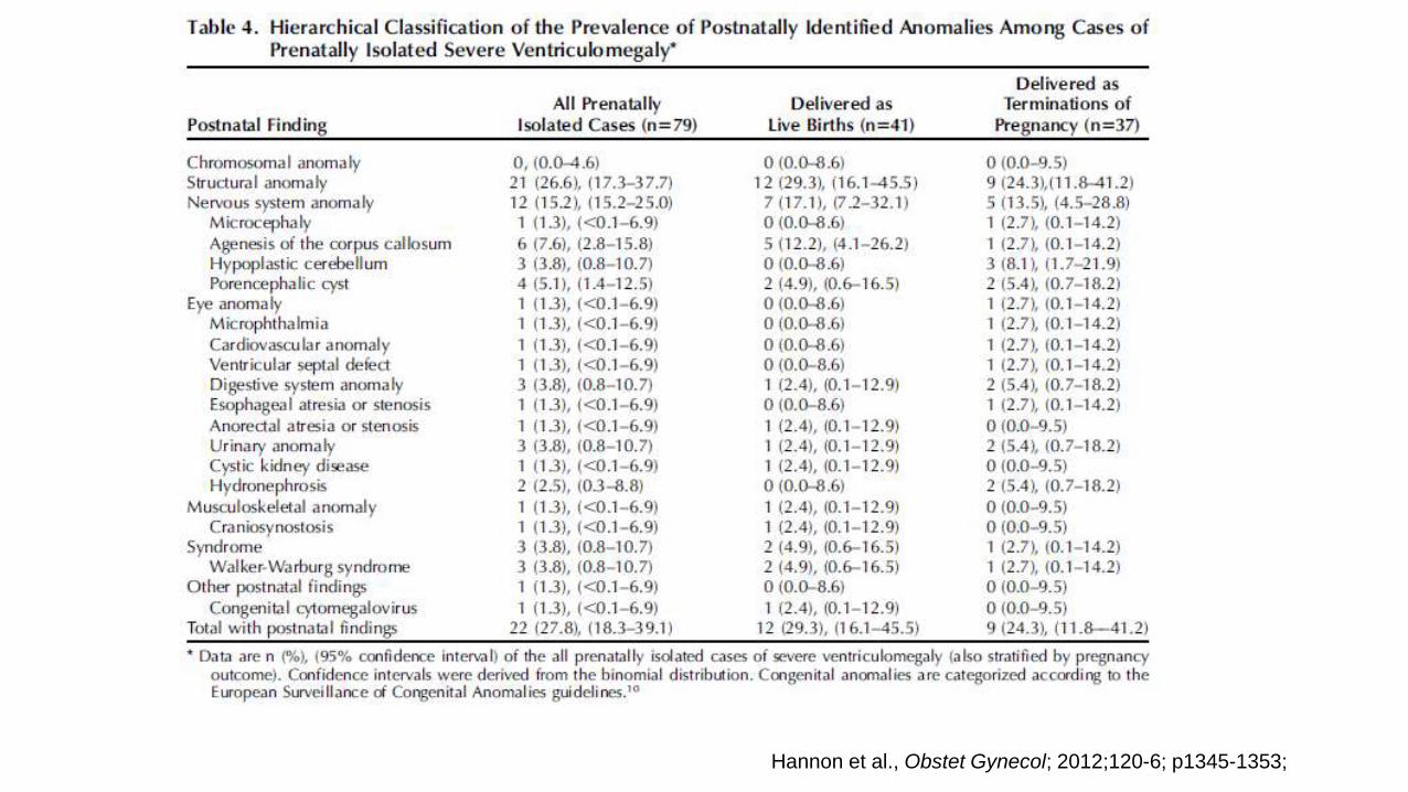
Hannon et al., Obstet Gynecol; 2012;120-6; p1345-1353;

Agenesis of the corpus callosum (ACC)
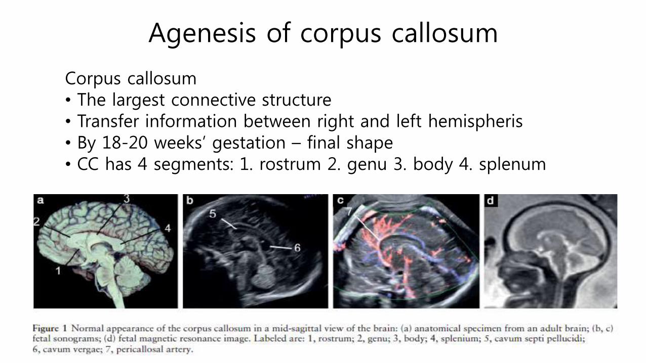
Agenesis of corpus callosum
Corpus callosum• The largest connective structure • Transfer information between right and left hemispheris• By 18-20 weeks’ gestation – final shape• CC has 4 segments: 1. rostrum 2. genu 3. body 4. splenum

Agenesis of corpus callosum
• Developmental absence of the corpus callosum: Agenesis (ACC)• Partial ACC
• Complete ACC
• Each of these may be:• Isolated: ACC with no other abnormalities
• Complex: ACC with other abnormalities
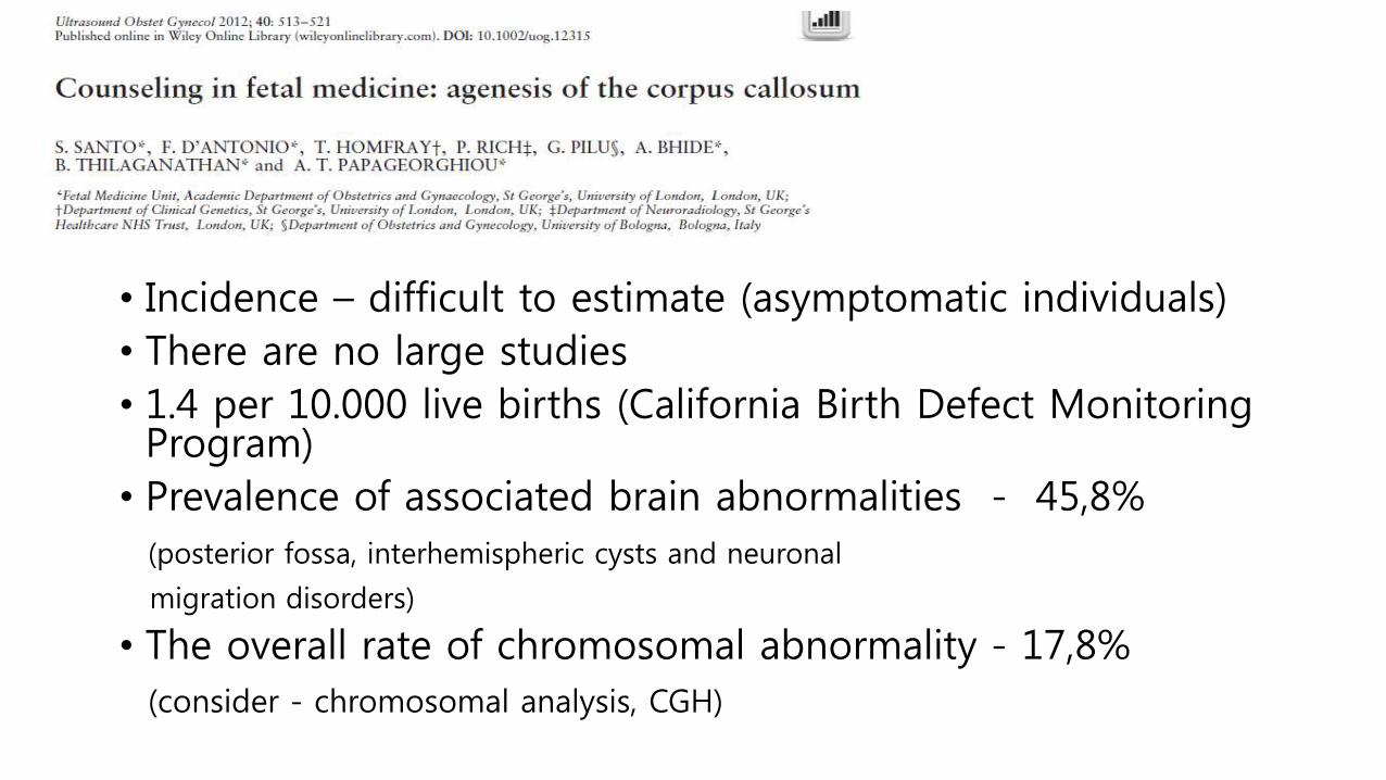
• Incidence – difficult to estimate (asymptomatic individuals)
• There are no large studies
• 1.4 per 10.000 live births (California Birth Defect Monitoring Program)
• Prevalence of associated brain abnormalities - 45,8%
(posterior fossa, interhemispheric cysts and neuronal
migration disorders)
• The overall rate of chromosomal abnormality - 17,8%
(consider - chromosomal analysis, CGH)
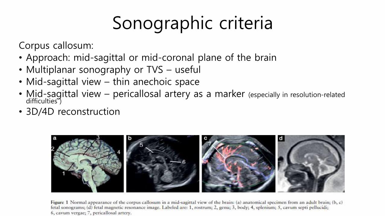
Sonographic criteria Corpus callosum:
• Approach: mid-sagittal or mid-coronal plane of the brain
• Multiplanar sonography or TVS – useful
• Mid-sagittal view – thin anechoic space
• Mid-sagittal view – pericallosal artery as a marker (especially in resolution-related difficulties )
• 3D/4D reconstruction
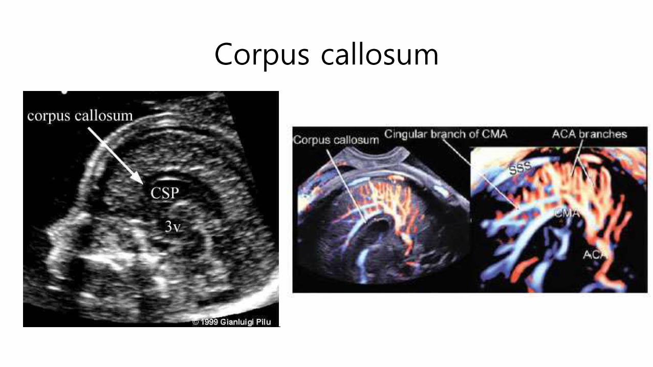
Corpus callosum

ACC – Sonographic criteria
• Measurement of CC size – recommended by some authors
Achiron R, Achiron A..Ultrasound Obstet Gynecol 2001; 18: 343–347.
ACC – key points on indirect features 1
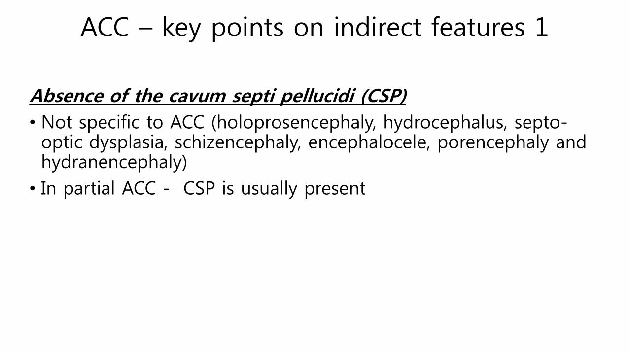
Absence of the cavum septi pellucidi (CSP)
• Not specific to ACC (holoprosencephaly, hydrocephalus, septo-optic dysplasia, schizencephaly, encephalocele, porencephaly and hydranencephaly)
• In partial ACC - CSP is usually present


ACC – key points on indirect features 2
Abnormalities of the ventricles
• Colpocephaly - dilatation of the atria and occipital horns of the lateral ventricles • Result of the absence of CC posterior portion, which allows
expansion of the occipital horns.
• Usually not associated with progressive ventriculomegaly

ACC – key points on indirect features 2
• Lateral displacement of LV on coronal views
• Upward displacement of the third ventricle, which reaches the level of the lateral ventricles
ACC – key points on indirect features 3
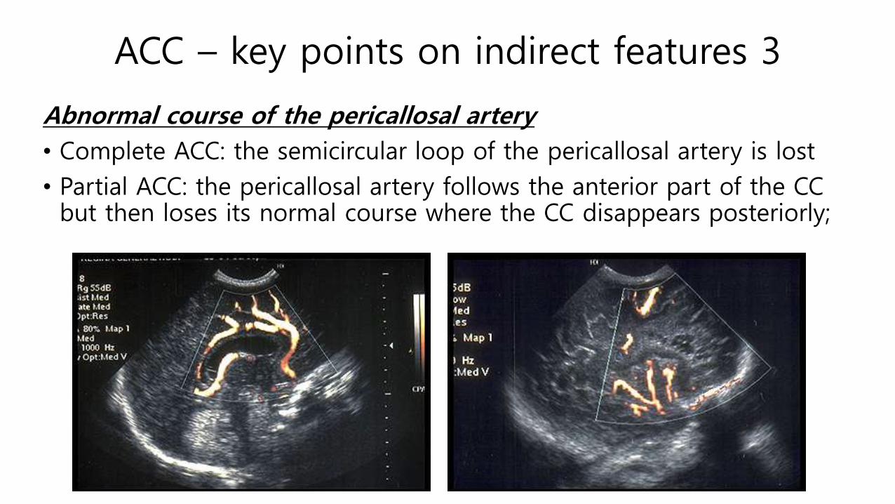
Abnormal course of the pericallosal artery
• Complete ACC: the semicircular loop of the pericallosal artery is lost
• Partial ACC: the pericallosal artery follows the anterior part of the CC but then loses its normal course where the CC disappears posteriorly;

ACC – key points on indirect features 4
Widening of the interhemisphericfissure
• Increased separation of the hemispheres
• 3 parallel echogenic lines (the middle - falx cerebri)
• The lateral ones representing the medial borders of the separated hemispheres
ACC – Prognosis
• Depends on coexistence of other abnormalities
• Association with cortical disorders – poor prognosis
• Normal or borderline intellectual development – in case of isolated
• Pediatric data suggest – more than 90% mental retardation or severe abnormalities


Dandy – Walker Malformation

Cranial posterior fossa
• The part of the intracranial cavity
• Located between the foramen magnum and tentorium cerebelli
• It contains the brainstem and cerebellum

Malformation of the posterior fossa
T. Chapman et al., Clinical imaging 39 (2015) 1-8

Definition • Dandy-Walker malformation
• Dandy-Walker variant 1. Marked cystic dilatation of the 4th
ventricle2. Hypogenesis or agenesis of the cerebellar
vermis3. Superior displacement of the tentorium
and lateral sinuses
• Dandy-Walker complex (or continuum)
• DWS malformation • Megacisterna magna (Benign enlargement of
subarachnoid spaces of PF)
• DWS variant • Blake’s pouch cyst (posterior ballooning of the
superior medullary velum into the cisterna magna)

Dandy – Walker malformation
1. Marked cystic dilatation of the 4th ventricle (may fill much of posterior fossa)
2. Hypogenesis or agenesis of the cerebellar vermis
3. Superior displacement of the tentorium and lateral sinuses
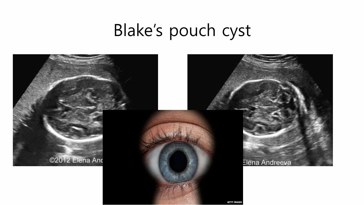
Blake’s pouch cyst
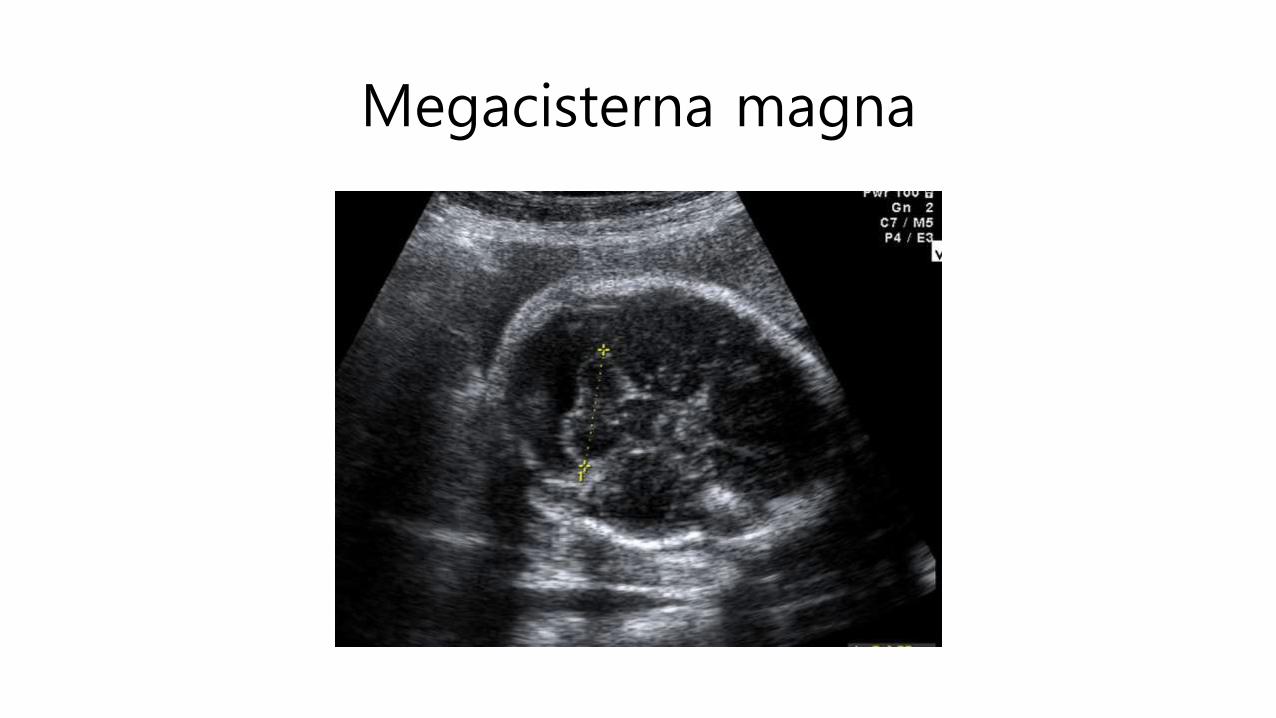
Megacisterna magna

Dandy – Walker malformation
• Incidence - 1:30.000 live births
• 4-12% of all cases of infantile hydrocephalus
• Frequently associated with
NTD
Midline anomalies (ACC, holoprosencephaly)
Extraneural defects (polycystic kidneys, cardiovascular defects, facial cleft…)

DWM – Sonographic diagnosis
• Transcerebellar and midsagittalplanes
• After 18 weeks’ gestation (complete cerebellar vermis development)
• Transcerebellar diameter (TCD)
• Cisterna magna
• + Nuchal fold
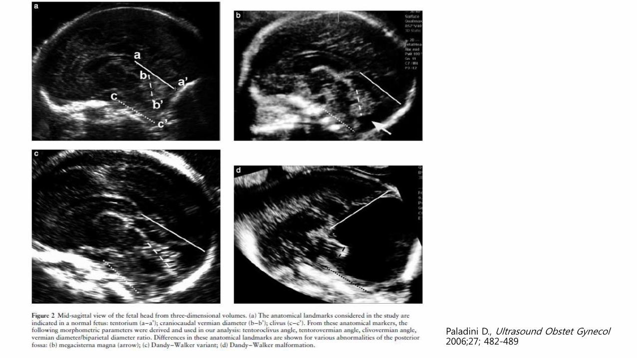
Paladini D., Ultrasound Obstet Gynecol2006;27; 482-489
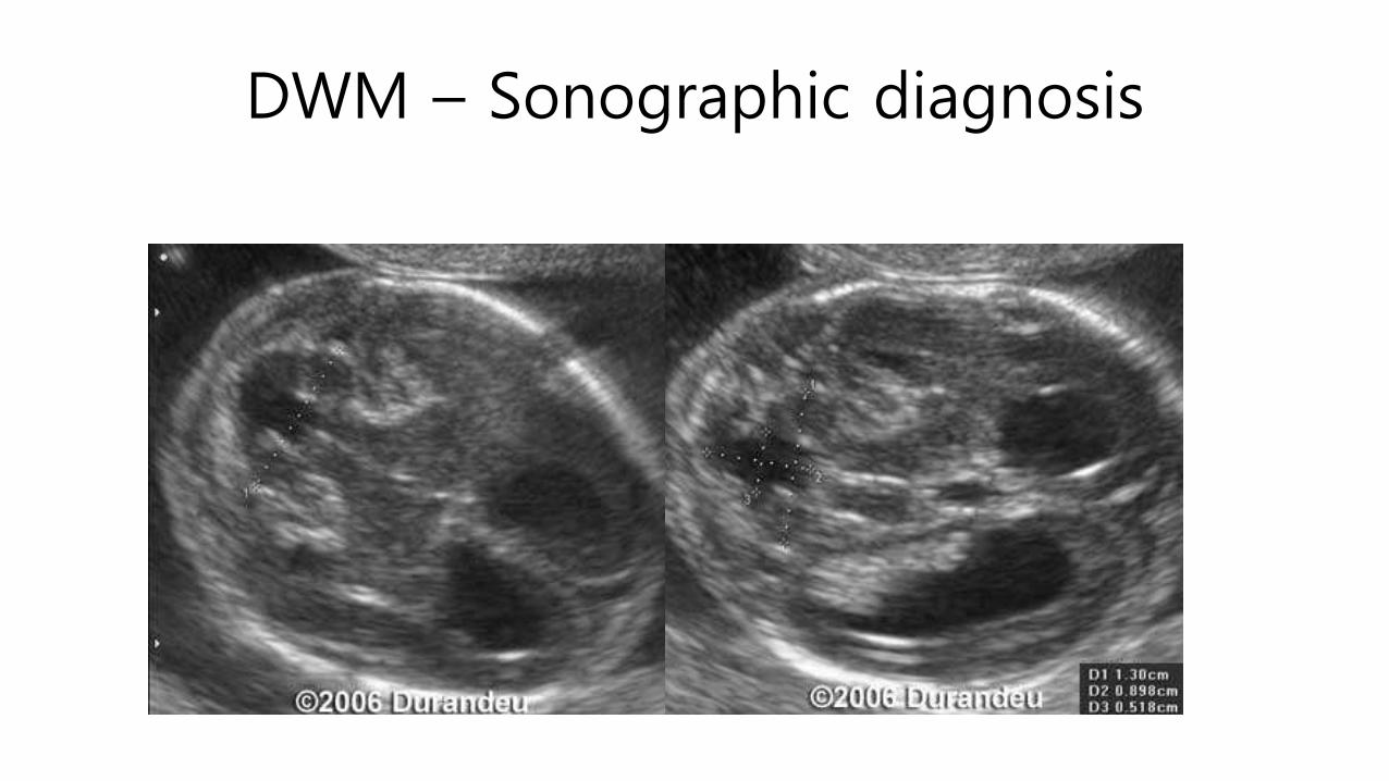
DWM – Sonographic diagnosis
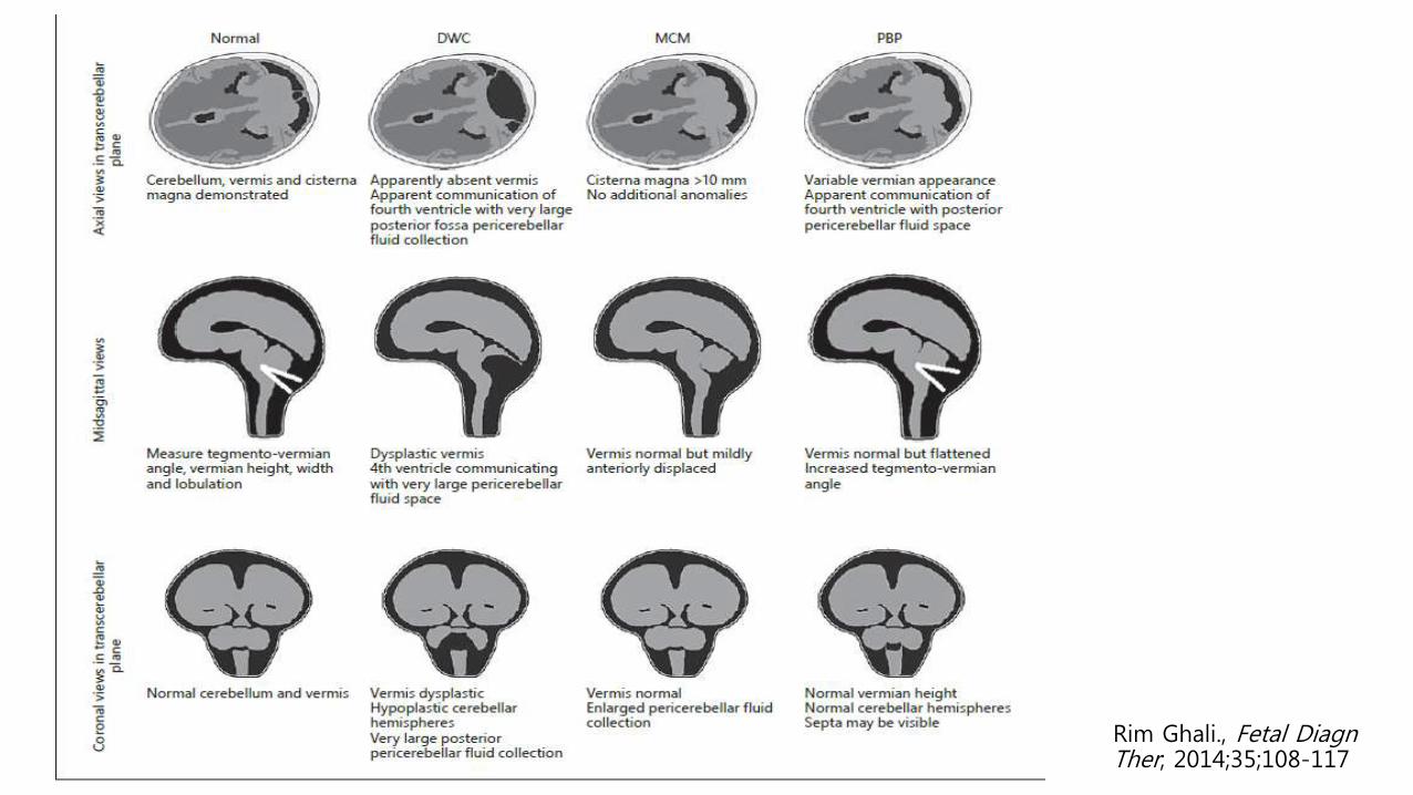
Rim Ghali., Fetal DiagnTher; 2014;35;108-117

DWM – Prognosis
• Largely dependent on type of condition, and associated anomalies
• Strongly related to other intracranial anomalies (degree of hydrocephalus, cortical malformations, and corpus callosal dysgenesis) and extracranial and chromosomal anomalies
• Severe cerebellar hypoplasia and vermian agenesis - poor intellectual outcome

DWM – Prognosis
• Isolated megacisterna magna and BPC without hydrocephalus - normal developmental outcome
• Isolated inferior vermian agenesis - variable prognosis

DWM – Prognosis
• Blake’s pouch cysts and megacisterna magna underwent spontaneous resolution in utero in one third of cases and over 90% of survivors without associated anomalies had normal developmental outcome at 1–5 years.
• Isolated Dandy–Walker malformation and vermian hypoplasia were associated with normal developmental outcome in only 50% of cases.
Gandolfi Coleoni et al., Ultrasound Obstet Gynecol; 2012;39; 625-631