fibrolipomawithosseousandcartilaginousmetaplasiaof …€™sfatpad:acasereport ioannisgigis1,2...
TRANSCRIPT

Hindawi Publishing CorporationCase Reports in OrthopedicsVolume 2012, Article ID 547963, 5 pagesdoi:10.1155/2012/547963
Case Report
Fibrolipoma with Osseous and Cartilaginous Metaplasia ofHoffa’s Fat Pad: A Case Report
Ioannis Gigis1, 2 and Panagiotis Gigis2, 3
1 Orthopaedics, Aristotle University of Thessaloniki, 54124 Thessaloniki, Greece2 Orthopaedic Department, Interbalkan Medical Center, Asklipiou 10, Pylaia, 57001 Thessaloniki, Greece3 Anatomy, Aristotle University of Thessaloniki, 54124 Thessaloniki, Greece
Correspondence should be addressed to Ioannis Gigis, [email protected]
Received 27 March 2012; Accepted 1 July 2012
Academic Editors: P. Lafforgue, M. G. Lykissas, J. Mayr, and A. Sakamoto
Copyright © 2012 I. Gigis and P. Gigis. This is an open access article distributed under the Creative Commons Attribution License,which permits unrestricted use, distribution, and reproduction in any medium, provided the original work is properly cited.
The most common benign tumors of the mesenchyme are the lipomas. Benign fatty tumors can arise in any location in which fat ispresent. Fibrolipomas are characterised by fat modules. Most patients affected by such tumors are in the fifth or sixth decade of life.When very close to vital structures such as joints, they may cause functional limitations as well as pain. Osseous and chondroidmetaplasia can infrequently manifest after chronic persistence. Given the rarity of this condition, a case of a big fibrolipoma ofHoffa’s fat pad with osseous and cartilaginous metaplasia is reported. A 44-year-old woman presented with an enlarging softmass on the right knee in the infrapatellar fat pad. After a thorough preoperative clinical and imaging examination, the mass wasremoved and sent to laboratory where the diagnosis was put. One year after surgery, both local and general condition of the patientwere good and no signs of recurrence were found.
1. Introduction
The most common [1] benign tumors of the mesenchymeare the lipomas. It is unclear if a soft-tissue lipoma representsa benign neoplasm, a local hyperplasia of fat cells, or acombination of both processes. They may arise in any loca-tion in which fat is present, the majority found in the upperhalf of the body, particularly the trunk and neck, though mayalso develop in other sites such as the hand [2, 3]. Benignlipomatous tumors have been classified by the World HealthOrganisation (WHO) in the following categories: classiclipoma, lipoblastoma, lipomatosis, angiolipoma, spindlecell/pleomorphic lipoma, angiomyolipoma, myelolipoma,hibernoma, and atypical lipoma [4]. The infrapatellar Hoffa’sfat pad is an intracapsular extrasynovial structure. Themost common tumour or tumour-like abnormalities ofthe infrapatellar fat pad reported are para-articular chon-droma/osteochondroma, focal pigmented villonodular syn-ovitis, synovial lipoma, synovial chondromatosis, synovialhaemangioma, ganglia/cysts, and intra-articular malignancy[5]. Some lipomas may exhibit morphological variations.These include fibrolipoma characterized by the presence of
prominent bundles of mature fibrous tissue traversing thefatty lobules [6]. It can be noticed that fibrolipomas areextremely rare variants excluded from the above classifica-tions and some times are also called benign mesenchymomas[7]. Most are subcutaneously located [8]. Uncommonly,they can manifest osseous and/or chondroid metaplasiaover extended periods of time, causing additional functionalproblems and pain compression syndromes like the case wepresent here [9]. Most patients affected by lipomas are intheir fifth or sixth decade of life, only rarely are childrenaffected. Given the rarity of this condition, a case of a bigfibrolipoma of Hoffa’s fat pad with osseous and cartilagenousmetaplasia is reported.
2. Case Report
A 44-year-old woman presented with an enlarging soft masson the right knee in infrapatellar fat pad; the mass had firstbeen noticed 1 year ago. A gradual increase of the size of themass had been observed by the patient during this year. Uponadmission the mass could be easily seen on the right kneeright below the patella in the infrapatellar region (Figure 1).

2 Case Reports in Orthopedics
Figure 1: Mass observed in the infrapatellar region.
Physical examination revealed a palpable mass in Hoffa’sfat pad. Flexion of the knee appeared to be limited andpain also occurred when trying to mobilize the joint. Thepatient complained for these limitations especially duringthe last few months when the mass increased in size. Astandard X-ray examination revealed osseous structure in theinfrapatellar site (Figure 2).
Furthermore, a CT, MRI, and a soft tissue ultrasonogra-phy were undertaken. According to the CT (Figure 3) andMRI (Figures 4(a) and 4(b)), a differential diagnosis betweensynovial osteochondromatosis, synovial sarcoma, and para-articular chondroma should be carried out.
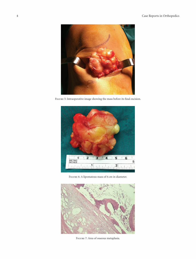
Additionally, the ultrasound showed a well-definedmature adipose tissue with the overlying patellar tendonintact. The patient was then operated and a mass of6 cm approximately in diameter was excised under generalanaesthesia along with its pedicle base (Figures 5 and 6).
Histopathological evaluation revealed that the lesion wascomposed of mature adipocytes with myxoid areas as wellas areas with osseous and chondroid metaplasia. The resultof this histological examination suggested a fibrolipoma withosseous and cartilagenous metaplasia (Figures 7 and 8).
To date, one year after operation, both local and generalcondition of the patient are good, the knee has no functionlimitations or pain, and there are no signs of recurrence ofthe lesion. Followup will continue with clinical examinationand imaging (radiographs and if necessary MRI or CT)annually.
3. Discussion
The aetiology of lipomas is unclear. They have been reportedto be both sporadic and inherited [10, 11]. Lipoma cells arebelieved to arise from mesenchymal primordial fatty tissuecells and tend to increase in size with body weight gain[12]. When located close to vital structures such as joints,giant lipomas may cause functional limitations on accountof their excessive size and weight [13] or lymphedema, pain,or nerve compression syndromes [12, 14]. For a lipomato be referred to as “giant”, the lesion should be at least10 cm in diameter [15]. In our case, although the lesionwas only 6 cm in diameter, given the narrow space of the
Figure 2: Osseous structure in the infrapatellar fat pad.
infrapatellar region, it caused serious limitations in kneeflexion as well as pain. The malignant transformation of alipoma into a liposarcoma is rare [16] as is the sarcomatoustransformation of giant lipomas [12, 17]. It is importantto differentiate giant lipomas from liposarcomas, malignantfibrous histiocytomas, and other benign soft-tissue lesions.The main concern should be the exclusion of malignancy. Ithas been suggested that a liposarcoma should be consideredwhen a fatty subcutaneous tumor is more than 10 cm indiameter and has grown rapidly in recent months [18].Fibrolipomas may develop in virtually any region thatcontains fat, but in general, they tend to appear on thetrunk, neck, and upper extremities. Thigh has also beenreported as rather uncommon region of origin [19, 20].Never before has a fibrolipoma been reported so close to ajoint, especially the knee joint. The clinical diagnosis of afibrolipoma is established upon presentation of a nontender,semisolid mass, which has grown over extended period oftime. In our case, we can only be sure that it was firstnoticed from the patient a year before. Radiologically, Katzer

Case Reports in Orthopedics 3
Figure 3: Computed tomography showing the mass with its osseous content in the infrapatellar region.
(a) (b)
Figure 4: (a) Axial T1-weighted image. (b) Axial T2-weighted image showing clearly the mass in Hoffa’s fat pad underlying the patellartendon and extra-articular.
reported that soft tissue ultrasound could display matureadipose tissue embedded within fibrous mesenchymal tissuecomponents, and even osseous metaplasia changes [9]. Theultrasound performed in our patient showed the above-mentioned mature adipose tissue but did not find theosseous metaplasia although some osseous structure waswell seen in the X-ray. Computed tomography (CT) aswell as magnetic resonance imaging (MRI) may be needed
in such cases to rule out malignancies such as liposar-comas [19]. Indeed, both examinations, in our case, setthe differential diagnosis of synovial osteochondromatosis,synovial sarcoma, and para-articular chondroma. The treat-ment of fibrolipomas involves surgical excision along withtheir well-defined pseudocapsule, followed by appropriatereconstruction. Recurrence of excised fibrolipomas has notbeen reported in the literature; however, there are reports
4 Case Reports in Orthopedics
Figure 5: Intraoperative image showing the mass before its final excision.
Figure 6: A lipomatous mass of 6 cm in diameter.
Figure 7: Area of osseous metaplasia.

Case Reports in Orthopedics 5
Figure 8: Area of cartilagenous metaplasia.
of neglected cases, which eventually developed malignanttransformation [7]. Dissection of these benign neoplasms isrelatively straightforward.
Acknowledgment
This study was carried out at the Interbalkan Medical Center.
References
[1] A. Rydholm and N. O. Berg, “Size, site and clinical incidenceof lipoma. Factors in the differential diagnosis of lipoma andsarcoma,” Acta Orthopaedica Scandinavica, vol. 54, no. 6, pp.929–934, 1983.
[2] D. Paarlberg, R. L. Linscheid, and E. H. Soule, “Lipomas of thehand. Including a case of lipoblastomatosis in a child,” MayoClinic Proceedings, vol. 47, no. 2, pp. 121–124, 1972.
[3] E. Hakim, Y. Kolander, Y. Meller, M. Moses, and A. Sagi,“Gigantic lipomas,” Plastic and Reconstructive Surgery, vol. 94,no. 2, pp. 369–371, 1994.
[4] S. W. Weiss, L. H. Sobin, and F. M. Enzinger, “Lipomatoustumours,” in Histological Typing of Soft Tissue Tumours (WorldHealth Organization International Histological Classification ofTumours), S. W. Weiss and L. H. Sobin, Eds., pp. 23–24,Springer, 2nd edition, 1994.
[5] C. Helpert, A. M. Davies, N. Evans, and R. J. Grimer,“Differential diagnosis of tumours and tumour-like lesionsof the infrapatellar (Hoffa’s) fat pad: pictorial review with anemphasis on MR imaging,” European Radiology, vol. 14, no.12, pp. 2337–2346, 2004.
[6] M. Mazzocchi, M. G. Onesti, P. Pasquini, R. La Porta, D.Innocenzi, and N. Scuderi, “Giant fibrolipoma in the leg—acase report,” Anticancer Research, vol. 26, no. 5 B, pp. 3649–3654, 2006.
[7] M. J. Kransdorf, R. P. Moser Jr., J. M. Meis, and C. A.Meyer, “Fat-containing soft-tissue masses of the extremities,”Radiographics, vol. 11, no. 1, pp. 81–106, 1991.
[8] A. Terzioglu, D. Tuncali, A. Yuksel, F. Bingul, and G. Aslan,“Giant lipomas: a series of 12 consecutive cases and a giantliposarcoma of the thigh,” Dermatologic Surgery, vol. 30, no. 3,pp. 463–467, 2004.
[9] B. Katzer, “Histopathology of rare chondroosteoblastic meta-plasia in benign lipomas,” Pathology Research and Practice, vol.184, no. 4, pp. 437–443, 1989.
[10] K. S. Pinski and H. H. Roenigk Jr., “Liposuction of lipomas,”Dermatologic Clinics, vol. 8, no. 3, pp. 483–492, 1990.
[11] D. J. Leffell and I. M. Braverman, “Familial multiple lipomato-sis: report of a case and a review of the literature,” Journal ofthe American Academy of Dermatology, vol. 15, no. 2 I, pp. 275–279, 1986.
[12] G. S. Phalen, J. I. Kendrick, and J. M. Rodriguez, “Lipomasof the upper extremity. A series of fifteen tumors in the handand wrist and six tumors causing nerve compression,” TheAmerican Journal of Surgery, vol. 121, no. 3, pp. 298–306, 1971.
[13] J. Guerrissi, D. Klersfeld, C. Sampietro, and J. Valdivieso,“Limitation of thigh function by a giant lipoma,” Plastic andReconstructive Surgery, vol. 94, no. 2, pp. 410–411, 1994.
[14] P. E. Higgs, V. L. Young, R. Schuster, and P. M. Weeks, “Giantlipomas of the hand and forearm,” Southern Medical Journal,vol. 86, no. 8, pp. 887–890, 1993.
[15] A. C. Harrington, J. Adnot, and R. S. Chesser, “Infiltratinglipomas of the upper extremities,” Journal of DermatologicSurgery and Oncology, vol. 16, no. 9, pp. 834–837, 1990.
[16] R. S. Cotran, V. Kumar, and S. L. Robbins, “Tumors of adiposetissue,” in Robbins’ Pathological Basis of Disesses, pp. 1374–1412, Saunders, Philadelphia, Pa, USA, 1989.
[17] H. Takagi, K. Kato, E. Yamada, and T. Suchi, “Six recentliposarcomas including largest to date,” Journal of SurgicalOncology, vol. 26, no. 4, pp. 260–267, 1984.
[18] C. Celik, C. P. Karakousis, R. Moore, and E. D. Holyoke,“Liposarcomas: prognosis and management,” Journal of Sur-gical Oncology, vol. 14, no. 3, pp. 245–249, 1980.
[19] T. Simsek, A. Sonmez, I. O. Aydogdu, L. Eroglu, and F.Karagoz, “Giant fibrolipoma with osseous metaplasia on thethigh,” Journal of Plastic, Reconstructive and Aesthetic Surgery,vol. 64, no. 5, pp. e125–e127, 2011.
[20] G. C. Zografos, I. Kouernis, P. Kalliopi, K. Karmen, M.Evagelos, and G. Androulakis, “Giant lipoma of the thighin a patient with morbid obesity,” Plastic and ReconstructiveSurgery, vol. 109, no. 4, pp. 1467–1468, 2002.

Submit your manuscripts athttp://www.hindawi.com
Stem CellsInternational
Hindawi Publishing Corporationhttp://www.hindawi.com Volume 2014
Hindawi Publishing Corporationhttp://www.hindawi.com Volume 2014
MEDIATORSINFLAMMATION
of
Hindawi Publishing Corporationhttp://www.hindawi.com Volume 2014
Behavioural Neurology
EndocrinologyInternational Journal of
Hindawi Publishing Corporationhttp://www.hindawi.com Volume 2014
Hindawi Publishing Corporationhttp://www.hindawi.com Volume 2014
Disease Markers
Hindawi Publishing Corporationhttp://www.hindawi.com Volume 2014
BioMed Research International
OncologyJournal of
Hindawi Publishing Corporationhttp://www.hindawi.com Volume 2014
Hindawi Publishing Corporationhttp://www.hindawi.com Volume 2014
Oxidative Medicine and Cellular Longevity
Hindawi Publishing Corporationhttp://www.hindawi.com Volume 2014
PPAR Research
The Scientific World JournalHindawi Publishing Corporation http://www.hindawi.com Volume 2014
Immunology ResearchHindawi Publishing Corporationhttp://www.hindawi.com Volume 2014
Journal of
ObesityJournal of
Hindawi Publishing Corporationhttp://www.hindawi.com Volume 2014
Hindawi Publishing Corporationhttp://www.hindawi.com Volume 2014
Computational and Mathematical Methods in Medicine
OphthalmologyJournal of
Hindawi Publishing Corporationhttp://www.hindawi.com Volume 2014
Diabetes ResearchJournal of
Hindawi Publishing Corporationhttp://www.hindawi.com Volume 2014
Hindawi Publishing Corporationhttp://www.hindawi.com Volume 2014
Research and TreatmentAIDS
Hindawi Publishing Corporationhttp://www.hindawi.com Volume 2014
Gastroenterology Research and Practice
Hindawi Publishing Corporationhttp://www.hindawi.com Volume 2014
Parkinson’s Disease
Evidence-Based Complementary and Alternative Medicine
Volume 2014Hindawi Publishing Corporationhttp://www.hindawi.com