first record of phlebotomine sandflies (diptera...
TRANSCRIPT

195Original contributionParasite, 2012, 19, 195-206
FIRST RECORD OF PHLEBOTOMINE SANDFLIES (DIPTERA: PSYCHODIDAE) IN THE COMOROS ARCHIPELAGO WITH DESCRIPTION OF SERGENTOMYIA (VATTIEROMYIA) PESSONI N. SP.
AND S. (RONDANOMYIA) GOODMANI COMORENSIS N. SSP.RANDRIANAMBININTSOA F.J.*,**,***, DEPAQUIT J.**, BRENGUES C.***, DHONDT C.**, YAHAYA I.****,
OULEDI A.*****, LÉGER N.****** & ROBERT V.***
Summary:
No Phlebotomine sandflies had ever been reported in the Comoros Archipelago, including the three islands of the Republic of the Union of Comoros (Grande Comore, Mohéli and Anjouan) and the French oversea department of Mayotte. During three field surveys carried out in 2003, 2007 and 2011, we provided the first record of Phlebotomine sandflies in this area. A total of 85 specimens belonging to three species were caught: a new species S. (Vattieromyia) pessoni n. sp. (two females from Grande Comore), a new subspecies of Sergentomyia (Rondanomyia) goodmani (80 specimens from Grande Comore and one from Anjouan) and Grassomyia sp. (two females from Mohéli). The individualisation of these taxa was inferred both from morphological criteria and sequencing of a part of the cytochrome b of the mitochondrial DNA. These taxa are closely related to Malagasy sandflies.
KEY WORDS: Phlebotomine sandfly, Sergentomyia, DNA barcoding, taxonomy, inventory, Comoros, Mayotte, Indian Ocean.
Résumé : PREMIÈRE MENTION DE PHLÉBOTOMES (DIPTERA : PSYCHODIDAE) DANS L’ARCHIPEL DES COMORES ET DESCRIPTION DE SERGENTOMYIA (VATTIEROMYIA) PESSONI N. SP. ET S. (RONDANOMYIA) GOODMANI COMORENSIS N. SSP.
Aucun phlébotome n’avait jamais été rapporté dans l’archipel des Comores, incluant les trois îles de l’Union des Comores (la Grande Comore, Mohéli et Anjouan) et le département français d’outre-mer, Mayotte. Au cours de trois campagnes d’études sur le terrain en 2003, 2007 et 2011, nous avons fait la première observation de phlébotomes dans cette zone. 85 spécimens appartenant à trois espèces ont été capturés : une nouvelle espèce, S. (Vattieromyia) pessoni n. sp. (deux femelles de la Grande Comore), une nouvelle sous-espèce de Sergentomyia (Rondanomyia) goodmani (80 spécimens de la Grande Comore et une d’Anjouan) et Grassomyia sp. (deux femelles à Mohéli). L’individualisation de ces taxa s’est faite à la fois à partir de critères morphologiques et du séquençage d’une partie du cytochrome b de l’ADN mitochondrial. Ces taxa sont très proches des phlébotomes de Madagascar.
MOTS-CLÉS : phlébotome, Sergentomyia, séquençage d’ADN, taxonomie, inventaire, Comores, Mayotte, Océan Indien.
* Département de Biologie Animale, Faculté des Sciences, Université d’Antananarivo, Madagascar.** Université de Reims Champagne-Ardenne, ANSES, JE2533 – USC “Transmission vectorielle et épidémiosurveillance de maladies parasitaires (VECPAR)”, 51, rue Cognacq-Jay, 51096 Reims Cedex, France.*** MIVEGEC, UMR IRD 224-CNRS 5290-UM1-UM2, 911, avenue Agropolis, BP 64501, 34394 Montpellier Cedex 5, France.**** CNDRS, BP 169, Moroni, Union des Comores.***** Coordonnateur des Programmes de recherches, Université des Comores, route de la Corniche, Moroni, Union des Comores.****** 63, avenue Pierre Sémart, 94210 La Varenne Saint Hilaire, France.Correspondence: Jérome Depaquit.Tel.: 33 (0)3 26 91 37 23 – Fax : 33 (0)3 26 91 35 97.E-mail: [email protected]
INTRODUCTION
To our knowledge, no record of Phlebotomine sandfly exists in the literature and there was no case of leishmaniasis in the Comoros Archipe-
lago. The latter includes the Islands of the Union of the Comoros (Grande Comore, Anjouan and Mohéli) and Mayotte (a French oversea department). In the neighbouring countries, little is known about Phlebo-tomine sandflies from South-eastern Africa (Abonnenc, 1972; Artemiev, 1985; Davidson, 1987) and from Mada-gascar (Depaquit et al., 2002, 2004a & b, 2007, 2008;
Léger et al., 2005). We performed three field works in order to inventory the four islands. A total of 85 adult specimens have been captured. All the speci-mens have been examined morphologically and some of them have been processed for molecular biology. All of them belong to the genera Sergentomyia and Grassomyia. Two new taxa for Science are described in the present study.
MATERIAL AND METHODS
Phlebotomine sandflies have been caught using CDC miniature light traps and UV miniature light traps in the four main islands of the Comoros
Archipelago: Grande Comore, Mohéli, Anjouan and Mayotte (Fig. 1, Table I).
The archipelago of the Comoros is located at the northern end of the Mozambique Channel in the Indian Ocean, halfway between the coasts of Mada-gascar and the African continent. The volcanic chain of the Comoros appears to be a “hot spot trace”. The Comoros are made up of volcanic rocks, prima-rily undersaturated alkali olivine basalts. Phonolitic and small volumes of trachytic lavas have also been
PAR2012-3_Randrianambinintsoa.indd 195 3/07/12 18:37:51

RANDRIANAMBININTSOA F.J., DEPAQUIT J., BRENGUES C. ET AL.
196 Original contributionParasite, 2012, 19, 195-206
reported (Pavlovsky & de Saint-Ours, 1953; Flower, 1973; Emerick & Duncan, 1982). The volcanic rocks have been differentiated into three volcanic phases, in which basaltic lavas prevail. Scorias and puzzolanic tuffs have been reported from Grande Comore, Mohéli and Anjouan. Phonolitic and trachytic rocks occur on Mayotte (Pavlovsky & de Saint-Ours, 1953). On the geo-chronological plan, the latest information on the age of the three islands is respectively as follows: 1.49, 0.48, 0.36 millions years (My) for Mayotte, Mohéli and Anjouan (Armstrong, 1972: Emerick & Duncan, 1982, 1983: Nougier et al., 1986) and 0.13 My for Grande Comore (Emerick & Duncan, 1982, 1983).
The catching sites, including mainly caves, are detailed below.1 – In Grande Comore:- Fassi cave (Mitsamiouli): a hole about 15 m deep, which divides into two long passages. One is about 10 m long and is of archaeological interest, the other probably leads to the sea;- Hahaya cave (Hamanvou): next to the runway at Hahaya airport;- Ynambeni cave (Nioumagama): a hole about 10 m deep, dividing into two parts along the main access on about 100 meters;- Idjinkondzi cave (Dimani), about 5 m long;- Ceiling of Dima primary school (Nioumagama);- Under the main mosk of the village of Mohoro (Itsahidi);- District of Zilimadzou (Moroni), in the east border of the city.
2 – In Anjouan:- Hamapoudre cave (Mromagi – Bambao), about 50 m long at the bottom of which lies a pond of about 4 m diameter;- Hapire cave (Limbi): it is more like an excavation with a rocky slope of about 30 m high next to a 10 m waterfall;- Bazimini cave (Bazimini): a hole about 5 m deep;- Primary school of Mirontsy (Mirontsy - Mutsamudu);- Ceiling of a social center of about 50 m × 20 m × 10 m, a hard brick building covered by a corrugated iron roof which is a shelter for little bats;- Next to the University of Patsy.3 – In Mohéli: Hamavouna, 20 m above sea level (a.s.l.), at the top of a steep slope at the base of a vertical stone cliff in which ten strange horizontal holes seem to have carved, 2 m deep and 10 to 30 cm diameter.4 – In Mayotte: Lake Karihani, Village of Kahani, Vil-lage of Bouyouni and the House of the Governor.
When available, some specimens from Madagascar have been added to this sampling (Table I). Those of Vattieromyia refer to Depaquit et al. (2008) and those of S. goodmani have been specially processed in the present study. Sandflies were stored in 96 % ethanol. The head and genitalia were cut off in a drop of ethanol, cleared in boiling Marc-André solution, and mounted between slide and cover slide for spe-cies identification. The body related to the specimen was stored dried in a vial at - 20 °C before DNA extraction. All the specimens were observed utilizing
Fig. 1. – Prospected area.
PAR2012-3_Randrianambinintsoa.indd 196 3/07/12 18:37:51

PHLEBOTOMINE SANDFLIES FROM COMOROS
197Original contributionParasite, 2012, 19, 195-206
Island Location Numberof traps
Catchingyear
Specimenscaught
Samples processed for molecular biology and their genbank accession numbers
Grande Comore Fassi
Idjinkoundzi
Hahaya
Ynambeni
Mohoro
Dima
Zilimadzou
6
6
6
6
6
2
2011
2011
2011
2011
2011
2011
2003
S. goodmani comorensis: 3
S. goodmani comorensis : 1
S. pessoni n. sp. : 2
S. goodmani comorensis : 1
S. goodmani comorensis : 1
S. goodmani comorensis : 39 & 35
JQ421008JQ421009JQ421010
JQ421012
JQ421019JQ421020
JQ421011
JQ421014JQ421017JQ421018
AnjouanPatsy
Mirontsy
Mutsamudu
Hamapoudre
Hapira
Bazimini
1
6
6
6
6
6
2011
2011
2011
2011
2011
2011
S. goodmani comorensis : 1 JQ421013
Moheli Hamavouna 2007 Grassomia sp. : 2 JQ421015JQ421016
Mayotte Lake of Karihani
Village of Kahani
Village of Bouyouni
Governor’s Home
9
6
8
8
2011
2011
2011
2011
Madagascar Namoroka
Ankarana
Ankiliefatra
2002
2003
2003
S. namo
S. goodmani
S. sclerosiphon
S. goodmani
S. anka
S. goodmani
EU143774EU143775EU143776
JQ421002JQ421005
EU143777EU143778EU143780EU143781EU143783EU143786EU143787EU143788EU143789EU143790
JQ434695
EU143779EU143782EU143784EU143785
JQ421006JQ421003JQ421007JQ421004
Table I. – Sampling species.
PAR2012-3_Randrianambinintsoa.indd 197 3/07/12 18:37:52

RANDRIANAMBININTSOA F.J., DEPAQUIT J., BRENGUES C. ET AL.
198 Original contributionParasite, 2012, 19, 195-206
a BX50 microscope and measured using the Perfect Image software (Aries Company, Chatillon, France) and a video camera connected to the microscope. Drawings have been done using the camera lucida installed on the microscope. Some specimens have been remounted in Canada balsam after complete processing of washing, dehydration in baths of ethanol 70 to 100, then beech creosote.
Genomic DNA was extracted from the thorax, wings, legs and abdomen of individual sandflies using the QIAmp DNA Mini Kit (Qiagen, Germany) following the manufacturer’s instructions, modified by crushing the sandfly tissues with a piston pellet (Treff, Swit-zerland), and using an elution volume of 200 μl, as detailed in Depaquit et al. (2004a). All the mtDNA amplifications were performed in a 50 μl volume using 5 μl of extracted DNA solution and 50 pmol of each of the primers. The PCR mix contained (final concentrations) 10 mM Tris HCl (pH 8.3), 1.5 mM MgCl2, 50 mM KCl, 0.01 % Triton X 100, 200 μM dNTP each base, and 1.25 units of 5 prime Taq polymerase (Eppendorf, Germany). The cycle begins with an initial denaturation step at 94 °C for 3 min and finishes by a final extension at 68 °C for 10 min. Amplification of a fragment of cytochrome B gene has been done by using the primers N1N-PDR: (5’-CA(T/C) ATT CAA CC(A/T) GAA TGA TA-3’) and C3B-PDR: (5’-GGT A(C/T)(A/T) TTG CCT CGA (T/A)TT CG(T/A) TAT GA-3’) following Esseghir et al. (1997): five cycles of (dena-turation at 94 °C for 30 s, annealing at 40 °C for 60 s and extension at 68 °C for 60 s), followed by 35 cycles of (denaturation at 94 °C for 60 s, annealing at 44 °C for 60 s and extension at 68 °C for 60 s). Amplicons were analysed by electrophoresis in 1.5 % agarose gel containing ethidium bromide. Direct sequencing in both directions was performed using the primers used for DNA amplification. The correction of sequences is done using Pregap and Gap softwares included in the Staden Package (Bonfield & Staden, 1996).
Molecular analyses are based on the sequence align-ment performed using the ClustalW routine included in the bioedit version 5 software (Hall, 1999) and checked by eye. According to the objective of this study, which is not a phylogenetical analysis, a Neighbor-Joining (NJ) analysis was performed using MEGA 5 software (Tamura et al., 2011), with the Kimura-2 parameter model and using uniform rates among sites.
RESULTS
A total of 85 Phlebotomine sandflies has been caught, on Grande Comore, Mohéli and Anjouan (82, two and one, respectively). No sandflies
have been caught in Mayotte (Table I). Most of the
specimens (81 specimens) belong to the subgenus Rondanomyia and appear to be a subspecies of Ser-gentomyia goodmani. Two female specimens of the subgenus Vattieromyia belong to a new species des-cribed below. Moreover, two specimens of Grassomyia sp. have been caught in Mohéli.
SERGENTOMYIA PESSONI N. SP. DEPAQUIT, RANDRIANAMBININTSOA & LÉGER
Genus Sergentomyia Rondani & Berté, in Rondani, 1840Subgenus Vattieromyia Depaquit, Léger & Robert, 2008Species Sergentomyia pessoni n. sp.The authors Depaquit, Randrianambinintsoa & Léger are responsible for satisfying the criteria of availability of the name Sergentomyia pessoni and should be cited as the sole authority of these taxa, according to the Article 50(1) of the lnternational Code of Zoological Nomenclature, 4th edition, 2000.
• Female (Fig. 2, Table II)
The description is based on the two specimens caught at Ynambeni, Grande Comore.
HeadInterocular suture almost complete.Cibarial armature with:- a posterior line with 44 to 45 teeth. The median ones are longer than the lateral ones. They are arched dorsoventrally;- a line of 11 to 12 V-shaped anterior denticles.Pigment patch large, with a triangular anterior exten-sion.Pharyngeal armature made up of small teeth pointed forward, in concentric rows.Palpal formula: 1, 2, 3, 4, 5. Newstead spines spatu-lated on the third palpal segment.Antennal formula: 2/III-X, 1/XI-XV, or 2/III-XI-1/XIII-XV. Ascoids don’t reach the next articulation. A III = 289-297 μm. A IV = 145-156 μm. A V = 142-149 μm. A III more or less equal to A IV + A V; A III/labrum = 1.53-1.55.
ThoraxMesanepisternum without setae. Wing: length = 1,600-1,692 μm; width = 475-505 μm; length/width = 3,35-3,37; a = 493-434 µm; b = 242-254 μm; d = 254-331 μm; g = 250-264 μm; width/g = 1.9; P = 48-65 μm.
SpermathecaeTypical of the subgenus Vattieromyia. Short cylindrical body, slightly striated on the edges nearly echinulated, more narrow in its median part than in its proximal and distal parts. Head made up of a few spear-like elements prolonged by some filaments, surrounded by a high collar. Ducts difficult to observe, slightly folded, relatively short, joining to form a common proximal
PAR2012-3_Randrianambinintsoa.indd 198 3/07/12 18:37:52

PHLEBOTOMINE SANDFLIES FROM COMOROS
199Original contributionParasite, 2012, 19, 195-206
duct. Individual ducts are strongly sclerotised over a limited length next in continuation with the ducts.
Type locality: cave of Ynambeni, Grande Comore, at 11°54.466’S, 43°28.615’E and 286 m a.s.l.
• Male
Unknown.
SERGENTOMYIA GOODMANI COMORENSIS N. SSP. DEPAQUIT, RANDRIANAMBININTSOA & LÉGER
Genus Sergentomyia Rondani & Berté, in Rondani, 1840Subgenus Rondanomyia Theodor, 1958
Species Sergentomyia goodmani Léger, Depaquit & Robert, 2005Subspecies Sergentomyia goodmani comorensis n. ssp.The authors Depaquit, Randrianambinintsoa & Léger are responsible for satisfying the criteria of availability of the name Sergentomyia goodmani comorensis and should be cited as the sole authority of these taxa, according to the Article 50(1) of the lnternational Code of Zoological Nomenclature, 4th edition, 2000.
• Female (Fig. 3, Table II)
HeadInterocular suture almost complete.
Fig. 2. – S. pessoni n. sp. female. A: pharynx and cibarium; B: antennal segments III, IV and V; C: palp; D: spermathe-cae; E: furca.
PAR2012-3_Randrianambinintsoa.indd 199 3/07/12 18:37:52
RANDRIANAMBININTSOA F.J., DEPAQUIT J., BRENGUES C. ET AL.
200 Original contributionParasite, 2012, 19, 195-206
Character
Female Male
S. pessonin. sp.
S.anka*
S.namo*
S.sclerosiphon*
S.goodmani**
S. goodmanicomorensis
ssp. nov. n = 27
S.goodmani**
S. goodmanicomorensis
ssp. nov. n = 39
Labrum
AIII
AIV
AV
AIII / labrum
AIII / AIV + AV
Wing length
Wing width
α
β
δ
γ
π
Wing length/width
Wing width/γ
Number of cibarial
teeth
Coxite length CL
Number of internal
setae of the coxite
style length
Aedeagus length
Genital filaments
Genital pump
Genital filaments/
genital pump
189-191
289-297
145-156
142-149
1.53-1.55
0.97-1
1,600-1,692
475-505
493-434
242-254
254-331
250-264
48-65
3.35-3.37
1.9
44-45
170-190
177-204
94-111
95-111
1.18-1.35
< 1
1,500-1,808
401-460
259-333
323-343
113-339
244-254
135-172
3.74-3.93
1.64-1.81
35-43
210-280
100-140
104-140
1.18-1.35
1
1,618-1,881
430-491
326-384
297-354
159-211
245-342
123-188
3.56-4.13
1.40-1.80
48-55
158-204
214-300
105-145
108-140
1.27-1.55
1
1,628-1,886
410-534
251-405
312-389
91-247
235-315
111-209
3.53-4.15
1.40-1.79
20-29
150-170
110-130
64-70
66-78
0.7-0.8
1,320
400
200
250
80
400
3.30
1.00
16-27
156-184
(171.93 ± 9.15)
119-158
(128.57 ± 12.49)
61-79
(68.98 ± 5.68)
58-87
(71.44 ± 7.98)
0.69-0.80
(0.73 ± 0.05)
0.88-0.92
(0.91 ± 0.04)
1,236-1,433
(1,349.79 ± 85.46)
320-418
(371.86 ± 33.98)
101 -218
(177.63 ± 46.88)
264-378
(304.95 ± 44.36)
11-82
(54.03 ± 30.41)
205-292
(256.40 ± 36.13)
109-177
(135.05 ± 31.96)
3.39-3.87
(3.64 ± 0.16)
1.61-1.26
(1.49 ± 0.14)
15-21
(16.93 ± 1.77)
110-140
104-132
64-75
74-80
0.9
1,280
330
200
220
90
150
15
65
70-90
250-380
75-104
3.57-4.24
118-147
(133.21 ± 6.57)
102-131
(118.27 ± 7.48)
64-76
(69.21 ± 4.19)
65-76
(71.89 ± 3.15)
0.75-1.02
(0.89 ± 0.06)
1,044-1,184
(1,136.27 ± 37.31)
223-309
(281.36 ± 21.78)
93-150
(120.11 ± 20.95)
206-273
(240.95 ± 27.86)
17-36
(25.31 ± 9.46)
226-238
(232.05 ± 4.50)
82-120
(101.85 ± 16.97)
3.82-4.69
(4.05 ± 0.20)
2.58-3.70
(3.08 ± 0.52)
127-149
(139.75 ± 7.83)
4-6
(4.60 ± 0.68)
56-72
(65.63 ± 4.21)
58-79
(65.81 ± 6.85)
331-409
(360.11 ± 25.80)
81-112
(90.60 ± 9.54)
3.73-4.68
(4.04 ± 0.29)
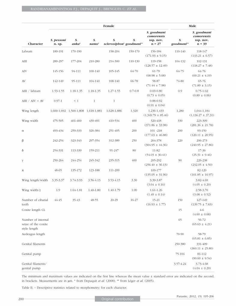
The minimum and maximum values are indicated on the first line whereas the mean value ± standard error are indicated on the second,
in brackets. Measurments are in μm. * from Depaquit et al. (2008); ** from Léger et al. (2005).
Table II. – Descriptive statistics related to morphometry; for each character.
PAR2012-3_Randrianambinintsoa.indd 200 3/07/12 18:37:53

PHLEBOTOMINE SANDFLIES FROM COMOROS
201Original contributionParasite, 2012, 19, 195-206
Cibarium with a line of 13 to 20 teeth like those of Sergentomyia schwetzi. The central ones are narrower than the lateral ones. No anterior denticles.Large pigment patch, mushroom-like, with an anterior extension.Pharynx narrow with i) in the back, few small teeth oriented backward displayed on concentric rounded rows, and ii) in the front, bigger teeth oriented forward.Palpal formula: 1, 2, 3, 4, 5. Newstead spines spatu-lated on the third segment.Antennal formula: 2/III-XV; short ascoids don’t reach the next articulation. Short antennal segments: AIII = 119-158 μm; AIV = 61-
79 μm ; AV = 55-87 μm. AIII/AIV+AV = 0.88 - 0.92.Labrum = 156-184 μm.
Thorax Mesanepisternum without setae.Wings: length = 1,236-1,433 μm; width = 320-418 μm; α = 101-218 μm; β = 264-378 μm; δ = 11-82 μm; γ = 205-292 μm; wing width/ = 1.26-1.61; P = 109-177 μm.No spines on the legs.
SpermathecaeTypical of the subgenus Rondanomyia: cylindrical body, smooth, covered with many spikes oriented towards the light of the spermathecae, thereby its incomplete ringed. Knob in a pit. Ducts very difficult to observe.
Fig. 3. – Sergentomyia goodmani comorensis n. ssp. female. A: pha-rynx and cibarium; B: antennal segments III, IV and V; C: palp; D: spermathecae and furca; E: wing.
PAR2012-3_Randrianambinintsoa.indd 201 3/07/12 18:37:53
RANDRIANAMBININTSOA F.J., DEPAQUIT J., BRENGUES C. ET AL.
202 Original contributionParasite, 2012, 19, 195-206
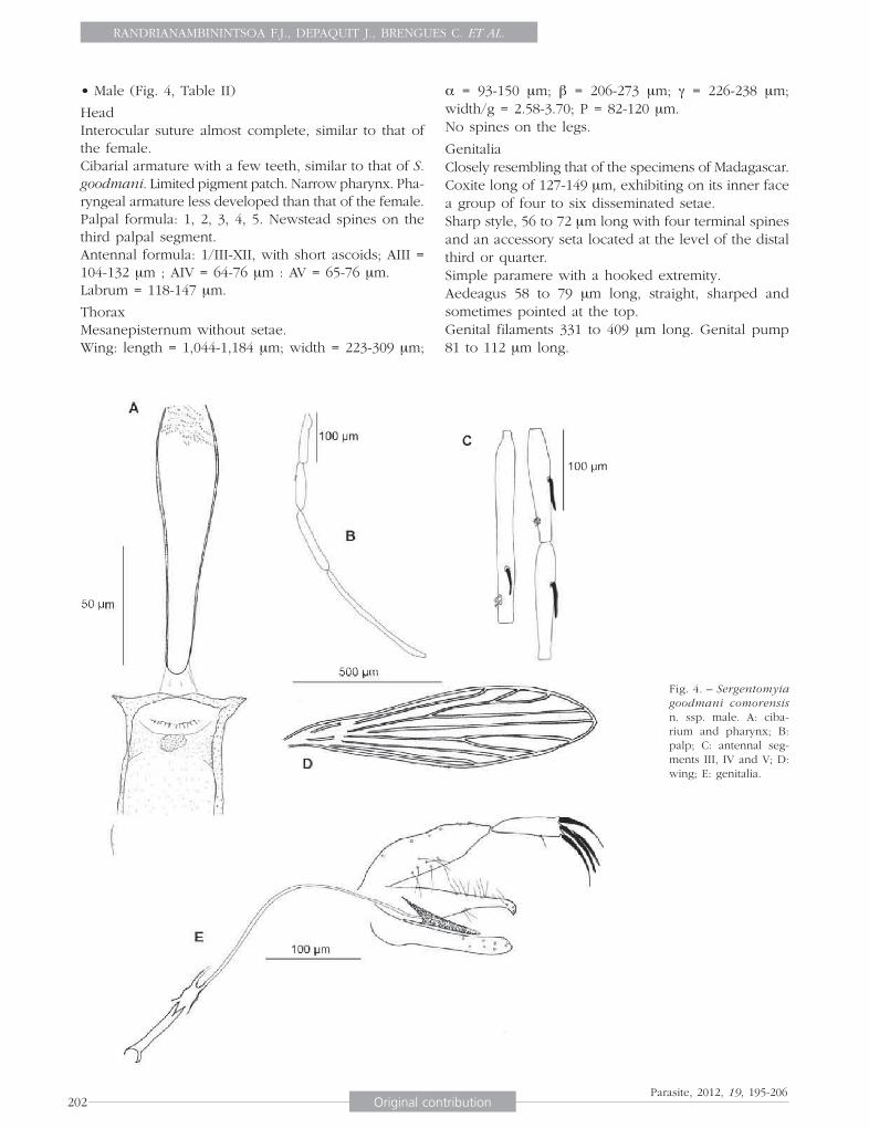
• Male (Fig. 4, Table II)
Head Interocular suture almost complete, similar to that of the female.Cibarial armature with a few teeth, similar to that of S. goodmani. Limited pigment patch. Narrow pharynx. Pha-ryngeal armature less developed than that of the female.Palpal formula: 1, 2, 3, 4, 5. Newstead spines on the third palpal segment. Antennal formula: 1/III-XII, with short ascoids; AIII = 104-132 μm ; AIV = 64-76 μm : AV = 65-76 μm. Labrum = 118-147 μm.
Thorax Mesanepisternum without setae. Wing: length = 1,044-1,184 μm; width = 223-309 μm;
α = 93-150 μm; β = 206-273 μm; γ = 226-238 μm; width/g = 2.58-3.70; P = 82-120 μm.No spines on the legs.
GenitaliaClosely resembling that of the specimens of Madagascar.Coxite long of 127-149 μm, exhibiting on its inner face a group of four to six disseminated setae.Sharp style, 56 to 72 μm long with four terminal spines and an accessory seta located at the level of the distal third or quarter.Simple paramere with a hooked extremity.Aedeagus 58 to 79 μm long, straight, sharped and sometimes pointed at the top.Genital filaments 331 to 409 μm long. Genital pump 81 to 112 μm long.
Fig. 4. – Sergentomyia goodmani comorensis n. ssp. male. A: ciba-rium and pharynx; B: palp; C: antennal seg-ments III, IV and V; D: wing; E: genitalia.
PAR2012-3_Randrianambinintsoa.indd 202 3/07/12 18:37:54

PHLEBOTOMINE SANDFLIES FROM COMOROS
203Original contributionParasite, 2012, 19, 195-206
Genital filaments/genital pump = 3.73 to 4.68.
Type locality: Zilimandzou, Grande Comore, at 11°42.583’S, 43°14.847’E and 26 m a.s.l.
DERIVATIO NOMINUM
S. pessoni is dedicated to our colleague Bernard Pesson.
The subspecies comorensis refers to the geographical origin of the studied population.
The holotype and the paratype of S. pessoni, and the holotype and nine paratypes (including four allotypes) of S. goodmani comorensis have been deposited in the Muséum national d’Histoire naturelle of Paris.
MOLECULAR RESULTS
The sequences studied were 497 to 503, depending on species and geographical origin (Table I). An alignment made by ClustalW included in the Bioedit package, and checked by eye, includes 507 posi- tions.
An analysis of the variable sites within the Vattiero-myia shows 68 variable sites (Fig. 5). An analysis of the variable sites within the Rondanomyia shows 51 variable sites (Fig. 6).
The pairwise distances between and within groups are showed on Table III.
The NJ tree based on cytochrome B sequences is given in Fig. 7. Within the Vattieromyia, the species Sergentomyia sclerosiphon, S. namo and S. anka are as strongly supported (bootstrap value: 100 %) as the two specimens caught in the Comoros islands.
Concerning the Rondanomyia, we observe a dicho-tomy between Malagasy and Comorian specimens. The Malagasy specimens are supported by 100 % bootstrap whereas Comorian specimens are supported by 88 % bootstrap. The specimen from Anjouan shows a haplotype differing by many nucleotides from the haplotypes of specimens caught in Grande Comore (Fig. 6). An additional NJ tree (data not shown) car-ried out without this specimen shows an increasing bootstrap value (96 %).
Fig. 5. – Variable positions observed wi th in spec ies belonging to the subgenus Vattie-romyia of Ser-gentomyia f o r c y t o ch rome b sequences.
Fig. 6. – Variable positions observed between Sergentomyia goodmani and for cytochrome b sequences.
PAR2012-3_Randrianambinintsoa.indd 203 3/07/12 18:37:54

RANDRIANAMBININTSOA F.J., DEPAQUIT J., BRENGUES C. ET AL.
204 Original contributionParasite, 2012, 19, 195-206
Fig. 7. – Neighbor-Joining tree based on cytochrome b sequences mtDNA. Bootstrap values indicated have been obtained after 1,000 replications.
S. goodmani madagascarensis
S. goodmani comorensis Grassomyia
S. pessoni
S. namo
S.scleosiphon
S. anka
Pairwise distance within taxa
S. goodmani madagascarensis
S. goodmani comorensis
Grassomyia
S. pessoni
S. namo
S. scleosiphon
S. anka
5.78
25.30
25.38
22.34
24.55
23.87
25.05
25.70
23.43
24.59
23.08
16.85
17.79
18.22
16.98
9.65
9.25
9.35
8.71
9.40 2.72
0.65
1.83
1.44
0.61
0.14
0.29
0.24
Table III. – Pairwise genetic distances (%) between and within taxa.
PAR2012-3_Randrianambinintsoa.indd 204 3/07/12 18:38:00
PHLEBOTOMINE SANDFLIES FROM COMOROS
205Original contributionParasite, 2012, 19, 195-206
DISCUSSION
Until an expected revision of its taxonomy has been done, the genus Sergentomyia França & Parrot, 1920 currently includes all the species
excluded from the other genera of the Old World (Phlebotomus, Idiophlebotomus, Chinius, Spelaeophle-botomus, Grassomyia, Parvidens, Spelaeomyia and Demeillonius). Concerning the Grassomyia, we refer to the position of Abonnenc & Léger (1976) conside-ring this group as a genus. The genus Sergentomyia is presently divided without any phylogenetical argument into six subgenera: Sergentomyia França & Parrot, 1920, Parrotomyia Theodor, 1948, Rondanomyia Theodor, 1948, Sintonius Nitzulescu, 1931, Capen-somyia Davidson, 1978 and Vattieromyia Depaquit, Léger & Robert 2007 (Duckhouse & Lewis, 1980; Léger & Depaquit, 1999; Depaquit, Léger & Robert, 2007). The males being difficult to identify, this classification is mainly based on the morphology of the female sper-mathecae. Moreover, some species are still ungrouped, due to their atypical spermathecae.
In 2007, Depaquit et al. defined the subgenus Vat-tieromyia by in the female i) the shape of the sper-mathecae and the sclerotized parts of their ducts, ii) the cibarial armature palisade-like, iii) an unusual antennal formula without ascoid on AIII. The males also have this original antennal formula shared only by Grassomyia spp. and S. majungaensis. At the light of S. pessoni n. sp., it appears that this species belong to the subgenus Vattieromyia according to the first two characters defining this group. This very original spermathecae has a more important weight than the original antennal formula. Consequently, we now exclude the antennal formula without ascoid on AIII from the definition of the subgenus Vattieromyia.
The morphological differences observed between the three Malagasy species belonging to the subgenus Vattieromyia and the two Comorian females appear to be of specific level. The females of S. pessoni n. sp. differ from all the Malagasy species by the presence of ascoids on the third antennal segment. Moreover, they are different from S. sclerosiphon by the number of cibarial teeth, from S. namo by the lack of cibarial ring and from S. anka by the shape of the cibarial armature and by the number of denticle ranges. The individualization of S. pessoni n. sp. is reinforced by the molecular data, also individualizing this new species.
Concerning S. goodmani, a strong molecular dicho-tomy is observed between the Malagasy specimens and the Comorian ones. However, there is little morphological divergence between Comorian and Malagasy specimens. This suggests an occurring spe-
ciation event linked with the insularity rather than a speciation fully achieved. Consequently, we consider S. goodmani comorensis n. ssp. as a subspecies of S. goodmani and not as a new species because both their molecular and morphological characters are more discreet and, linked to insularity, perfectly correspond to the concept of subspecies sensu Mayr et al. (1953). Although we are unable to test the interfecondity which could exist between Malagasy and Comorian populations. To individualize this subspecies, we consider i) in females the smaller number of cibarial teeth (15 to 21) and some morphometrical characters of the wing and the antennae and ii) in males, mainly the very small number of coxal setae in the Comorian specimens (four to six without real tuft versus about 15 in specimens from Madagascar).
We can’t identify the Grassomyia females caught during the present study at a specific level. The two specimens have been mounted for molecular biology processing (Table III) and consequently, their thoraces have been crushed for DNA extraction. The main characters used for the identification of the species of the genus Grassomyia are the absence/presence (and number) of setae on the mesanepimerum and the absence/presence of spines on the anterior and medium femurs (G. dreyfussi Parrot). According to the well developed pharyngeal armature of our specimens from Mohéli, their number of cibarial teeth (34 and 39), and to the characters revised by Abonnenc (1969, 1972), it cannot belong to G. inermis Theodor, 1938 (lack of pharyngeal armature, 19 to 25 cibarial teeth), or to G. squamipleuris Newstead, 1912 (48 to 55 ciba-rial teeth). Even though we have not observed the femurs of the specimens from Mohéli, the latter cannot be G. dreyfussi Parrot, 1933 (42 to 55 cibarial teeth and femurs with spines). Consequently, the Comorian specimens could be G. madagascarensis Abonnenc, 1969 (36 to 43 cibarial teeth), or G. ghesquierei Parrot, 1929 (27 to 37 cibarial teeth, lack of setae on the mesanepimerum). A complete revision of this group should be carried out in the future.
Nothing is known about the vectorial competence of the three species found in Comoros islands. Although no leishmaniasis cases have been reported, further stu-dies are needed to evaluate the risk of autochtonous transmission of Leishmania.
ACKNOWLEDGEMENTS
Funds had been provided from the FRB-AIRD-CNRS 2007 project “Biodiversité dans les Îles de l’Océan
Indien” and the GDRI Biodiversité et Développement Durable à Madagascar.
PAR2012-3_Randrianambinintsoa.indd 205 3/07/12 18:38:02
RANDRIANAMBININTSOA F.J., DEPAQUIT J., BRENGUES C. ET AL.
206 Original contributionParasite, 2012, 19, 195-206
REFERENCES
ABONNENC E. Sur Phlebotomus squamipleuris Newstead, 1912 et espèces voisines (Diptera : Psychodidae). Cahiers ORSTOM, série Entomologie Médicale et Parasitologie, 1969, 7, 307-323.
ABONNENC E. Les Phlébotomes de la région éthiopienne (Dip-tera, Psychodidae). Mémoires ORSTOM, 1972, 55, 239 p.
ABONNENC E. & LÉGER N. Sur une classification rationnelle des Diptères Phlebotomidae. Cahiers ORSTOM, Série Entomo-logie Médicale et Parasitologie, 1976, 14, 69-78.
ARTEMIEV M.M. Sandfly fauna (Phlebotominae) of the Peo-ple’s Republic of Mozambique [in Russian]. Parazitologiia, 1985, 19, 464-469.
BONFIELD J.K. & STADEN R. Experiment files and their appli-cation during large-scale sequencing projects. DNA Seq, 1996, 6, 109-117.
DAVIDSON I.H. New species of the subgenus Sergentomyia (Diptera: Psychodidae: Phlebotominae) from East Africa. Journal of the Entomological Society of Southern Africa, 1987, 50, 343-349.
DEPAQUIT J., FERTÉ H. & LÉGER N. Systématique moléculaire des Phlebotominae (Diptera, Psychodidae) : corollaires biogéographiques. Annales de la Société Française d’En-tomologie, 1999, 35 (S), 27-32.
DEPAQUIT J., FERTÉ H., LÉGER N. & ROBERT V. Les Phlébotomes de Madagascar. II – Description de la femelle de Phlebo-tomus (Anaphlebotomus) fertei Depaquit, Léger & Robert, 2002 ; description du mâle et redescription de la femelle de Phlebotomus (Anaphlebotomus) berentiensis (Léger et Rodhain, 1978) comb. nov. Parasite, 2004a, 11, 201-209.
DEPAQUIT J., LÉGER N. & ROBERT V. Première mention de Phlebotomus à Madagascar (Diptera : Psychodidae). Des-cription de Phlebotomus (Anaphlebotomus) fertei n. sp. et de Phlebotomus (Anaphlebotomus) huberti n. sp. Parasite, 2002, 9, 325-331.
DEPAQUIT J., LÉGER N. & ROBERT V. Les Phlébotomes de Madagascar. III – Description de Phlebotomus (Anaphlebo-tomus) fontenillei n. sp. Parasite, 2004b, 11, 261-265.
DEPAQUIT J., LÉGER N. & ROBERT V. Les Phlébotomes de Madagascar (Diptera – Psychodidae). V – Description de Sergentomyia majungaensis n. sp. Parasite, 2007, 14, 219- 223.
DEPAQUIT J., LÉGER N. & ROBERT V. Les Phlébotomes de Mada-gascar. VI – Un sous-genre nouveau (Vattieromyia) avec trois espèces nouvelles : Sergentomyia (V.) sclerosiphon, S. (V.) namo et S. (V.) anka. Parasite, 2008, 15, 15-26.
DUCKHOUSE D. & LEWIS D.J. Psychodidae, in: Catalogue of the Diptera of the Afrotropical region. Ctosskey R.W. (Ed.), British Museum (Natural History), Londres, 1980, 93-105.
EMERICK C.M. & DUNCAN R.A. Age progressive volcanism in the Comoro Archipelago, western Indian Ocean and implications for Somali plate tectonics. Earth and Plane-tary Science Letters, 1982, 60, 415-428.
ESSEGHIR S., READY P., KILLICK-KENDRICK R. & BEN-ISMAIL R. Mitochondrial haplotypes and phylogeography of Phle-botomus vectors of Leishmania major. Insect Molecular Biology, 1997, 6, 211-225.
FLOWER M.F.J. Evolution of basaltic and differentiated lavas from Anjouan, Comores Archipelago. Contributions to Mineralogy and Petrology, 1973, 38, 237-260.
HAJASH A. & ARMSTRONG R.L. Paleomagnetic and radiometric evidence for the ages of the Comores Island, west central Indian Ocean. Earth and Planetary Science Letters, 1972, 16, 231.
HALL T.A. BioEdit: a user-friendly biological sequence align-ment editor and analysis program for Windows 95/98/NT. Nucleic Acids Symposium Series, 1999, 41, 95-98.
LÉGER N. & DEPAQUIT J. Les phlébotomes, in: Les leishma-nioses. Dedet J.P. (Ed.), Ellipses, Paris, 1999, 89-108.
LÉGER N., DEPAQUIT J. & ROBERT V. Les Phlébotomes de Madagascar (Diptera – Psychodidae). IV – Description de Sergentomyia (Rondanomyia) goodmani n. sp. Rétablis-sement du sous-genre Rondanomyia Theodor. Parasite, 2005, 12, 51-57.
MAYR E., LINSLEY E.G. & USINGER R.L. (EDS). Methods and Principles of Systematic Zoology. McGraw-Hill, New York, 1953, 328 p.
NOUGIER J., CANTAGREL J.M. & KARCHE J.P. The Comoro archi-pelago in the western Indian ocean: volcanology, geo-chronology and geodynamic setting. Journal of African Earth Sciences, 1986, 5, 135-144.
PAVLOVSKY R. & DE SAINT-OURS J. Étude géologique de l’Ar-chipel des Comores. Service Géologique, Tananarive, Madagascar, 1953, 55 p.
TAMURA K., PETERSON D., PETERSON N., STECHER G., NEI M. & KUMAR S. MEGA5: molecular evolutionary genetics analysis using maximum likelihood, evolutionary distance, and maximum parsimony methods. Molecular Biology and Evolution, 2011, 28, 2731-2739.
Received on January 30th, 2012Accepted on April 23rd, 2012
PAR2012-3_Randrianambinintsoa.indd 206 3/07/12 18:38:02