fluids & lytes - algonquin college
TRANSCRIPT

ADVANCEDLIFE SUPPORT
PRECOURSEFLUIDS & ELECTROLYTES
SECTION FIVE
2005 Update byOntario Base Hospital Group Education Subcommittee
Copyright 2005, 1985 Ministry of Health and Long Term Care
ONTARIOBASE HOSPITAL GROUP

____________________________________________________________________________________________________OBHG Education Subcommittee 148
OBJECTIVES: FLUIDS & ELECTROLYTESThe objectives indicate what you should know, understand and be prepared to explain uponcompletion of this module. The self-assessment questions and answers will enable you to judgeyour understanding of the material.
Upon completion of this module, the student should be able to:
1. Name and describe the body fluid compartments.
2. Describe the distribution of body fluid in the intracellular and extracellular compartments.
3. Define and state the role of the following in fluid and electrolyte movement.
a) Diffusionb) Active Transportc) Osmosisd) Osmotic Pressuree) Oncotic Pressure (colloidal osmotic pressure)
4. State the approximate volume of body water in the normal adult.
5. State the distribution (as % of body weight) of fluid volumes in the normal adult and thepediatric patient.
6. Identify significant fluid loss (as % of body weight) in the adult and the pediatric patient.
7. Briefly explain the use of normal saline and 5% D/W in management of fluid depletion.
8. Describe briefly the role of the kidney (including the hormones involved) in the maintenanceof fluid and electrolyte balance.
9. Define:
a) Dehydrationb) Edemac) Volume depletion.
10. State the physiological roles of Na+, K+, Cl-, Ca++ and HCO3-.
If you have studied this subject previously, you may test your ability using the self-assessmentquestions at the end of each section. If you are able to obtain 90% or greater, you may choosenot to do the unit and merely review the section, or parts of sections, where weakness may exist.If you obtain less than 90%, it is recommended that the module be done in its entirety, stressingareas where more review is needed.

____________________________________________________________________________________________________OBHG Education Subcommittee 149
GLOSSARYACTIVE TRANSPORT The passage of molecules or ions across a semi-permeable
membrane against the concentration gradient, i.e. from an areaof lower to higher concentration of that substance; requiresexpenditure of energy.
ANION An ion with a negative (-) charge
ANOXIA Absence of oxygen in the body tissues
CATION An ion with a positive (+) charge.
COLLOID Molecules which are invisible to the naked eye but are too largeto dissolve and are therefore suspended in solution. In theblood, this refers to large protein molecules.
CONCENTRATIONGRADIENT
The tendency for substances to move from an area of higher tolower concentration with two solutions of differentconcentrations are separated by a semi-permeable membrane.
DEHYDRATION A condition in which there is a net loss of water from the fluidcompartments of the body.
DIFFUSION The movement of molecules of ions from an area of higher tolower concentration of that substance, i.e. with theconcentration gradient; a passive transport system.
EDEMA A condition in which the interstitial (tissue) spaces contain anexcessive amount of extracellular fluid; characterized byswelling and puffiness of the tissues.
ELECTROLYTE A substance capable of dissociating into ions and which insolution will conduct an electrical current.
HOMEOSTASIS The maintenance of a steady state in the body, and a relativelyconstant concentration of ions, pH, and osmotic pressure in thevarious body fluids.
EXTRACELLULARY FLUID(ECF)
A solution, primarily saline (NaCl), which occupies the areasoutside the cells. (“extra” = outside; “cellular” = the cells)
INTRACELLULAR FLUID (ICF) A solution of water, electrolytes, and proteins which circulateswithin the cells (“intra” = inside; “cellular” = the cells)

____________________________________________________________________________________________________OBHG Education Subcommittee 150
GLOSSARY
INTERSTITIAL FLUID (ISF) Extracellular fluid composed of water and electrolytes whichcirculates between and around the cells. (“inter” = between;“stitial” – spaces)
INTRAVASCULAR FLUID (IVF) Extracellular fluid composed of water, electrolytes and proteinswhich is contained within the blood vessels. (“intra” = inside;“vascular” = blood vessels)
METABOLIC ACIDOSIS A disturbance of the acid-base balance in which the body pHdecreases (becomes more acidic) due to decreased bloodlevels of bicarbonate ions.
METABOLIC ALKALOSIS A disturbance of the acid-base balance in which the body pHincreases (becomes more alkaline) due to increased bloodlevels of bicarbonate ions.
ONCOTIC PRESSURE Osmotic pressure due to the presence of plasma colloids.
OSMOSIS The passage of water (solvent) across a semi-permeablemembrane from a more dilute solution (lower soluteconcentration, higher water concentrations) to a moreconcentrated solution (higher solute concentration, lower waterconcentration), i.e. with the concentration gradient; a passivetransport system.
OSMOTIC PRESSURE The amount of pressure that would be required to prevent themovement of water (osmosis) across semi-permeablemembrane when two solutions of different concentrations areseparated by that membrane.
PASSIVE TRANSPORT The movement of ions or molecules in the direction of theconcentration gradient, thereby requiring no energyexpenditure.
SEMI-PERMEABLEMEMBRANE
A membrane that only permits the passage of certainmolecules and not others.
SKIN TURGOR The degree of tension of the skin.
SOLUTE The substance/compound that is dissolved in a solution.
SOLVENT The liquid in which another substance (the solute) is dissolvedto form a solution.
____________________________________________________________________________________________________OBHG Education Subcommittee 151
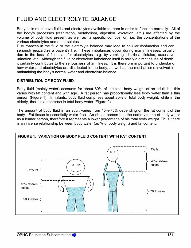
FLUID AND ELECTROLYTE BALANCEBody cells must have fluids and electrolytes available to them in order to function normally. All ofthe body's processes (respiration, metabolism, digestion, excretion, etc.) are affected by thevolume of body fluid present as well as its specific composition, i.e. the concentrations of thevarious electrolytes and other solutes.Disturbances in the fluid or the electrolyte balance may lead to cellular dysfunction and canseriously jeopardize a patient's life. These imbalances occur during many illnesses, usuallydue to the loss of fluids and/or electrolytes, e.g. by vomiting, diarrhea, fistulas, excessiveurination, etc. Although the fluid or electrolyte imbalance itself is rarely a direct cause of death,it certainly contributes to the seriousness of an illness. It is therefore important to understandhow water and electrolytes are distributed in the body, as well as the mechanisms involved inmaintaining the body's normal water and electrolyte balance.
DISTRIBUTION OF BODY FLUID
Body fluid (mainly water) accounts for about 60% of the total body weight of an adult, but thisvaries with fat content and with age. A fat person has proportionally less body water than a thinperson (Figure 1). In infants, body fluid comprises about 80% of total body weight, while in theelderly, there is a decrease in total body water (Figure 2).
The amount of body fluid in an adult varies from 45%-75% depending on the fat content of thebody. Fat tissue is essentially water-free. An obese person has the same volume of body wateras a leaner person, therefore it represents a lower percentage of his total body weight. Thus, thereis an inverse relationship between body water (as % of body weight) and fat content.
FIGURE 1: VARIATION OF BODY FLUID CONTENT WITH FAT CONTENT
32% fat
18% fat-freesolids
50% water
4% fat
26% fat-freesolids
70% water

____________________________________________________________________________________________________OBHG Education Subcommittee 152
The thin man pictured in Figure 1 contains less fat and therefore more water in proportion to histotal body weight. The fat man contains proportionally less water as a percentage of his bodyweight because of the large amount of adipose (fat) tissue present.
In the newborn, body fluid comprises about 80% of the total body weight, with more than onehalf of this as extracellular fluid. As the child grows, proportions and total volume graduallyapproximate the adult fluid distribution.
Water is found both inside and outside the cells, and is usually considered to be divided intotwo main "compartments"* or spaces. Of the total body water, approximately 2/3 of the wateris contained inside the cells themselves. This water is collectively referred to as theINTRACELLULAR FLUID (ICF), and is said to be found in the intracellular compartment.The remaining 1/3 of total body water is distributed throughout the body as theEXTRACELLULAR FLUID (ECF). This refers to all the fluid found outside the cells and is saidto be located in the extracellular compartment. The extracellular fluid is further divided into twomain categories. The INTRAVASCULAR FLUID (IVF) is that part of the extracellular fluidlocated within the blood vessels as blood plasma. The remainder of the extracellular fluid,referred to as the INTERSTITIAL FLUID (ISF), is found between the cells and the bloodvessels and in tissue spaces.
FIGURE 2: VARIATION OF BODY FLUID CONTENT WITH AGEIntracellular fluid
Extracellular fluid
100
80
60
40
20
0Newborn One year Adult
Body
wei
ght (
%)

____________________________________________________________________________________________________OBHG Education Subcommittee 153
* It is important to understand that the term "compartment" does not refer to one specificcontained space, like an organ, but rather it is a convenient abstraction used to describewhere fluid is found throughout the body.
The relative distribution of body fluid is summarized as follows:
The average adult has approximately 42 liters of body water, which represents about 60%of his/her total body weight.
Of this 60%, 40% (28 L) is present as intracellular fluid (ICF) and 20% (14 L) is present asextracellular fluid(ECF).
The extracellular fluid can be further divided as intravascular fluid or plasma 5%, (3.5 L)and interstitial fluid, 15% (10.5 L).
FIGURE 3: BODY FLUID COMPARTMENTS
Intravascular fluid (IVF) 8%
Interstitial fluid (ISF) 25%
Extracellular fluid (ECF) 33%
Intracellular fluid (ICF) 67%

____________________________________________________________________________________________________OBHG Education Subcommittee 154
MOVEMENT OF FLUIDS AND ELECTROLYTES
The various fluid "compartments" of the body are separated by semi-permeable (or selectively-permeable) membranes. These membranes allow some molecules to pass through freely,while restricting or preventing the passage of other molecules. In health, the total volume andcomposition of the fluid in each compartment remains remarkably stable in spite of the fact thatthe water and solute molecules are in constant motion, moving from one compartment toanother. There are several mechanisms by which this movement may occur.
DIFFUSION
Many solute molecules move between fluid compartments by means of simple diffusion. Thisterm refers to the natural tendency of all substances to move about in a solution in an effort todistribute themselves evenly throughout the solution. If a membrane is permeable to a certainsubstance, i.e. if it allows free passage of that substance across it, then those molecules willmove through the membrane in an effort to equalize their concentration on either side of themembrane.
The direction of movement in diffusion is said to be "with the concentration gradient". Thismeans that molecules follow the natural tendency to equalize concentration by moving from anarea of higher concentration to an area of lower concentration of that substance. Because itfollows the "natural" direction of flow, no energy is required for simple diffusion to take place.Therefore, diffusion is referred to as a passive transport system.
Diffusion, then, is a general term referring to the passive movement of any substance froman area of higher to lower concentration of that substance. When discussing biologicalsystems, the term diffusion is usually used loosely in reference to the movement of solutemolecules. Of course, solvent molecules diffuse as well. In fact, in the human body, themajority of molecules diffusing across semi-permeable membranes are solvent molecules, e.g.
FIGURE 4: DIFFUSION
High Concentration Membrane Low Concentration

____________________________________________________________________________________________________OBHG Education Subcommittee 155
water. Because the diffusion of water molecules is such an important process, it is given thespecial name of osmosis.
OSMOSIS
In osmosis, water moves across a semi-permeable membrane from the more dilute solution(lower solute concentration) to the more concentrated solution (higher solute concentration). Itmay appear at first glance that this is a contradiction of the earlier statement that diffusion ismovement from an area of higher to lower concentration, i.e. with the concentration gradient.However, remember that the definition refers to the concentration of the substance that isdoing the diffusing.
The process of osmosis does occur in the direction of the concentration gradient since water ismoving from an area of higher concentration of water (more dilute solution; lower soluteconcentration) to an area of lower concentration of water (more concentrated solution; highersolute concentration). Because movement is with the concentration gradient, osmosis is alsoa passive transport system, i.e. no energy required. Therefore, osmosis refers specifically tothe movement of water (solvent) molecules across a semi-permeable membrane from an areaof higher to lower concentration of water (lower to higher concentration of solute). Osmosis isillustrated in Figures 5 and 6.
In Figure 5, solution “A” has fewer solute particles per unit volume than solution “B”.Therefore, solvent molecules move from solution A to solution B. Osmosis occurs until thenumber of solute particles per unit volume is the same on both sides of the membrane.
The movement of solvent results in diluting the number of solute particles per unit volume insolution B. When the number of solute particles per unit volume is the same on both sides ofthe membrane, the movement of solvent molecules occurs equally in both directions, i.e. adynamic equilibrium exists (Figure 6). The two solutions are said to be isotonic to each other.
FIGURE 5: OSMOSIS
Semi-permeable Membrane
Solution A Solution B
Solvent

____________________________________________________________________________________________________OBHG Education Subcommittee 156
Osmosis and diffusion occur simultaneously. The processes work together in an attempt toequalize the concentrations and balance the osmotic pressures of the two solutions by movingsolute out of the more concentrated solution and water out of the more dilute solution.
When two solutions of differing concentrations are separated by a semi-permeable membranethere is a 'pulling force' which draws water through to the more concentrated side, i.e. highersolute concentration. The amount of pressure that would be required to prevent thismovement of water is referred to as the OSMOTIC PRESSURE of a solution.
The osmotic pressure is determined by the number of particles of solute on the moreconcentrated side, relative to the side with the lower concentration. The greater the number ofparticles in the concentrated solution, the more 'pull' there will be to draw the water through themembrane and therefore, the greater the pressure required to prevent that movement.
In Figure 7, solution A has fewer solute particles per unit volume (lower osmotic pressure) thansolution B. Solvent will move from solution A to solution B, unless pressure is applied tosolution B to prevent osmosis.
FIGURE 6: TWO SOLUTIONS IN DYNAMIC EQUILIBRIUM
Semi-permeable Membrane
Solution A
Solution B
Solvent

____________________________________________________________________________________________________OBHG Education Subcommittee 157
The osmotic pressure due to plasma colloids (protein molecules mainly) is specifically referredto as the COLLOIDAL OSMOTIC PRESSURE or ONCOTIC PRESSURE.
ACTIVE TRANSPORT
The processes of diffusion and osmosis follow the natural tendency of molecules or ions tomove from areas of higher to areas of lower concentrations in an attempt to equalizeconcentrations. However, it is often necessary for the body to move substances in theopposite direction, against the concentration gradient, in order to maintain a higherconcentration of a substance on one side of a membrane. The process which movessubstances against the concentration gradient is referred to as ACTIVE TRANSPORT.
In order to move substances from an area of lower concentration to an area of higherconcentration, a carrier substance* is often needed (Figure 8). For example, insulin acts as acarrier substance to transport glucose across the cell membrane, from the blood (ECF) into thecell (ICF).
Energy, in the form of adenosine triphosphate (ATP), must be expended to facilitate thetransport (hence the term "active transport").
FIGURE 7: PREVENTION OF OSMOSIS BY OSMOTIC PRESSURE
Semi-permeableMembrane
Solution A Solution B
OsmoticPressure

____________________________________________________________________________________________________OBHG Education Subcommittee 158
Therefore, active transport refers to the movement of molecules or ions from an area of lowerconcentration to an area of higher concentration of that substance, against the concentrationgradient. Some important substances actively transported in the body include ions of sodium,potassium, chloride, hydrogen, calcium and iron, amino acids, and some sugars.
All of the processes discussed are responsible for carrying the molecules of water, foods,gases, wastes, and many kinds of ions between compartments, in and out of the body's cells.Together, they act to maintain a proper chemical and osmotic balance between theintracellular and extracellular fluids.
* The carrier molecule undergoes a structural change as it picks up the substance to betransported. This change requires energy.
FLUID LOSS
In order to maintain fluid balance in the body, the daily intake of fluids must match the dailyoutput. The kidneys excrete the largest quantity of fluid, but fluid also leaves the body throughthe lungs, skin and gastrointestinal tract. Fluid is replenished in the body by ingestion ofliquids and by digestion of foods.
There is a basic minimum daily requirement for fluid of approximately 2500 mL. In thehealthy individual the total intake by all routes is equal to the total output by all routes.
Injury or disease can cause abnormal increases in water loss and seriously upset the fluid andelectrolyte balance of the body. Water loss via the lungs and skin is increased in conditionscausing fever or an increased respiratory rate, in hot or dry environments and in skin injuries,e.g. burns. Water loss via the kidneys is increased in conditions involving increased soluteexcretion such as diabetes mellitus and in conditions in which there is a decrease inantidiuretic hormone (ADH) levels. Serious water and electrolyte loss can also occur from theG.I. tract in the case of severe vomiting or diarrhea.
FIGURE 8: ACTIVE TRANSPORT
Carrier Molecule
High Concentration
Low Concentration ENERGY

____________________________________________________________________________________________________OBHG Education Subcommittee 159
Clinical vignette
1. Normal saline is used to fill the intravascular space for patients who are hypovolemic or hypotensive.Because normal saline is isotonic, it will equilibrate in both the intravascular and extravascular space.Hypovolemic patients have first lost fluid from the intravascular space. Fluid is then shifted from theextravascular space into the intravascular space in an attempt to maintain blood pressure. If necessary,intracellular fluid will shift into the extracellular space as well. In fluid resuscitation, only one third ofnormal saline will remain in the vascular space.
2. 5% dextrose in water (D5W) is a hypotonic solution and therefore is not held within the intravascularspace. Rather it diffuses into the extravascular space where the sugar is metabolized by the cells. It isused as a vehicle for administering certain drugs and should not be used in patients who require fluidresuscitation.
As previously mentioned, the total fluid volume of the average adult is approximately 42 liters -28 liters as intracellular fluid (ICF) and 14 liters as extracellular fluid (ECF). Any change in theamount of composition of these fluids may cause serious problems. In the adult, a water lossof 5% by body weight (≈ 2 L) is considered unfavourable, a loss of 10% ( ≈ 4 L) is consideredserious, and a loss of 20% ( ≈ 8 L) is usually fatal.
Infants and small children need a proportionately larger fluid intake and output in relation toadults since they have a greater body surface area in proportion to mass and an increasedmetabolic rate. Also, infants have immature kidneys which require proportionately more waterto excrete metabolic wastes. The younger the child, the smaller his fluid reserve, andtherefore the greater his vulnerability to water deficit. Volume depletion in infants and smallchildren can also be estimated based on body weight loss. A water loss of 2-4% by bodyweight is considered mild, a loss of 5-9% is considered moderate, and a water loss of over10% is considered to be severe.
____________________________________________________________________________________________________OBHG Education Subcommittee 160
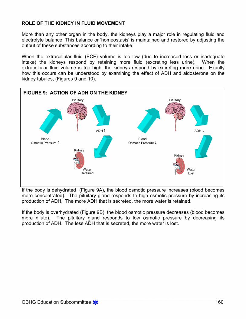
ROLE OF THE KIDNEY IN FLUID MOVEMENT
More than any other organ in the body, the kidneys play a major role in regulating fluid andelectrolyte balance. This balance or 'homeostasis' is maintained and restored by adjusting theoutput of these substances according to their intake.
When the extracellular fluid (ECF) volume is too low (due to increased loss or inadequateintake) the kidneys respond by retaining more fluid (excreting less urine). When theextracellular fluid volume is too high, the kidneys respond by excreting more urine. Exactlyhow this occurs can be understood by examining the effect of ADH and aldosterone on thekidney tubules, (Figures 9 and 10).
If the body is dehydrated (Figure 9A), the blood osmotic pressure increases (blood becomesmore concentrated). The pituitary gland responds to high osmotic pressure by increasing itsproduction of ADH. The more ADH that is secreted, the more water is retained.
If the body is overhydrated (Figure 9B), the blood osmotic pressure decreases (blood becomesmore dilute). The pituitary gland responds to low osmotic pressure by decreasing itsproduction of ADH. The less ADH that is secreted, the more water is lost.
FIGURE 9: ACTION OF ADH ON THE KIDNEY
BloodOsmotic Pressure ↑
ADH ↑
WaterRetained
Kidney
Pituitary Pituitary
Kidney
BloodOsmotic Pressure ↓
ADH ↓
WaterLost

____________________________________________________________________________________________________OBHG Education Subcommittee 161
If the body is dehydrated, there is a resultant decrease in blood volume and blood flow. Thisdecrease in blood volume and flow is sensed by specialized cells in the kidney and results inthe increased secretion of aldosterone from the adrenal glands. Aldosterone causes thekidneys to retain sodium (and with it, water) to correct the volume deficit. The reverse processoccurs in the case of overhydration, resulting in the loss of sodium and water by the kidney.
FIGURE 10: ACTION OF ALDOSTERONE ON THE KIDNEY
BloodVolume ↓
Aldosterone
Kidney
Renal Retention ofWater and Na+

____________________________________________________________________________________________________OBHG Education Subcommittee 162
CONDITIONS OF FLUID IMBALANCEVOLUME DEPLETION
Volume depletion is the loss of water from all fluid compartments of the body. As the degree ofvolume depletion increases, fluid is lost first from the interstitial fluid (ISF), then theintravascular fluid (IVF; plasma), and finally from the intracellular fluid (ICF). Volume depletionusually results from either an inadequate fluid intake or an excessive fluid loss. Some of theseconditions are listed in Table 1.
TABLE 1
CONDITIONS LEADING TO VOLUME DEPLETIONINADEQUATE FLUID INTAKE EXCESSIVE FLUID LOSS
inability to swallow coma unavailability of water extreme debilitation and illness mechanical devices and intubation
chronic vomiting severe diarrhea diabetes mellitus diabetes insipidus kidney failure fever hemorrhage hyperventilation drainage from wounds and suctioning burns
Two entirely different disorders can occur when one is assessing a patient with a fluid and/orelectrolyte disorder:
One is a disorder of total volume, e.g. volume overload, volume depletion The other is a disorder of water balance, e.g. water relative to salt, as in hyponatremia
(↓Na+) or hypernatremia (↑Na+).
The diagnosis of volume status is made clinically, while the diagnosis of water balancedisorders is made in a laboratory.
Dehydration is a vague term but in its strictest sense refers to a loss of water from the body.
When assessing a patient clinically we are concerned about his volume status and a patient istherefore more accurately described as being volume depleted rather than dehydrated (waterdepleted).

____________________________________________________________________________________________________OBHG Education Subcommittee 163
Clinical vignetteThe most reliable way of confirming volume depletion is by testing postural or orthostatic vital signs. Thepulse and blood pressure are tested first with the patient in a supine position and then in an upright position.If volume depletion is present, the pulse will increase and the blood pressure will decrease when the patientis put in an upright position.
Volume depletion can be recognized by watching for changes in body temperature (increased),postural vital signs (decreased blood pressure, increased pulse rate), skin turgor, and byobserving the appearance of the skin and the mucous membranes of the mouth for dryness.
Signs and symptoms of volume depletion to watch for include:
thirst dry skin dry mucous membranes sunken eyes (especially in infants) sunken fontanel (infants) low grade fever increased pulse rate poor skin turgor (turgor in the skin of the forehead may be a better gauge of fluid status
than the back of the hand in the elderly) hypotension altered mental status
Especially susceptible to volume depletion are individuals with a relatively low proportion oftotal body water, such as infants, the elderly, and the obese. Volume depletion is potentiallyserious clinically because it involves not only a change in water balance, but a change inelectrolyte balance as well (most importantly - sodium, potassium, chloride and bicarbonate).
VOLUME OVERLOAD
Edema refers to the presence of excess extracellular fluid (from plasma) in the interstitialspaces or fluid compartment. This abnormal accumulation of fluids produces noticeableswelling or "puffiness" in some tissues, particularly in the lower extremities or dependant areasof the body. By the time edema is noticeable, the adult patient will have accumulated about4.5 kg (≈ 4.5 L) of extra fluid. Any condition which results in excessive retention of salt andwater or in a decrease in plasma proteins (especially albumin) can lead to edema. In eachcase, excess water moves from the plasma into the tissues in an attempt to equalize theosmotic pressure. Some conditions leading to edema are listed in Table 2.

____________________________________________________________________________________________________OBHG Education Subcommittee 164
TABLE 2CONDITIONS LEADING TO EDEMA
LOW PROTEIN LEVELS EXCESSIVE FLUID/SALT RETENTION
malnutrition (↓ intake)
liver disease (↓ synthesis) kidney disease (loss in urine) e.g.
nephrosis severe burns
heart disease e.g. congestive heart failure kidney disease pregnancy anti-diuretic medications
PHYSIOLOGICAL ROLES OF ELECTROLYTESBody fluids contain two types of dissolved substances:
those that dissociate or ionize in solution, called electrolytes, e.g. NaCl- those that do not dissociate, called non-electrolytes, e.g. glucose
The term electrolyte refers specifically to the fact that solutions of these substances willconduct an electric current.
Each of the fluid compartments of the body has a particular composition by electrolytes whichare unique to that fluid. The principal extracellular electrolytes are ions of sodium, chloride,calcium, and bicarbonate, while the principal intracellular electrolytes are ions of potassium,magnesium, and phosphates and ionized proteins. In the extracellular fluid (plasma and ISF),the main cation is sodium and the main anion is chloride. Inside the cell (ICF) the main cationis potassium and the main anion is monohydrogen phosphate. The distribution of theseelectrolytes is summarized in Table 3.

____________________________________________________________________________________________________OBHG Education Subcommittee 165
TABLE 3RELATIVE DISTRIBTION OF ELECTROLYTES
EXTRACELLULAR FLUIDCATIONS (+) ANIONS (-)
Sodium *Na+ Chloride *Cl-
Calcium Ca++ BicarbonateHCO3-Potassium K+ Protein-Magnesium Mg++ Biphosphate PO4
2-
Sulphate SO42-
Organic acids-
INTRACELLULAR FLUIDCATIONS (+) ANIONS (-)
Potassium *K+ Biphosphate *HPO42-
Magnesium Mg2+ Protein-Sodium Na+ Sulphate SO4
2-
BicarbonateHCO3-Chloride Cl-
* Major or dominant anions and cations in each fluid.
In clinical usage, the term electrolyte is used to refer to the four ions in plasma that mostgreatly affect water and acid-base balance (Na+, K+, Cl- and HCO3
-), and increasingly, Ca++.These electrolytes profoundly influence water distribution, osmotic pressure, acid-basebalance and neuromuscular irritability. Each has its own special functions in the body andalthough some play larger roles than others, all are necessary for the maintenance of health.
Sodium (Na+) is the major extracellular cation and therefore plays a major role in determiningextracellular fluid volume and osmotic pressure. Because it represents about 90% of all theextracellular cations, it is especially important in the transmission of electrical impulses in nerveand muscle fibers. Since sodium ions are exchanged for hydrogen ions in the acidification ofthe urine, sodium has a role in pH regulation. It is also involved to some degree in regulatingcell membrane permeability. Excessive levels of sodium ions in the blood (hypernatremia)produce symptoms of extreme muscle irritability, dry, sticky mucous membranes, flushed skinand intense thirst; while symptoms of low blood sodium levels (hyponatremia) include lethargy,muscle weakness, edema, decreased urinary output, and mental confusion leading to coma.
Potassium (K+) is the major intracellular cation, and it plays a major role in the regulation ofmuscle irritability. Potassium ions are crucial to the normal functioning of the heart muscle byallowing the muscle to contract properly and to rest properly in the diastolic phase (betweencontractions). K+ is also somewhat associated with pH balance in that it is freely exchanged

____________________________________________________________________________________________________OBHG Education Subcommittee 166
with hydrogen ions when the body is responding to an acid-base disturbance. Both high andlow blood potassium levels are potentially threatening because of their effect on cardiacmuscle. High levels of potassium in the blood (hyperkalemia) cause heart arrhythmias(progression: peaked T → flattening of the P waves → QRS widening → slowing of the heartrate, heart blocks, etc), weakening of cardiac contractility and eventually heart failure. Lowblood potassium levels (hypokalemia) produce symptoms of cardiac excitability includingtachycardia and ectopy (e.g. premature ventricular complexes), improper heart contractions,poor circulation, muscle cramps and eventually weakness and loss of muscle tone.Hypokalemia may also lead to cardiac arrest (usually due to anoxia created by paralysis of therespiratory muscles).
Chloride (Cl-) is the major extracellular anion, and like sodium, plays an important role in themaintenance of extracellular fluid volume and osmotic pressure. The excretion andreabsorption of chloride ions is also related to acid-base regulation.
Calcium (Ca++) is well-known for its important role in the formation of bones and teeth. It is acrucial factor in blood coagulation and in the activation of some enzymes. It also assists in thetransmission of nerve impulses and the proper contraction of muscle fibers. It acts to decreaseneuromuscular activity.

____________________________________________________________________________________________________OBHG Education Subcommittee 167
Bicarbonate (HCO3-) acts as a buffer-base in the bicarbonate/carbonic acid buffer system.
This is the most important buffer in the blood and plays the key role in maintaining the body'sacid-base balance. Bicarbonate ions (HCO3
-) react with free hydrogen ions (H+) in the bodyfluids to form undisassociated carbonic acid (H2CO3
-). In this way, acid (H+) can be neutralizedand carried to the kidneys for excretion. High levels of bicarbonate in the blood lead to acondition of metabolic alkalosis and symptoms of nausea, vomiting, diarrhea, confusion,irritability and agitation leading to coma. Low blood levels of bicarbonate lead to a condition ofmetabolic acidosis and the accompanying symptoms of headache, drowsiness, nausea,vomiting, diarrhea, stupor, and eventually, coma.

____________________________________________________________________________________________________OBHG Education Subcommittee 168
ADVANCED LIFE SUPPORTPRECOURSE
FLUIDS AND ELECTROLYTES
SELF-ASSESSMENT
MARKS
1. Differentiate between:
[2] a) intracellular fluid (ICF) and extracellular fluid (ECF)
[2] b) intravascular fluid (IVF) and interstitial fluid (ISF)
2. Define the terms:
[1] a) osmotic pressure
[1] b) oncotic pressure.

____________________________________________________________________________________________________OBHG Education Subcommittee 169
[3] 3. Complete the following chart:
Diffusion Osmosis Active Transport
Type of substancemoving
Direction of movement
Energy requirements
[4] 4. Fill in the blanks:
Body fluid accounts for about (a) % of the total body weight of an
adult. Expressed in liters, the total fluid volume of the average adult is approximately
(b) (L). The amount of body fluid as a percentage of total
body weight varies from person to person with (c) and with
(d) . In infants, body fluid comprises about (e)
% of total body water.
In disease conditions, water loss can be estimated based on percentage of body weight
lost. In an adult, a fluid loss of (f) % is considered serious, and a loss of
20% is usually fatal. Infants and small children are especially susceptible to volume
depletion due to their smaller fluid reserve. In an infant, a fluid loss of (g)
% is considered moderate, while a loss of (h) % is considered to be
severe.
[1] 5. Name the two main hormones which act on the kidney to regulate fluid andelectrolyte balance.

____________________________________________________________________________________________________OBHG Education Subcommittee 170
[3] 6. Define the terms:
a) dehydration
b) edema
c) volume depletion.
[2] 7. Complete the following chart:
EXTRACELLULAR FLUID INTRACELLULAR FLUID
Major Cation
Major Anion
[8] 8. State two physiological functions for each of the following:
a) sodium
b) potassium

____________________________________________________________________________________________________OBHG Education Subcommittee 171
c) chloride
d) calcium
[1] 9. State the major physiological function of bicarbonate.
[2] 10. Explain the rationale for using normal salinerather than 5% D/W for infusion on thevolume-depleted patient.
30 Total

____________________________________________________________________________________________________OBHG Education Subcommittee 172
ADVANCED LIFE SUPPORTPRECOURSE
FLUIDS AND ELECTROLYTES
SELF-ASSESSMENT ANSWERS
1. a) Intracellular fluid: The fluid which circulates inside the body's cells.
Extracellular fluid: The fluid which circulates outside the body's cells.
b) Intravascular fluid: The portion of extracellular fluid located within the bloodvessels (blood plasma).
Intestinal fluid: The portion of extracellular fluid found between the cells andblood vesels and in the tissue spaces.
2. a) Osmotic Pressure: The amount of pressure that would be required to prevent themovement of water (osmosis) across a semi-permeable membrane when twosolutions of different concentrations are separated by that membrane.
b) Oncotic Pressure: The osmotic pressure due specifically to the presence ofplasma colloids, e.g. proteins. Also referred to as "Colloidal Osmotic Pressure".
1. (1 mark for each column correct).
Diffusion Osmosis Active Transport
Type of substancemoving
Molecules & ions(usually solute)
Water (solvent) Molecules & ions(solute)
Direction of movement With the concentrationgradient; from area ofhigher to lower conc.Of that substance.
With the concentrationgradient; from area ofhigher to lower conc.of water (i.e. frommore dilute to moreconcentrated solution)
Against theconcentration gradient;fram areof lower tohigher conc. Of thatsubstance.
Energy requirements Passive process (noenergy required)
Passive process (noenergy required)
Active process(energy is required)
4. a) 60%

____________________________________________________________________________________________________OBHG Education Subcommittee 173
b) 42 Lc) age (or fat content)d) fat content (or age)e) 80%f) 10%g) 5-9%h) over 10%
5. antidiuretic hormone (ADH) and aldosterone.
6. a) Dehydration: A condition in which there is a net loss of water from the fluidcompartments of the body.
b) Edema: A condition in which the interstitial (tissue) spaces contain an excessiveamount of extracellular fluid; characterized by swelling and puffiness of thetissues.
c) Volume depletion: A clinical term, referring to findings which indicate thatcirculatng volume is diminished.
7.
EXTRACELLULAR FLUID INTRACELLULAR FLUID
Major Cation Na+ K+
Major Anion HPO4-
8. a) Sodium (any two of):
maintenance of osmotic pressure and ECF volume ` transmission of nerve andmuscle impulses
pH regulation (acidification of urine) regulation of cell membrane permeability
b) Potassium:
regulation of muscle irritability (especially cardiac muscle) pH regulation (acidification of urine)
c) Chloride:
maintenance of osmotic pressure and ECF volume ` acid-base regulation(excretion with NH4+ ions and reabsorption alternately to HCO3- ions)

____________________________________________________________________________________________________OBHG Education Subcommittee 174
d) Calcium (any two of)
formation of bones and teeth blood coagulation enzyme activation transmission of nerve impulses contraction of muscle fibers control of neuromuscular activity
9. Bicarbonate - part of the major blood buffer system, (i.e. the bicarbonate/carbonic acidbuffer system)
10. More normal saline than 5% D/W stays in the intravascular space, since normal salineis isotonic and 5% D/W becomes hypotonic once the dextrose component is taken upby the cells. Fluid remaining in the intravascular space then increases the circulatingvolume.

____________________________________________________________________________________________________OBHG Education Subcommittee 175
ADVANCED LIFE SUPPORTPRECOURSE
FLUIDS AND ELECTROLYTES
EVALUATION
Upon completion of this module, please fill in and return this form to your base hospitalco-ordinator.
Your comments will help to ensure that this unit is a useful learning module. Please indicate anyproblems that you may have encountered. All suggestions for improvement are welcomed.
1. How long did it take to complete this module? Please estimate.
Reading hoursSelf assessment hoursTotal time hours
2. Were the objectives of the module clearly stated?
[ ] yes [ ] noIf no, please comment.
3. Did you see any of the resource materials?
[ ] yes [ ] noIf yes, which items
Were they helpful?
4. Were the reference notes adequate?
[ ] yes [ ] noIf no, please comment.
5. Were the reference notes easy to follow?
____________________________________________________________________________________________________OBHG Education Subcommittee 176
[ ] yes [ ] noIf no, please comment.
6. Were the examples provided satisfactory?
[ ] yes [ ] noIf no, please comment.
7. Were any of the self-assessment questions poorly worded?
[ ] yes [ ] noIf yes, please specify.
1. Was the level of the module satisfactory for your program of study?
[ ] yes [ ] noIf no, please comment.
Base Hospital
9. General comments or suggested improvements.