following optogenetic dimerizers and quantitative prospectspages.jh.edu/~inouelab/pdfs/r6.pdf ·...
TRANSCRIPT
Biophysical Perspective
Following Optogenetic Dimerizers and QuantitativeProspects
Jacqueline Niu,1,* Manu Ben Johny,1 Ivy E. Dick,1,5 and Takanari Inoue1,2,3,4,*1Department of Biomedical Engineering, School of Medicine, 2Department of Cell Biology, School of Medicine, and 3The Center for CellDynamics, Institute for Basic Biomedical Sciences, The Johns Hopkins University, Baltimore, Maryland; 4Precursory Research for EmbryonicScience and Technology, Japan Science and Technology Agency, Saitama, Japan; and 5Department of Physiology, University of MarylandSchool of Medicine, Baltimore, Maryland
ABSTRACT Optogenetics describes the use of genetically encoded photosensitive proteins to direct intended biologicalprocesses with light in recombinant and native systems. While most of these light-responsive proteins were originallydiscovered in photosynthetic organisms, the past few decades have been punctuated by experiments that not only comman-deer but also engineer and enhance these natural tools to explore a wide variety of physiological questions. In addition, theability to tune dynamic range and kinetic rates of optogenetic actuators is a challenging question that is heavily explored withcomputational methods devised to facilitate optimization of these systems. Here, we explain the basic mechanisms of a fewpopular photodimerizing optogenetic systems, discuss applications, compare optogenetic tools against more traditionalchemical methods, and propose a simple quantitative understanding of how actuators exert their influence on targetedprocesses.
Optogenetic systems in their broader context
By harnessing the power of genetically encoded light-sensi-tive proteins, optogenetics seeks to interrogate biologicalprocesses via two complementary methods: sensing andactuating (1). Sensors, derived mainly from fluorescent pro-teins, measure a plethora of outputs. Actuators, composedchiefly of photoreceptor proteins, exert distinctive anddiverse cellular functions.
Two major approaches for designing sensors aremanipulating fluorescent protein spectral properties andinducing Forster resonance energy transfer efficiencychanges under specified conditions (2). While bothengineering blueprints translate protein conformationalchanges into functional measurements of fluorescence,the general principle of design for the first type of sensorrelies upon the signal changes of a single fluorescent pro-tein typically arising from b-barrel structural rearrange-ments when it senses the intended target (3). In contrast,the second type of sensor correlates its conformationalchanges in the absence or presence of the intended targetto an effective measurement of Forster resonance energytransfer efficiency between two fluorescent proteins(4,5). Well-known sensors report changes in intracel-
Submitted December 1, 2015, and accepted for publication July 22, 2016.
*Correspondence: [email protected] or [email protected]
Editor: Brian Salzberg.
1132 Biophysical Journal 111, 1132–1140, September 20, 2016
http://dx.doi.org/10.1016/j.bpj.2016.07.040
� 2016
lular calcium (Ca2þ) (6), voltage (7), and secondarymessengers (8).
Actuators may also be subdivided into two major groups:microbial opsins (9) and photomultimerizing proteins.From microbial opsins, rhodopsins have come to the fore-front of optogenetics with channelrhodopsins (10) andhalorhodopsins, which depolarize to activate (11) and hy-perpolarize to inhibit action potential firing in the cell,respectively (12). Pioneering work with these Chlamydo-monas reinhardtii proteins show that they can fine-tuneneuronal responses through single-cell excitation (13).Other design tactics with photoswitchable receptors (14)also corroborate the usefulness of optogenetics for manypurposes including brain-mapping and studying neurolog-ical diseases (15).
While the combinatorial use of sensors and microbial op-sins revolutionized many aspects of neuroscience and basicscience research, this article will not proceed beyond citingcomprehensive reviews for these topics. We will also offerlittle practical guidance for using optogenetic switchespast citing relevant reviews as there are many interestingdiscussions on how to choose an optogenetic system basedupon dynamic range (16), kinetics (17), and experimentalrequirements (18). Instead, we focus on understandingbroader concepts for the various categories of uses forphotomultimerizing proteins in probing and engineeringbiology and medicine today. More specifically, we willdiscuss the origin and development of optogenetic tools

Optogenetics and its Prospects
and then present a simple quantitative understanding of op-togenetic systems derived from these proteins.
Development of optogenetic designs
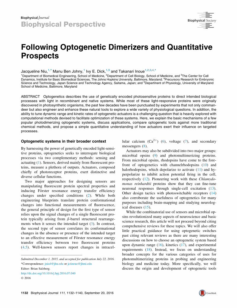
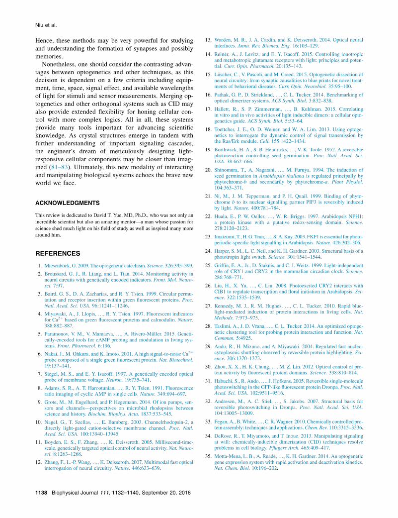
Since the dawn of evolution, primary producers have beentranslating light into cellular signals. Accordingly, themore primitive forms of these photomultimerizing actuatorsoriginated from scientists studying the regulation of tran-scription pathways in plants as well as similar mechanismsin other photosynthetic organisms such as algae and cyano-bacteria. The mustard plant Arabidopsis thaliana is recog-nized for phytochrome B (PhyB)/phytochrome-interactingfactor (PIF) (Fig. 1 A), light-oxygen-voltage (LOV) domain
FIGURE 1 Organism of origin and relevant lifecycle is shown for various
and flowering of Arabidopsis thaliana (84). (B) Cartoon depiction of PhyB
etioliation C) PhyB/PIF-induced lamellipodia formation (red circle) (86). (D) LO
opening in Arabidopsis (84). (E) Atomic structures show two light-dependent co
Bank (PDB): 2V0U; right, PDB: 2V0W) without the Ja helix (87). An alternate
(F) Lov2-Jamediated nuclear localization (88). (G) CRY2 is an important playe
CRY2/CIB1 dimerization with blue light (89). Light-dependent oligomerization
localization of CRY2-Akt in response to blue light (44). (J) Dronpa is a homo
dimerization of Dronpa (left, tetramer PDB: 2POX (32); right, monomers PDB: 2
tional exemplar of light-regulated photoswitching of Dronpa (30). For visualiz
monitored. Images in (A), (D), and (G) are reproduced from Kramer (91). To s
(Fig. 1 D), and cryptochrome2 (CRY2)/cryptochrome-inter-acting basic-helix-loop-helix (CIB1)-based (Fig. 1 G) opto-genetic systems while isolation of mutant GFP-like proteinsfrom the Pectiniidae coral family, also known as chalicecoral, brought about Dronpa (Fig. 1 J)-based systems.
The oldest characterized system from the Arabidopsisgroup is the PhyB/PIF system, which may be traced as farback as the 1950s. Lettuce seeds were observed to germinateat differing speeds; red light promoted germination whileinfrared light repressed germination (19). From these find-ings, it was assumed that a pigment was chemically chang-ing depending on the wavelength of light. Approximatelyfour decades later, this pigment was identified as PhyB(20). Upon light stimulation, PhyB binds to its partner
optogenetic actuators. (A) PhyB is known to be involved in germination
/PIF photoswitching mechanism with chromophore structure (85). (de-
V domain of phototropin is a key component of phototropism and stomatal
nfigurations of LOV domain isolated from Avena sativa (left, Protein Data
mechanism is LOV photodimerization with an EL222 mutation and VVD.
r in flowering and deetiolation of Arabidopsis (84). (H) Cartoon rendition of
of CRY2 is another powerful optogenetic mode of operation. (I) Membrane
log of GFP found in coral Pectiniidae. (K) Crystal structures show reverse
IOV (90)). (Green) Fluorescent state; (gray) dark state of Dronpa. (L) Func-
ation of relocalization, the fluorescence of mNeptune tagged to Dronpa is
ee this figure in color, go online.
Biophysical Journal 111, 1132–1140, September 20, 2016 1133
Niu et al.
PIF3, which is a transcription factor with a helix-loop-helixstructure, and begins DNA transcription to signal seedgermination (Fig. 1 B) (21).
Similarly, the LOV domain was found to be an importantphotosensing protein when it was discovered that exposureto blue light resulted in heavy phosphorylation of the proteinencoded in the NPH1 gene of Arabidopsis (22). When LOVis irradiated with blue light, the flavin cofactor forms a co-valent bond with a key cysteine residue and changes theconformation of the LOV domain (Fig. 1 E). Many studiesfocus on optimizing this system as the different types ofLOV domains offer a large selection of kinetic rates fromhighly stable conformations in both the dark state and lightstate as seen with FKF1 (23) to fast on- and off-rates as withAsLOV2 (24).
CRY2 is a member of the cryptochrome family, andsome argue that it takes part in regulating the circadianrhythm like its homolog CRY1 (25). More important forthe points discussed here, however, is CRY2’s integralrole with its partner CIB1 (Fig. 1 H) in modulating tran-scription leading to Arabidopsis flowering (26). Recently,the pair garnered a lot of attention due to their intermediatekinetic rates with an on-rate of milliseconds and an off-rateof minutes (27). The CRY2 mutation E490G also dramati-cally increases the oligomerization affinity of CRY2(CRY2olig) and induces clustering at the expense of slowerkinetics (28). Nonetheless, CRY2olig provides anotherpowerful flavor of optical signaling through clusteringcompared to colocalization of CRY2 with its bindingpartner CIB1.
Lastly, Dronpa was initially found as a homolog of GFPin the coral family Pectiniidae (Fig. 1 J) (29). Monomersof Dronpa bind to one another to form dimers, and thenthese dimers come together to form tetramers. A pointmutation, K145N, was introduced to weaken tetramer for-mation and thereby enhance the probability of findingDronpa dimers (30). With blue light, Dronpa switchesfrom a fluorescent multimer to dark monomers and reverseswith ultraviolet light (Fig. 1 K). Unlike the PhyB/PIFsystem, Dronpa reverses dimerization, allowing it tophoto-uncage enzymes (Fig. 1 L). In addition, the fasci-nating property of fluorophore switching (31,32) makesDronpa unique from its fellow optogenetic proteins in itcan also serve as a sensor of its own activity (30).
Topics of achievement in discovering andconstructing cellular processes
With the elucidation of these systems, many have selectedtheir favorite optogenetic tool to examine their preferredbiological problems. Some common themes that emergethrough nature and converge with other multimerizationsystems, such as chemically inducible dimerization (CID)systems (33,34), include manipulating gene expressionand toggling molecular concentrations with localization
1134 Biophysical Journal 111, 1132–1140, September 20, 2016
and clustering-based logic. Classic problems that havebeen explored include activation of DNA transcription andother well-known molecular pathways, while newer ven-tures focus on other modes of intracellular signaling suchas Ca2þ regulation.
There is much success associated with using these tools tocontrol gene expression as many of these systems werederived from light-sensitive transcription pathways. In onecase, the EL222 transcription factor, which contains aLOV domain and helix-turn-helix (HTH) motif, was usedto upregulate the production of luciferase in HEK293Tcells. While LOV binds HTH in the dark, blue light stimu-lation causes LOV to undock HTH, which leads to HTHdimerization, then HTH dimer-DNA binding, and finally,DNA transcription (35). Another example is using CRY2/CIB1 with epigenetic modifiers such as LITE (36) or perma-nent genetic modifiers such as TALEN and CRISPR-Cas9 topattern perpetual transcription profiles in cells spatially(37,38). Similar experiments have also been performedwith split-Cas9 and Magnets, a variant of LOV domains,so that upon blue light stimulation, magnets dimerize andproduce a functional Cas9 (39). Outside of single-cell appli-cations, these tools have also been implemented in modelorganisms. The CRY2/CIB1 system has been shown to driveluciferase expression in zebrafish (40) as well as increaseGFP levels in Drosophila (41) under their respective tran-scription systems.
More phenotypical assessments of cellular output withoptogenetic systems target well-understood molecular path-ways such as Rac activation in membrane ruffling and cellmotility. A photoactivatable version of Rac1 with LovJa-directed cell movement with blue light stimulation (42).Comparably, the PhyB/PIF system was also used to showthat it is not only possible to direct lamellipodia and filopo-dia formation (Fig. 1 C) (43) but also possible to generate atrain of signals and subsequently evaluate how the func-tional output of cell survival and cell growth changes withstimulation frequency of the Ras/ERK pathway (18). More-over, the achievements of optogenetic techniques havenot been limited to cell motility. Akt localization tothe membrane with CRY2/CIB1 (Fig. 1 I) uncovered theimportance of Akt in mechanistic target of the rapamycingrowth pathway and the forkhead box O metabolismpathway for the generation of Atrogin1 implicated in muscleatrophy (44).
One powerful notion is using optogenetics to controlCa2þ signaling. Ca2þ regulation is important for numerousyet dissimilar physiological processes including transcrip-tion activation, cell death, and neurological development(45,46). Ca2þ release-activated channels (later mapped tothe ORAI gene) and stromal interaction molecules (STIM)were first discovered because of their importance in T cellactivation. When Ca2þ stores are depleted, it is believedthat STIM EF-hands undergo a conformational changethat allows aggregation of the C-terminal domain of STIM
Optogenetics and its Prospects
(CCD) to prompt the opening of ORAI (47). Recently, mul-tiple groups confirmed that it is possible to increase cyto-solic Ca2þ levels by opening ORAI channels througheither clustering CCD with CRY2olig (48) or releasingCCD with the LOV domain (49). Thus, these studies provideadditional steps toward examining more complex processesinvolving Ca2þ signaling.
Explicit differences between optogenetics andCID techniques
One established rival technology we would like to consideris CID (34). A few points of contemplation important for un-derstanding the differences between optogenetics and CIDinclude inherent properties of the systems themselves andexperimental equipment. Explicitly, these two factors influ-ence the reversibility, spatial specificity, temporal precision,and dynamic range of biological signals generated by thesesystems during experiments.
Generally, after light-sensitive proteins absorb photons,they enter a high-energy state with a different conformationdue to energy transfer. Then, when the light source isremoved, they relax and revert to their original conforma-tion. As most CID processes appear irreversible underexperimental conditions, optogenetic systems are attractivefor their transient responses that enable reversible cyclingof signals. One possible resolution for the irreversibilityof CID systems, however, is the idea of using a mixture ofdifferent CID systems to create antagonistic reversal ofthe original signal (50). A process that has been highlightedwith both optogenetics and CID is KCNQ2/3 channel activ-ity. In 2006, it was proved with CID that PIP2 levels in theplasma membrane directly modulate KCNQ2/3 so that cur-rent decreases upon PIP2 depletion by an engineered PIP2phosphatase (51). Shortly after the popularization of optoge-netic techniques, the CRY2/CIB1 system was also applied tothe regulation of KCNQ2/3 channels. In addition to repli-cating the attenuation of current from the original CIDexperiment, a measurement of the recovery of current andPIP2 concentrations at the membrane could also be madewithin 10 min (52).
Another set of experiments for comparison is probingprocesses that require spatial signaling differences such ascell polarity. Creating molecular concentration gradients isa challenging problem that requires proper equipment forboth CID and optogenetic systems. For CID, careful designof microfluidic devices or biomimetic scaffolding is imper-ative to correctly shape a cell’s chemical environment suchas with graded rapamycin activation of Rac1 (43). On theother hand, technological advances of microscope setupswith optical filters, pinholes (53), digital micromirror de-vices (54), and even endoscopy tools (55) provide a varietyof effective solutions for defining the field of view stimu-lated with light. Examples include controlling the polarityof yeast for budding with Bem1 (56), using CRY2olig to
cluster receptor tyrosine kinases (57), and using CRY2/CIB1 to manipulate G-protein-coupled receptors that pro-mote lamellipodia and cell migration (58). LOV domainshave also been used to optogenetically control proteindegradation (59).
Speed of stimulus delivery is an area in which optoge-netics exceeds its chemical competitors. Most optogeneticsystems produce the desired effect within seconds whileCID systems require tens of seconds due to limited deliv-ery of rapamycin. One example of this phenomenon isseen with organelle transport by kinesins and dyneinwhere directed migration of peroxisomes using the LOVdomain linked to the respective cytoskeleton motor (60)resulted in an approximately twofold faster recruitmentcompared to its CID analog (61). Regardless, both sys-tems offer great promise for answering a myriad of bio-logical questions and contributing to the synthetic cellbiology toolbox (62), but still require optimization andfurther specific design to reduce unwanted chemical andphototoxicity effects as well as undesired basal cellularperturbations.
Dynamic range in relation to kinetics
On the topic of dynamic range and speed, it may be useful tocontemplate a general scheme for how photomultimerizingproteins behave as optogenetic actuators. Upon photoex-citation, light-sensitive proteins undergo conformationalchanges (Fig. 2 A) that result in functional outputs. Forthe photomultimerizing optogenetic tools described above,this conformational change often exposes a hidden surfaceon the light-sensitive protein that allows it to interact withits target. This bimolecular reaction converts the targetinto its active form (Fig. 2 B). These actuators may alsointeract with their target in the dark, albeit with a substan-tially weaker affinity, leading to basal activity. Thus, theoutput of target’s activity in a cell is directly proportionalto the fraction of targets bound to an actuator (fT,b). If we as-sume rapid and complete photoconversion of the optoge-netic actuator (free concentration, ½A�), then f hnT;b after lightactivation is governed by
df hnT;bdt
¼ khyon , ½A� ��khyon , ½A� þ khyoff
�fT;b: (1)
Solving Eq. 1 for steady state yields
f hyT;bðNÞ ¼ ½A�½A� þ khyoff
.khyon
¼ ½A�½A� þ Khy
d
; (2)
where khyoff is off-rate, khyon is on-rate, and the ratioKhyd ¼ khyoff=k
hyon represents the affinity of the actuator for
the target molecule in the presence of light. Because thefree concentration of optogenetic actuator in a cell is
Biophysical Journal 111, 1132–1140, September 20, 2016 1135
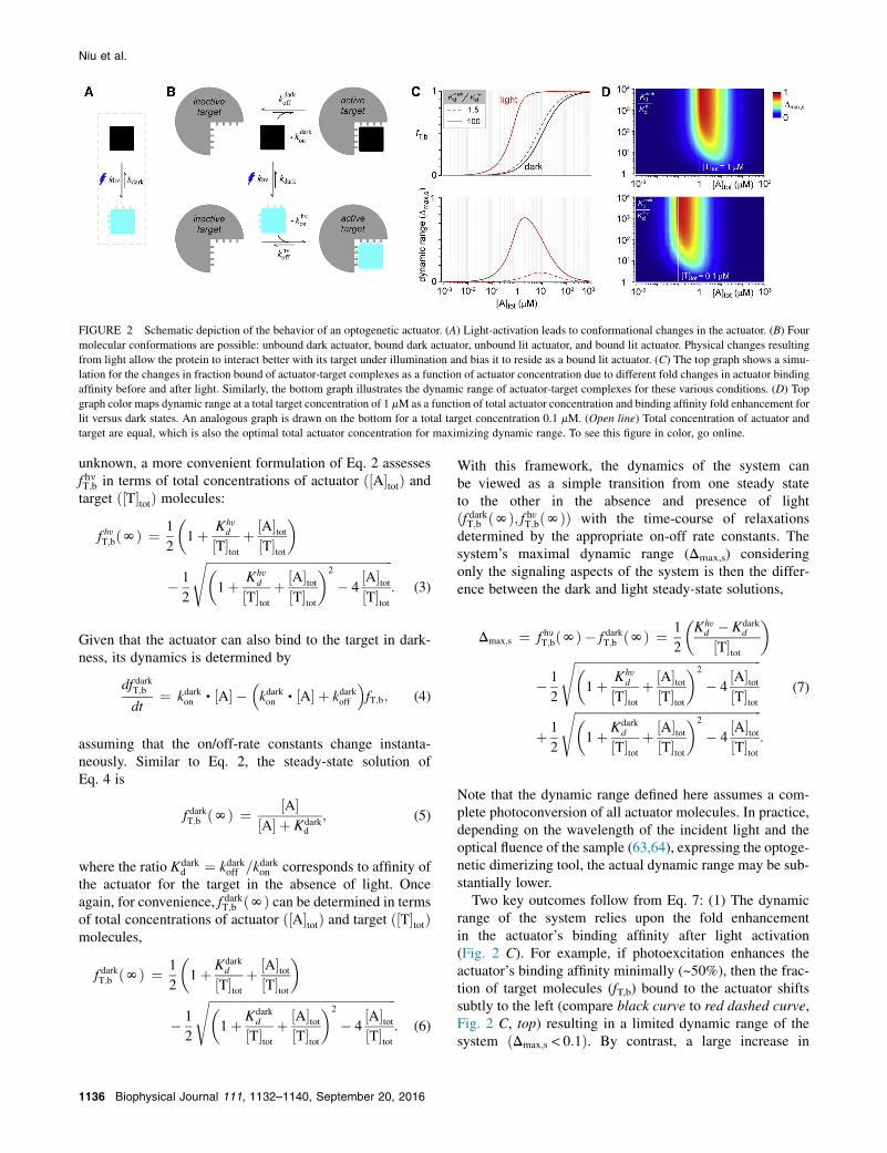
FIGURE 2 Schematic depiction of the behavior of an optogenetic actuator. (A) Light-activation leads to conformational changes in the actuator. (B) Four
molecular conformations are possible: unbound dark actuator, bound dark actuator, unbound lit actuator, and bound lit actuator. Physical changes resulting
from light allow the protein to interact better with its target under illumination and bias it to reside as a bound lit actuator. (C) The top graph shows a simu-
lation for the changes in fraction bound of actuator-target complexes as a function of actuator concentration due to different fold changes in actuator binding
affinity before and after light. Similarly, the bottom graph illustrates the dynamic range of actuator-target complexes for these various conditions. (D) Top
graph color maps dynamic range at a total target concentration of 1 mM as a function of total actuator concentration and binding affinity fold enhancement for
lit versus dark states. An analogous graph is drawn on the bottom for a total target concentration 0.1 mM. (Open line) Total concentration of actuator and
target are equal, which is also the optimal total actuator concentration for maximizing dynamic range. To see this figure in color, go online.
Niu et al.
unknown, a more convenient formulation of Eq. 2 assessesf hnT;b in terms of total concentrations of actuator ð½A�totÞ andtarget ð½T�totÞ molecules:
f hyT;bðNÞ ¼ 1
2
�1þ Khy
d
½T�totþ ½A�tot
½T�tot
�
� 1
2
ffiffiffiffiffiffiffiffiffiffiffiffiffiffiffiffiffiffiffiffiffiffiffiffiffiffiffiffiffiffiffiffiffiffiffiffiffiffiffiffiffiffiffiffiffiffiffiffiffiffiffiffiffiffiffiffiffiffiffiffiffi�1þ Khy
d
½T�totþ ½A�tot
½T�tot
�2
� 4½A�tot½T�tot
s: (3)
Given that the actuator can also bind to the target in dark-ness, its dynamics is determined by
df darkT;b
dt¼ kdarkon , ½A� �
�kdarkon , ½A� þ kdarkoff
�fT;b; (4)
assuming that the on/off-rate constants change instanta-neously. Similar to Eq. 2, the steady-state solution ofEq. 4 is
f darkT;b ðNÞ ¼ ½A�½A� þ Kdark
d
; (5)
where the ratio Kdarkd ¼ kdarkoff =kdarkon corresponds to affinity of
the actuator for the target in the absence of light. Onceagain, for convenience, f darkT;b ðNÞ can be determined in termsof total concentrations of actuator ð½A�totÞ and target ð½T�totÞmolecules,
f darkT;b ðNÞ ¼ 1
2
�1þ Kdark
d
½T�totþ ½A�tot
½T�tot
�
� 1
2
ffiffiffiffiffiffiffiffiffiffiffiffiffiffiffiffiffiffiffiffiffiffiffiffiffiffiffiffiffiffiffiffiffiffiffiffiffiffiffiffiffiffiffiffiffiffiffiffiffiffiffiffiffiffiffiffiffiffiffiffiffi�1þ Kdark
d
½T�totþ ½A�tot
½T�tot
�2
� 4½A�tot½T�tot
s: (6)
1136 Biophysical Journal 111, 1132–1140, September 20, 2016
With this framework, the dynamics of the system canbe viewed as a simple transition from one steady stateto the other in the absence and presence of lightðf darkT;b ðNÞ; f hyT;bðNÞÞ with the time-course of relaxationsdetermined by the appropriate on-off rate constants. Thesystem’s maximal dynamic range (Dmax,s) consideringonly the signaling aspects of the system is then the differ-ence between the dark and light steady-state solutions,
Dmax;s ¼ f hyT;bðNÞ � f darkT;b ðNÞ ¼ 1
2
�Khy
d � Kdarkd
½T�tot
�
� 1
2
ffiffiffiffiffiffiffiffiffiffiffiffiffiffiffiffiffiffiffiffiffiffiffiffiffiffiffiffiffiffiffiffiffiffiffiffiffiffiffiffiffiffiffiffiffiffiffiffiffiffiffiffiffiffiffiffiffiffiffiffiffi�1þ Khy
d
½T�totþ ½A�tot
½T�tot
�2
� 4½A�tot½T�tot
s
þ 1
2
ffiffiffiffiffiffiffiffiffiffiffiffiffiffiffiffiffiffiffiffiffiffiffiffiffiffiffiffiffiffiffiffiffiffiffiffiffiffiffiffiffiffiffiffiffiffiffiffiffiffiffiffiffiffiffiffiffiffiffiffiffi�1þ Kdark
d
½T�totþ ½A�tot
½T�tot
�2
� 4½A�tot½T�tot
s:
(7)
Note that the dynamic range defined here assumes a com-plete photoconversion of all actuator molecules. In practice,depending on the wavelength of the incident light and theoptical fluence of the sample (63,64), expressing the optoge-netic dimerizing tool, the actual dynamic range may be sub-stantially lower.
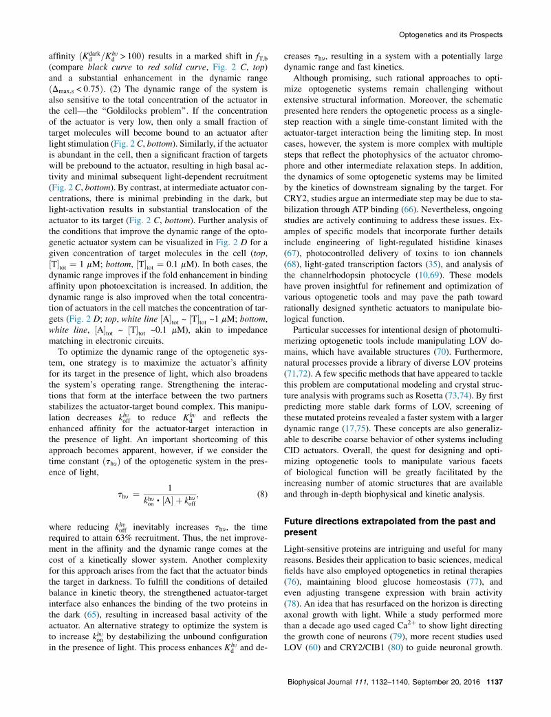
Two key outcomes follow from Eq. 7: (1) The dynamicrange of the system relies upon the fold enhancementin the actuator’s binding affinity after light activation(Fig. 2 C). For example, if photoexcitation enhances theactuator’s binding affinity minimally (~50%), then the frac-tion of target molecules (fT,b) bound to the actuator shiftssubtly to the left (compare black curve to red dashed curve,Fig. 2 C, top) resulting in a limited dynamic range of thesystem ðDmax;s < 0:1Þ. By contrast, a large increase in
Optogenetics and its Prospects
affinity ðKdarkd =Khy
d > 100Þ results in a marked shift in fT,b(compare black curve to red solid curve, Fig. 2 C, top)and a substantial enhancement in the dynamic rangeðDmax;s < 0:75Þ. (2) The dynamic range of the system isalso sensitive to the total concentration of the actuator inthe cell—the ‘‘Goldilocks problem’’. If the concentrationof the actuator is very low, then only a small fraction oftarget molecules will become bound to an actuator afterlight stimulation (Fig. 2 C, bottom). Similarly, if the actuatoris abundant in the cell, then a significant fraction of targetswill be prebound to the actuator, resulting in high basal ac-tivity and minimal subsequent light-dependent recruitment(Fig. 2 C, bottom). By contrast, at intermediate actuator con-centrations, there is minimal prebinding in the dark, butlight-activation results in substantial translocation of theactuator to its target (Fig. 2 C, bottom). Further analysis ofthe conditions that improve the dynamic range of the opto-genetic actuator system can be visualized in Fig. 2 D for agiven concentration of target molecules in the cell (top,½T�tot ¼ 1 mM; bottom, ½T�tot ¼ 0.1 mM). In both cases, thedynamic range improves if the fold enhancement in bindingaffinity upon photoexcitation is increased. In addition, thedynamic range is also improved when the total concentra-tion of actuators in the cell matches the concentration of tar-gets (Fig. 2 D; top, white line ½A�tot ~ ½T�tot ~1 mM; bottom,white line, ½A�tot ~ ½T�tot ~0.1 mM), akin to impedancematching in electronic circuits.
To optimize the dynamic range of the optogenetic sys-tem, one strategy is to maximize the actuator’s affinityfor its target in the presence of light, which also broadensthe system’s operating range. Strengthening the interac-tions that form at the interface between the two partnersstabilizes the actuator-target bound complex. This manipu-lation decreases khyoff to reduce Khy
d and reflects theenhanced affinity for the actuator-target interaction inthe presence of light. An important shortcoming of thisapproach becomes apparent, however, if we consider thetime constant ðthyÞ of the optogenetic system in the pres-ence of light,
thy ¼ 1
khyon , ½A� þ khyoff; (8)
where reducing khyoff inevitably increases thy, the timerequired to attain 63% recruitment. Thus, the net improve-ment in the affinity and the dynamic range comes at thecost of a kinetically slower system. Another complexityfor this approach arises from the fact that the actuator bindsthe target in darkness. To fulfill the conditions of detailedbalance in kinetic theory, the strengthened actuator-targetinterface also enhances the binding of the two proteins inthe dark (65), resulting in increased basal activity of theactuator. An alternative strategy to optimize the system isto increase khyon by destabilizing the unbound configurationin the presence of light. This process enhances Khy
d and de-
creases thy, resulting in a system with a potentially largedynamic range and fast kinetics.
Although promising, such rational approaches to opti-mize optogenetic systems remain challenging withoutextensive structural information. Moreover, the schematicpresented here renders the optogenetic process as a single-step reaction with a single time-constant limited with theactuator-target interaction being the limiting step. In mostcases, however, the system is more complex with multiplesteps that reflect the photophysics of the actuator chromo-phore and other intermediate relaxation steps. In addition,the dynamics of some optogenetic systems may be limitedby the kinetics of downstream signaling by the target. ForCRY2, studies argue an intermediate step may be due to sta-bilization through ATP binding (66). Nevertheless, ongoingstudies are actively continuing to address these issues. Ex-amples of specific models that incorporate further detailsinclude engineering of light-regulated histidine kinases(67), photocontrolled delivery of toxins to ion channels(68), light-gated transcription factors (35), and analysis ofthe channelrhodopsin photocycle (10,69). These modelshave proven insightful for refinement and optimization ofvarious optogenetic tools and may pave the path towardrationally designed synthetic actuators to manipulate bio-logical function.
Particular successes for intentional design of photomulti-merizing optogenetic tools include manipulating LOV do-mains, which have available structures (70). Furthermore,natural processes provide a library of diverse LOV proteins(71,72). A few specific methods that have appeared to tacklethis problem are computational modeling and crystal struc-ture analysis with programs such as Rosetta (73,74). By firstpredicting more stable dark forms of LOV, screening ofthese mutated proteins revealed a faster system with a largerdynamic range (17,75). These concepts are also generaliz-able to describe coarse behavior of other systems includingCID actuators. Overall, the quest for designing and opti-mizing optogenetic tools to manipulate various facetsof biological function will be greatly facilitated by theincreasing number of atomic structures that are availableand through in-depth biophysical and kinetic analysis.
Future directions extrapolated from the past andpresent
Light-sensitive proteins are intriguing and useful for manyreasons. Besides their application to basic sciences, medicalfields have also employed optogenetics in retinal therapies(76), maintaining blood glucose homeostasis (77), andeven adjusting transgene expression with brain activity(78). An idea that has resurfaced on the horizon is directingaxonal growth with light. While a study performed morethan a decade ago used caged Ca2þ to show light directingthe growth cone of neurons (79), more recent studies usedLOV (60) and CRY2/CIB1 (80) to guide neuronal growth.
Biophysical Journal 111, 1132–1140, September 20, 2016 1137
Niu et al.
Hence, these methods may be very powerful for studyingand understanding the formation of synapses and possiblymemories.
Nonetheless, one should consider the contrasting advan-tages between optogenetics and other techniques, as thisdecision is dependent on a few criteria including equip-ment, time, space, signal effect, and available wavelengthsof light for stimuli and sensor measurements. Merging op-togenetics and other orthogonal systems such as CID mayalso provide extended flexibility for honing cellular con-trol with more complex logics. All in all, these systemsprovide many tools important for advancing scientificknowledge. As crystal structures emerge in tandem withfurther understanding of important signaling cascades,the engineer’s dream of meticulously designing light-responsive cellular components may be closer than imag-ined (81–83). Ultimately, this new modality of interactingand manipulating biological systems echoes the brave newworld we face.
ACKNOWLEDGMENTS
This review is dedicated to David T. Yue, MD, Ph.D., who was not only an
incredible scientist but also an amazing mentor—a man whose passion for
science shed much light on his field of study as well as inspired many more
around him.
REFERENCES
1. Miesenbock, G. 2009. The optogenetic catechism. Science. 326:395–399.
2. Broussard, G. J., R. Liang, and L. Tian. 2014. Monitoring activity inneural circuits with genetically encoded indicators. Front. Mol. Neuro-sci. 7:97.
3. Baird, G. S., D. A. Zacharias, and R. Y. Tsien. 1999. Circular permu-tation and receptor insertion within green fluorescent proteins. Proc.Natl. Acad. Sci. USA. 96:11241–11246.
4. Miyawaki, A., J. Llopis, ., R. Y. Tsien. 1997. Fluorescent indicatorsfor Ca2þ based on green fluorescent proteins and calmodulin. Nature.388:882–887.
5. Paramonov, V. M., V. Mamaeva, ., A. Rivero-Muller. 2015. Geneti-cally-encoded tools for cAMP probing and modulation in living sys-tems. Front. Pharmacol. 6:196.
6. Nakai, J., M. Ohkura, and K. Imoto. 2001. A high signal-to-noise Ca2þ
probe composed of a single green fluorescent protein. Nat. Biotechnol.19:137–141.
7. Siegel, M. S., and E. Y. Isacoff. 1997. A genetically encoded opticalprobe of membrane voltage. Neuron. 19:735–741.
8. Adams, S. R., A. T. Harootunian, ., R. Y. Tsien. 1991. Fluorescenceratio imaging of cyclic AMP in single cells. Nature. 349:694–697.
9. Grote, M., M. Engelhard, and P. Hegemann. 2014. Of ion pumps, sen-sors and channels—perspectives on microbial rhodopsins betweenscience and history. Biochim. Biophys. Acta. 1837:533–545.
10. Nagel, G., T. Szellas, ., E. Bamberg. 2003. Channelrhodopsin-2, adirectly light-gated cation-selective membrane channel. Proc. Natl.Acad. Sci. USA. 100:13940–13945.
11. Boyden, E. S., F. Zhang, ., K. Deisseroth. 2005. Millisecond-time-scale, genetically targeted optical control of neural activity.Nat. Neuro-sci. 8:1263–1268.
12. Zhang, F., L.-P. Wang,., K. Deisseroth. 2007. Multimodal fast opticalinterrogation of neural circuitry. Nature. 446:633–639.
1138 Biophysical Journal 111, 1132–1140, September 20, 2016
13. Warden, M. R., J. A. Cardin, and K. Deisseroth. 2014. Optical neuralinterfaces. Annu. Rev. Biomed. Eng. 16:103–129.
14. Reiner, A., J. Levitz, and E. Y. Isacoff. 2015. Controlling ionotropicand metabotropic glutamate receptors with light: principles and poten-tial. Curr. Opin. Pharmacol. 20:135–143.
15. Luscher, C., V. Pascoli, and M. Creed. 2015. Optogenetic dissection ofneural circuitry: from synaptic causalities to blue prints for novel treat-ments of behavioral diseases. Curr. Opin. Neurobiol. 35:95–100.
16. Pathak, G. P., D. Strickland, ., C. L. Tucker. 2014. Benchmarking ofoptical dimerizer systems. ACS Synth. Biol. 3:832–838.
17. Hallett, R., S. P. Zimmerman, ., B. Kuhlman. 2015. Correlatingin vitro and in vivo activities of light inducible dimers: a cellular opto-genetics guide. ACS Synth. Biol. 5:53–64.
18. Toettcher, J. E., O. D. Weiner, and W. A. Lim. 2013. Using optoge-netics to interrogate the dynamic control of signal transmission bythe Ras/Erk module. Cell. 155:1422–1434.
19. Borthwick, H. A., S. B. Hendricks, ., V. K. Toole. 1952. A reversiblephotoreaction controlling seed germination. Proc. Natl. Acad. Sci.USA. 38:662–666.
20. Shinomura, T., A. Nagatani, ., M. Furuya. 1994. The induction ofseed germination in Arabidopsis thaliana is regulated principally byphytochrome-b and secondarily by phytochrome-a. Plant Physiol.104:363–371.
21. Ni, M., J. M. Tepperman, and P. H. Quail. 1999. Binding of phyto-chrome b to its nuclear signalling partner PIF3 is reversibly inducedby light. Nature. 400:781–784.
22. Huala, E., P. W. Oeller, ., W. R. Briggs. 1997. Arabidopsis NPH1:a protein kinase with a putative redox-sensing domain. Science.278:2120–2123.
23. Imaizumi, T.,H.G.Tran,., S.A.Kay. 2003. FKF1 is essential for photo-periodic-specific light signalling in Arabidopsis. Nature. 426:302–306.
24. Harper, S. M., L. C. Neil, and K. H. Gardner. 2003. Structural basis of aphototropin light switch. Science. 301:1541–1544.
25. Griffin, E. A., Jr., D. Staknis, and C. J. Weitz. 1999. Light-independentrole of CRY1 and CRY2 in the mammalian circadian clock. Science.286:768–771.
26. Liu, H., X. Yu, ., C. Lin. 2008. Photoexcited CRY2 interacts withCIB1 to regulate transcription and floral initiation in Arabidopsis. Sci-ence. 322:1535–1539.
27. Kennedy, M. J., R. M. Hughes, ., C. L. Tucker. 2010. Rapid blue-light-mediated induction of protein interactions in living cells. Nat.Methods. 7:973–975.
28. Taslimi, A., J. D. Vrana,., C. L. Tucker. 2014. An optimized optoge-netic clustering tool for probing protein interaction and function. Nat.Commun. 5:4925.
29. Ando, R., H. Mizuno, and A. Miyawaki. 2004. Regulated fast nucleo-cytoplasmic shuttling observed by reversible protein highlighting. Sci-ence. 306:1370–1373.
30. Zhou, X. X., H. K. Chung,., M. Z. Lin. 2012. Optical control of pro-tein activity by fluorescent protein domains. Science. 338:810–814.
31. Habuchi, S., R. Ando,., J. Hofkens. 2005. Reversible single-moleculephotoswitching in the GFP-like fluorescent protein Dronpa. Proc. Natl.Acad. Sci. USA. 102:9511–9516.
32. Andresen, M., A. C. Stiel, ., S. Jakobs. 2007. Structural basis forreversible photoswitching in Dronpa. Proc. Natl. Acad. Sci. USA.104:13005–13009.
33. Fegan,A., B.White,., C. R.Wagner. 2010. Chemically controlled pro-tein assembly: techniques and applications.Chem. Rev. 110:3315–3336.
34. DeRose, R., T. Miyamoto, and T. Inoue. 2013. Manipulating signalingat will: chemically-inducible dimerization (CID) techniques resolveproblems in cell biology. Pflugers Arch. 465:409–417.
35. Motta-Mena, L. B., A. Reade,., K. H. Gardner. 2014. An optogeneticgene expression system with rapid activation and deactivation kinetics.Nat. Chem. Biol. 10:196–202.
Optogenetics and its Prospects
36. Konermann, S., M. D. Brigham, ., F. Zhang. 2013. Optical controlof mammalian endogenous transcription and epigenetic states. Nature.500:472–476.
37. Polstein, L. R., and C. A. Gersbach. 2015. A light-inducible CRISPR-Cas9 system for control of endogenous gene activation. Nat. Chem.Biol. 11:198–200.
38. Nihongaki, Y., S. Yamamoto, ., M. Sato. 2015. CRISPR-Cas9-basedphotoactivatable transcription system. Chem. Biol. 22:169–174.
39. Nihongaki, Y., F. Kawano, ., M. Sato. 2015. PhotoactivatableCRISPR-Cas9 for optogenetic genome editing. Nat. Biotechnol.33:755–760.
40. Liu, H., G. Gomez, ., C. Lin. 2012. Optogenetic control of transcrip-tion in zebrafish. PLoS One. 7:e50738.
41. Chan, Y.-B., O. V. Alekseyenko, and E. A. Kravitz. 2015. Optogeneticcontrol of gene expression in Drosophila. PLoS One. 10:e0138181.
42. Wu, Y. I., D. Frey, ., K. M. Hahn. 2009. A genetically encodedphotoactivatable Rac controls the motility of living cells. Nature.461:104–108.
43. Lin, B., W. R. Holmes, ., A. Levchenko. 2012. Synthetic spatiallygraded Rac activation drives cell polarization and movement. Proc.Natl. Acad. Sci. USA. 109:E3668–E3677.
44. Katsura, Y., H. Kubota,., T. Ozawa. 2015. An optogenetic system forinterrogating the temporal dynamics of Akt. Sci. Rep. 5:14589.
45. Clapham, D. E. 2007. Calcium signaling. Cell. 131:1047–1058.
46. Greer, P. L., and M. E. Greenberg. 2008. From synapse to nucleus:calcium-dependent gene transcription in the control of synapse devel-opment and function. Neuron. 59:846–860.
47. Prakriya, M., and R. S. Lewis. 2015. Store-operated calcium channels.Physiol. Rev. 95:1383–1436.
48. Kyung, T., S. Lee,., W. D. Heo. 2015. Optogenetic control of endog-enous Ca2þ channels in vivo. Nat. Biotechnol. 33:1092–1096.
49. Ishii, T., K. Sato,., T. Nakata. 2015. Light generation of intracellularCa2þ signals by a genetically encoded protein BACCS. Nat. Commun.6:8021.
50. Miyamoto, T., R. DeRose, ., T. Inoue. 2012. Rapid and orthogonallogic gating with a gibberellin-induced dimerization system. Nat.Chem. Biol. 8:465–470.
51. Suh, B.-C., T. Inoue, ., B. Hille. 2006. Rapid chemically inducedchanges of PtdIns(4,5)P2 gate KCNQ ion channels. Science. 314:1454–1457.
52. Idevall-Hagren, O., E. J. Dickson,., P. De Camilli. 2012. Optogeneticcontrol of phosphoinositide metabolism. Proc. Natl. Acad. Sci. USA.109:E2316–E2323.
53. Swedlow, J. R. 2012. Innovation in biological microscopy: current sta-tus and future directions. BioEssays. 34:333–340.
54. Shin, S., K. Kim, ., Y. Park. 2015. Active illumination using a digitalmicromirror device for quantitative phase imaging.Opt. Lett. 40:5407–5410.
55. Klimas, A., and E. Entcheva. 2014. Toward microendoscopy-inspiredcardiac optogenetics in vivo: technical overview and perspective.J. Biomed. Opt. 19:08070-1–14.
56. Jost, A. P.-T., and O. D. Weiner. 2015. Probing yeast polarity withacute, reversible, optogenetic inhibition of protein function. ACS Synth.Biol. 4:1077–1085.
57. Bugaj, L. J., D. P. Spelke, ., D. V. Schaffer. 2015. Regulation ofendogenous transmembrane receptors through optogenetic Cry2 clus-tering. Nat. Commun. 6:6898.
58. O’Neill, P. R., and N. Gautam. 2014. Subcellular optogenetic inhibitionof G proteins generates signaling gradients and cell migration. Mol.Biol. Cell. 25:2305–2314.
59. Renicke, C., D. Schuster, ., C. Taxis. 2013. A LOV2 domain-basedoptogenetic tool to control protein degradation and cellular function.Chem. Biol. 20:619–626.
60. van Bergeijk, P., M. Adrian,., L. C. Kapitein. 2015. Optogenetic con-trol of organelle transport and positioning. Nature. 518:111–114.
61. Kapitein, L. C., M. A. Schlager, ., C. C. Hoogenraad. 2010. Probingintracellular motor protein activity using an inducible cargo traffickingassay. Biophys. J. 99:2143–2152.
62. Kim, A. K., R. DeRose, ., T. Inoue. 2016. Toward total synthesisof cell function: reconstituting cell dynamics with synthetic biology.Sci. Signal. 9:re1.
63. Cheong, W. F., S. A. Prahl, and A. J. Welch. 1990. A review of the op-tical properties of biological tissues. IEEE J. Quantum Electron.26:2166–2185.
64. Stujenske, J. M., T. Spellman, and J. A. Gordon. 2015. Modeling thespatiotemporal dynamics of light and heat propagation for in vivooptogenetics. Cell Reports. 12:525–534.
65. Jackson, M. B. 2006. Molecular and Cellular Biophysics. CambridgeUniversity Press, Cambridge, UK.
66. Cailliez, F., P. Muller,., A. de la Lande. 2014. ATP binding and aspar-tate protonation enhance photoinduced electron transfer in plant cryp-tochrome. J. Am. Chem. Soc. 136:12974–12986.
67. Moglich, A., R. A. Ayers, and K. Moffat. 2009. Design and signalingmechanism of light-regulated histidine kinases. J. Mol. Biol. 385:1433–1444.
68. Schmidt, D., P. W. Tillberg,., E. S. Boyden. 2014. A fully geneticallyencoded protein architecture for optical control of peptide ligand con-centration. Nat. Commun. 5:3019.
69. Schneider, F., C. Grimm, and P. Hegemann. 2015. Biophysics of chan-nelrhodopsin. Annu. Rev. Biophys. 44:167–186.
70. Halavaty, A. S., and K. Moffat. 2007. N- and C-terminal flanking re-gions modulate light-induced signal transduction in the LOV2 domainof the blue light sensor phototropin 1 from Avena sativa. Biochemistry.46:14001–14009.
71. Pudasaini, A., K. K. El-Arab, and B. D. Zoltowski. 2015. LOV-basedoptogenetic devices: light-driven modules to impart photoregulatedcontrol of cellular signaling. Front Mol Biosci. 2:18.
72. Strickland, D., Y. Lin, ., M. Glotzer. 2012. TULIPs: tunable, light-controlled interacting protein tags for cell biology. Nat. Methods.9:379–384.
73. Das, R., and D. Baker. 2008. Macromolecular modeling with Rosetta.Annu. Rev. Biochem. 77:363–382.
74. Kaufmann, K. W., G. H. Lemmon,., J. Meiler. 2010. Practically use-ful: what the Rosetta protein modeling suite can do for you. Biochem-istry. 49:2987–2998.
75. Guntas, G., R. A. Hallett, ., B. Kuhlman. 2015. Engineering animproved light-induced dimer (iLID) for controlling the localizationand activity of signaling proteins. Proc. Natl. Acad. Sci. USA.112:112–117.
76. Tomita, H., E. Sugano, ., M. Tamai. 2010. Channelrhodopsin-2 genetransduced into retinal ganglion cells restores functional vision ingenetically blind rats. Exp. Eye Res. 90:429–436.
77. Ye, H., M. Daoud-El Baba, ., M. Fussenegger. 2011. A synthetic op-togenetic transcription device enhances blood-glucose homeostasis inmice. Science. 332:1565–1568.
78. Folcher, M., S. Oesterle, ., M. Fussenegger. 2014. Mind-controlledtransgene expression by a wireless-powered optogenetic designer cellimplant. Nat. Commun. 5:5392.
79. Zheng, J. Q. 2000. Turning of nerve growth cones induced by localizedincreases in intracellular calcium ions. Nature. 403:89–93.
80. Kakumoto, T., and T. Nakata. 2013. Optogenetic control of PIP3: PIP3is sufficient to induce the actin-based active part of growth cones and isregulated via endocytosis. PLoS One. 8:e70861.
81. Volgraf, M., P. Gorostiza,., D. Trauner. 2006. Allosteric control of anionotropic glutamate receptor with an optical switch. Nat. Chem. Biol.2:47–52.
82. Airan, R. D., K. R. Thompson, ., K. Deisseroth. 2009. Temporallyprecise in vivo control of intracellular signalling. Nature. 458:1025–1029.
Biophysical Journal 111, 1132–1140, September 20, 2016 1139

Niu et al.
83. Xu, Y., Y.-M. Hyun,., M. Kim. 2014. Optogenetic control of chemo-kine receptor signal and T-cell migration. Proc. Natl. Acad. Sci. USA.111:6371–6376.
84. Sullivan, J. A., and X. W. Deng. 2003. From seed to seed: therole of photoreceptors in Arabidopsis development. Dev. Biol.260:289–297.
85. Burgie, E. S., A. N. Bussell, ., R. D. Vierstra. 2014. Crystal structureof the photosensing module from a red/far-red light-absorbing plantphytochrome. Proc. Natl. Acad. Sci. USA. 111:10179–10184.
86. Levskaya, A., O. D. Weiner, ., C. A. Voigt. 2009. Spatiotemporalcontrol of cell signalling using a light-switchable protein interaction.Nature. 461:997–1001.
1140 Biophysical Journal 111, 1132–1140, September 20, 2016
87. Mitra, D., X. Yang, and K. Moffat. 2012. Crystal structures of Aureo-chrome1 LOV suggest new design strategies for optogenetics. Struc-ture. 20:698–706.
88. Niopek, D., D. Benzinger, ., B. Di Ventura. 2014. Engineering light-inducible nuclear localization signals for precise spatiotemporal con-trol of protein dynamics in living cells. Nat. Commun. 5:4404.
89. Xing, W., L. Busino,., N. Zheng. 2013. SCF(FBXL3) ubiquitin ligasetargets cryptochromes at their cofactor pocket. Nature. 496:64–68.
90. Stiel, A. C., S. Trowitzsch, ., M. C. Wahl. 2007. 1.8 A bright-statestructure of the reversibly switchable fluorescent protein Dronpa guidesthe generation of fast switching variants. Biochem. J. 402:35–42.
91. Kramer, U. 2015. Planting molecular functions in an ecological contextwith Arabidopsis thaliana. eLife. 4:1–13.