food research international - ترجمه...
TRANSCRIPT

Food Research International 53 (2013) 96–103
Contents lists available at SciVerse ScienceDirect
Food Research International
j ourna l homepage: www.e lsev ie r .com/ locate / foodres
Co- encapsulation of Lactobacillus acidophilus with inulin orpolydextrose in solid lipid microparticles provides protection andimproves stability
Paula K. Okuro ⁎, Marcelo Thomazini, Júlio C.C. Balieiro, Roberta D.C.O. Liberal, Carmen S. Fávaro-TrindadeUniversidade de São Paulo, Faculdade de Zootecnia e Engenharia de Alimentos, Pirassununga-SP, Brazil
⁎ Corresponding author. Tel.: +55 19 3565 4282.E-mail address: [email protected] (P.K. Okuro).
0963-9969/$ – see front matter © 2013 Elsevier Ltd. Allhttp://dx.doi.org/10.1016/j.foodres.2013.03.042
a b s t r a c t
a r t i c l e i n f oArticle history:Received 14 January 2013Accepted 29 March 2013
Keywords:ProbioticsMicroencapsulationEncapsulationSpray-chillingSpray-coolingStructural evaluationFat
The aims of this study were to produce and evaluate solid lipid microparticles (SLM) in which Lactobacillusacidophilus (La), a probiotic, was co-encapsulated with a prebiotics, either inulin (Inu) or polydextrose(Poly) using spray chilling technology. Morphological, chemical, and thermal characterisation of SLMs wereconducted, along with survival assays to evaluate the resistance of the probiotic to the microencapsulationprocess, its resistance to exposure to simulated gastric fluids (SGF) and simulated intestinal fluids (SIF),and its stability throughout storage for 120 days at −18, 7 and 22 °C in a vacuum or with controlled relativehumidity. Cell viability was not affected by the spray-chilling process. All of the microcapsules produced inthe present study increased the survival rate of La exposed to SGF and SIF compared to that of free probioticcells. Promising results were obtained when these microcapsules were stored refrigerated and frozen with acontrolled relative humidity. This study indicated that combined spray chilling process, combined with theaddition of a prebiotic component, specifically polydextrose is an interesting technology for the protection,delivery and improve stability of probiotics, which increases the potential of symbiotic SLMs. Scaling upthe spray chilling technique will allow efficient encapsulation of probiotics in a lipid matrix.
© 2013 Elsevier Ltd. All rights reserved.
1. Introduction
Probiotics are endowed with the ability to modulate of the intesti-nal microbiota, and the presence of prebiotics, ingredients that areselectively fermentable, exert a beneficial effect on the growth and/or activity of bacteria in the colon (Gibson, Probert, Van Loo, Rastall,& Roberfroid, 2004; McCartney & Gibson, 2006; Roberfroid, 2007;Wells, Saulnier, & Gibson, 2008). There is a synergistic relationshipbetween probiotics and prebiotics in which the prebiotics are con-sumed by probiotics as sources of carbon and energy, favouringtheir colonisation of the intestinal tract over colonisation by pathogenicmicroorganisms (Vernazza, Rabiu, & Gibson, 2006; Homayouni, Azizi,Ehsani, Yarmand, & Razavi, 2008).
Microencapsulation has emerged as an alternative for protection ofprobiotics, providing a particular and convenient micro-environmentfor the encapsulated microorganism, enhancing their viability (Anal &Singh, 2007; Favaro-Trindade & Grosso, 2000; Rodrigues et al., 2011;Shah & Ravula, 2000; Sohail, Turner, Coombes, Bostrom, & Bhandari,2011), and enabling controlled release of cells in the intestinaltract (Cook, Tzortzis, Charalampopoulos, & Khutoryanskiy, 2012;Favaro-Trindade, Heinemann, & Pedroso, 2011; Mandal, Puniya, &
rights reserved.
Singh, 2006; Oliveira et al., 2007a; Shoji et al., 2013). The choices ofthemethod andmaterials are essential for an effective probiotic encap-sulation strategy, and the use of gentle techniques, such as cold-inducedgelation described by Nag, Han, and Singh (2011), as well as the appro-priate materials, such as gastro-resistant polymers, biopolymers, andstearic acid (hydrophilic retardants), among others, are essential forefficient microencapsulation (Kanmani et al., 2011; Pimentel-González,Camposmontiel, Lobato-Calleros, Pedroza-Islas, & Vernoncarte, 2009).
Spray chilling is a microencapsulation technique that is based onthe addition of the bioactive component to the molten carrier viadissolution, emulsion or dispersion. The mixture is passed throughan atomizer nozzle, and when the nebulised material contacts anatmosphere refrigerated below themelting point of thematrix material(cold air chamber or liquid nitrogen), heat exchange occurs and thevehicle solidifies, creating solid lipid microparticles (SLMs) (Ilić et al.,2009).
Packaging probiotic microorganisms within solid lipid microparti-cles may protect them and is an interesting alternative to otherconveyance systems, such as polymers and polysaccharides, becausethe microparticles will deliver the bioactive compound at approxi-mately the melting point of the carrier material. SLMs producedwith lipid materials are easily digested by the lipases in the intestines,releasing the probiotic in the vicinity of the intestines where they arerequired. The viability of microorganisms within lipid matrices was

97P.K. Okuro et al. / Food Research International 53 (2013) 96–103
reported to be greater than that of free microorganisms (Lahtinen,Ouwehand, Salminen, Forssell, & Myllärinen, 2007; Pedroso, Thomazini,Heinemann, & Favaro-Trindade, 2012; Picot & Lacroix, 2003).
Encapsulation with lipid matrices is promising because lipids arelikely to stabilise probiotics. Mandal, Hati, Puniya, Singh, and Singh(2012) found that number of viable free Lactobacillus casei NCDC298 and Lactobacillus casei NCDC 298 encapsulated in a milk choco-late matrix did not differ during up to 60 days of storage under refrig-erated conditions. They attributed this result to the high total solids inthis matrix, including the fats and suggested that the fats had theability to protect the probiotic cells. Hou, Lin, Wang, and Tzen (2003)encapsulated Lactobacillus delbrueckii ssp. in an artificial emulsion ofsesame oil and observed that encapsulated bacteria performed betterunder simulated gastrointestinal conditions than did free cells. Theyconcluded that the emulsionwas an effective biocapsule for dairy prod-ucts and Lahtinen et al. (2007) suggested that lipid matrix may protectthe cells by blocking H+ ions.
Pedroso et al. (2012) produced SLMs containing B. lactis andL. acidophilus using the spray chilling method. While analyzing theSLMs, they observed that encapsulated L. acidophilus cells were resis-tant to simulated gastric and intestinal fluids and that they had ashelf life of 30 and 60 days of storage at 37 °C and 7 °C, respectively.Therefore, the aims of this study were to produce solid lipid micro-particles containing L. acidophilus and a prebiotic compound, to eval-uate the ability of inulin and polydextrose to increase the viability ofthe probiotic throughout 120 days of storage at different tempera-tures, and to investigate probiotic survival in simulated gastric andintestinal fluids.
2. Materials and methods
2.1. Materials
A lipid carrier consisting of the fats obtained upon interesterificationof fully hydrogenated palm and palm-kernel oil (GPPI) was used. Thefats were kindly provided by Vigor (São Caetano do Sul, Brazil), theirmelting point is 43.34 °C.
A culture of Lactobacillus acidophilus (LAC-04) kindly provided byDanisco (Cotia, Brazil) was used as the active material. The prebioticsco-incorporated in the microparticles were inulin (Raftiline® ST,Beneo Orafti, Tienen, Belgium) and polydextrose (Litesse®, Danisco,Cotia, São Paulo, Brazil), which were kindly provided by Clariant(Suzano, Brazil) and Danisco (Cotia, Brazil), respectively.
2.2. Methods
2.2.1. Preparation of bacterial cell inoculumThe L. acidophilus culture was activated in sterilised MRS broth at
37 °C for 18 h. An aliquot of this culture was transferred to MRSbroth and incubated for other 18 h at 37 °C in jars using an anaerobi-osis system (Anaerobac, Probac, São Paulo-Brazil). This culture wascentrifuged at 2400 g for 9 min at 4 ± 3 °C. The washed cells wereresuspended in 2% sodium citrate at a concentration of approximately109–1010 CFU/mL.
2.2.2. L. acidophilus cell countsThe number of viable La cells was determined using the pour plate
technique, with MRS agar plates (DeMan Rogosa and Sharp) obtainedfrom Acumedia (Indaiatuba, São Paulo, Brazil), according to themethoddescribed byGrosso and Favaro-Trindade (2004), with themodificationsuggested by Pedroso et al. (2012). One gram of SLMs was placed in9 mL of 2% sodium citrate warmed at 52 ± 1 °C to completely dissolvethe lipid matrix. The released cells were serially diluted in tubescontaining warm 2% sodium citrate. Microaerophilic conditions wereproduced in anaerobiosis jars using the anaerobiosis generator systems.The jars were incubated at 37 °C ± 1°C for 72 h.
2.2.3. Preparation of solid lipid microparticles (SLM)SLMs were produced according to the method described by
Chambi, Alvim, Barrera-Arellano, and Grosso (2008), with somemodifications. The inoculum (4%) was mixed with the prebiotic(3%), and the molten carrier was added. Suspensions of the mixtures(probiotic + prebiotic + lipid matrix) or (probiotic + lipid matrix)were produced using an Ultra-Turrax IKA® T-25 homogeniser (IKA,Staufen, Germany) at 7000 rpm for 60 s. The suspensions wereatomised using a spray chiller (Labmaq, Ribeirão Preto, Brazil)equipped with a double fluid atomiser (Ø = 1.2 mm) and a coldchamber at 15 ± 2 °C (environmental variables: temperature andhumidity), at a pressure of 5 bar.
Three formulations were produced: F1 (La without prebiotic), F2(La + Inu), and F3 (La + Poly).
2.2.4. SLM characterisation
2.2.4.1. Microscopy. The morphologies of the SLMs were analysedusing scanning electron microscopy (SEM). The SLMs were placedon pieces of double-faced carbon tape (Ted Pella, Inc., Redding-USA) that were fixed on aluminum stubs. Images were captured at avoltage of 5 kV and current of 1.750 mA. To analyse the internalmorphologies of SLMs, they were frozen with liquid N2 and sectioned.For confocal microscopy, the samples were examined using a ZeissLSM 780-NLO confocal system with an Axio Observer Z.1 microscope(Carl Zeiss AG, Germany) and a 63x/1.4 NA oil plan apochromatic DICobjective lens. Images in a 1024 × 1024 format were captured using605 nm laser lines for excitation, with pinholes set to 1 airy unit foreach channel. The bacteria were stained with SYTO 9, a componentof the LIVE/DEADs BacLight bacterial viability kit (Molecular Probes,Eugene, Oregon USA).
2.2.4.2. Particle size. A Shimadzu Sald-201V particle size analyser(Kyoto, Japan) that employs laser diffraction was used to determinethe sizes and size distributions of the SLMs. The SLMs were dispersedin ethyl alcohol (Synth, Brazil) and stabilised for 5 min before theassay.
2.2.4.3. Differential scanning calorimetry (DSC). Differential scanningcalorimetry was conducted using a DSCM2010 system (TA Instruments,Newcastle, USA). Approximately 10 mg of each sample (GPPI/SLM) wasplaced in an aluminum capsule and gradually heated (10 °C/min)to temperatures between −50 and 100 °C in an inert atmosphere(45 mL/min of N2). An empty capsule was used as the reference. Thedata were analysed using Universal Analysis 2000 version 3.9a (TAInstruments) (Favaro-Trindade, Santana, Monterrey-Quintero, Trindade,& Netto, 2010).
2.2.4.4. Fourier transform infrared (FTIR) spectroscopy. The pure ingre-dients and the SLMs were characterised by FTIR spectroscopy in the4000 to 600 cm−1 region using a Perkin Elmer FTIR spectrometer(Massachusetts/USA) and Spectrum One version 5.3.1 software. Theprobiotic sample was suspended in 2% sodium citrate.
2.2.4.5. X-ray powder diffraction. The individual components andmicroparticles were analysed using the X-ray powder diffractiontechnique. An AXSAnalytical X-Ray Systems Siemens D 5005 (Germany)diffractometer with Cu K alpha radiation (λ = 1.54056A°) was usedwith the voltage was 40 kV and current set at 40 mA. The scanningangle ranged from 3 to 90° of 2θ, 2°/min and 0.033°/s.
2.2.5. Viability of encapsulated microorganisms during storageFor the stability studies, the microparticles were stored at three
different temperatures (−18, 7, and 22 °C) in a vacuum or with therelative humidity controlled with lithium chloride (LiCl), as describedby Heidebach, Forst, and Kulozik (2010). The SLMs were stored in
98 P.K. Okuro et al. / Food Research International 53 (2013) 96–103
hermetically sealed glass flasks or in open flasks in hermeticallysealed containers containing LiCl, and later on stored in a BOD thatwas set at the three temperatures. The number of viable cells wascounted after 7, 30, 60, 90, and 120 days of storage.
2.2.6. In vitro evaluation of gastric and intestinal fluidsThe in vitro resistance of free andmicroencapsulatedmicroorganisms
to simulated gastric fluid (SGF) and simulated intestinal fluid (SIF)was evaluated using the methods described by Gbassi, Vandamme,Ennahar, and Marchioni (2009). The SGF was composed of 9 g/L ofsodium chloride (Synth, Diadema, Brazil) and 3 g/L of porcine stomachpepsin (Sigma-Aldrich, St. Louis, MO, USA) in distilled water and wasadjusted with HCl to pH 1.8. The SIF was prepared with 9 g/L of sodiumchloride (Synth, Diadema, Brazil), 10 g/L of pancreatin, the same amountof bovine pancreas trypsin (Sigma-Aldrich, St. Louis, MO, USA), and 3 g/Lof bile salts (Oxgall, Difco, Hampshire, UK) suspended in distilled water.The pH was adjusted to 6.5 with sodium hydroxide (Synth, Diadema,Brazil). The microorganisms were counted after 0, 60, and 120 min inSGF, and after 0, 90, and 180 min in SIF.
2.2.7. Statistical analysisA completely randomised design (CRD) using a 3 × 2 × 3 × 6
factorial treatment combination was adopted to evaluate the variable-dependent numbers of viable L. acidophilus cells determined in thestability assays according to the prebiotic (without prebiotic or withinulin or polydextrose), the type of storage (vacuum or relative hu-midity), the temperature (−18, 7 or 22 °C), and the length of storage(0, 7, 30, 60, 90 or 120 days). Because this statistical model showedsignificant effects for the quadruple interactions, developments wereassessed using Tukey's test within each prebiotic/type of storage/temperature combination, prebiotic/temperature/length of storage,type of storage/temperature/length of storage and prebiotic/type ofstorage/length of storage combination. All of the tests were performedusing Statistical Analysis System© software (SAS, 2005), and the PROCMIXED procedure.
3. Results and discussion
3.1. SLM characterisation
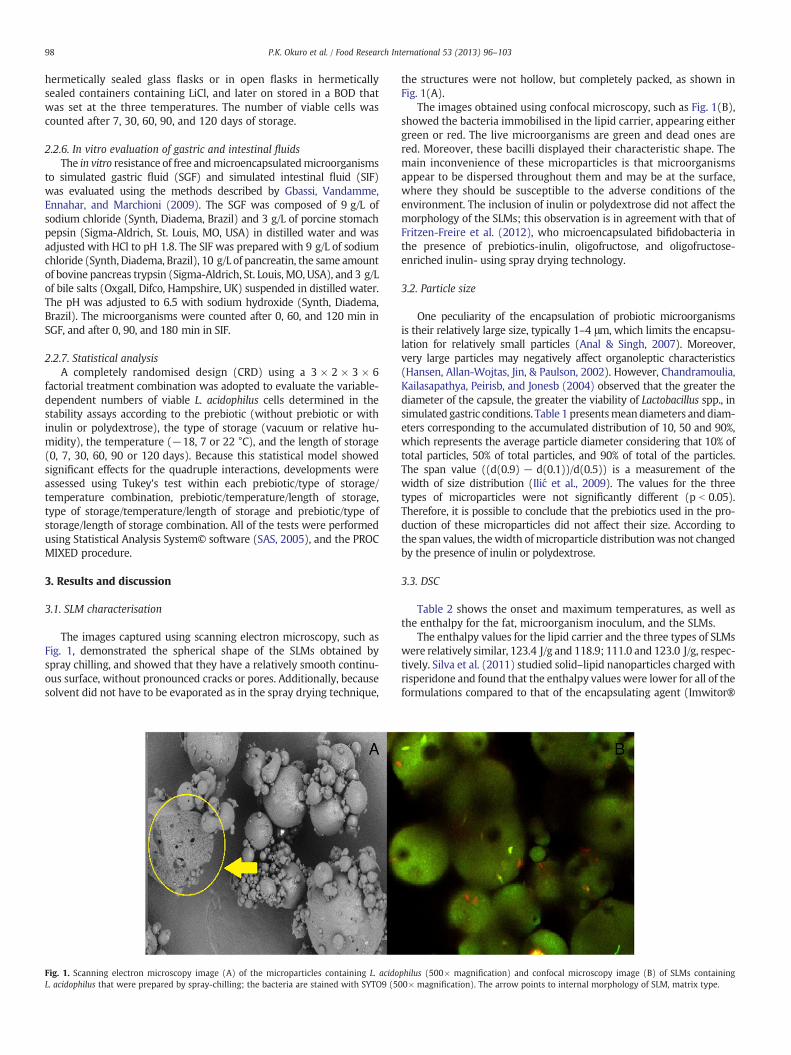
The images captured using scanning electron microscopy, such asFig. 1, demonstrated the spherical shape of the SLMs obtained byspray chilling, and showed that they have a relatively smooth continu-ous surface, without pronounced cracks or pores. Additionally, becausesolvent did not have to be evaporated as in the spray drying technique,
Fig. 1. Scanning electron microscopy image (A) of the microparticles containing L. acidoL. acidophilus that were prepared by spray-chilling; the bacteria are stained with SYTO9 (5
the structures were not hollow, but completely packed, as shown inFig. 1(A).
The images obtained using confocal microscopy, such as Fig. 1(B),showed the bacteria immobilised in the lipid carrier, appearing eithergreen or red. The live microorganisms are green and dead ones arered. Moreover, these bacilli displayed their characteristic shape. Themain inconvenience of these microparticles is that microorganismsappear to be dispersed throughout them and may be at the surface,where they should be susceptible to the adverse conditions of theenvironment. The inclusion of inulin or polydextrose did not affect themorphology of the SLMs; this observation is in agreement with that ofFritzen-Freire et al. (2012), who microencapsulated bifidobacteria inthe presence of prebiotics-inulin, oligofructose, and oligofructose-enriched inulin- using spray drying technology.
3.2. Particle size
One peculiarity of the encapsulation of probiotic microorganismsis their relatively large size, typically 1–4 μm, which limits the encapsu-lation for relatively small particles (Anal & Singh, 2007). Moreover,very large particles may negatively affect organoleptic characteristics(Hansen, Allan-Wojtas, Jin, & Paulson, 2002). However, Chandramoulia,Kailasapathya, Peirisb, and Jonesb (2004) observed that the greater thediameter of the capsule, the greater the viability of Lactobacillus spp., insimulated gastric conditions. Table 1 presentsmeandiameters anddiam-eters corresponding to the accumulated distribution of 10, 50 and 90%,which represents the average particle diameter considering that 10% oftotal particles, 50% of total particles, and 90% of total of the particles.The span value ((d(0.9) − d(0.1))/d(0.5)) is a measurement of thewidth of size distribution (Ilić et al., 2009). The values for the threetypes of microparticles were not significantly different (p b 0.05).Therefore, it is possible to conclude that the prebiotics used in the pro-duction of these microparticles did not affect their size. According tothe span values, the width of microparticle distributionwas not changedby the presence of inulin or polydextrose.
3.3. DSC
Table 2 shows the onset and maximum temperatures, as well asthe enthalpy for the fat, microorganism inoculum, and the SLMs.
The enthalpy values for the lipid carrier and the three types of SLMswere relatively similar, 123.4 J/g and 118.9; 111.0 and 123.0 J/g, respec-tively. Silva et al. (2011) studied solid–lipid nanoparticles charged withrisperidone and found that the enthalpy values were lower for all of theformulations compared to that of the encapsulating agent (Imwitor®
philus (500× magnification) and confocal microscopy image (B) of SLMs containing00× magnification). The arrow points to internal morphology of SLM, matrix type.

Table 1Effect of different formulations on the particle size distribution of the microparticles(average ± SD; n = 3).
Formulation D(4,3)*(μm)
D(0,1)**(μm)
D(0,5)***(μm)
D(0,9)****(μm)
SPAN
F1 65.2 ± 8.1ª 38.2 ± 11.6 70.0 ± 3.7 99.7 ± 0.4 0.879 ± 0.198F2 63.7 ± 5.1ª 37.4 ± 5.6 68.9 ± 2.5 99.9 ± 1.3 0.886 ± 0.099F3 66.1 ± 3.9ª 41.8 ± 6.3 70.0 ± 1.7 98.0 ± 3.9 0.848 ± 0.148
F1: L. acidophilus; F2: L. acidophilus + inulin; F3: L. acidophilus + polydextrose.
Fig. 2. X-ray diffraction patterns of F1-0 day (A), F1-90 days (B), F2-0 day (C), F2-90 days(D), F3-0 day (E) and F3-90 days (F).
99P.K. Okuro et al. / Food Research International 53 (2013) 96–103
900K). However, in the study of Silva et al. (2011), nanocapsules wereproduced by first adding surfactants to obtain pre-emulsions and thenusing two different techniques ultrasound (US) and high-pressurehomogenisation (HPH). The melting temperatures of the formulationswere very similar to each other and slightly higher than the meltingtemperature of pure fat, which is 43.7 °C. In fact, the melting tempera-tures for the formulations were expected to be similar because thelipid matrix used was the same for all of the formulations. Neither theinoculum, or the inoculum and the prebiotic, interact with the lipidmatrix of the SLMs and, therefore, they did not change the polymorphicbehavior of the mixtures used to produce them.
As for the possibility of melting, prematurely, the melting tempera-tures observed for the formulations were between 47.27 and 47.58 °C,which would make them physically stable at room temperature andin the mouth.
3.4. X-ray powder diffraction
Additional information on the solid state structures of SLMs wasobtained using X-ray powder diffraction. Fig. 2 shows the diffractionprofiles of the three formulations tested, when they were freshly pre-pared and after 90 days of storage. Because with the SLM have a lipidmatrix, a trend for polymorphic reorganisation should be consideredfor more energetically favourable levels, and this behavior may leadto the expulsion of the bioactive ingredient when it attains a morecrystalline arrangement (Gamboa, Gonçalves, & Grosso, 2011;Jenning, Thünemann, & Gohla, 2000; Müller, Radtke, & Wissing,2002a, 2002b; Schubert & Müller-Goymann, 2005). Fig. 2 showsthat the refractograms had similar shapes, independently of theformulations or the period of storage. The diffraction patterns of theSLMs are very similar to each other, indicating that no polymorphicchanges occurred during storage. The behavior of lipid materialobserved in this study was similar to that reported in the literature,which is generally associated with a polymorphic β form that is char-acteristic of triacylglycerol and fatty acids (Gamboa et al., 2011).
3.5. FTIR
The FTIR assay detected possible interactions between the encapsu-lated materials and the carrier. Fig. 3 shows peaks of interesterifiedpalm and palm-kernel oil at 1738, 2851, and 2919 cm−1 in the5(B)spectrum. These peaks are associatedwith the presence of carbonylated
Table 2DSC parameters for the encapsulating matrix, bioactive material (inoculum) andformulations prepared with L. acidophilus with or without addition of a prebiotic.
Sample Peak (°C) OnSet (°C) Enthalpy (J/g)
Fat 43.34 32.28 123.4Inoculum 47.82 47.24 2.89F1 47.43 39.29 118.9F2 47.27 34.98 111.0F3 47.58 34.52 123.0
F1: L. acidophilus; F2: L. acidophilus + inulin; F3: L. acidophilus + polydextrose.
compounds, more precisely, the vibration of COOH groups, and thepeak at 1738 cm−1 is related to C_O vibration. Passerini, Albertini,Perissutti, and Rodriguez (2006) used the lipid matrix Gelucire 50/13to encapsulate a drug (Praziquantel), and observed a large band in the3650–3100 cm−1 region that was related to the vibration of the O\Hbonds in the COOH groups.
The infrared spectrum of inulin 3 (C-solid line) demonstrated thepolysaccharide nature of the C\O\C bond that is characteristic ofcarbohydrates between 1200 and 900 cm−1, which confirmed thatthe monomers had bound to one another to form polymers. Theband for the O\H bond in the monosaccharide structure is observedbetween 3600 and 3000 cm−1. An angular deformation of the bondat 1639 cm−1 is also observed. Asymmetric stretching of the CH2
group is observed between 2900 and 2950 cm−1 (Silverstein, Webster,& Kiemle, 2006).
The spectrum for polydextrose 3(C-broken line) displays apronounced vibration related to C\O\C glycosidic bond (1202–927 cm−1), C_O stretching vibration of aldehyde (1659 cm−1),O\H stretching vibrations (3640–2978 cm−1) and a C\H stretchingvibration at 2946 cm−1 (Mickova, Copikova, & Synytsya, 2007).
Analysis of the infrared spectra of the SLMs, shown in Fig. 3(D,E,F),revealed that none of the peaks characteristic of the encapsulatedmaterial (prebiotic and probiotic) or the carrier were changed,suggesting the absence of significant interactions.
3.6. Resistance to encapsulation by spray chilling
Table 3 shows the values pertaining of resistance to the produc-tion process in all the formulations. Based on these results, the micro-organisms are highly resistant to the spray chilling process, that is,atomisation and cooling of the molten mixture to maintain the viabil-ity of the L. acidophilus cells, both in the mixture with molten carrierand in the SLMs. It is conceivable that spray chilling had very littleeffect on cellular integrity. A similar behavior was reported byPedroso et al. (2012), who employed the same encapsulation process.Ultraturrax homogenisation, as well as the exposure to heat when thecells were added to the molten fat, did not seem to have had strongimpact on cell survival, considering the initial count of the microor-ganisms in the inoculum and their dilution at 4 g/100 mL in the mol-ten mixture.

Fig. 3. FT-IR spectra of L. acidophilus (A), palm oil and palm-kernel oil (GPPI) (B), Inulin(solid line) and polydextrose (broken line) (C), F1(D), F2(E) and F3 (F).
Table 3Resistance of L. acidophilus to the spray-chilling process.
Formulation Viable cells (count in log10 cfu g−1)
Inoculum Suspension a SLMsb
F1 10.46 ± 0.30 8.67 ± 0.31 8.34 ± 0.38F2 8.53 ± 0.34 8.29 ± 0.27F3 8.57 ± 0.26 8.32 ± 0.09
F1: L. acidophilus; F2: L. acidophilus + inulin; F3: L. acidophilus + polydextrose.a Before atomisation.b
100 P.K. Okuro et al. / Food Research International 53 (2013) 96–103
3.7. Resistance to gastrointestinal fluids
In the present study, microencapsulation protected La from simu-lated gastrointestinal conditions (Fig. 4). The free cells count reachedthe limit of the method (10−2 CFU/g) by 210 min in the assay, dem-onstrating that the cells were susceptible to the simulated conditions.Although free viable cells were not detectable after 210 min, approx-imately 60% of the La cells in the SLMs produced with or without aprebiotic were found to be viable. There was a reduction of 2.99;2.79; 2.84 log cycles for formulations F1, F2, and F3, respectively, by300 min of treatment.
Light microscopy (Fig. 4) showed that the SLMs were intact,well-defined spheres at the start of the simulated gastrointestinalconditions assays. The most significant disintegration of SLMs oc-curred when they were exposed to simulated intestinal fluid (SIF),where the presence of pancreatin, trypsin, and bile salts, togetherwith increased pH (6.5), created conditions favourable for the rup-ture/disintegration of SLMs, due to their lipid content.
The release of the active components (bacteria and prebiotics)was correlated with a more drastic reduction in the viable cell countbecause probiotics that are not protected in the SLMs are more sus-ceptible to external conditions.
SGF-treatment did not significantly reduce (p ≤ 0.05) the viablecell count associated with the SLMs containing polydextrose (60 to120 min) or the SLMs lacking a prebiotic (0 to 60 min), but a signifi-cant reduction in cell viability was observed for all of the SLMs thatpassed from SGF to SIF and for those incubated in intestinal fluid(120 to 210 min and 210 to 300 min). At the end of the experiment,the cell counts for the F2 and F3 formulations and for the F1 and F2formulations were not significantly different, whereas the cells inthe F2 and F3 formulations demonstrated better resistance to thefluids than did those in the F1 formulation. These results showedthat the F2 and F3 formulations could be used to improve the resis-tance of La to the simulated gastrointestinal conditions.
There was a reduction of 2.19, 1.75 and 1.73 log cycles for F1, F2,and F3, respectively. There was a significant reduction in viable cellscounts when probiotics were exposed to bile salts. The antimicrobialnature of bile salts is related to its detergent property, which dissolvesmicroorganismmembranes, and its amphiphilic naturemakes it stronglyinhibitory for the gastrointestinal tract (Madureira, Amorim, Gomes,Pintado, & Malcata, 2011; Senaka Ranadheera, Evans, Adams, & Baines,2012).
Kim et al. (2008) observed similar results. They reported a 3log-cycle reduction in viable cells when microparticles of L. acidophilusATCC 43121 microencapsulated with sodium alginate were exposedto artificial gastric juices at pH 1.2 and 1.5. Other studies also foundbetter survival for encapsulated L. acidophilus than for free cells whenthey were exposed to simulated gastric and intestinal solutions(Favaro-Trindade & Grosso, 2002; Kim et al., 2008; Sabikhi, Babu,Thompkinson, & Kapila, 2010).
3.8. Viability
High relative humidity negatively affects the viability of microor-ganisms in capsules during storage because the increase in water
After the spray-chilling process.

0
1
2
3
4
5
6
7
8
9
10
0
20
40
60
80
100
0 60 120 210 300
Via
ble
cel
ls lo
g10
(CF
U.g
-1)
Via
ble
cel
ls (
%)
Time (minutes)
La_free cells La_T1 La_T2 La_T3 La_free cells La_T1 La_T2 La_T3
Fig. 4. Survival of free and microencapsulated Lactobacillus acidophilus cells after their exposure to simulated gastric fluid (0–120 min) and simulated intestinal fluid(120–300 min). The bars represent the survival of the probiotic as percentages and the lines represent the survival in Log (CFU/g).
101P.K. Okuro et al. / Food Research International 53 (2013) 96–103
content is detrimental to the microorganisms due to the accelerationof oxidative process (Rodrigues et al., 2011; Teixeira, Castro, Malcata,& Kirby, 1995).
Therefore, in this study, we attempted to decrease Aw of themicroparticles by incorporating prebiotics to inhibit the metabolic
Table 4Effects of the storage parameters on the stability of microencapsulated L. acidophilus throug
Preb Storageconditions
T (°C) Viable cells (log cfu g−1)
0 days 7 days 30 days
WP UR (11%) −18 8.381 ± 0.640 a A 7.513 ± 0.107 b A 7.028 ± 0.15WP UR (11%) 7 8.381 ± 0.640 a A 7.155 ± 0.295 b A 5.021 ± 0.82WP UR (11%) 22 8.381 ± 0.640 a A 4.233 ± 0.487 b B 3.470 ± 0.72WP Vácuo −18 8.381 ± 0.640 a A 7.519 ± 0.372 b A 6.725 ± 0.17WP Vácuo 7 8.381 ± 0.640 a A 4.993 ± 0.090 b B 2.941 ± 0.65WP Vácuo 22 8.381 ± 0.640 a A 3.709 ± 0.176 b C 2.319 ± 0.58Inu UR (11%) −18 8.194 ± 0.444 a A 7.815 ± 0.268 a A 6.751 ± 0.12Inu UR (11%) 7 8.194 ± 0.444 a A 6.993 ± 0.090 b B 6.361 ± 0.17Inu UR (11%) 22 8.194 ± 0.444 a A 6.249 ± 0.083 b C 5.079 ± 0.19Inu Vácuo −18 8.194 ± 0.444 a A 7.746 ± 0.265 ab A 7.121 ± 0.05Inu Vácuo 7 8.194 ± 0.444 a A 4.992 ± 0.450 b B 2.464 ± 1.41Inu Vácuo 22 8.194 ± 0.444 a A 3.759 ± 0.153 b C 1.737 ± 0.74Poly UR (11%) −18 8.964 ± 0.331 a A 8.472 ± 0.545 ab A 7.979 ± 0.39Poly UR (11%) 7 8.964 ± 0.331 a A 8.314 ± 0.225 b A 7.491 ± 0.37Poly UR (11%) 22 8.964 ± 0.331 a A 8.111 ± 0.407 b A 6.915 ± 0.85Poly Vácuo −18 8.964 ± 0.331 a A 7.975 ± 0.273 b A 7.523 ± 0.11Poly Vácuo 7 8.964 ± 0.331 a A 6.006 ± 0.963 b B 2.386 ± 1.37Poly Vácuo 22 8.964 ± 0.331 a A 4.330 ± 0.402 b C 2.007 ± 1.25
Viable cell counts for each prebiotic-storage condition- temperature combination were afferows are significantly different (Tukey test: p ≤ 0.05). Viable cell counts for each prebiotemperature; values denoted with different capital letters within the columns are significanNS: no surviving cells.
activity of the immobilised probiotic microorganisms and therebyimprove their stability during storage. A determinate number of viableprobiotic cells must be consumed (106–107 CFU/g) for them to play asignificant role when they eventually reach the intestine (Sabikhi etal., 2010; Talwalkar, Miller, Kailasapathy, & Nguyen, 2004).
hout 120 days of storage.
60 days 90 days 120 days
8 b A 5.639 ± 0.142 c A 5.271 ± 0.237 c A 4.024 ± 0.088 d A9 c B 4.529 ± 0.824 cd B 4.061 ± 0.851 d B 3.085 ± 0.176 e B3 c C 2.314 ± 0.160 d C 1.435 ± 0.298 e C NS f C2 c A 5.830 ± 0.082 d A 5.081 ± 0.093 e A 3.757 ± 0.541 f A0 c B 2.000 ± 0.990 d B NS e B NS e B4 c B 1.724 ± 0.345 c B NS d B NS d B1 b A 6.432 ± 0.193 bc A 6.063 ± 0.211 cd A 5.708 ± 0.204 d A5 c A 4.843 ± 0.146 d B 4.450 ± 0.161 de B 3.954 ± 0.041 e B7 c B 4.554 ± 0.149 cd B 4.256 ± 0.091 d B 3.603 ± 0.512 e B9 b A 5.654 ± 0.097 c A 5.117 ± 0.199 c A 4.462 ± 0.153 d A5 c B 0.956 ± 1.105 d B NS e B NS e B4 c C NS d C NS d B NS d B3 bc A 7.618 ± 0.192 cd A 7.125 ± 0.063 d A 6.997 ± 0.085 d A0 c AB 7.397 ± 0.199 cd A 7.068 ± 0.037 cd A 6.812 ± 0.039 d A3 c B 6.009 ± 0.724 d B 5.554 ± 0.444 de B 4.939 ± 0.586 e B8 bc A 6.895 ± 0.170 cd A 6.388 ± 0.250 d A 5.517 ± 0.400 e A0 c B NS d B NS d B NS d B9 c B NS d B NS d B NS d B
cted by duration of storage; values denoted with different lowercase letters within thetic-storage condition–duration of storage combination were affected by the storagetly different (Tukey test: p ≤ 0.05).

Table 5Effects of the storage parameters on the stability of microencapsulated L. acidophilus throughout 120 days of storage.
Preb Storageconditions
T (°C) Viable cells (log cfu g−1)
0 days 7 days 30 days 60 days 90 days 120 days
WP UR (11%) −18 8.381 ± 0.640 a A 7.513 ± 0.107 b A 7.028 ± 0.158 b A 5.639 ± 0.142 c A 5.271 ± 0.237 c A 4.024 ± 0.088 c AInu UR (11%) −18 8.194 ± 0.444 b A 7.815 ± 0.268 b A 6.751 ± 0.121 b A 6.432 ± 0.193 b A 6.063 ± 0.211 b A 5.708 ± 0.204 b APoly UR (11%) −18 8.964 ± 0.331 a A 8.472 ± 0.545 a A 7.979 ± 0.393 a A 7.618 ± 0.192 a A 7.125 ± 0.063 a A 6.997 ± 0.085 a AWP UR (11%) 7 8.381 ± 0.640 ab A 7.155 ± 0.295 b A 5.021 ± 0.829 c A 4.529 ± 0.824 b A 4.061 ± 0.851 b A 3.085 ± 0.176 c AInu UR (11%) 7 8.194 ± 0.444 b A 6.993 ± 0.090 b A 6.361 ± 0.175 b A 4.843 ± 0.146 b A 4.450 ± 0.161 b A 3.954 ± 0.041 b APoly UR (11%) 7 8.964 ± 0.331 a A 8.314 ± 0.225 a A 7.491 ± 0.370 a A 7.397 ± 0.199 a A 7.068 ± 0.037 a A 6.812 ± 0.039 a AWP UR (11%) 22 8.381 ± 0.640 ab A 4.233 ± 0.487 c A 3.470 ± 0.723 c A 2.314 ± 0.160 c A 1.435 ± 0.298 c A NSInu UR (11%) 22 8.194 ± 0.444 b A 6.249 ± 0.083 b A 5.079 ± 0.197 b A 4.554 ± 0.149 b A 4.256 ± 0.091 b A 3.603 ± 0.512 b APoly UR (11%) 22 8.964 ± 0.331 a A 8.111 ± 0.407 a A 6.915 ± 0.853 a A 6.009 ± 0.724 a A 5.554 ± 0.444 a A 4.939 ± 0.586 a AWP Vácuo −18 8.381 ± 0.640 ab A 7.519 ± 0.372 a A 6.725 ± 0.172 b A 5.830 ± 0.082 b A 5.081 ± 0.093 b A 3.757 ± 0.541 c AInu Vácuo −18 8.194 ± 0.444 b A 7.746 ± 0.265 a A 7.121 ± 0.059 ab A 5.654 ± 0.097 b B 5.117 ± 0.199 b B 4.462 ± 0.153 b BPoly Vácuo −18 8.964 ± 0.331 a A 7.975 ± 0.273 a A 7.523 ± 0.118 a A 6.895 ± 0.170 a B 6.388 ± 0.250 a B 5.517 ± 0.400 a BWP Vácuo 7 8.381 ± 0.640 ab A 4.993 ± 0.090 b B 2.941 ± 0.650 a B 2.000 ± 0.990 a B NS NSInu Vácuo 7 8.194 ± 0.444 b A 4.992 ± 0.450 b B 2.464 ± 1.415 a B 0.956 ± 1.105 b B NS NSPoly Vácuo 7 8.964 ± 0.331 a A 6.006 ± 0.963 a B 2.386 ± 1.370 a B NS NS NSWP Vácuo 22 8.381 ± 0.640 ab A 3.709 ± 0.176 a A 2.319 ± 0.584 a B 1.724 ± 0.345 a A NS NSInu Vácuo 22 8.194 ± 0.444 b A 3.759 ± 0.153 a B 1.737 ± 0.744 a B NS NS NSPoly Vácuo 22 8.964 ± 0.331 a A 4.330 ± 0.402 a B 2.007 ± 1.259 a B NS NS NS
Viable cell counts for each storage conditions–temperature-duration of storage combinations were affected by prebiotic; values denoted with different lowercase letters within therows are significantly different (Tukey test: p ≤ 0.05). Viable cell counts for each prebiotic-temperature-duration of storage combination were affected by the storage condition;values denoted with different capital letters within the columns are significantly different (Tukey test: p ≤ 0.05).NS: no surviving cells.
102 P.K. Okuro et al. / Food Research International 53 (2013) 96–103
In the present study, according to the results presented in Tables 4and 5, the F3 formulation stored in UR at −18 °C and 7 °C for120 days would provide the number of viable cells required, andthey presented a logarithmic cycle reduction of 1.967 and 2.152,respectively. The good results obtained for low temperature storagemay be explained by the microorganisms being maintained in a latentstate and to the low temperature preventing crystal rearrangementsin the lipid matrix and thus expulsion of the bioactive component(Pedroso et al., 2012).
In a previous study, L. acidophilus retained greater viability at astorage temperature of 7 °C (120 days), as observed in this studyfor the F3 formulation, although in the previous study, the microcap-sules were produced by complex coacervation and were dehydratedby the spouted bed method (Oliveira et al., 2007b).
The effects of adding inulin or polydextrose on the survival ofLa are presented in Table 5. The data show that the presence ofpolydextrose increased cell viability (p ≤ 0.05) compared with thepresence of inulin or no prebiotic during storage with controlled rel-ative humidity at −18, 7 and 22 °C. The cell viability values for F1, F2and F3 formulations stored under vacuum for 30 days at 7 or 22 °Cwere not significantly different, whereas the cell viability after frozenstorage for 30 to 120 days was significantly greater for the F3formulation.
For all the formulations tested, it was observed that storage at thelowest temperature studied led to the best cell viability, as shown inTable 4. There is a consensus that temperatures close to 0 °C improvethe rates of cell viability because lower temperatures reduce the ratesof chemical reactions that are detrimental to the microorganisms,such as fatty acid oxidation (Corcoran, Ross, Fitzgerald, & Stanton,2004; Teixeira et al., 1995).
For F1, the storage at freezing temperature (−18 °C) showed thebest rate of viable cells, with counts greater than 6 log cycles, bothunder vacuum and controlled relative humidity up to 30 days.However, for the other temperatures analysed, stability was inade-quate, once there was a great reduction in the number of viablecells in 7 days, except for relative humidity at 7 °C, whose countsdecreased in 30 days, this result is possibly related to greater meta-bolic activity of the microorganisms when stored at 7 and 37 °C.High temperatures can lead to production of metabolic acids and bac-teriocins, and/or the loss of substrates, which would explain the inac-tivation of the viable cells in the microparticles during storage.
F2 showed the best performance at −18 °C, under controlled rel-ative humidity, with satisfactory counts up to 90 days; up to 30 dayswhen stored under controlled relative humidity at 7 °C; and undervacuum at −18 °C. Finally, F3 seemed to present the best responsesin viability during storage in controlled relative humidity (11%),where counts over 106 were recorded up to 120 days at −18 °Cand 7 °C, and up to 60 days at 22 °C, whereas under vacuum countsremained acceptable for 90 days at −18 °C.
It is known that not only temperature, but also relative humidity,is determinant for probiotic survival during storage. Moreover, theAw values are balanced at room relative humidity. Thus, storage athigh relative humidity increases the water concentration, which isdetrimental to the survival of probiotics. On the other hand, at verylow relative humidity, removal of water from the cells may increasethe possibility of damage. Therefore, intermediate relative humiditylevels, between 7 and 11%, were reported to be the ideal values forbacterial survival (Castro, Teixeira, & Kirby, 1995).
Pedroso et al. showed that microencapsulated L. acidophilus had ashelf life of 30 and 60 days at 7 and 37 °C, respectively. Although thisstudy used the same encapsulation process and lipid matrix theyused, they did not add prebiotic components or store the microcap-sules with a controlled relative humidity, which explains the lowerstability observed in their study. The presence of a prebiotic and thecontrolled storage conditions improved the stability of L. acidophilus.
4. Conclusions
Symbiotic solid lipid microparticles are potential vehicles ofprobiotic microorganisms and prebiotic compounds. The symbioticSLM-based systems could protect to L. acidophilus cells from theeffects of gastric and intestinal fluids and release them in the intestinesduring fat digestion.
SLMs improve the viability of L. acidophilus during storage at freezingor refrigeration temperatures with controlled relative humidity (11%).The best formulation studied combined L. acidophilus and polydextrosebecause this system maintained the viability of the stored microorgan-isms viable for 120 days.
The SLMs produced in this study are an interesting vehicle thatcan be applied by the food industry, given their specific morphologyand insoluble nature. Issues remain to be investigated, such as theeffects of different levels of prebiotics on the morphology of SLMs

103P.K. Okuro et al. / Food Research International 53 (2013) 96–103
and the probiotic cell viability as well as the effect of other lipidmatrices that may prolong probiotic cell viability during storage,thereby favouring the application of SLMs in food products.
Acknowledgements
The authors thank FAPESP for the financial support (Process 09/11713-2) and the scholarship that was granted (Process 10/13026-0).
References
Anal, A. K., & Singh, H. (2007). Recent advances in microencapsulation of probiotics forindustrial applications and targeted delivery. Trends in Food Science and Technology,18(5), 240–251.
Castro, H. P., Teixeira, P. M., & Kirby, R. (1995). Storage of lyophilized cultures ofLactobacillus bulgaricus under different relative humidities and atmospheres. AppliedMicrobiology and Biotechnology, 44, 172–176.
Chambi, H. N.M., Alvim, I. D., Barrera-Arellano, D., &Grosso, C. R. F. (2008). Solid lipidmicro-particles containingwater-soluble compounds of differentmolecularmass: production,characterization and release profiles. Food Research International, 41, 229–236.
Chandramoulia, V., Kailasapathya, K., Peirisb, P., & Jonesb, M. (2004). An improvedmethod of microencapsulation and its evaluation to protect Lactobacillus spp. insimulated gastric conditions. Journal of Microbiological Methods, 56, 27–35.
Cook,M. T., Tzortzis, G., Charalampopoulos, D., & Khutoryanskiy, V. V. (2012).Microencap-sulation of probiotics for gastrointestinal delivery. Journal of Controlled Release, 162,56–67.
Corcoran, B. M., Ross, R. P., Fitzgerald, G. F., & Stanton, C. (2004). Comparative survivalof probiotic lactobacilli spray-dried in the presence of prebiotic substances. Journalof Applied Microbiology, 96, 1024–1039.
Favaro-Trindade, C. S., & Grosso, C. R. F. (2000). The effect of the immobilization ofL. acidophilus and B. lactis in alginate on their tolerance to gastrointestinal secretions.Michwissenschaft, 55, 496–499.
Favaro-Trindade, C. S., & Grosso, C. R. F. (2002). Microencapsulation of L. acidophilus(La-05) and B. lactis (Bb-12) and evaluation of their survival at the values of stomachand in bile. Journal of Microencapsulation, 19(4), 485–494.
Favaro-Trindade, C. S., Heinemann, R. J. B., & Pedroso, D. L. (2011). Developments inprobiotic encapsulation. CAB Reviews: Perspectives in Agriculture, Veterinary Science,Nutrition and Natural Resources, 6, 1–8.
Favaro-Trindade, C. S., Santana, A. S., Monterrey-Quintero, E. S., Trindade, M. A., &Netto, F. M. (2010). The use of spray drying technology to reduce bitter taste ofcasein hydrolysate. Food Hydrocolloids, 24, 336–340.
Fritzen-Freire, C. B., Prudêncio, E. S., Amboni, R. D. M. C., Pinto, S. S., Negrão-Murakami,A. N., & Murakami, F. S. (2012). Microencapsulation of bifidobacteria by spray dryingin the presence of prebiotics. Food Research International, 45, 306–312.
Gamboa, O. D., Gonçalves, L. G., & Grosso, C. F. (2011). Microencapsulation of tocoph-erols in lipid matrix by spray chilling method. Procedia Food Science, 1, 1732–1739.
Gbassi, G. K., Vandamme, T., Ennahar, S., & Marchioni, E. (2009). Microencapsulationof Lactobacillus plantarum spp in an alginate matrix coated with whey proteins.International Journal of Food Microbiology, 129, 103–105.
Gibson, G. R., Probert, H. M., Van Loo, J., Rastall, R. A., & Roberfroid, M. B. (2004). Dietarymodulation of the human colonic microbiota: updating the concept of prebiotics.Nutrition Research Reviews, 17(2), 259–275.
Grosso, C. R. F., & Favaro-Trindade, C. S. (2004). Stability of free and immobilizedLactobacillus acidophilus and Bifidobacterium lactis in acidified milk and immobilizedB. lactis in yoghurt. Brazilian Journal of Microbiology, 35, 151–156.
Hansen, L. T., Allan-Wojtas, P. M., Jin, L. A., & Paulson, A. T. (2002). Survival ofCa-alginate microencapsulated Bifidobacterium spp. in milk and simulated gastro-intestinal conditions. Food Microbiology, 19, 35–45.
Heidebach, T., Forst, P., & Kulozik, U. (2010). Influence of casein-based microencapsu-lation on freeze-drying and storage of probiotic cells. Journal of Food Engineering,98, 309–316.
Homayouni, A., Azizi, A., Ehsani, M. R., Yarmand, M. S., & Razavi, S. H. (2008). Effect ofmicroencapsulation and resistant starch on the probiotic survival and sensoryproperties of synbiotic ice cream. Food Chemistry, 111, 50–55.
Hou, R. C. W., Lin, M. Y., Wang, M. C., & Tzen, J. T. C. (2003). Increase of viability ofentrapped cells of Lactobacillus delbrueckii ssp. Bulgaricus in artificial sesame oilemulsions. Journal of Dairy Science, 86(2), 424–428.
Ilić, I., Dreu, R., Burjak, M., Homar, M., Kerč, J., & Srčič, S. (2009). Microparticle sizecontrol and glimepiride microencapsulation using spray congealing technology.International Journal of Pharmaceutics, 381, 176–183.
Jenning, V., Thünemann, A. F., & Gohla, S. H. (2000). Characterisation of a novel solidlipid nanoparticle carrier system based on binary mixtures of liquid and solidlipids. International Journal of Pharmaceutics, 199, 167–177.
Kanmani, P., Kumar, R. S., Yuvaraj, N., Paari, K. A., Pattukumar, V., & Arul, V. (2011).Cryopreservation and microencapsulation of a probiotic in alginate chitosan capsulesimproves survival in simulated gastrointestinal conditions. Biotechnology and BioprocessEngineering, 16(6), 1106–1114.
Kim, S. J., Cho, S. Y., Kim, S. H., Song, O. J., Shin, S., Cha, D. S., et al. (2008). Effect ofmicroencapsulation on viability and other characteristics in Lactobacillus acidophilusATCC 43121. LWT- Food Science and Technology, 41, 493–500.
Lahtinen, S. J., Ouwehand, A. C., Salminen, S. J., Forssell, P., & Myllärinen, P. (2007).Effect of starch and lipid-based encapsulation on the culturability of twoBifidobacterium longum strains. Letters in Applied Microbiology, 44, 500–505.
Madureira, A. R., Amorim, M., Gomes, A. M., Pintado, M. E., & Malcata, F. X. (2011).Protective effect of whey cheese matrix on probiotic strains exposed to simulatedgastrointestinal conditions. Food Research International, 44(1), 465–470.
Mandal, S., Hati, S., Puniya, A. K., Singh, R., & Singh, S. (2012). Development of synbioticmilk chocolate using encapsulated Lactobacillus casei NCDC 298. Journal of FoodProcessing and Preservation, 1–7.
Mandal, S., Puniya, A. K., & Singh, K. (2006). Effect of alginate concentrations on survivalof microencapsulated Lactobacillus casei NCDC-298. International Dairy Journal, 16,1190–1195.
McCartney, A. L., & Gibson, G. R. (2006). The normal microbiota of the human gastroin-testinal tract: history of analysis, succession, and dietary influences. In A. C.Ouwehand, & E. E. Vaughan (Eds.), Gastrointestinal Microbiology (pp. 51–73).New York: Taylor and Francis.
Mickova, K., Copikova, J., & Synytsya, A. (2007). Determination of polydextrose as a fatreplacer in butter. Czechoslovak Journal of Food Sciences, 25, 25–31.
Müller, R. H., Radtke, M., & Wissing, S. A. (2002b). Solid lipid nanoparticles (SLN) andnanostructured lipid carriers (NLC) in cosmetic and dermatological preparations.Advanced Drug Delivery Reviews, 54(1), 131–155.
Müller, R. H., Radtke, M., & Wissing, S. A. (2002a). Nanostructured lipid matrices forimproved microencapsulation of drugs. International Journal of Pharmaceutics,242, 121–128.
Nag, A., Han, K. S., & Singh, H. (2011). Microencapsulation of probiotic bacteria usingpH-induced gelation of sodium caseinate and gellan gum. International Dairy Journal,21, 247–253.
Oliveira, A. C., Moretti, T. S., Boschini, C., Balieiro, J. C. C., Freitas, L. A. P., Freitas, O., et al.(2007). Microencapsulation of B. lactis (BI 01) and L. acidophilus (LAC 4) by complexcoacervation followed by spouted-bed drying. Drying Technology, 25, 1687–1693.
Oliveira, A. C., Moretti, T. S., Boschini, C., Baliero, J. C. C., Freitas, O., & Favaro-Trindade, C.S. (2007). Stability of microencapsulated B. lactis (BI 01) and L. acidophilus (LAC 4)by complex coacervation followed by spray drying. Journal of Microencapsulation,24, 685–693.
Passerini, N., Albertini, B., Perissutti, B., & Rodriguez, L. (2006). Evaluation of meltgranulation and ultrasonic spray congealing as techniques to enhance the dissolutionof praziquantel. International Journal of Pharmaceutics, 318, 92–102.
Pedroso, D. L., Thomazini, M., Heinemann, R. J. B., & Favaro-Trindade, C. S. (2012). Protectionof Bifidobacterium lactis and Lactobacillus acidophilus by microencapsulation usingspray-chilling. International Dairy Journal, 26, 127–132.
Picot, A., & Lacroix, C. (2003). Production of multiphase water-insoluble microcapsulesfor cell microencapsulation using an emulsification/spray-drying technology.Journal of Food Science, 68(9), 2693–2700.
Pimentel-González, D. J., Camposmontiel, R. G., Lobato-Calleros, C., Pedroza-Islas, R., &Vernoncarte, J. (2009). Encapsulation of Lactobacillus rhamnosus in double emulsionsformulated with sweet whey as emulsifier and survival in simulated gastrointestinalconditions. Food Research International, 42(2), 292–297.
Roberfroid, M. B. (2007). Inulin-type fructans: functional food ingredients. The Journalof Nutrition, 137(11), 2493–2502.
Rodrigues, D., Sousa, S., Rocha-Santos, T., Silva, J. P., Sousa Lobo, J. M., Costa, P., et al.(2011). Influence of L-cysteine, oxygen and relative humidity upon survivalthroughout storage of probiotic bacteria in whey protein-based microcapsules.International Dairy Journal, 21, 869–876.
Sabikhi, L., Babu, R., Thompkinson, D. K., & Kapila, S. (2010). Resistance ofmicroencapsulated Lactobacillus acidophilus LA1 to processing treatments and sim-ulated gut conditions. Food and Bioprocess Technology, 3, 586–593.
SAS Institute Inc. (2005). SAS OnlineDoc, v. 9.1.3. Cary, North Carolina: SAS Institute Inc.Schubert, M. A., & Müller-Goymann, C. C. (2005). Characterisation of surfacemodified
solid lipid nanoparticles (SLN): influence of lecithin and nonionic emulsifier.European Journal of Pharmaceutics and Biopharmaceutics, 61, 77–86.
Senaka Ranadheera, C., Evans, C. A., Adams, M. C., & Baines, S. K. (2012). In vitro analysisof gastrointestinal tolerance and intestinal cell adhesion of probiotics. Food ResearchInternational, 49, 619–625.
Shah, N. P., & Ravula, R. R. (2000). Microencapsulation of probiotic bacteria and theirsurvival in frozen fermented dairy desserts. Australian Journal of Dairy Technology,55, 127–131.
Shoji, A. S., Oliveira, A. C., Balieiro, J. C. C., Freitas, O., Thomazini, M., Heinemann, R. J. B.,et al. (2013). Viability of L. acidophilus microcapsules and their application tobuffalo milk yoghurt. Food and Bioproducts Processing, 91, 83–88.
Silva, A. C., González-Mira, E., García, M. L., Egea, M. A., Fonseca, J., Silva, R., et al. (2011).Preparation, characterization and biocompatibility studies on risperidone-loadedsolid lipid nanoparticles (SLN): high pressure homogenization versus ultrasound.Colloids and Surfaces. B, Biointerfaces, 86, 158–165.
Silverstein, R. M., Webster, F. X., & Kiemle, D. J. (2006). Identificação espectrométrica decompostos orgânicos (7th ed.). Rio de Janeiro: LTC.
Sohail, A., Turner, M. S., Coombes, A., Bostrom, T., & Bhandari, B. (2011). Survivability ofprobiotics encapsulated in alginate gel microbeads using a novel impinging aerosolsmethod. International Journal of Food Microbiology, 145, 162–168.
Talwalkar, A., Miller, C. W., Kailasapathy, K., & Nguyen, M. H. (2004). Effect of packagingmaterials and dissolved oxygen on the survival of probiotic bacteria in yoghurt.International Journal of Food Science and Technology, 39, 605–611.
Teixeira, P. C., Castro, M. H., Malcata, F. X., & Kirby, R. M. (1995). Survival of Lactobacillusdelbrueckii ssp.bulgaricus following spray-drying. Journal of Dairy Science, 78, 1025–1031.
Vernazza, C. L., Rabiu, B. A., & Gibson, G. R. (2006). Human colonic microbiology and therole of dietary intervention: Introduction to prebiotics. Prebiotics: Development andapplication (pp. 1–28). Chichester: John Wiley & Sons Press.
Wells, A. L., Saulnier, D. M. A., & Gibson, G. R. (2008). Gastrointestinal microflora andinteractions with gut flora. In G. R. Gibson, & M. B. Roberfroid (Eds.), Handbook ofPrebiotics (pp. 14–38). New York: CRC Press.