formulation of ciprofloxacin hydrochloride loaded biodegradable...
TRANSCRIPT

Journal of Pharmaceutical Research and Opinion 3: 11 (2013) 72 - 81.
www.innovativejournal.inContents lists available at
JOURNAL OF PHARMACEUTICAL RESEARCH AND OPINION
http://www.innovativejournal.in/index.php/jpro Journal homepage:
72
RESEARCH
FORMULATION OF CIPROFLOXACIN HYDROCHLORIDE LOADED BIODEGRADABLE NANOPARTICLES: OPTIMIZATION OF THE FORMULATION VARIABLES.
Elkheshen S.A.,*1 Mobarak D.H.,2 Salah S., 2and Essam T 3
1Professor of Pharmaceutics and Industrial Pharmacy and Dean, Faculty of Pharmaceutical Sciences and Pharmaceutical Industries, Future University in Egypt, FUE.
2Department of Pharmaceutics and Industrial Pharmacy, Faculty of Pharmacy, Cairo University, Kasr El-aini street, Cairo 11562, Egypt.
3 Department of Microbiology, Faculty of Pharmacy, Cairo University, Kasr El-aini street,Cairo 11562,Egypt.
ARTICLE INFO
ABSTRACT
Received 20th Nov 2013 Accepted 26th Nov2013
Corresponding Author: Seham A. Elkheshen Professor of Pharmaceutics and Industrial Pharmacy and Dean, Faculty of Pharmaceutical Sciences and harmaceutical Industries, Future University in Egypt, FUE, New Cairo, Egypt KeyWords: Ciprofloxacin HCl, Nanoparticles, Biodegradable polymers, Factorial design, In-vitro release, Microbiological study.
The aim of this work was to formulate nanoparticles with longer sustainment since it can play an important role in treatment of chronic osteomyelitis. A factorial design 23 was used to optimize the formulation variables of ciprofloxacin hydrochloride (CP) loaded biodegradable nanoparticles (NPS). The effect of three independent variables, namely; the polymer type (Poly D, L-lactide-co-glycolide 50/50& Poly (D, L-lactide), the polymer molecular weight(M.Wt) and the drug to polymer ratio on the particle size, encapsulation efficiency (EE%), zeta potential and the time required for 90% of ciprofloxacin to be released (t90%) was investigated . The selected drug loaded nanoparticles was subjected to further investigations including; Scanning electron microscope (SEM), DSC, x-ray powder diffractometry and FTIR spectroscopy. Moreover the antimicrobial effectiveness of the selected formula was investigated. The results revealed that the mean particle size ranged from 180-355 nm while the EE% ranged from 52-70%. The selected formula retained its antimicrobial activity throughout the preparation process.
©2013, JPRO, All Right Reserved.
1 INTRODUCTION The use of a locally applied carrier that would sustain the release of the antibiotic for a longer period of time thus prolonging its antimicrobial effect was recommended by Lesic et al( 1). Yet sustainment of a high serum concentration for an extended period would consequently subject the patient to higher incidence of side effects and result in poor patient compliance ( 2). The use of localized dosage form was recommended in various literature sites since it would enable high drug concentration at the site of infection with low serum drug level and reducing unwanted toxic side effects reflecting higher patient compliance( 2,3) In a preceding study (4) Poly lactide-co –glycolic acid (PLGA 502 H) nanoparticles incorporating ciprofloxacin HCl (CP) were prepared by double emulsion solvent diffusion technique to improve and sustain the availability of the drug for the treatment of chronic infectious diseases after parenteral administration. Optimization of the formulation steps using High pressure Homogenizer was carried out using a factorial design. The rationale of the presented paper was to formulate nanoparticles with longer sustainment since it can play an important role in treatment of chronic osteomyelitis as it increases the patient compliance and more importantly is
expected to decrease the duration of treatment (1). Different factors that were expected to increase sustainment were tried including the use of more hydrophobic polymer such as PLA, increasing the M.Wt. of the polymer in addition to increasing the polymer to drug ratio. The aforementioned factors were tested in the preparation of NPs targeting some characteristics including: size above 100 nm, satisfactory EE%, controlled release and preserved antimicrobial activity of the encapsulated drug. 2.Materials and Methods: 2.1.Materials: Ciprofloxacin hydrochloride was kindly supplied by EPICO Company,Cairo, Egypt, 50:50 Poly D, L-lactide-co-glycolic acid, inherent viscosity: 0.16-0.24 dL/g, M.Wt.: 7000-17000 (Resomer RG 502 H), 50:50 Poly D, L-lactide-co-glycolic acid, inherent viscosity: 0.45-0.6 dL/g, M.Wt.: 38,000-54,000 were purchased from Sigma Aldrich Inc. AL., USA, Poly (D, L-lactide) ester, inherent viscosity: 0.34 dL/g. was purchased from LACTEL Absorbable Polymers Inc. AL., USA, Poly (D, L-lactide) ester, inherent viscosity: 1.6 dL/g. was purchased from Boehringer Ingelheim Inc., Germany, Poly vinyl alcohol: M.Wt.: 22000 was purchased from Sigma Aldrich Inc.AL., USA, Ethyl acetate, anhydrous

Elkhesan et.al/ Formulation of ciprofloxacin hydrochloride loaded biodegradable nanoparticles: Optimization of the formulation variables.
73
disodium hydrogen phosphate, Potassium dihydrogen orthophosphate, Sodium chloride and mannitol were purchased from El-Nasr Pharmaceutical Company, Cairo, Egypt. 2.2.Methods: 2.2.1.Optimization of the formulation variables of Ciprofloxacin Hcl loaded biodegradable nanoparticles using double emulsion solvent diffusion technique 2.2.1.1. Design of the Experiment: A factorial 23 design was used for the optimization of the preparation of CP biodegradable NPs. The study design involved the investigation of the effect of three independent variables, namely; the polymer type, the polymer M.Wt. and the drug to polymer ratio on the particle size, EE%, zeta potential and t90% of the in vitro drug release. The variables were investigated at two different levels. The levels of the variables are presented in table (1). 2.2.1.2. Preparation of ciprofloxacin hydrochloride loaded nanoparticles using double emulsion solvent diffusion technique: Ciprofloxacin hydrochloride NPs dispersion was prepared according to the double emulsion solvent diffusion technique. Briefly; 50 mg of CP hydrochloride was dissolved in 2 ml of 1% PVA aqueous solution. A specified amount of different M.Wt. of biodegradable polymers namely; 50/50 PLGA and DL-PLA was dissolved in 7 ml of ethyl acetate. The aqueous phase was poured on the organic phase and was immediately sonicated with the probe sonicator with diameter of 6 mm and a 130 W high-intensity ultrasonic processor(ultrasonic processor, GE130 ,Newton, USA) operating at 20 kHz. Samples were placed in ice bath and ultrasonicated. Ultrasonication was carried out at a specified amplitude of 80% over a duration of 2 min (5,6) to produce the primary emulsion. The primary emulsion was added to 10 ml of 1% PVA aqueous solution after which the dispersion step was made using the high pressure homogenizer (Model SPCH-10 , Stansted Fluid Power Ltd., Essex, UK ) at pressure of 600 bar(8702 psi) for 5 cycles at room temperature (7)
followed by the probe sonication for 2 minutes at amplitude of 80% while cooling in ice bath. The final emulsion was poured onto 50 ml of 0.5% PVA solution in phosphate buffer of pH 7.4 containing 0.1M Nacl. To remove the extracted ethyl acetate, the emulsified system was stirred using magnetic stirrer at room temperature for 1-2 hours. Finally to collect the NPs, centrifugation was done at 15000 rpm at room temperature for 30 minutes then the NP precipitate was redispersed and washed with distilled water and centrifuged afterwards to remove any residual free drug or free surfactant .(5) The washing step was repeated three times. After the final washing nanospheres were resuspended in 5 ml of water solution containing 5%(w/v) mannitol as cryoprotectant and freeze dried (Novolyphe-NL 500, Savant instruments Corp. USA) for 72 hours. The lyophilized nanoparticles were kept for further investigation. 2.2.1.3. Statistical Analysis: All statistical analysis were made using ANOVA test. The tests were followed by Fisher’s PLSD (pair wise least significance difference) for multiple comparisons at P ≤ 0.05 with Stat View statistical software program version 4.57.
Table(1):Composition of ciprofloxacin HCl loaded nanoparticles prepared according to 2 3 factorial design.
Formula No.
Code Factors studied at two levels each: present (+) and Absent (-) Polymer type
Drug:Polymer weight ratio
Polymer molecular weight Low (-)
High (+)
F1 PLGA/L/5 _ _ _
F2 PLGA/L/10 _ + _
F3 PLGA/H/5 _ _ +
F4 PLGA/H/10 _ + +
F5 PLA/L/5 + _ _
F6 PLA/L/10 + + _
F7 PLA/H/5 + + _
F8 PLA/H/10 + + +
2.2.2. Evaluation of the prepared ciprofloxacin hydrochloride biodegradable NPs: 2.2.2.1.Determination of particle size: Particle size analysis was carried out using Malvern Zetasizer Nanoseries; (Malvern Inst. Limited, UK). This relies on dynamic light scattering that measures the Brownian motion and relates it to the particle size by Photon Correlation Spectroscopy. All values were based on the intensity distribution, measurements were made at 25 ˚C and readings were done at angle 173˚. A portion of the freshly prepared suspension was diluted with distilled water before measurement. Measurements were carried out in duplicates. 2.2.2.2. Determination of drug encapsulation efficiency: To determine drug encapsulation efficiency, 15 mg of lyophilised nanoparticles were dissolved in 10 ml of dichloromethane and then 15 ml of deionised water was added. The mixture was stirred magnetically for 4 hours and then 1 ml of water phase was taken to measure drug EE% (8) ). Absorbance of aqueous phase was measured at 277 nm using UV-spectrophotometer (UV spectrophotometer 1601 PC; Shimadzu, Kyoto, Japan). Measurements were carried out in duplicates. Drug EE% was as follows:

Elkhesan et.al/ Formulation of ciprofloxacin hydrochloride loaded biodegradable nanoparticles: Optimization of the formulation variables.
74
EE% = (drug weight in the nanoparticles /Wt of initial drug)*100
2.2.2.3. Zeta potential measurments
The zeta potential that gives an indication on the surface charge of the nanoparticles was measured using Zetasizer Nanoseries (Malvern Inst. Limited, UK). This Zetasizer Model uses a combination of laser Doppler velocimetry and phase analysis light scattering (PALS) in a patented technique called M3-PALS. All readings were made at 25˚ C. A portion of the freshly prepared suspension was diluted with distilled water before measurement and was injected in the zeta particle sizer capillary cell with electrodes at both ends. Measurements were carried out in duplicates. 2.2.2.4. In vitro release and kinetic analysis of the release data of CP from its biodegradable NPs: The in vitro drug release study was performed using a dialysis membrane diffusion technique. A dialysis membrane of 12000-14000 M.Wt. cut off was used to retain the nanoparticles, while enabling the free drug to diffuse freely into the release media(8-11). Briefly, 1ml of CP nanoparticles suspension, containing an amount equivalent to 2.5mg of CP was transferred into the dialysis membrane, both ends were sealed. Afterwards it was immersed in a tightly capped glass cylinder (7x2.8 cm) containing 14 ml of phosphate buffer saline of pH 7.4 (PBS, pH 7.4). The release test was performed by placing the glass cylinder in a thermostatically controlled shaking water bath adjusted at 37±0.5˚C with constant shaking rate of 100 rpm. At predetermined time points the whole release medium was withdrawn and replaced with fresh release medium to maintain the sink condition (12). The concentration of released drug was measured with UV spectrophotometer (UV-1601 PC; Shimadzu, Japan) at 272 nm against PBS as blank. All experiments were done in duplicates. The data of the in vitro drug release of CP was treated according to different kinetic orders to determine the mechanism of drug release. Data were fitted to Zero- and First- order kinetics, as well as, to Higuchi’s diffusion model (13). The kinetic parameters and correlation coefficient were calculated for the in-vitro release of CP from nanoparticles. 2.2.3. Choosing the formula of superior characteristics: Four of the main characteristics of nanoparticles are discussed in this chapter, which are the particle size, zeta potential, EE% and release profile presented by the t90%. Each character is as important as the other. In order to choose the formula of choice a group of mathematical equations were applied to rank all the formulae within each of the aforementioned responses with value between (0-1), according to the proximity of each formula to the targeted value. The targeted value is chosen to be the highest value or the lowest value according to the studied response. The lowest value was of choice in case of particle size and zeta potential while the highest value was chosen in case of the EE% and t90%. Finally the total ranks for each formula was calculated from its individual rank values in each studied previously mentioned responses. The formulae were sorted from highest to lowest and the formula with the highest rank value was chosen to be the formula of choice. The equations applied in each case were reported by Mashru et al (14):
The equation applied in the case where the lowest value was targeted is: Di= Ymax-Yi / Y max-Ytarget The equation applied in the case where the highest value was targeted is: Di=Yi-Ymin/Ytarget-Ymin
Where Di is the rank for each formula within each response, Yi is the actual response value, Ytarget is the response value of choice, Ymax is the maximum response value, and Ymin is the minimum response value. The total rank of each formula was calculated using the following equation: D total= (di*di+1*di+n) 1/n
Where D total is the sum of all di (the individual rank) for each formula, n is the number of studied responses. For the nanoparticles formula of choice further investigations were performed including DSC, FTIR, X-ray diffraction and SEM. 2.2.4. Further investigation for the NPs formula of choice: 2.2.4.1. Differential scanning calorimetry (DSC): The thermal analysis of plain drug, PLGA 502 H and lyophilized drug loaded NPs “F1” were performed to determine any changes in the melting drug temperature.Differential scanning calorimetry was performed using (Shimadzu differential scanning calorimeter, DSC-50). Samples of 3-4 mg were placed in a flat bottomed aluminum pan and heated at a constant rate of 10 degree/min, using dry atmosphere of nitrogen as carrier gas, in a temperature range of 20-400˚C. The instrument temperature and energy scales were calibrated using purified indium (99.9%) as the standard reference material. 2.2.4.2. Fourier transforms Infrared (FTIR)
spectroscopy: The Fourier transform IR spectra were recorded for CP, PLGA 502 H and lyophilized drug loaded NPs “F1” to determine if the drug retained its chemical characteristic and peaks after encapsulation. Samples of (2-3mg) were grounded with 100 mg of dry potassium bromide powder and compressed into a disc using a hydrostatic press. The scanning range was 450 -4000 cm-1 at ambient temperature. The spectra were saved using a Lotus 123 computer program. 2.2.4.3. X-ray diffraction (XRD): Crystallinity of CP after encapsulation was evaluated by by XR diffraction patterns that were recorded at room temperature using Scintag XGEN-4000 diffractometer. The drug (CP), polymer (PLGA 502 H) and lyophilized loaded nanoparticles “F1” were irradiated with Ni filtered Cu K alpha radiation, at 45 K voltage and 40 mA current. The scanning rate employed was 2 ˚/ minute over a diffraction angle range (2 Ɵ) of 3-70˚. 2.2.4.4. Scanning Electron Microscope: Drug formula “F1” was subjected to examination by scanning electron microscope. A scanning electron microscope was used to confirm the nano-size range of the prepared nanoparticles. The sample was prepared on sample holder. On a holder, one drop of sample suspension was dried as non-aggregated particles. The holder was coated with gold palladium using sputter coater for one minute under argon gas before scanning electron microscopy.

Elkhesan et.al/ Formulation of ciprofloxacin hydrochloride loaded biodegradable nanoparticles: Optimization of the formulation variables.
75
2.2.5. Determination of the antimicrobial activities of free and CP loaded biodegradable nanoparticles :
The minimum inhibitory concentrations (MICs) and minimum bactericidal concentrations (MBCs) of free ciprofloxacin and CP loaded NPs “F1” were determined against 2 reference bacterial strains; Pseudomonas aeruginosa ATCC 9027 and Staphylococcus aureus ATCC 6538P. All assays were conducted in triplicates under strict aseptic conditions. 2.2.5.1. Determination of the Minimum Inhibitory Concentrations (MICs): The preliminary MICs were firstly determined by the microbroth dilution method (15). Briefly, 100 µl of double strength trypcase soy broth (Oxoid, USA) were placed in each well of a 96-well microtiter plate. Aliquot of 100 µl of the solutions to be tested were added to the first column. Then 2-fold dilutions were carried out from one column to the next up to column 10. All these columns were inoculated with 20 µl of bacterial suspension (108 CFU ml-1). Columns 11 was used as a sterility control (without adding neither product nor bacteria) and column 12 was used as a growth control (inoculated with bacterial suspension but without adding the test antimicrobial agent). Plates were then incubated at 37o C for 24 h under aerobic conditions. MIC was determined as the lowest concentration with no visible growth. MICs were then determined using agar diffusion technique. 200 µl of each bacterial suspension (108 CFU ml-1) were placed into sterilized petridishes. A total of 15 ml molten (45 o C) trypcase soya agar (Oxoid, USA) were added, properly mixed and allowed to solidify. Wells with 10 mm diameters were prepared by punching a sterile cork borer to remove the agar creating the wells. Finally 200 µl of each dilution of the tested solution were placed into each well and the plates were allowed to stand for 10 min and then all plates were transferred and incubated at 37o C for 24 hours under aerobic conditions. The zones of inhibitions around the wells were then measured in millimeters using calipers (16) and a correlation was constructed between logarithm concentration of CP and the zone of inhibition in mm. 2.2.5.2. Determination of the Minimum Bactericidal Concentrations (MBCs): MBCs were determined in 96 well microtiter plate where a 100 µl of trypcase soya broth (Oxoid, USA) were placed in each well. A proper amount of the stock solution of either free ciprofloxacin or drug loaded nanoparticles was added to reach the desired concentration (1-12 µg/ml). All columns were then inoculated with 20 µl of bacterial suspension (108 CFU ml-1) and incubated for 6 h. An aliquot of 100 µl from each well was transferred into another pre-supplemented with 100 µl f Dey-Engly broth medium (Fluka, USA) and allowed to stand for 10 min to neutralize any antimicrobial activities. Then these neutralized solutions were subjected to proper dilutions and streaked onto trypcase soya agar plates to determine the viable count. (15) Controls were done for sterility and growth and subjected to the same regimen of treatment. MBC was determined as the lowest concentration which decreased the number of viable bacteria by 3 log. 2.2.6.Microbiological assay of ciprofloxacin released from its selected nanoparticles: The selected drug loaded nanoparticles(F1) was subjected to in vitro drug release study as detailed in section (2.2.2.4). In this study, the entire release medium was
taken at once every 24 hours and was replaced with fresh medium. This step was repeated for 5 days (t90%). The CP concentration in the daily taken release medium was determined by both techniques namely; spectrophotometrically (at the predetermined ƛ max) and microbiolgically. Microbiological assay was conducted using the same 2 reference bacterial strains (Pseudomonas aeruginosa ATCC 9027 and Staphylococcus aureus ATCC 6538P). Collected samples were diluted 5, 10 and 20 times using sterilized water. An Aliquot of 200 µl of each of these dilutions was placed into well, made in solidified trypcase soya agar and inoculated with 200 µl of bacterial suspension (108 CFU ml-1). All plates were allowed to stand for 10 min then were incubated at 37˚C for 24 h. The resulted zones of inhibitions were measured in mm and the corresponding log concentration was determined from the correlation equations. 3. Results and Discussion: 3.1. Preparation of ciprofloxacin loaded nanoparticles using double emulsion solvent diffusion technique: Different polymers of different M.Wt., and various drug-to-polymer ratios have been used in the formulation of CP loaded nanoparticles using the optimized preparation conditions deduced from the previous work (4) . In this study the objective was to sustain the release of the drug aiming to reach a longer t90% by: increasing the polymer hydrophobicity through using poly lactic acid, increasing the polymer M.Wt and increasing the polymer to drug ratio. 3.2.Evaluation of the prepared ciprofloxacin
hydrochloride biodegradable nanoparticles: 3.2.1. Particle size analysis Figure (1) shows the mean particle size of the prepared drug loaded nanoparticles. It is evident that the resultant particle size range (183.15-354.5 nm) is still suitable for all formulations to target the macrophages. The effect of different formulation variables, namely; polymer type, polymer M.Wt., and drug to polymer ratio on the particle size was assessed using 23 factorial design. Concerning the polymer type, it showed that the obtained particle sizes upon using the PLGA polymers were significantly smaller than that obtained upon using the PLA polymers (figure 1). This result may be attributed to the increased hydrophobicity on moving from PLGA to PLA leading to dispersed phase viscosity increment. The higher viscosity may decrease the net stress applied for particles subdivision, which was consequently reflected in particle size increment (17,18) .The polymer M.Wt. significantly affected the particles size as shown in figure( 1). The obtained particle size with applying the low M.Wt. polymer was smaller than that upon applying the high M.Wt. polymer. This result may be explained on the basis of the direct relationship between the intrinsic viscosity of polymer and their M.Wt ( 19). The effect of different drug to polymer ratios on the mean particle size emphasized the postulated expectation that as the drug: polymer ratio increases the particle size increases. Thus the particle size of the higher ratio was bigger in comparison to that of the lower drug: polymer ratio as could be seen in figure 1.The effect of polymer M.Wt. and its ratio could be interpreted by the direct relationship between the M.Wt. and ratio with the viscosity of the organic polymer solution. Increasing polymer M.Wt. or its ratio increase the viscosity thus prevent optimal shearing which subsequently lead to

Elkhesan et.al/ Formulation of ciprofloxacin hydrochloride loaded biodegradable nanoparticles: Optimization of the formulation variables.
76
less efficient particle size reduction ( 20). Moreover low viscosity polymer solutions can be dispersed in the external aqueous phase to a greater extent than high viscosity solutions leading to a reduction in the size of the nanospheres in case of solvent diffusion technique (21,22).
This can be explained by the fact that organic phase with higher viscosity provide a higher mass transfer resistance, hence the diffusion of polymer –solvent phase into the external phase is reduced and larger NPs were formed. On the other hand the decrease in organic phase viscosity increases the distribution efficiency of the polymer –solvent phase into the external phase leading to smaller NPs formation (9). The particle size distribution indicated by the poly-dispersity index for all the nanoparticles formulae ranged between 0.05-0.17 which was an acceptable range. However it could be seen that in case of the DL-PLA polymer, the PDI increase with increasing the polymer M.Wt. and its ratio. The aforementioned result may be due to the increased viscosity that leads to less efficient energy distribution. The presence of a linear direct relationship between the polymer concentration and ratio and polydispersity of the formed NPs was reported by different authors (19,20,23)
Figure(1): The mean particle size of ciprofloxacin hydrochloride NPs applying 23 factorial design.
3.2.2. Encapsulation efficiency: Figure (2) shows the encapsulation efficiency (EE %) of ciprofloxacin-loaded nanoparticles.The encapsulation efficiency range was 50.71-69%. The effect of the independent formulation variables on the encapsulation efficiency was evaluated using 23 factorial design. The ANOVA test revealed that both the polymer M.Wt. and polymer ratio significantly affected the EE% while the polymer type had no effect on this response. Concerning the effect of polymers M.Wt. and their ratios on the EE%, it was found that there was an inverse relationship between these polymers parameters (M.Wt. & ratio) and the EE%. Both results could be explained by knowing that the low polymer M.Wt. and ratio result in lower viscosity thus better shear forces distribution enabling smaller particle size production. The reduction in particle size lead to increasing the surface area, allowing faster ethyl acetate diffusion and thus faster polymer solidification and low probability for drug leakage which was reflected in EE% increment ( 24). On the contrary of the above mentioned data increasing the viscosity might increase the EE% in some cases as a result of decreasing
the drug diffusion to the aqueous phase by means of lowered drug diffusion coefficient and increased drug/matrix dispersion stability (25). This might be helpful in case of long solidification period as in case of solvent evaporation, where the rate of evaporation determines the rate of polymer precipitation and consequently solidification. However in case of solvent diffusion technique increasing the viscosity of organic phase hinders the rapid polymer solidification where the rate of solvent extraction is the determining rate for solidification which is usually very short in comparison to solvent evaporation. Consequently any reason that leads to longer solidification would lead to longer soft polymer state duration and in turn drug leakage.
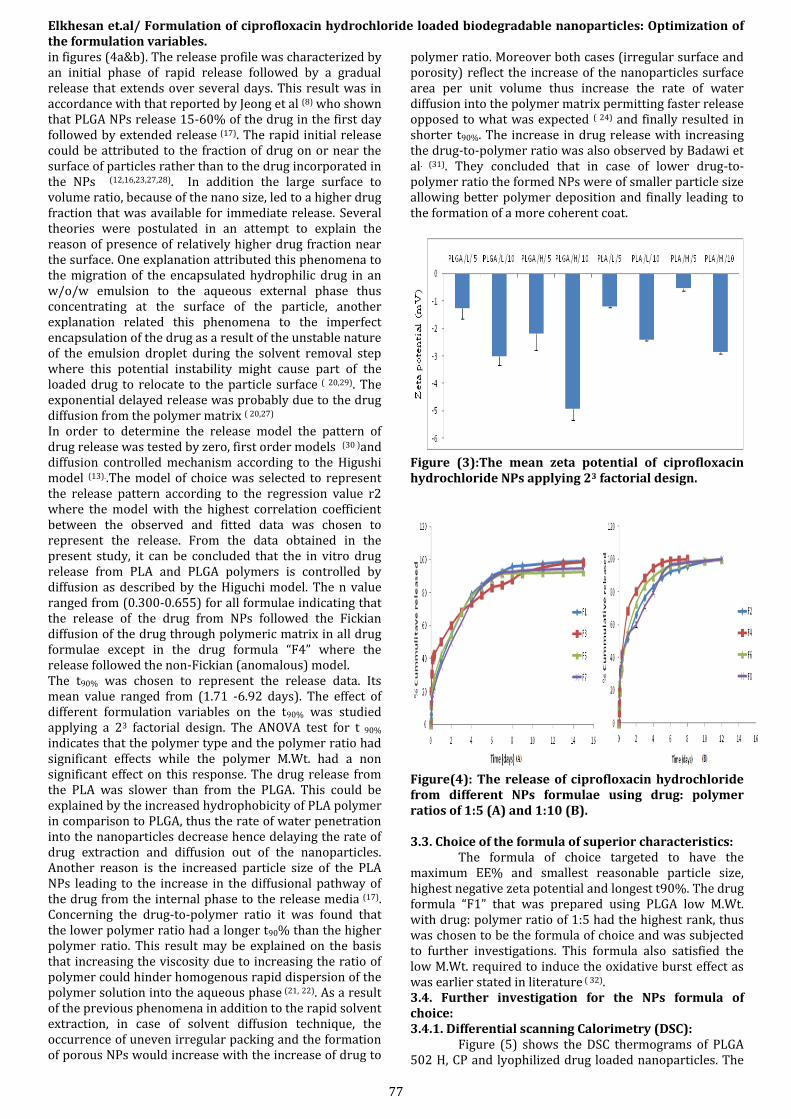
Figure(2): The mean encapsulation efficiency of ciprofloxacin hydrochloride NPs applying 23 factorial design. 3.2.3. Zeta potential: Figure(3) shows the values of the mean zeta potential of the prepared nanoparticles. It was obvious that all formulae showed a surface negative charge with mean values ranging from -0.521 to -4.915mV. The ANOVA test revealed that all the studied variables had significantly affected the zeta potential of the NPs. The polymer type indicated that PLGA had a higher negative value than PLA polymer. This could be explained by the presence of free carboxylic acid end on the PLGA ( 6 )contrary to the PLA polymer that had an ester end. It is worth mentioning that in case of PLA the slightly negative charge on the surface may be attributed to the hydroxyl group of the PVA that was anchored on the surface of the nanoparticles (5). The amphiphillic nature of the PVA leads to its entanglement in the polymer phase with their polar head group extruding out from the surface. This interaction is recorded to be strong and makes it difficult to be removed from the surface (5). Concerning the polymer M.Wt. and polymer ratio, it was shown that the higher polymer M.Wt. had a higher negative zeta potential as well as the higher polymer ratio (26). This might be explained by the significant increase in particle size due to the use of higher polymer M.Wt. and ratio thus increasing the exposed charge on the surface of particles. 3.2.4. In vitro release of ciprofloxacin hydrochloride from nanoparticles and kinetic analysis: The cumulative percentage release profile of CP from the nanoparticles formulae prepared with different polymers types, polymer M.Wt. and drug:polymer ratios are shown
Elkhesan et.al/ Formulation of ciprofloxacin hydrochloride loaded biodegradable nanoparticles: Optimization of the formulation variables.
77
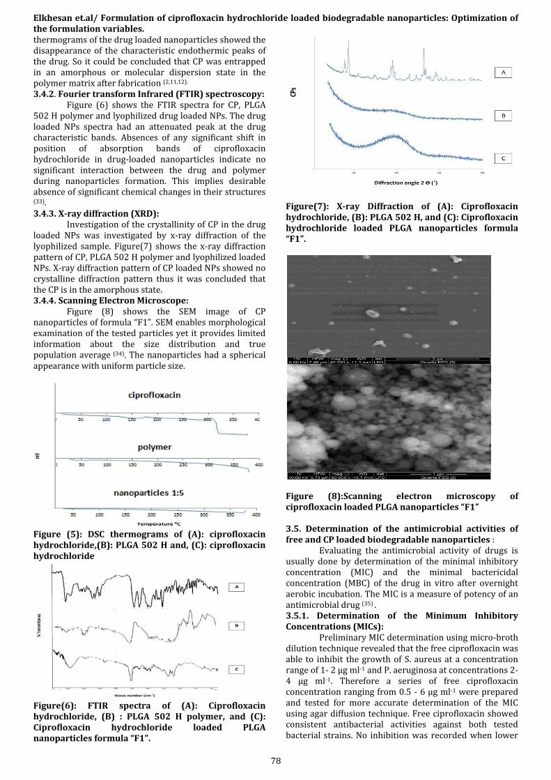
in figures (4a&b). The release profile was characterized by an initial phase of rapid release followed by a gradual release that extends over several days. This result was in accordance with that reported by Jeong et al (8) who shown that PLGA NPs release 15-60% of the drug in the first day followed by extended release (17). The rapid initial release could be attributed to the fraction of drug on or near the surface of particles rather than to the drug incorporated in the NPs (12,16,23,27,28). In addition the large surface to volume ratio, because of the nano size, led to a higher drug fraction that was available for immediate release. Several theories were postulated in an attempt to explain the reason of presence of relatively higher drug fraction near the surface. One explanation attributed this phenomena to the migration of the encapsulated hydrophilic drug in an w/o/w emulsion to the aqueous external phase thus concentrating at the surface of the particle, another explanation related this phenomena to the imperfect encapsulation of the drug as a result of the unstable nature of the emulsion droplet during the solvent removal step where this potential instability might cause part of the loaded drug to relocate to the particle surface ( 20,29). The exponential delayed release was probably due to the drug diffusion from the polymer matrix ( 20,27)
In order to determine the release model the pattern of drug release was tested by zero, first order models (30 )and diffusion controlled mechanism according to the Higushi model (13)..The model of choice was selected to represent the release pattern according to the regression value r2 where the model with the highest correlation coefficient between the observed and fitted data was chosen to represent the release. From the data obtained in the present study, it can be concluded that the in vitro drug release from PLA and PLGA polymers is controlled by diffusion as described by the Higuchi model. The n value ranged from (0.300-0.655) for all formulae indicating that the release of the drug from NPs followed the Fickian diffusion of the drug through polymeric matrix in all drug formulae except in the drug formula “F4” where the release followed the non-Fickian (anomalous) model. The t90% was chosen to represent the release data. Its mean value ranged from (1.71 -6.92 days). The effect of different formulation variables on the t90% was studied applying a 23 factorial design. The ANOVA test for t 90% indicates that the polymer type and the polymer ratio had significant effects while the polymer M.Wt. had a non significant effect on this response. The drug release from the PLA was slower than from the PLGA. This could be explained by the increased hydrophobicity of PLA polymer in comparison to PLGA, thus the rate of water penetration into the nanoparticles decrease hence delaying the rate of drug extraction and diffusion out of the nanoparticles. Another reason is the increased particle size of the PLA NPs leading to the increase in the diffusional pathway of the drug from the internal phase to the release media (17). Concerning the drug-to-polymer ratio it was found that the lower polymer ratio had a longer t90% than the higher polymer ratio. This result may be explained on the basis that increasing the viscosity due to increasing the ratio of polymer could hinder homogenous rapid dispersion of the polymer solution into the aqueous phase (21, 22). As a result of the previous phenomena in addition to the rapid solvent extraction, in case of solvent diffusion technique, the occurrence of uneven irregular packing and the formation of porous NPs would increase with the increase of drug to
polymer ratio. Moreover both cases (irregular surface and porosity) reflect the increase of the nanoparticles surface area per unit volume thus increase the rate of water diffusion into the polymer matrix permitting faster release opposed to what was expected ( 24) and finally resulted in shorter t90%. The increase in drug release with increasing the drug-to-polymer ratio was also observed by Badawi et al. (31). They concluded that in case of lower drug-to-polymer ratio the formed NPs were of smaller particle size allowing better polymer deposition and finally leading to the formation of a more coherent coat.
Figure (3):The mean zeta potential of ciprofloxacin hydrochloride NPs applying 23 factorial design.
Figure(4): The release of ciprofloxacin hydrochloride from different NPs formulae using drug: polymer ratios of 1:5 (A) and 1:10 (B). 3.3. Choice of the formula of superior characteristics: The formula of choice targeted to have the maximum EE% and smallest reasonable particle size, highest negative zeta potential and longest t90%. The drug formula “F1” that was prepared using PLGA low M.Wt. with drug: polymer ratio of 1:5 had the highest rank, thus was chosen to be the formula of choice and was subjected to further investigations. This formula also satisfied the low M.Wt. required to induce the oxidative burst effect as was earlier stated in literature ( 32). 3.4. Further investigation for the NPs formula of choice: 3.4.1. Differential scanning Calorimetry (DSC): Figure (5) shows the DSC thermograms of PLGA 502 H, CP and lyophilized drug loaded nanoparticles. The
Elkhesan et.al/ Formulation of ciprofloxacin hydrochloride loaded biodegradable nanoparticles: Optimization of the formulation variables.
78
thermograms of the drug loaded nanoparticles showed the disappearance of the characteristic endothermic peaks of the drug. So it could be concluded that CP was entrapped in an amorphous or molecular dispersion state in the polymer matrix after fabrication (2,11,12). 3.4.2. Fourier transform Infrared (FTIR) spectroscopy: Figure (6) shows the FTIR spectra for CP, PLGA 502 H polymer and lyophilized drug loaded NPs. The drug loaded NPs spectra had an attenuated peak at the drug characteristic bands. Absences of any significant shift in position of absorption bands of ciprofloxacin hydrochloride in drug-loaded nanoparticles indicate no significant interaction between the drug and polymer during nanoparticles formation. This implies desirable absence of significant chemical changes in their structures (33). 3.4.3. X-ray diffraction (XRD): Investigation of the crystallinity of CP in the drug loaded NPs was investigated by x-ray diffraction of the lyophilized sample. Figure(7) shows the x-ray diffraction pattern of CP, PLGA 502 H polymer and lyophilized loaded NPs. X-ray diffraction pattern of CP loaded NPs showed no crystalline diffraction pattern thus it was concluded that the CP is in the amorphous state. 3.4.4. Scanning Electron Microscope: Figure (8) shows the SEM image of CP nanoparticles of formula “F1”. SEM enables morphological examination of the tested particles yet it provides limited information about the size distribution and true population average (34). The nanoparticles had a spherical appearance with uniform particle size.
Figure (5): DSC thermograms of (A): ciprofloxacin hydrochloride,(B): PLGA 502 H and, (C): ciprofloxacin hydrochloride
Figure(6): FTIR spectra of (A): Ciprofloxacin hydrochloride, (B) : PLGA 502 H polymer, and (C): Ciprofloxacin hydrochloride loaded PLGA nanoparticles formula “F1”.
Figure(7): X-ray Diffraction of (A): Ciprofloxacin hydrochloride, (B): PLGA 502 H, and (C): Ciprofloxacin hydrochloride loaded PLGA nanoparticles formula “F1”.
Figure (8):Scanning electron microscopy of ciprofloxacin loaded PLGA nanoparticles “F1” 3.5. Determination of the antimicrobial activities of free and CP loaded biodegradable nanoparticles : Evaluating the antimicrobial activity of drugs is usually done by determination of the minimal inhibitory concentration (MIC) and the minimal bactericidal concentration (MBC) of the drug in vitro after overnight aerobic incubation. The MIC is a measure of potency of an antimicrobial drug (35) . 3.5.1. Determination of the Minimum Inhibitory Concentrations (MICs): Preliminary MIC determination using micro-broth dilution technique revealed that the free ciprofloxacin was able to inhibit the growth of S. aureus at a concentration range of 1- 2 µg ml-1 and P. aeruginosa at concentrations 2-4 µg ml-1. Therefore a series of free ciprofloxacin concentration ranging from 0.5 - 6 µg ml-1 were prepared and tested for more accurate determination of the MIC using agar diffusion technique. Free ciprofloxacin showed consistent antibacterial activities against both tested bacterial strains. No inhibition was recorded when lower

Elkhesan et.al/ Formulation of ciprofloxacin hydrochloride loaded biodegradable nanoparticles: Optimization of the formulation variables.
79
concentrations (0.5 µg ml-1) were tested for S. aureus and (0.5 and 1 µg ml-1) for P. aeruginosa. The recorded antibacterial activities had a proper linear range of 2-4 µg ml-1 with a linear correlation coefficient r2 > 0.93. Interestingly, P. aeruginosa showed slightly higher resistance to ciprofloxacin. Application of the previously estimated equation revealed that free ciprofloxacin had MIC of 0.85 and 1.2 µg ml-1 against S. aureus and P. aeruginosa respectively. Similarly, the formulated ciprofloxacin had antimicrobial activities of 2-4 and 4-6 µg ml-1 against S. aureus and P. aeruginosa respectively. Therefore a series of different concentrations of drug loaded nanoparticles ranging from 2 - 8 µg ml-1 were prepared and tested for more accurate determination of the MIC using agar diffusion technique. Linear correlations were recorded between log concentrations and the zone of inhibitions when the formulated ciprofloxacin "F1" was tested against S. aureus and P. aeruginosa. The recorded antibacterial activities had a proper linear range of 2-8 µg ml-1 with a linear correlation coefficient r2 > 0.94. Again P. aeruginosa showed slightly higher resistance to ciprofloxacin. Application of the previously estimated equation revealed that formulated ciprofloxacin had MIC of 0.8 and 0.97 µg ml-1 against S. aureus and P. aeruginosa respectively. 3.5.2. Determination of the Minimum Bactericidal Concentrations (MBCs): When free ciprofloxacin was tested to determine the MBC, all concentrations lower than 6 µg ml-1 didn't reduce the microbial count of S. aureus by 3 log cycles. Higher concentrations reduced this microbial count by 3 cycles or more. Similarly, the reduction of microbial count of P. aeruginosa was lower than 3 log cycles at concentrations lower than 8 µg ml-1. When these concentrations increased to 8 µg ml-1 or higher the count was reduced by 3 log cycles and more. Therefore, the MBC was calculated to be 5.5 and 7.5 µg ml-1 against S. aureus and P. aeruginosa respectively as shown in table (2). Similarly for the ciprofloxacin loaded nanoparticles ,the MBC was calculated to be 5.5 and 8.5 µg ml-1 against S. aureus and P. aeruginosa respectively as shown in table (3). The recorded MBCs of both free and formulated ciprofloxacin were 5-8 times higher than corresponding MICs. Although Dillen et al. have reported that MICs were almost the same as MBCs (11) another study by Page-Clisson et al. have revealed that the MBC was almost 10 times the recorded MIC (36). In a previous study, Esmaeili et al (16) have reported that the nanoparticles preparation significantly lower the MBC compared to the free drug. However, other studies (11, 36) have reported that nanoparticles preparations did not change either MICs or MBCs. This was the case in the present study, where both tested NPs formulations had neither beneficial nor detrimental effect on both MICs and MBCs for S. aureus or P. aeruginosa.
Bacteria Concentration (µg ml-1)
1 2 3 4 5 6 7 8 9 10
S. aureus - - - - - + + + + + P. aeruginosa - - - - - - - + + +
(-) means the reduction in the viable count was less than 3 log cycles (+) means the reduction in the viable count was higher than 3 log cycles Table(2): The recorded effect of the tested concentrations of free ciprofloxacin on the viable count of 106 -107 CFU ml-1 of tested reference bacteria.
Bacteria Concentration (µg ml-1)
1 2 3 4 5 6 7 8 9 10
S. aureus - - - - - + + + + + P. aeruginosa - - - - - - - - + +
(-) means the reduction in the viable count was less than 3 log cycles (+) means the reduction in the viable count was higher than 3 log cycles Table (3): The recorded effect of the tested concentrations of Ciprofloxacin NPs “F1” on the viable count of 106 -107 CFU ml-1 of tested reference bacteria.
3.5.3. Microbiological assay of ciprofloxacin released from its selected nanoparticles: The spectrophotometric assay determines the drug concentration; however it can’t ensure the drug potency. Accordingly microbiological assay was done to the daily released amount of drug loaded nanoparticles “F 1” to determine that the drug potency was encountered till the t90% of this formula i.e. 5 days. The released ciprofloxacin from formula “F 1” was assayed using spectrophotometric and microbiological assay techniques. High concentration of free ciprofloxacin was determined after one day of release by both techniques. These concentrations were decreased gradually by time however; all determined concentrations were higher than the highest determined MIC against P. aeruginosa. It is evident from the above mentioned results that the CP encapsulated in PLGA NPs in formula “F1” retained its antimicrobial activity throughout the preparation process. A good correlation between the concentrations determined spectrophotometrically and microbiologically was obtained in case of both bacterial strains, where the determination coefficient (r2) value was 0.96 and 0.92 for S.A. and P.A respectively.
4.CONCLUSION
In the present study,the prepration and characterization of biodegradable nanoparticles containing ciprofloxacin HCL was investigated. To analyze the influence of different factors on the properties of nanoparticles and to find optimum formulation, a 23factorial experimental design was carried out. The polymer type had a significant effect on the particle size, t
90%, zeta-potential and none significant effect on the EE% of different NPs formulations. The drug to the polymer ratio had a significant influence on all the studied parameters of the prepared formulations. The drug loaded nanoparticles prepared by using PLGA of low M.Wt. and drug to polymer ratio of 1:5 had the superior characteristics including a particle size of 183.2 nm, EE% of 69 %, zeta potential of -1.26 mV and t 90% of 5.59 days. The MIC value for both formulae and CP solution were comparable in case of the two tested organisms. The microbiological characterization demonstrates that the CP loaded NPs of the selected formula retained its antimicrobial activity throughout the manufacturing process .The antimicrobial activity of the encapsulated CP against the two studied organisms was observed until the end of the t 90% of the selected formula.
5. DECLARATION OF INTEREST
The authors report no declaration of interest 6. REFERENCES 1-Lesic A.,Marko B.,Vladimir B.,Nikola K et al. Artificially
contaminated open tibial fractures: Therapy with cloxacillin sodium. Acta veterinaria.2004,54(5-6),pp.457-466. Available at: http://www.doiserbia.nb.rs/Article.aspx?id=0567-

Elkhesan et.al/ Formulation of ciprofloxacin hydrochloride loaded biodegradable nanoparticles: Optimization of the formulation variables.
80
83150406457L&AspxAutoDetectCookieSupport=1 [Accessed April 19, 2012].
2- Cevher E, Orhan Z, Sensoy D, Ahiskali R, Kan PL, Sağirli O, et al. Sodium fusidate-poly(D,L-lactide-co-glycolide) microspheres: preparation, characterisation and in vivo evaluation of their effectiveness in the treatment of chronic osteomyelitis. Journal of microencapsulation.2007, Sep;24(6):577–95.
3- Pillai, R.R.,Somayaji S.N.,Robinovich M,Hudson M.C.,Gonsalves K.E., Nafcillin-loaded PLGA nanoparticles for treatment of osteomyelitis. Biomedical materials.2008, 3(3), p.034114. Available at: http://www.ncbi.nlm.nih.gov/pubmed/18708713 [Accessed March 31, 2012].
4- Mobarak,D.H.,Salah,S., Elkheshen,S.A. Formulation of ciprofloxacin hydrochloride loaded biodegradable nanoparticles: optimization of technique and process variables. Pharm Dev Technol.2013, ISSN: 1083-7450 Early Online: 1–10.
5- Xu Q, Crossley A, Czernuszka JAN. Preparation and Characterization of Negatively Charged Poly ( Lactic-co-Glycolic Acid ) Microspheres.J.Pharm.Sci.2009,Jul;98(7):2377-89.
6-Sahoo S.K, Panyam J, Prabha S, Labhasetwar V. Residual polyvinyl alcohol associated with poly (D,L-lactide-co-glycolide) nanoparticles affects their physical properties and cellular uptake. Journal of controlled release .2002, Jul 18;82(1):105–14.
7- Mundargi R.C, Babu V.R, Rangaswamy V, Patel P. Aminabhavi TM. Nano/micro technologies for delivering macromolecular therapeutics using poly(D,L-lactide-co-glycolide) and its derivatives. Journal of controlled release.2008, Feb 11;125(3):193–209.
8- Jeong Y-I, Na H-S, Seo D-H, Kim D-G, Lee H-C, Jang M-K, et al. Ciprofloxacin-encapsulated poly(DL-lactide-co-glycolide) nanoparticles and its antibacterial activity. International journal of pharmaceutics.2008, Mar 20;352(1-2):317–23.
9- Dillen K, Vandervoort J, Van den Mooter G, Ludwig A. Evaluation of ciprofloxacin-loaded Eudragit RS100 or RL100/PLGA nanoparticles. International journal of pharmaceutics.2006, May 11;314(1):72–82.
10- Yu W, Wong J, Chang T. Biodegradable polylactic acid nanocapsules containing ciprofloxacin: preparation and characterization. Artificial cells, blood substitutes and immobilization biotechnology.1999, May;27(3):263–78.
11- Dillen K, Vandervoort J, Van den Mooter G, Verheyden L, Ludwig A. Factorial design, physicochemical characterisation and activity of ciprofloxacin-PLGA nanoparticles. International journal of pharmaceutics.2004, May 4;275(1-2):171–87.
12- Sant S, Nadeau V, Hildgen P. Effect of porosity on the release kinetics of propafenone-loaded PEG-g-PLA nanoparticles. Journal of controlled release .2005, Oct 3;107(2):203–14.
13- Higuchi T. Rate of release of medicaments from ointment bases containing drugs in suspension. Journal of Pharmaceutical Sciences.1961, Oct;50(10):874–5.
14- Mashru, R.C.,Sutariya V.B.,Sankalia M.G.,Parikh P.P. Development and Evaluation of Fast-Dissolving Film of Salbutamol Sulphate. Drug Development and Industrial Pharmacy.2005,,Jan; 31(1), pp.25-34.
15- Azéma J., Guidetti B., Korolyov A.,Kiss R.,Roques C.,Constant P.,et al. Synthesis of lipophilic dimeric C-7/C-7-linked ciprofloxacin and C-6/C-6-linked levofloxacin derivatives. Versatile in vitro biological evaluations of monomeric and dimeric fluoroquinolone derivatives as potential antitumor, antibacterial or antimycobacte. European journal of medicinal chemistry.2011, 46(12), pp.6025-38. Available at: http://www.ncbi.nlm.nih.gov/pubmed/22036229 [Accessed April 12, 2012].
16- Esmaeili F.,Hosseini-Nasr M.,Rad-Malekshahi M.,Samadi N.,Atyabi F.,Dinarvand R. Preparation and antibacterial activity evaluation of rifampicin-loaded poly lactide-co-glycolide nanoparticles. Nanomedicine.2007,Jun;3(2): pp.161-7. Available at: http://www.ncbi.nlm.nih.gov/pubmed/17468055 [Accessed January 6, 2012].
17- Song X, Zhao Y, Hou S, Xu F, Zhao R, He J, et al. Dual agents loaded PLGA nanoparticles: systematic study of particle size and drug entrapment efficiency. European journal of pharmaceutics and biopharmaceutics.2008, Jun;69(2):445–53.
18- Jafari S, Assadpoor E, He Y, Bhandari B. Re-coalescence of emulsion droplets during high-energy emulsification. Food Hydrocolloids.2008, Oct;22(7):1191–202.
19 Jain R.A., The manufacturing techniques of various drug loaded biodegradable poly(lactide-co-glycolide) (PLGA) devices. Biomaterials.2000,Dec; 21(23), pp.2475-90.
20- Li X, Xu Y, Chen G, Wei P, Ping Q. PLGA nanoparticles for the oral delivery of 5-Fluorouracil using high pressure homogenization-emulsification as the preparation method and in vitro/in vivo studies. Drug development and industrial pharmacy.2008, Jan;34(1):107–15.
21- Blanco M.D., Alonso M.J., Development and characterization of protein-loaded poly(lactide-co-glycolide) nanospheres. European Journal of Pharmaceutics and Biopharmaceutics.1997, 43(3), pp.287-294.
22- Gao H.,Yang Y.W.,Fan Y. G.,Ma J.B., Conjugates of poly(DL-lactic acid) with ethylenediamino or diethylenetriamino bridged bis(beta-cyclodextrin)s and their nanoparticles as protein delivery systems. Journal of controlled release. 2006, 112(3), pp.301-11.
23- Lamprecht A.,Ubrich N.,Hombeiro Perez N.,Lehr C.,Hoffman M.,Maincent P., Influences of process parameters on nanoparticle preparation performed by a double emulsion pressure homogenization technique. International Journal of Pharmaceutics.2000, 196(2), pp.177-182.
24- Dawes G.J.,Fratila –Apachitei L.E.,Mulia K.,Apachitei I.,Wikamp G.J.,Duszczyk J., Size effect of PLGA spheres on drug loading efficiency and release profiles. Journal of materials science. Materials in medicine.2009, 20(5), pp.1089-94.
25- Freitas S., Merkle H.P.,Gander, B., Microencapsulation by solvent extraction/evaporation: reviewing the state of the art of microsphere preparation process technology. Journal of controlled release.2005, 102(2), pp.313-32.
26- Mohammadi G, Valizadeh H, Barzegar-Jalali M, Lotfipour F, Adibkia K, Milani M, et al. Development of

Elkhesan et.al/ Formulation of ciprofloxacin hydrochloride loaded biodegradable nanoparticles: Optimization of the formulation variables.
81
azithromycin-PLGA nanoparticles: physicochemical characterization and antibacterial effect against Salmonella typhi. Colloids and surfaces. B, Biointerfaces.2010, Oct 1;80(1):34–9.
27- Soppimath, K.S., Aminabhavi T.M., Anandrao K.R., Rudzinski W.E., Biodegradable polymeric nanoparticles as drug delivery devices. Journal of controlled release.2001, 70(1-2), pp.1-20.
28- Hans M.L, Lowman A.M., Biodegradable nanoparticles for drug delivery and targeting. Journal of Controlled Release.2002,6(4):319–27.
29- Yeo, Y., Park K., Control of encapsulation efficiency and initial burst in polymeric microparticle systems.Archives of pharmacal research.2004, 27(1), pp.1-12. Available at: http://www.ncbi.nlm.nih.gov/pubmed/14969330.
30- Kim H., Fassihi R. Application of a binary polymer system in drug release rate modulation. 1. Characterization of release mechanism. Journal of pharmaceutical sciences.1997,Mar;86(3):316–22.
31- Badawi, A.A., El-samaligy M., Badawy S.S. Aziz, R.L., Controlled release theophylline delivery systems using Eudragit R S 100. Die pharmazie.1998, 58(8): p. 556-558.
32- Lecároz C., Blanco-Prieto M.J., Burrell M. A., Gamazo C. Intracellular killing of Brucella melitensis in human macrophages with microsphere-encapsulated gentamicin. The Journal of antimicrobial chemotherapy.2006, Sep;58(3):549–56.
33- Kumar G., Sharma S., Shafiq N., Khuller G.K., Malhotra S. Optimization , In Vitro – In Vivo Evaluation and Short-Term Tolerability of Novel Levofloxacin-Loaded PLGA Nanoparticle. Journal of Pharmaceutical Sciences., 2012;1–12
34- Pal S.L, Jana U., Manna P.K., Mohanta G.P., Manavalan R. Nanoparticle : An overview of preparation and characterization. Journal of Applied Pharmaceutical Science.2011, 01(06):228–34.
35- Levison, M.E., 2004. Pharmacodynamics of antimicrobial drugs. Infectious disease clinics of North America.2004, 18(3), pp.451-65. Available at: http://www.ncbi.nlm.nih.gov/pubmed/15308272 [Accessed September 7, 2011].
36- Page-Clisson M.E.,Pinto-Alphandary H.,Ourevitch M.,Andremont A.,Couvreur P., Development of ciprofloxacin-loaded nanoparticles: physicochemical study of the drug carrier.Journal of controlled release .1998,56(1-3), pp.23-32. Available at: http://www.ncbi.nlm.nih.gov/pubmed/9801426.